Isolation and identification of M. tuberculosis from sheep tissue samples and sero-diagnosis study in an organized sheep farm
Bạn đang xem bản rút gọn của tài liệu. Xem và tải ngay bản đầy đủ của tài liệu tại đây (178.74 KB, 5 trang )
Int.J.Curr.Microbiol.App.Sci (2018) 7(1): 2740-2744
International Journal of Current Microbiology and Applied Sciences
ISSN: 2319-7706 Volume 7 Number 01 (2018)
Journal homepage:
Original Research Article
/>
Isolation and Identification of M. tuberculosis from Sheep Tissue Samples
and Sero-Diagnosis Study in an Organized Sheep Farm
K. Arunmozhivarman1, R. Radhika1, P. Kannan2, V. Maroudam1,
K. Vijayalakshmi1, P. Valentina Claudet1 and G. Dhinakar Raj1*
1
Translational Research Platform for Veterinary Biologicals, Tamil Nadu Veterinary and
Animal Sciences University, Chennai -600 051, India
2
Department of Immunology, National Institute for Research in Tuberculosis,
Chennai-31, India
*Corresponding author
ABSTRACT
Keywords
Sheep, M.
tuberculosis, PCR,
ELISA,
Seroprevalence
Article Info
Accepted:
20 December 2017
Available Online:
10 January 2018
A 2 years old female Madras Red sheep with the medical history of reduction in feed
intake, poor weight gain and emaciation was found dead in an organised farm. The
sheep did not have any obvious respiratory symptoms. Edematous and caseous lesions
were observed in mesenteric, bronchial, mediastinal and prescapular lymph nodes of
the sheep during post mortem examination. Other internal organs were free of any
specific lesions. The lymph node samples were decontaminated and cultured by
inoculating into BACTEC Mycobacteria Growth Indicator Tube (MGIT) system and
Lowenstein Jensen slants. The cultures turned positive and acid fast staining of the
bacterial culturerevealed the presence of Mycobacteria. The bacteria was further
confirmed as Mycobacterium tuberculosis by multiplex PCR and nucleotide
sequencing. A Tuberculosis sero-diagnostic study was conducted for all the animals in
the farm using commercially available ELISA kit to know the incidence of
tuberculosis in the farm. Three sheep out of the total 205 sheep were positive for
tuberculosis by ELISA with the estimated 1.5% positivity. This shows the active
circulation of tuberculosis in sheep farm and there may be possibility of human to
animal transmission and vice versa. The role of sheep in the epidemiology and
transmission of tuberculosis needs further study.
Introduction
Tuberculosis (TB) is a chronic bacterial
disease caused by Mycobacterium tuberculosis
complex (MTC) leading to decreased
productivity, economic losses and poses a
significant threat to human health. Among
MTC organisms, the major agents are M.
tuberculosis and M. bovis. The primary host
for M. bovis is cattle and M. tuberculosis is
human. However, occurrence of M.
tuberculosis in animals and M. bovis infection
in humans has been reported previously
(Ocepek et al., 2005).
2740
Int.J.Curr.Microbiol.App.Sci (2018) 7(1): 2740-2744
In ovine, the occurrence of tuberculosis is very
rare although there are few reports indicating
the presence of M. bovis in sheep and goat
(Kassa et al., 2012; Marianelli et al., 2010).
This primarily occurs in areas with high
intensity sheep population and when there
exists close contact between infected cattle
and sheep facilitating transmission between
these species. India accounts for one fourth of
the global TB burden (Central TB division,
GOI, 2017).
Tuberculosis in animals is not well studied in
India; the lack of nation-wide epidemiological
studies makes the disease burden largely
unknown (Neeraja et al., 2014a). Few studies
have documented the prevalence of TB in
animals in India (Parmer et al., 2014;
Srivastava, 2008). Tuberculosis causes huge
economic loss in farm animals and the
production loss in infected animals will be 10
to 20 percent (Verma et al., 2004).
transported to laboratory
mycobacterial culture.
on
ice
for
Isolation Mycobacterium Sp. from tissue
samples
The samples were decontaminated and
processed following the modified Petroff’s
method (Kent and Kubica, 1985). A portion of
the decontaminated sediments were inoculated
into Mycobacterial Growth Indicator Tubes
(MGIT)TM from Becton Dickinson (BD) and
incubated in BACTEC MGIT 960 instrument
for 49 days at 37 °C.
The remaining sediments were inoculated into
one tube each of OADC-supplemented
Middlebrook 7H10 agar and LowensteinJensen (LJ) medium with sodium pyruvate and
glycerol and each tube was incubated for 8
weeks at 37 °C.
Acid fast staining
Tuberculosis is often unnoticed in animals and
the infected animals continue to spread the
disease to other susceptible animals and
human by excreting the organisms through
milk, faeces and respiratory droplets. Hence to
control tuberculosis both animals and human
has to be monitored for disease prevalence. In
this study Mycobateria was isolated from a TB
infected sheep and M. tuberculosis was
identified by multiplex PCR and gene
sequencing. Then all the sheep in the farm
were screened for TB sero-positivity.
Materials and Methods
Sample collection
Post mortem examination was carried out on
one Madras red sheep that had died in an
organized farm. The mesenteric, pre-scapular,
bronchial and mediastinal lymphnodes were
edematous and caseous. Samples from these
lymphnodes were collected in sterile PBS and
Heat fixed smears prepared from the sediment
and MGIT cultures declared as positive by the
BACTEC 960 and typical growths on
Middlebrook 7H10 and LJ media were
screened for presence of acid fast bacilli. The
heat-fixed smears were stained for acid fast
bacilli as per the standard protocol.
Polymerase chain reaction confirmation
and sequencing
The DNA extraction from MGIT liquid
culture and colonies on 7H10 agar/LJ media
was performed according to the CTAB –NaCl
method. These DNA samples were subjected
to conventional polymerase chain reaction
(PCR) with specific primers reported by
Zumarrga et al., (1999) and Bakshi et al.,
(2005). Then amplified PCR products were
sequenced to confirm the mycobacterium
species.
2741
Int.J.Curr.Microbiol.App.Sci (2018) 7(1): 2740-2744
Sero prevalence study using ELISA
Sheep sera samples from study farm were
screened for tuberculosis antibodies using the
commercial ELISA kit (IDEXX), USA as per
manufacturer’s instruction.
Result and Discussion
The post mortem caseous, edematous
lymphnode tissue samples collected from the
tuberculosis-suspected sheep were subjected
to acid fast staining, bacterial culture and
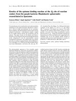
PCR. Staining of tissue smear from sheep
lymph node revealed that presence of rod
shaped, acid fast bacilli indicating the
presence of mycobacterium infection (Figure
1a).
Bacterial culture study is the gold standard for
laboratory confirmation of TB. Hence the
tissue samples were cultured in LJ medium
resulting in colonies that were rough, granular
and whitish initially and later on the colonies
turned yellowish (Figure 1b).
DNA amplification by PCR provides a rapid
and sensitive method for the detection of M.
tuberculosis complex (MTC) from postmortem samples and cultures (Clarridge et al.,
1993). DNA extracted from LJ medium
culture were subjected to multiplex PCR
method. PCR product was further analyzed by
agarose gel electrophoresis. There was no
band around 168 bp which is M. bovis specific
whereas M. tuberculosis specific band around
337 bp was visualized (Figure 2).
Fig.1a Acid Fast bacilli in Ziehl-Neelsen staining; Fig.1b Characteristic Mycobacterium
colonies on Lowenstein Jensen medium
1a
1b
2742
Int.J.Curr.Microbiol.App.Sci (2018) 7(1): 2740-2744
The PCR product was subjected to gene
sequencing
and
confirmed
as
M.
Tuberculosis.
Further, the circulation TB in the sheep farm
was identified by using ELISA to estimate
sero-prevalence. Generally humans are the
maintenance hosts for M. tuberculsois. The
sheep is considered to be the spill-over hosts
for M. bovis, can maintain the organism only
when its population density is high and is
generally considered very rare in small
ruminants (Tschopp et al., 2011).
However, presence of MTB in sheep indicates
a possible transmission of infection from
human to animal. In this study out of 205
sheep 3 were sero-positives and indicates
1.5% sero-prevalence of TB was observed in
study population. Lack of a robust animal TB
surveillance system and vaccine use in
animals aids in the transmission of TB
between animals and from animals to human
or vice versa. Thus there is an urgent and
unmet need for implementation of animal TB
control programs in developing countries
through extensive surveillance. The license
for the use of BCG vaccine in animals also
warrants further studies.
References
Bakshi, C. S., D. H. Shah, R. Verma, R. K.
Singh and M. Malik, 2005. Rapid
differentiation of Mycobacterium
bovis and Mycobacterium tuberculosis
based on a 12.7-kb fragment by a
single tube multiplex-PCR. Vet
Microbiol., 109: 211-6.
Clarridge, J. E., R. M. Shawaz, T. M.
Shinnick and B. B. Plikaytis, 1993.
Large scale use of PCR for detection
of Mycobacterium tuberculosis in
routine mycobacteriology laboratory.
J. Clin. Microbiol., 31: 2049-2056.
Gezahegne MamoKassa, Fekadu Abebe,
Yalelet Worku, Mengistu Legesse,
Girmay Medhin, Gunnar Bjune and
Gobena Ameni, 2012. Tuberculosis in
Goats and Sheep in Afar Pastoral
Region of Ethiopia and Isolation of
Mycobacterium tuberculosis from
Goat. Vet Med Int., 2012: 869146.
Kent. P. T., and Kubica G. P., 1985. Public
Health Mycobacteriology a guide for
the level III laboratory. Centre for
Disease Control Manual, Pp. 21-44.
Marianelli, C., N. Cifani and M. T.
Capucchio, 2010. A case of
generalized bovine tuberculosis in a
sheep. Journal
of
Veterinary
Diagnostic Investigation, 22(3): 445–
448.
Neeraja, D., B. M. Veeregowda, M. S. Rani,
D. Rathnamma, R. Bhaskaran, L.
Gowda, S. H. Somshekhar, M.
Saminathan, K. Dhama and S.
Chakraborty, 2014. Comparison of
Single Intradermal Test, Gamma
Interferon Assay and Indirect ELISA
for the Diagnosis of Tuberculosis in a
Dairy Farm. Asian Journal of Animal
and Veterinary Advances, 9: 593-598.
Ocepek, M., M. Pate, M. Zolnir-Dovc and M.
Poljak, 2005. Transmission of
Mycobacterium tuberculosis from
human to cattle. J. Clin. Microbiol.,
43: 3555-3557.
Parmar, B. C., M. N. Brahmbhatt, J. B.
Nayak, A. J. Dhami and Y. A. Chatur,
2014. Prevalence of tuberculosis in
men and animals: Confirmation by
cultural examinations, tuberculin tests
and PCR technique. Journal of
Foodborne and Zoonotic Diseases, 2:
36-44.
Srivastava, K., D. S. Chauhan, P. Gupta, H.
B. Singh, V. D. Sharma and V. S.
Yadav,
2008.
Isolation
of
Mycobacterium
bovis
and
M.
tuberculosis from cattle of some farms
in north India - possible relevance in
2743
Int.J.Curr.Microbiol.App.Sci (2018) 7(1): 2740-2744
human health. Indian J Med Res., 128:
26–31.
Tschopp, R., K. Bobosha, A. Aseffa, E.
Schelling, M. Habtamu, R. Iwnetu, E.
Hailu, R. Firdessa, J. Hussein, D.
Young and J. Zinsstag, 2011. Bovine
tuberculosis
at
a
cattle-small
ruminant-human interface in Meskan,
Gurage region, Central Ethiopia. BMC
Infectious Diseases, 11: 318.
Verma, S., N. K. Mahajan and G. Malik,
2004. An epidemiological study on
bovine H.S. in Haryana, Indian J Anim
Res., 38(1): 14-19.
Zumarraga, M., F. Bigi, A. Alito, M. I.
Romano and A. Cataldi, 1999. A 12.7
kb fragment of the Mycobacterium
tuberculosis genome is not present in
Mycobacterium bovis. Microbiology,
145: 893-897.
How to cite this article:
Arunmozhivarman, K., R. Radhika, P. Kannan, V. Maroudam, K. Vijayalakshmi, P. Valentina
Claudet and Dhinakar Raj, G. 2018. Isolation and Identification of M. tuberculosis from Sheep
Tissue Samples and Sero-Diagnosis Study in an Organized Sheep Farm.
Int.J.Curr.Microbiol.App.Sci. 7(01): 2740-2744.
doi: />
2744