Aortic dissection
Bạn đang xem bản rút gọn của tài liệu. Xem và tải ngay bản đầy đủ của tài liệu tại đây (106.69 KB, 3 trang )
Aortic Dissection
Amritpal Singh Nat, M.D., and Dinesh Subedi, M.D.
September 18, 2014 N Engl J Med 2014; 371:e17
DOI: 10.1056/NEJMicm1313897
An 81-year-old man with no documented medical
history presented to the emergency department [khoa
cấp cứu] with suprapubic pain [đau vùng trên mu] and
urinary retention [bí tiểu] resulting from benign
prostatic hyperplasia [tăng sinh lành tính tuyến tiền
liệt]. He was incidentally found to have an elevated
troponin I level, at 0.17 ng per milliliter (normal value,
<0.08). He reported no chest or back pain [đau ngực
hay đau lưng] or shortness of breath [khó thở]. There
was no evidence of ischemic changes [các thay đổi của
thiếu máu cục bộ] on electrocardiography [điện tâm
đồ]. Chest radiography [phim X-quang ngực] showed
1
widening of the mediastinum [trung thất].
Transthoracic echocardiography [siêu âm tim qua
thành ngực] showed aneurysmal dilatation [giãn phình
mạch] and a dissection flap [mảng bóc tách] in the
ascending aorta [động mạch chủ lên] (video).
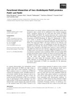
Computed tomographic angiography [chụp mạch cắt
lớp vi tính] of the thorax [ngực] and abdomen [bụng]
revealed an ascending aortic aneurysm [phình động
mạch chủ lên] (Panel A, blue arrows) and a type I
DeBakey aortic dissection [bóc tách động mạch chủ
type I theo phân loại DeBakey] . The dissection
involved the ascending aorta (Panel B, white arrow),
aortic arch [cung động mạch chủ] (Panel C, blue
arrow), and descending aorta [động mạch chủ xuống]
(Panels B and C, red arrows), terminating just below
the origin [nơi xuất phát (nguyên uỷ)] of the renal
arteries [động mạch thận]. Several branch vessels
[mạch máu nhánh] were involved as well, including the
brachiocephalic artery [động mạch cánh tay – đầu], lef
subclavian artery [động mạch dưới đòn trái] (Panel C,
green arrow), and superior mesenteric artery [động
2
mạch mạc treo tràng trên]. Given the absence of
associated symptoms [triệu chứng], the dissection was
thought to be chronic [mạn tính]. The patient declined
consideration of surgical intervention [can thiệp bằng
phẫu thuật] but agreed to treatment [điều trị] with a
beta-blocker [thuốc chẹn beta]. One year later, the
patient's condition had not worsened; he continued to
be treated with a beta-blocker and to receive routine
follow-up care.
Amritpal Singh Nat, M.D. Dinesh Subedi, M.D.
SUNY Upstate Medical University, Syracuse, NY
3