Scanning electron microscopy (SEM) of seed infected with seed borne fungi
Bạn đang xem bản rút gọn của tài liệu. Xem và tải ngay bản đầy đủ của tài liệu tại đây (273.67 KB, 6 trang )
Int.J.Curr.Microbiol.App.Sci (2018) 7(7): 4096-4101
International Journal of Current Microbiology and Applied Sciences
ISSN: 2319-7706 Volume 7 Number 07 (2018)
Journal homepage:
Original Research Article
/>
Scanning Electron Microscopy (SEM) of Seed Infected
with Seed Borne Fungi
A. R. Gulhane, G. K. Giri and Shilpa V. Khambalkar*
Sorghum Research Unit, Dr. PDKV, Akola - 444 104, Maharashtra, India
*Corresponding author
ABSTRACT
Keywords
Curvularia lunata,
Drechslera
tetramera, Bipolaris
sorghicola
Article Info
Accepted:
28 June 2018
Available Online:
10 July 2018
The scanning electron micrograph of various fungi growing over sorghum
and wheat seeds showed that, the conidia of Curvularia lunata were
slightly curved, three septate, central cell were broad having surface
ornamentation, darker than the end cell and arranged on sympodially
branched conidiophores on sorghum seed. The mycelial structure of
Drechslera tetramera on wheat showed network of hyphae, roughed
conidiophores and cluster of conidia arranged on geniculate conidiophores.
Conidia of Bipolaris sorghicola were smooth walled, straight and slightly
curved attached to the geniculate conidiophores. Lytic mycelium of B.
sorghicola, was also observed on sorghum seed.
Introduction
Scanning
Graminicolous spp. such as paddy, wheat,
pearl millet and sorghum is the most important
crop in India. In recent years the changes of
cropping practices in to intensive systems
leads rice, wheat, sorghum and pearl millet
crop to more diseases problems such as blast,
sheath blight, grain, discolouration, grain
mould, leaf spot. Curvularia spp. occasionally
caused leaf spot in several plants, C. lunata
has been implicated as one of the most
important grain mould in rice, wheat, pearl
millet and sorghum. The pathogen causes
discolouration on the seed, degenarate
endosperm and also infected embryo resulting
in almost 100% loss in variability of seed (Rao
and William, 1978). Recently Deshmukh and
Raut (1993) confirmed that C. lunata can
infect all the part of seeds of sorghum.
Bipolaris sorokiniana causing spot blight is
one of the most important foliar pathogens of
wheat. Causing several losses in wheat
production. It is capable of causing damage
from the primary leaf stage, through the plant
tends to become more susceptible after
flowering. The incidence of B. sorokiniana
was found highest in grains the leaf blight and
seedling blight disease caused by B.
sorokiniana was reported on leaf of wheat
crop (Malekar et al., 2010; Malekar and Main,
4096
Int.J.Curr.Microbiol.App.Sci (2018) 7(7): 4096-4101
2007; Azad et al., 2009). Twenty seven fungal
species associated with rice seed samples
among them most prominent fungi was
Bipolaris oryzae (82.08%). The leaf blight/
brown spot caused by Bipolaris oryzae and C.
lunata were recorded in rice crop (Bharti and
Raut, 2009; Archana and Prakash, 2013;
Safari and Kaviani, 2008; Kamaluddem et al.,
2013).
Seed health study mostly has been conducted
to measure yield losses as well as pathogen the
aim of this work is to describe morphological
and cytological features of vegetative and
reproductive structures of fungi observed with
the Scanning Electron Microscope (SEM).
were dehydrated with normal dry air. Infected
seeds were fixed with 2% glutaraldehyde 0.1
M solution and then treated with 4% osmium
tetra oxide (OsO4) vapor for two hours
respectively. For dehydration step, a serial
fixation was used with cold ethanol (70% 100%) and further it was dried by using
vacuum evaporation. Seeds of sorghum and
wheat was mounted in aluminium stub, and it
was sealed with colloidal silver paste, then
sputter-coated with gold particle (15 nm
thickness). The ready to use preparation of
specimens was observed under JEOL (JSM
6380A) SEM. The visual examination of
growing structure of fungi over seed was
recorded by photography using black and
white film.
Materials and Methods
The scanning electron microscopy was
performed at Department of Metallurgical and
Material Engineering, Visvesvaraya National
Institute
of
Technology,
Nagpur
(Maharashtra). The pure culture of
Curvularia, Drechslera and Bipolaris species
were inoculated to the seed and incubated for
seven days by blotter paper method. The
morphological characters of fungi growing
over seed was studied through scanning
electron microscopy.
Seed inoculation
Seeds were surface disinfected with mercuric
chloride (0.1%), washed repeatedly with
distilled water and inoculated with pure
culture of respective fungi to the entire seed
surface. Inoculated seed were incubated for
seven days by employing blotter paper method
and used for further observations under
scanning electron microscope.
SEM observations
The Curvularia, Drechslera and Bipolaris
species infected seed of sorghum and wheat
As observed under JEOL (JSM 6380A) SEM.
The visual examination of growing structure
of fungi over seed was recorded by
photography using black and white film.
Results and Discussion
The seed borne fungi, Curvularia lunata,
Drechslera
tetramera
and
Bipolaris
sorghicola growing over the seed of sorghum
and wheat were observed under Scanning
Electron Microscope (SEM).The Scanning
Electron Microscopy showed different
development stages of these fungi. The plate
12 showed that Curvularia lunata, Drechslera
tetramera and Bipolaris sorghicola were
predominantly covered the seed with
mycelium and conidiophore bearing conidia.
Similarly, colonies of Curvularia lunata
produced well developed mycelium on the
seed surface.
The mycelium was mostly immerse, forming
straight and smooth conidiophore and larger
roughened central cell of conidia arranged on
sympodially branched conidiophores. In case
of sorghum seed, the conidia of Curvularia
lunata were slightly curved and three septate.
4097
Int.J.Curr.Microbiol.App.Sci (2018) 7(7): 4096-4101
Plate.1 Scanning electron micrographs showing conidia and mycelium of Curvularia lunata on
surface of wheat seed
Plate.2 Scanning electron micrographs of Drechslera tetramera growing on surface of wheat
seed
4098
Int.J.Curr.Microbiol.App.Sci (2018) 7(7): 4096-4101
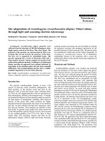
Plate.3 Scanning electron micrographs of Bipolaris sorghicola growing on surface of sorghum
seed
4099
Int.J.Curr.Microbiol.App.Sci (2018) 7(7): 4096-4101
In general, central cells were broad having
surface ornamentation and darker than end
cells (Plate 1). Some mature, slightly curved
conidia of Curvularia lunata were also being
liberated from the conidiophores on surface of
wheat seed.
The scanning electron micrograph of mycelial
structure of Drechslera tetramera growing on
wheat seed showed the network of hyphae
and also showed roughened conidiophores,
conidia with apical germpors and germinating
conidia. A cluster of conidia arranged on
geniculate conidiophores were also seen.
(Plate 2)
The conidia of Bipolaris sorghicola were
smooth walled, straight or slightly curved
arranged on geniculate conidiophore.
Terminal germ pores were visible on smooth
conidia of B. sorghichola and the lytic
mycelium was also observed on sorghum seed
(Plate 3).
The conidial attachment of Bipolaris
sorghicola with conidiophores, conidial
septation, conidial hilum, and older and
senescent conidia were also noticed.
Mukherjee et al., (2013) reported that under
scanning electron micrograph, the conidia of
Curvularia species was developed from
septate branched hyphae and conidia were
curved shaped, 3 to 4 celled. These findings
are also similar with those of Freire et al.,
(1998) who reported that, conidia of
Curvularia pallescens observed under
scanning electron micrograph was curved
shaped and three septate. The present findings
are in accordance with findings of Aggarwal
et al., (2002) who also observed the conidia
of Bipolaris sorokiniana under scanning
electron microscope in the form of smooth
walled, terminal germ pores attached with
conidiophores. Similar results have also been
reported by Domiciano et al., (2013) under
scanning electron micrograph, Bipolaris
sorokiniana formed a dense network of
conidiophores hyphae and conidia formed a
germ tube.
Scanning Electronic Microscopy (SEM) of C.
lunata, B. sorghicola and D. tetramera
growing over sorghum and wheat seed
revealed the presence of conidial surface
ornamentation, roughened conidiophore,
senescent conidia, geniculate conidiophore,
lytic mycelium, hilum and apical germpore on
conidia.
References
Aggarwal, R, S. Das, D.V. Singh and K.D.
Srivastava, 2002. SEM studies on spore
morphology and infection process of
spot blotch pathogen in wheat. Indian
Phytopath. 55(2): 197-199.
Carison, H., U. Stenram, M. Gustafsson and
H.B.
Jansoon,
1991.
Electron
microscopy of barley root infection by
the
fungal
pathogen
Bipolaris
sorokiniana. Canadian J. of Botany.
69(12): 2724-2731.
Domiciano, G.P., F.A. Rodrigues, A.M.N.
Guerra and F.X.R. Vale, 2013. Infection
process of Bipolaris sorokiniana on
wheat leaves is affected by silicon.
Trop. Plant Pathol. 38(3).
Falloon, R.E., 1976. Curvularia trifolii as a
high temperature turfgrass pathogen.
N.Z. Journal of Agricultural Research.
19: 243-248.
Mukherjee, A., A. Bandhyopadhyay and S.
Dutta, 2013. New Report of leaf spot
disease of Clerodendrum indicum
caused by Curvularia lunata. Int. J.
Pharm Bio. Sci. 4(3): 808-812.
Singh, U.P., S.K. Singh, Koya Sugawara, J.S.
Srivastava, B.K. Sarma and B.
Prithiviraj, 2001. Studies on Sclerotium
formation in Curvularia species.
Mycobiology. 29(3): 154-159.
4100
Int.J.Curr.Microbiol.App.Sci (2018) 7(7): 4096-4101
Waheeb Heneen and Kerstin Brismar, 1987.
Scanning electron microscopy of mature
grains of rye, wheat and triticale with
emphasis
on
grain
Hereduas, 107: 147-162.
shrivelling.
How to cite this article:
Gulhane A. R., G. K. Giri and Shilpa V. Khambalkar. 2018. Scanning Electron Microscopy
(SEM) of Seed Infected with Seed Borne Fungi. Int.J.Curr.Microbiol.App.Sci. 7(07): 40964101. doi: />
4101