Substrate thiophosphorylation by Arabidopsis mitogen-activated protein kinases
Bạn đang xem bản rút gọn của tài liệu. Xem và tải ngay bản đầy đủ của tài liệu tại đây (1.97 MB, 11 trang )
Leissing et al. BMC Plant Biology (2016) 16:48
DOI 10.1186/s12870-016-0731-6
RESEARCH ARTICLE
Open Access
Substrate thiophosphorylation by
Arabidopsis mitogen-activated protein
kinases
Franz Leissing1, Mika Nomoto2, Marco Bocola3, Ulrich Schwaneberg3, Yasuomi Tada2,4, Uwe Conrath1
and Gerold J. M. Beckers1*
Abstract
Background: Mitogen-activated protein kinase (MPK) cascades are important to cellular signaling in eukaryotes.
They regulate growth, development and the response to environmental challenges. MPK cascades function via
reversible phosphorylation of cascade components, MEKK, MEK, and MPK, but also by MPK substrate phosphorylation.
Using mass spectrometry, we previously identified many in vivo MPK3 and MPK6 substrates in Arabidopsis thaliana, and
we disclosed their phosphorylation sites.
Results: We verified phosphorylation of several of our previously identified MPK3/6 substrates using a nonradioactive
in vitro labeling assay. We engineered MPK3, MPK4, and MPK6 to accept bio-orthogonal ATPγS analogs for
thiophosphorylating their appropriate substrate proteins. Subsequent alkylation of the thiophosphorylated amino
acid residue(s) allows immunodetection using thiophosphate ester-specific antibodies. Site-directed mutagenesis
of amino acids confirmed the protein substrates’ site-specific phosphorylation by MPK3 and MPK6. A combined
assay with MPK3, MPK6, and MPK4 revealed substrate specificity of the individual kinases.
Conclusion: Our work demonstrates that the in vitro-labeling assay represents an effective, specific and highly
sensitive test for determining kinase-substrate relationships.
Keywords: Mitogen-activated protein kinase, (thio-)phosphorylation, MPK3/4/6, Arabidopsis thaliana, Analog-sensitive
kinase
Background
Mitogen-activated protein kinase (MPK) cascades are conserved signaling modules in eukaryotes. They transduce
external signals to intracellular responses via phosphorylation. MPK cascades contain three sequential types of
protein kinases. These are MPKs, MPK-activating kinases
(MKKs or MEKs), and MKK/MEK-activating kinases
(MEKKs). Genetic and biochemical analyses identified
distinct MEKK/MKK/MPK modules with overlapping
functions in development, immunity, and abiotic stress responses [1–3]. In addition to MKKs, MPK activity is regulated by MPK phosphatases that dephosphorylate, and
thereby inactivate their target MPKs. By now, the identity
* Correspondence:
1
Department of Plant Physiology, Aachen Biology and Biotechnology, RWTH
Aachen University, Aachen 52056, Germany
Full list of author information is available at the end of the article
of many MPK substrates and the nature of the MPKsubstrate interaction have remained elusive, especially in
plants. Their disclosure is essential to understanding
MEKK/MKK/MPK-mediated cell signaling.
Over the past decade, research on plant MPKs focused
on the large-scale identification of MPK substrate proteins. For example, protein and peptide microarrays were
probed with recombinant MPKs in the presence of radiolabeled ATP to identify novel MPK substrate proteins
[4–6]. Another in vitro screen used a synthetic peptide
library that was incubated with purified kinases before
phosphorylation site identification by mass spectrometry
[7]. Together, these screens revealed a large number of
potential kinase-substrate relationships.
Novel protocols in phosphoproteomics enable the enrichment even of low-abundant MPK substrate proteins
thus making them accessible to mass spectrometry [8, 9].
© 2016 Leissing et al. Open Access This article is distributed under the terms of the Creative Commons Attribution 4.0
International License ( which permits unrestricted use, distribution, and
reproduction in any medium, provided you give appropriate credit to the original author(s) and the source, provide a link to
the Creative Commons license, and indicate if changes were made. The Creative Commons Public Domain Dedication waiver
( applies to the data made available in this article, unless otherwise stated.
Leissing et al. BMC Plant Biology (2016) 16:48
These protocols describe dual enrichment strategies
that include the enrichment of phosphoproteins by
Al(OH)3-based metal-oxide affinity chromatography
(MOAC). The Al(OH)3-based MOAC is either preceded
by an ammonium-sulfate prefractionation step [9], or
followed by a specific enrichment of phosphopeptides
using TiO2. We referred to the latter approach as tandem MOAC [8]. The tandem approach enables the direct recording of site-specific phosphorylation of several
known and many unknown substrate candidate proteins
of MPK3 and MPK6 in Arabidopsis thaliana.
Here, we verify selected, previously in vivo identified
MPK substrate proteins of Arabidopsis using a novel nonradioactive in vitro labeling assay for determining plant
MPK substrate phosphorylation [8]. We use ATPγS analogs that cannot enter the ATP-binding pocket of a wildtype kinase, but can do so when the kinase’s ATP-binding
pocket is enlarged. These so-called analog-sensitive (AS)
kinases use bulky ATPγS analogs as cofactors during catalysis. We engineered the proline-directed serine/threonine kinases MPK3, MPK4, and MPK6 of Arabidopsis by
mutating the gatekeeping amino acid in the ATP-binding
pocket of the appropriate kinases. The mutation enlarges
the ATP-binding pocket thus allowing the AS kinase to
catalyze thiophosphorylation of its substrate proteins.
Subsequent alkylation of thiophosphorylated serine and/
or threonine residues provides a semisynthetic epitope for
a monoclonal thiophosphate ester-specific (anti-TPE) antibody [10]. In addition to verifying previously identified in
vivo MPK3/6 substrates in Arabidopsis, we demonstrate
that these MPK3/6 substrates are poor substrates for the
closely related Arabidopsis MPK4 [5, 8].
Results
Arabidopsis MPKs use ATPγS to thiophosphorylate myelin
basic protein
In conventional in vitro kinase assays, a kinase and its substrate are incubated in the presence of [γ-32P] or [γ-33P]-labeled ATP. Upon incubation, 32P/33P-radiolabeled substrate
is being detected as a measure for kinase activity or suitability of a protein or peptide to serve as a substrate for the
kinase in question. To determine the in vitro activity of
Arabidopsis MPK3, MPK4, and MPK6 towards selected
proteinaceous candidate substrates in the absence of radioactive labeling, we expressed MPK3, MPK4, and MPK6 as
GST-fusions in Escherichia coli. Fusion proteins were purified and incubated, in catalyzing conditions, in the presence
of the generic MPK substrate myelin basic protein (MBP)
[11]. MPK activity was ensured by phosphorylation of
column-bound GST-MPKs (before elution) using purified,
constitutively active versions of upstream MKKs in the
presence of ATP. GST-MPK3 and GST-MPK6 were
activated with constitutively active MKK4 and MKK5
(subsequently referred to as MKK4DD and MKK5DD),
Page 2 of 11
whereas column-bound GST-MPK4 was activated by
MKK1DD and MKK2DD. Before elution of the GST-fused
MPKs, the column was extensively washed with buffer to
remove any residual MKK. As controls we used equally
treated kinase-death versions of MPK3, MPK4, and MPK6
(subsequently referred to as MPK3KD, MPK4KD, and
MPK6KD). Pre-incubation of MPK3KD/4KD/6KD with their
appropriate upstream MKKDDs induced dual phosphorylation of the TEY motif in the activation loop of kinases,
but did not stimulate MPK3KD/4KD/6KD to phosphorylate
MBP (Fig. 1a). In contrast, on-column pre-incubation of
wild-type MPKs with their upstream MKKDDs not only
enhanced dual phosphorylation of the MPK’s TEY motif,
but it also strongly induced MPK3/4/6 activity (Fig. 1a).
Next, we asked whether MPK3/4/6 accept ATPγS as a
cofactor to thiophosphorylate their generic substrate
MBP. We also wondered whether a commercial antiTPE antibody can be used to detect thiophosphorylated
substrate proteins of MPK3/4/6. To answer these questions, pre-phosphorylated wild-type and kinase-death
versions of MPK3/4/6 were incubated with MBP in the
presence or absence of ATPγS. After adding p-nitrobenzyl mesylate (PNBM) for alkylating the potential thiophosphoryl group on MBP, the reaction mixture was
subjected to standard western blotting analysis and
immunodetection with anti-TPE antibody. No signal was
detected in control reactions without ATPγS, or when
kinase-death MPKs were used in the assay with ATPγS
(Fig. 1b). However, the anti-TPE antibody cross-reacted
with MBP in reaction mixtures containing only active
wild-type MPK3/4/6 and ATPγS (Fig. 1b). This result
strongly suggested that MPK3/4/6 all accept ATPγS as a
phosphoryldonor to thiophosporylate MBP and supposedly also other substrate proteins. It also disclosed that
the commercial anti-TPE antibody can be used to detect
thiophosphoryl groups on MPK3/4/6 substrate proteins
(Fig. 1b).
Engineered Arabidopsis AS-MPKs use N6-benzyl-ATPγS as
cofactor
Next we asked whether AS-MPK3/4/6 would accept
synthetic N6-benzyl-ATPγS (Bn-ATPγS) as a thiophosphate donor to exclude thiophosphorylation of substrates by contaminating kinases and to also prevent
substrates from potential autophosphorylation. Earlier
studies identified two tyrosine residues (Y124 in MPK4
and Y144 in MPK6) as the gatekeeper amino acid in
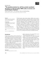
these two kinases [12, 13]. To identify the gatekeeping
residue in MPK3, we built a 3D atomic structure of
MPK3 and Bn-ADP (Fig. 2a). The model predicts an
atomic clash of the large threonine-119 (T119) amino
acid residue in MPK3 with the bulky side chain of BnADP (Fig. 2b). Mutation of T119 to a smaller amino
acid, such as alanine (A), would probably allow Bn-ATP
Leissing et al. BMC Plant Biology (2016) 16:48
Page 3 of 11
A
B
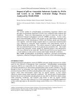
Fig. 1 MBP Phosphorylation and Thiophosphorylation by MPK3/4/6. a Kinase activity assay of purified wild-type (Wt) and kinase-death (KD) forms
of MPK3/4/6 with (+) and without (-) pre-phosphorylation by upstream MKKs. b In vitro thiophosphorylation of MBP by Wt and KD forms of
MPK3/4/6 in the presence (+) or absence (-) of ATPγS. CBB: Coomassie Brilliant Blue
A
B
C
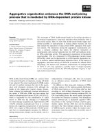
Fig. 2 Model of Wild-type and Mutant Variants of MPK3 with Bn-ADP. a Ribbon diagram of wild-type MPK3 with its ligands ADP (shown in stick
mode and colored by atom type) and Mg2+ (yellow). The backbone and amino acid side chain of T119 in MPK3 are highlighted in red and shown
in stick mode. Blue outlined rectangle highlights the ATP-binding site of MPK3. b Close-up of Bn-ADP modeled into the ATP-binding site of
wild-type MPK3. c Same as in (b) but with MPK3T119A
Leissing et al. BMC Plant Biology (2016) 16:48
to access the ATP-binding site of MPK3 (Fig. 2c). Thus,
T119 seems to be the gatekeeper amino acid residue in
Arabidopsis MPK3.
To test our in silico model (Fig. 2) in vitro, we purified
GST-MPK3T119A that was expressed in E.coli. We also
purified a mutant version of AS-MPK4 (GST-MPK4Y124A)
and AS-MPK6 (GST-MPK6Y144A) that we had expressed
in E. coli. First, the relative activity of these AS kinases to
phosphorylate MBP was compared to the activity of their
appropriate wild-type versions in the presence of either
[γ-32P] ATP (Fig. 3a) or ATPγS (Fig. 3b). Mutation of
Y124 to alanine did not affect (Fig. 3b) MPK4’s ability to
use [γ-32P] ATP (Fig. 3a) or ATPγS (Fig. 3b). MPK6Y144A
phosphorylated MBP to an about same (Fig. 3b) or slightly
lower extent (Fig. 3a) than the MPK6 wild-type protein.
MPK3T119A also catalyzed MBP phosphorylation which
was lower (Fig. 3a) or somewhat higher (Fig. 3b) than it
was with the wild type.
In another set of experiments, the ability of ASMPK3/4/6 to use Bn-ATPγS as a cofactor during catalysis was tested in in vitro substrate labeling assays
(Fig. 3c). Wild-type MPK3/4/6 did not use Bn-ATPγS as
a thiophosphate donor, whereas all AS kinases used the
bio-orthogonal Bn-ATPγS analog to thiophosphorylate
MBP (Fig. 3c).
AS-MPKs can be specifically inhibited by purine analogs
that do not affect the activity of wild-type kinases. For
example, previous studies revealed that 4-amino-1-tertbutyl-3-(1′-naphthyl)pyrazolo[3,4-d]pyrimidine (NA-PP1)
specifically inhibits AS kinases but not their appropriate
wild-type kinase because its bulkier side chain prevents
NA-PP1 from accessing the ATP-binding pocket of wildtype kinases [12, 13]. Consistent with this AS-MPK3/4/6
seem to be sensitive to NA-PP1 because NA-PP1 addition
to the substrate labeling reaction completely abolished
MPK3T119A, MPK4Y124A, and MPK6Y144A activity (Fig. 3d).
Site-specific phosphorylation of MPK targets by in vitro
substrate labeling
Next, we used in vitro substrate labeling reactions with
active AS-MPK3/4/6 to verify previously identified in
vivo MPK3/6 substrates and their target phosphorylation
sites. We randomly selected four MPK3/6-specific in
vivo substrates which we identified by tandem MOAC in
previous work [8]: two proteins of unknown function
(AT2G26530 and AT1G78150), a putative translation
initiation factor (AT4G38710), and PIRL9, a member of
the Plant Intracellular Ras group-related leucine-rich repeat (LRR) proteins (AT3G11330). In addition to the
wild-type version of proteins, we cloned phosphosite
mutants in which the previously recorded phosphorylated serine and/or threonine residues were mutated to
alanine. FLAG-tagged wild-type and mutant proteins
were immunoprecipitated with anti-FLAG agarose resin
Page 4 of 11
after successful in vitro translation in wheat germ extracts. While bound to the affinity gel, proteins were incubated with active AS-MPK3/4/6 or the appropriate
wild-type form of kinase in the presence of Bn-ATPγS.
As shown in Fig. 4, wild-type AT1G78150, AT2G26530,
and AT4G38710 were phosphorylated by all three MPKs.
However, in contrast to MPK3/6, these proteins seem to
be only weakly phosphorylated by MPK4Y124A (Fig. 4c).
AT3G11330 is a good substrate of MPK3/6 but not
phosphorylated by MPK4 (Fig. 4a–c). In most cases, mutation of previously identified phosphosites in the investigated proteins strongly reduced the extent of
phosphorylation by MPK3/4/6 in vitro. These findings
indicate that the previously identified serine/threonine
residues are specifically targeted by MPK3/6 and to a
lesser extent by MPK4.
The results in Fig. 4 indicated that AT1G78150,
AT2G26530, AT3G11330, and AT4G38710 are good
substrates for MPK3/6 but are not, or only weakly phosphorylated by MPK4. To directly compare the potential
of MPK3/4/6 to phosphorylate the four proteins, we repeated the substrate labeling reactions with all three
MPKs (Fig. 5a). We tested the wild-type version of
AT1G78150, AT2G26530, AT3G11330, and AT4G38710
and loaded the samples of each of these proteins in combination with MPK3/4/6 on a separate gel. The results
of this experiment support the previous finding arguing
that the assayed proteins represent good substrates of
MPK3/6, but are marginally phosphorylated by MPK4.
To exclude the possibility that the lower level of phosphorylation of these MPK3/6 substrates by MPK4 is due
to a lower in vitro activity of MPK4, we expressed and
purified the known MPK4 substrate MAP kinase substrate
1 (MKS1) from E. coli, and found that MKS1 was equally
well phosphorylated by MPK3/6 and 4 (Fig. 5b) [14].
Together, these data verify AT1G78150, AT2G26530,
AT3G11330, and AT4G38710 as substrate proteins of
MPK3/6, and they suggest specificity in the in vitro
substrate-labeling reactions.
Specificity of the in vitro substrate labeling assay
Arabidopsis MPKs are not only related in sequence but
they also share substrates [4, 5]. Substrate overlaps have
been reported mainly for MPK3 and MPK6, but also for
MPK3 and MPK4. To analyze the specificity of the in
vitro substrate labelling reaction, we first examined
whether the mutation of the gatekeeper amino acids of
MPK3/4/6 to alanine leads to a change in substrate selectivity. Therefore we compared substrate specificity of wildtype versus AS-MPKs in in vitro labelling reactions containing ATPγS. As MPK-substrates we chose two members of the VQ-motif-containing protein family, the
MPK3/6 substrate VQ4, and the well described MPK4specific substrate MKS1 (also known as VQ21) [8, 14, 15].
Leissing et al. BMC Plant Biology (2016) 16:48
Page 5 of 11
A
B
C
D
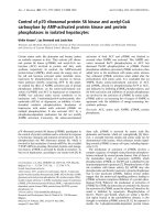
Fig. 3 Activity of AS-MPKs and Inhibition by NA-PP1. a Activity assay of wild-type and AS variants of MPK3/4/6 with MBP as substrate and [γ-32P]
ATP as cofactor. b Same as in (A) but with ATPγS as cofactor in the substrate labeling reaction. c Same as in (B) but with Bn-ATPγS as cofactor.
d Activity assay of AS-MPK3/4/6 with MBP as substrate and Bn-ATPγS as cofactor in the presence (+) or absence (-) of NA-PP1
Leissing et al. BMC Plant Biology (2016) 16:48
Page 6 of 11
A
B
C
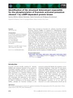
Fig. 4 In vitro Labeling Assay of Wild-type and Mutated MPK Substrates. a Thiophosphorylation assays of native (Wt) and phosphosite mutant
(SA) forms of FLAG-tagged MPK substrates AT1G78150, AT2G26530, AT3G11330 and AT4G38710 in the presence of Bn-ATPγS as cofactor and
using either AS-MPK3 or Wt-MPK3 as a negative control. b Same as in (A) but using AS-MPK6 and Wt-MPK6. c Same as in (a) but using AS-MPK4
and Wt-MPK4
Whereas AS-MPK4 and wild-type MPK4 could only
markedly thiophosphorylate MKS1, both VQ4 and MKS1
were thiophosphorylated by wild-type and mutated MPK3
and MPK6 (Fig. 6). Thus these results indicate that mutation of the gatekeeper amino acid to alanine does not influence substrate specificity of MPK3/4/6. In a final
experiment, to compare the substrate preference of ASMPK3, AS-MPK4, and AS-MPK6 using bio-orthogonal
Bn-ATPγS, we again tested VQ4 and MKS1 but also included the MPK3/6-specific substrate AT1G78150 [8, 14].
The three MPK substrates were expressed and purified
from E. coli as native His6-tagged fusion proteins with
an additional N-terminal T7 epitope for immunodetection. To evaluate the substrate preference of AS-MPK3,
AS-MPK4, and AS-MPK6 we performed in vitro labeling assays using equal amounts of the above mentioned
protein substrates. The samples were loaded on a same
gel to allow for the direct comparison of signal intensities.
Consistent with the results in Figs. 4 and 5, the unknown
protein AT1G78150 was well phosphorylated by MPK3
and MPK6 and only weakly by MPK4 (Fig. 7). Likewise,
VQ4, a specific MPK3/6 substrate protein [8] also was
only weakly phosphorylated by MPK4 (Fig. 7). In contrast,
the known MPK4 substrate MKS1 was not only phosphorylated by MPK4, as previously reported [14, 16], but is
equally well phosphorylated by MPK3/6 (Figs. 5b and 7).
This result shows that by including known substrates of
the three major plant MAP kinases as positive controls on
the same gel, the in vitro labeling reactions enable assessment of the specificity of kinase-substrate interactions.
Discussion and conclusions
Traditional in vitro kinase assays employ kinases, their
substrates and γ-32P or γ-33P-labeled ATP. Nowadays,
the radiolabeled nucleotide can be substituted by nonradioactive ATPγS resulting in thiophosphorylation of
Leissing et al. BMC Plant Biology (2016) 16:48
Page 7 of 11
A
B
Fig. 5 Substrate Preference of MPK3/4/6. a Thiophosphorylation assays of native, FLAG-tagged AT1G78150, AT2G26530, AT3G11330 and
AT4G38710 in the presence of Bn-ATPγS as cofactor and using either AS-MPK3/4/6 or Wt-MPK3/4/6 as a negative control. b Same as in (a) but
using E. coli expressed T7-tagged MKS1 as a substrate. Sample wells that were left empty are indicated by an asterisk (*)
the kinase substrate. Following alkylation with PNBM, the
thiophosphorylated substrate can be detected by western
blotting analysis and immunodetection with an anti-TPE
antibody [10]. Specific substrate labeling is achieved by engineering the kinase of interest to accept bulky ATPγS analogs that, because of steric hindrance cannot be used by
naive kinases. Until now, this approach for studying
kinase-substrate interactions in vitro has not been applied
in plant biology research. We used the approach to show
that AS-MPK3/4/6 are able to use Bn-ATPγS to
thiophosphorylate substrates, and that there is specificity
in these in vitro reactions. In addition, we validated the
phosphorylation of previously identified in vivo MPK3/6
protein substrates, and we demonstrated that these substrates are rather poor substrates for MPK4.
The substrate thiophosphorylation assay is simple, effective, and has high sensitivity. It does not need work with
hazardous material or problematic waste disposal. Another
advantage of using AS kinases and bio-orthogonal ATPanalogs is the specificity of the kinase-substrate interaction.
Leissing et al. BMC Plant Biology (2016) 16:48
Page 8 of 11
Fig. 6 Comparison of Substrate Specificity of Wild-type and AS-MPKs. Thiophosphorylation assays of T7-tagged VQ4 and MKS1 in the presence of
ATPγS as cofactor and using wild-type, AS-MPK, and KD-MPK versions of MPK3/4/6
This is of particular relevance when (i) the kinase reaction
is performed with substrate proteins of suboptimal purity,
(ii) when an additional upstream activating kinase is required for the reaction, or (iii) if the putative substrate is a
protein with kinase activity. The latter is true, for example,
for the MPK3/6-specific substrate PIRL9 (AT3G11330)
(Figs. 4 and 5) [17]. Using AS kinases and ATP analogs thus
avoids the undesired detection of such autophosphorylation
and/or phosphorylation by contaminating kinases in the reaction mixture.
Until now, AS kinases have been widely exploited when
studying cell signaling in yeast and mammals [10, 18]. In
Arabidopsis, AS-MPK4 or AS-MPK6 were used to genetically complement the mpk4 or mpk6 mutant [12, 13].
The authors mutated the gatekeeper amino acid to
glycine. The exchange leads to a specific inhibition of the
kinases in vivo upon application of NA-PP1. In the
present work, we for the first time applied AS-MPK3/4/6
in substrate labeling reactions. Since glycine lacks an
amino acid side chain, it often causes a sharp turn of the
polypeptide backbone [19] and, thus, may result in a collapse of the ATP-binding pocket and the associated loss of
kinase activity. By contrast, introducing an alanine at the
gatekeeper position not only preserved the enzyme activity
(Fig. 3), and substrate specificity (Figs. 6 and 7) of ASMPK3/4/6, but also maintained the possibility to block
their activity by binding of NA-PP1 in the enlarged active
site pocket (Fig. 3).
Our previous work identified in vivo MPK target
proteins using tandem-MOAC combined with dexamethasone-inducible expression of a constitutively active
tobacco MPK-kinase (NtMEK2DD) in Arabidopsis [8].
NtMEK2 phosphorylates and thus activates Arabidopsis
MPK3 and MPK6 [20]. However, we cannot exclude that
the activity of other MPKs is affected as well in these
plants. Here, we validated in vitro that AT2G26530,
AT1G78150, AT4G38710, and AT3G11330 are phosphorylated by MPK3/6, but are poor substrates for Arabidopsis
MPK4 (Figs. 4 and 5). Except for AT2G26530, knocking
out the phosphorylation-targeted residue by site-directed
mutagenesis of the serine or threonine amino acid within
the serine-proline or threonine-proline dipeptide motif resulted in a major decrease in the phosphorylation of these
proteins (Fig. 4). However, besides the identified phosphorylation site of AT2G26530, the protein contains eight
additional putative MPK phosphorylation sites suggesting
that MPK3/6 might target additional phosphosites in
AT2G26530 (Additional file 1: Figure S1). Together, these
findings disclose the power of our tandem-MOAC analyses and the high confidence of phosphosite localization
probability. To assess substrate preferences, we not only
assayed MPK3/4/6 phosphorylation of each substrate independently (Figs. 4 and 5), but we also directly compared
phosphorylation of several substrates by any of these
MPKs (Fig. 7). Consistent with results in other laboratories, MKS1 is a good substrate for MPK4 (Figs. 5, 6 and 7)
Fig. 7 Specificity of Substrate Phosphorylation by AS-MPK3/4/6. Thiophosphorylation assays of T7-tagged AT1G78150, VQ4 and MKS1 in the
presence of Bn-ATPγS as cofactor and using either AS-MPK3/4/6 or Wt-MPK3/4/6 as negative controls. Sample wells that were left empty are
indicated by an asterisk (*)
Leissing et al. BMC Plant Biology (2016) 16:48
[14, 21]. However, based on our data we conclude that
MKS1 also is a good substrate for MPK3/6, which contrast
recent reports by Sörensson et al. (2012) and Pecher et al.
(2014) [16, 21].
Page 9 of 11
After 30 mins, the reaction was stopped by adding SDS
loading buffer. The phosphorylation of MBP was visualized after SDS-PAGE by autoradiography. Loading of
MBP was visualized by PageBlue™ Protein Staining Solution (Thermo Scientific).
Methods
Cloning and site-directed mutagenesis
Substrate labeling reactions
Coding regions of MPK3, MPK4, and MPK6 were amplified
by PCR, ligated in frame into pGEX5x-3 vector (GE
Healthcare) and sequenced. MKK1, MKK2, MKK4, MKK5,
MKS1, VQ4 and AT1G78150.1 were cloned into pETλHIS
[22] using restriction enzymes listed in Additional file 1:
Table S1 and transformed into E. coli BL21. Coding
regions of the MPK substrates AT2G26530, AT1G78150,
AT4G38710, and AT3G11330 were amplified by PCR and
cloned into pJET1.2 (Thermo Scientific). Site-directed
mutagenesis (Additional file 1: Table S2) was performed
either by double joint PCR as described [23] or using the
QuickChangeII site-directed mutagenesis kit (Stratagene).
Substrate labeling reactions were performed as described
[10]. In brief, 100 ng recombinant active GST-MPKs
were mixed either with 3 μg MBP, 1 μg recombinant T7tagged MPK substrate, or 10 μL immunoprecipitated
FLAG-tagged substrates in kinase reaction including either 1 mM ATPγS (Sigma Aldrich) or 1 mM N6-BnATPγS (Biolog). For immunocomplex substrate labeling,
in vitro translated FLAG-tagged proteins were immunopreciptated with 40 μL EZviewTM Red ANTI-FLAG M2
affinity Gel (Sigma Aldrich) according to manufacturer’s
instructions. While still binding the FLAG-tagged substrate, the affinity gel was washed twice with kinase buffer and the resin was resuspended in 40 μL kinase
buffer. For each substrate labeling reaction 10 μL of the
bead suspension was used. The reaction was stopped by
adding 20 mM EDTA after 1 h and the thiophosphorylated substrate was alkylated with 2.5 mM PNBM
(Abcam) in 5 % (v/v) DMSO for 2 h. The alkylation reaction was stopped by adding SDS loading buffer. Samples were subjected to SDS-PAGE, transferred to a
nitrocellulose membrane (Carl Roth), and used for
immunodetection as described [24]. Primary rabbit antibodies against the thiophosphatester (α-TPE, Abcam)
were used for detection of thiophosphorylation. The
anti-phospho-p44/42 MPK (Thr202/Tyr204) antibody,
which detects doubly phosphorylated MPK3/4/6, was
from New England Biolabs. Rabbit anti-T7 (Cell Signaling Technologies) and mouse monoclonal anti-FLAG
M2 (Sigma Aldrich) epitope antibodies served as loading
control of FLAG-tagged and T7-tagged MPK substrates.
Rabbit anti-GST (Cell Signaling Technologies) antibodies were used to check equal amounts of kinase in
each reaction. Antigen-antibody complexes were detected with horseradish peroxidase-coupled anti-rabbit
or anti-mouse secondary antibodies (Cell Signaling
Technologies) followed by chemiluminescence detection
with Luminata Crecendo HRP substrate (Millipore). Using
independent protein preparations, all substrate labelling
reactions were repeated at least twice with similar results.
Protein expression and purification
For recombinant protein expression 2.5 mL of an E. coli
overnight culture was diluted in 250 mL LB medium.
The culture was grown at 37 °C to an OD600 of 0.8, supplemented with 1 mM IPTG and incubated at 28 °C for
another 3 h. Cells were harvested by centrifugation at
4000 × g and 4 °C for 15 min and stored at -80 °C until
further processing. Proteins were purified either using
GSTrap FF (GE Healthcare) columns for purification of
GST-tagged proteins or Ni2+-NTA columns (Qiagen) for
the purification of His-tagged proteins. For phosphorylation
of GST-MPKs by their respective, constitutively active
MKKDD (MKK4/5DD for MPK3/6; MKK1/2DD for MPK4),
on-column immobilized GST-MPK was incubated for 2 h
with 1 μg purified His-tagged MKKDD in kinase buffer
(50 mM Tris-HCl pH 7.5, 10 mM MgCl2, 1 mM DTT,
1 mM ATP). After an additional washing step, the
phosphorylated GST-MPK was eluted according to manufacturer’s instructions. Protein concentrations were determined with the Bradford protein assay kit (Bio-Rad) using
BSA as the standard.
In vitro transcription and translation
FLAG-tagged AT1G78150, AT2G26530, AT3G11330
and AT4G38710 were synthesized using the IN VITRO
Transcription/Translation Reagents kit following the
manufacturer’s instructions (BioSieg).
Bioinformatics
Radioactive kinase activity assay
Radioactive kinase assays were performed as described
[20]. Briefly, 100 ng recombinant active GST-MPK3, 4
or 6 were mixed with 3 μg MBP in kinase reaction buffer
(50 mM Tris-HCl pH 7.5, 10 mM MgCl2, 1 mM DTT)
with 25 μM ATP and [γ-32P]-ATP (1 μCi per reaction).
The homology model of MPK3 was generated with
HHpred 2.0 and MODELLER [25, 26] based on the Xray crystal structure of human MPK7/ERK5, PDB: 4ic7,
sequence identity of 51 % and similarity of 0.890; human
MPK12, PDB: 1 cm8, sequence identity of 41 % and
similarity of 0.818; yeast FUS3, PDB: 2b9h, sequence
Leissing et al. BMC Plant Biology (2016) 16:48
identity of 50 % and similarity of 0.922; human MPK8,
PDB: 2xrw, sequence identity of 41 % and similarity of
0.717 and human, CDK7, PDB: 1ua2, sequence identity of
41 % and similarity of 0.609. The model structure of
MPK3 was overlayed by MUSTANG [27] with the ADP
and Mg2+ bound to the FUS3 structure (PDB: 2b9h) using
YASARA structure version 14.7.17 [28] and the initial
binding mode of the Mg2+/ADP cofactor was introduced
into the apo-model structure of MPK3. Based on this initial MPK3 Mg2+/ADP bound model, the binding mode of
the bulky N6-benzyl-ATP was manually build and energy
minimized using YASARA [29]. To remove atomic clashes
and correct the covalent geometry, first a short steepest
descent minimization was performed. After removal of
conformational stress the procedure continued by simulated annealing (timestep 2 fs, atom velocities scaled down
by 0.9 every 10th step) until convergence was reached, i.e.
the energy improved by less than 0.05 kJ/mol per atom
during 200 steps. We applied the AMBER03 [30] force
field for protein residues and the general amber force field
GAFF [31] utilizing AM1BCC [32] calculated partial
charges and a force cutoff of 0.786 Å and particle mesh
Ewald [33] for exact treatment of long range electrostatics
using periodic boundary conditions. The same procedure
was performed with the active site mutation T119A.
Conclusion
Our data show that the in vitro-labeling assay represents
an effective, specific and highly sensitive test for determining kinase-substrate relationships using Arabidopsis
MPKs. By applying the analog-sensitive MPK3 and
MPK6 we confirm previously identified in vivo phosphorylation of MPK3/6 substrates and demonstrate that
these substrates are poor targets for the closely related
Arabidopsis MPK4.
Availability of data and materials
All supporting data can be found within the manuscript
and its additional files.
Highlight
We describe a novel nonradioactive in vitro labeling
assay for determining plant MPK protein substrate phosphorylation. The assay is effective, specific, and highly
sensitive for determining kinase-substrate relationships.
Additional file
Additional file 1: Figure S1. Protein sequences of selected MPK3/6
substrates. Table S1. List of primers used for cloning. Table S2. List of
primers used for site-directed mutagenesis. (DOCX 21 kb)
Competing interests
The authors declare that they have no competing interests.
Page 10 of 11
Authors’ contributions
FL did the biochemical experiments and analyses; MN and YT provided in vitro
translated proteins; MB and US guided the molecular modelling of MPK3. GB
designed the study and supervised the work; UC and GB coordinated and helped
to draft the manuscript. All authors read and approved the final manuscript.
Acknowledgements
This work was supported by a Grant-in-Aid for Scientific Research on Innovative
Areas [No. 23120520 and 25120718 to YT] from the Ministry of Education,
Culture, Sports, Science and Technology (Japan) and the German Research
Foundation (DFG) [BE4054/2-1 to GJMB and CO186/9-1 to UC]. FL is supported
by an RFwN Scholarship of RWTH Aachen University.
Author details
1
Department of Plant Physiology, Aachen Biology and Biotechnology, RWTH
Aachen University, Aachen 52056, Germany. 2Division of Biological Science,
Graduate School of Science, Nagoya University, Furo-cho, Chikusa-ku,
Nagoya, Aichi 464-8602, Japan. 3Department of Biotechnology, Aachen
Biology and Biotechnology, RWTH Aachen University, Aachen 52056,
Germany. 4The Center for Gene Research, Division of Biological Science,
Nagoya University, Furo-cho, Chikusa-ku, Nagoya, Aichi 464-8602, Japan.
Received: 27 October 2015 Accepted: 6 February 2016
References
1. Meng X, Zhang S. MAPK cascades in plant disease resistance signaling.
Annu Rev Phytopathol. 2013;51:245–66.
2. Rodriguez MCS, Petersen M, Mundy J. Mitogen-activated protein kinase
signaling in plants. Annu Rev Plant Biol. 2010;61:621–49.
3. Cargnello M, Roux PP. Activation and function of the MAPKs and their
substrates, the MAPK-activated protein kinases. Microbiol Mol Biol Rev.
2011;75:50–83.
4. Feilner T, Hultschig C, Lee J, Meyer S, Immink RG, Koenig A, et al. High
throughput identification of potential Arabidopsis mitogen-activated protein
kinases substrates. Mol Cell Proteomics. 2005;4:1558–68.
5. Popescu SC, Popescu GV, Bachan S, Zhang Z, Gerstein M, Snyder M, et al.
MAPK target networks in Arabidopsis thaliana revealed using functional
protein microarrays. Genes Dev. 2009;23:80–92.
6. Stulemeijer IJE, Stratmann JW, Joosten MHAJ. Tomato mitogen-activated
protein kinases LeMPK1, LeMPK2, and LeMPK3 are activated during the Cf4/Avr4-induced hypersensitive response and have distinct phosphorylation
specificities. Plant Physiol. 2007;144:1481–94.
7. Ahsan N, Huang Y, Tovar-Mendez A, Swatek KN, Zhang J, Miernyk JA, et al.
A versatile mass spectrometry-based method to both identify kinase clientrelationships and characterize signaling network topology. J Proteome Res.
2013;12:937–48.
8. Hoehenwarter W, Thomas M, Nukarinen E, Egelhofer V, Röhrig H,
Weckwerth W, et al. Identification of novel in vivo MAP kinase substrates in
Arabidopsis thaliana through use of tandem metal oxide affinity
chromatography. Mol Cell Proteomics. 2013;12:369–80.
9. Lassowskat I, Böttcher C, Eschen-Lippold L, Scheel D, Lee J. Sustained
mitogen-activated protein kinase activation reprograms defense
metabolism and phosphoprotein profile in Arabidopsis thaliana.
Front Plant Sci. 2014. doi:10.3389/fpls.2014.00554.
10. Allen J, Li M, Brinkworth CS, Paulson JL, Wang D, Hübner A, et al. A
semisynthetic epitope for kinase substrates. Nat Methods. 2007;4:511–6.
11. Eichberg J, Iyer S. Phosphorylation of myelin proteins: recent advances.
Neurochem Res. 1996;21:527–35.
12. Brodersen P, Petersen M, Nielsen HB, Zhu S, Newman M-A, Shokat KM, et al.
Arabidopsis MAP kinase 4 regulates salicylic acid- and jasmonic acid/
ethylene-dependent responses via EDS1 and PAD4. Plant J. 2012;47:532–46.
13. Xu J, Xie J, Yan C, Zou X, Ren D, Zhang S. A chemical genetic approach
demonstrates that MPK3/MPK6 activation and NADPH oxidase-mediated
oxidative burst are two independent signaling events in plant immunity.
Plant J. 2014;77:222–34.
14. Andreasson E, Jenkins, Brodersen P, Thorgrimsen S, Petersen NH, Zhu S, et al.
The MAP kinase substrate MKS1 is a regulator of plant defense responses.
EMBO J. 2005;24:2579–89.
Leissing et al. BMC Plant Biology (2016) 16:48
Page 11 of 11
15. Cheng Y, Zhou Y, Yang Y, Chi YJ, Zhou J, Chen JY, et al. Structural and
functional analysis of VQ motif-containing proteins in Arabidopsis as interacting
proteins of WRKY transcription factors. Plant Physiol. 2012;159:810–25.
16. Pecher P, Eschen-Lippold L, Herklotz S, Kuhle K, Naumann K, Bethke G, et al.
The Arabidopsis thaliana mitogen-activated protein kinases MPK3 and MPK6
target a subclass of ‘VQ-motif’-containing proteins to regulate immune
responses. New Phytol. 2014;203:592–606.
17. Nemoto K, Seto T, Takahashi H, Nozawa A, Seki M, Shinozaki K, et al.
Autophosphorylation profiling of Arabidopsis protein kinases using the
cell-free system. Phytochemistry. 2011;72:1136–44.
18. Lo HC, Hollingsworth NM. Using the semi-synthetic epitope system to
identify direct substrates of the meiosis-specific budding yeast kinase, Mek1.
Methods Mol Biol. 2011;745:135–49.
19. Ho BK, Brasseur R. The Ramachandran plots of glycine and pre-proline.
BMC Struct Biol. 2005. doi:10.1186/1472-6807-5-14.
20. Liu Y, Zhang S. Phosphorylation of 1-aminocyclopropane-1-carboxylic acid
synthase by MPK6, a stress-responsive mitogen-activated protein kinase,
induces ethylene biosynthesis in Arabidopsis. Plant Cell. 2004;16:3386–99.
21. Sörensson C, Lenman M, Veide-Vilg J, Schopper S, Ljungdahl T, Grøtli M, et al.
Determination of primary sequence specificity of Arabidopsis MAPKs MPK3
and MPK6 leads to identification of new substrates. Biochem J. 2012;446:271–8.
22. Groot AJ, Verheesen P, Westerlaken EJ, Gort EH, van der Groep P,
Bovenschen N, et al. Identification by phage display of single-domain
antibody fragments specific for the ODD domain in hypoxia-inducible
factor 1alpha. Lab Invest. 2006;86:345–56.
23. Ho SN, Hunt HD, Horton RM, Pullen JK, Pease LR. Site-directed mutagenesis by
overlap extension using the polymerase chain reaction. Gene. 1989;77:51–9.
24. Beckers GJM, Jaskiewicz M, Liu Y, Underwood WR, He SY, Zhang S, et al.
Mitogen-activated protein kinases 3 and 6 are required for full priming of
stress responses in Arabidopsis thaliana. Plant Cell. 2009;21:944–53.
25. Söding J, Biegert A, Lupas AN. The HHpred interactive server for protein
homology detection and structure prediction. Nucleic Acids Res. 2005;33:W244–8.
26. Sali A, Potterton L, Yuan F, van Vlijmen H, Karplus M. Evaluation of
comparative protein modelling by MODELLER. Proteins. 1995;23:318–26.
27. Konagurthu AS, Whisstock JC, Stuckey PJ, Lesk AM. MUSTANG: a multiple
structural alignment algorithm. Proteins. 2006;64:559–74.
28. Krieger E, Vriend G. YASARA view - molecular graphics for all devices - from
smartphones to workstations. Bioinformatics. 2014;30:2981–2.
29. Krieger E, Darden T, Nabuurs S, Finkelstein A, Vriend G. Making optimal use
of empirical energy functions: force field parameterization in crystal space.
Proteins. 2004;57:678–83.
30. Duan Y, Wu C, Chowdhury S, Lee MC, Xiong G, Zhang W, et al. A point-charge
force field for molecular mechanics simulations of proteins. J Comput Chem.
2003;24:1999–2012.
31. Wang J, Wolf RM, Caldwell JW, Kollman PA, Case DA. Development and
testing of a general amber force field. J Comput Chem. 2004;25:1157–74.
32. Jakalian A, Jack DB, Bayly CI. Fast, efficient generation of high-quality atomic
charges. AM1-BCC model: II. Parameterization and validation. J Comput
Chem. 2002;23:1623–41.
33. Krieger E, Nielsen JE, Spronk CA, Vriend G. Fast empirical pKa prediction by
Ewald summation. J Mol Graph Model. 2006;25:481–6.
Submit your next manuscript to BioMed Central
and we will help you at every step:
• We accept pre-submission inquiries
• Our selector tool helps you to find the most relevant journal
• We provide round the clock customer support
• Convenient online submission
• Thorough peer review
• Inclusion in PubMed and all major indexing services
• Maximum visibility for your research
Submit your manuscript at
www.biomedcentral.com/submit