Analysis of wheat microspore embryogenesis induction by transcriptome and small RNA sequencing using the highly responsive cultivar “Svilena”
Bạn đang xem bản rút gọn của tài liệu. Xem và tải ngay bản đầy đủ của tài liệu tại đây (1.83 MB, 16 trang )
Seifert et al. BMC Plant Biology (2016) 16:97
DOI 10.1186/s12870-016-0782-8
RESEARCH ARTICLE
Open Access
Analysis of wheat microspore
embryogenesis induction by transcriptome
and small RNA sequencing using the highly
responsive cultivar “Svilena”
Felix Seifert1, Sandra Bössow2, Jochen Kumlehn3, Heike Gnad2* and Stefan Scholten1,4*
Abstract
Background: Microspore embryogenesis describes a stress-induced reprogramming of immature male plant
gametophytes to develop into embryo-like structures, which can be regenerated into doubled haploid plants
after whole genome reduplication. This mechanism is of high interest for both research as well as plant breeding.
The objective of this study was to characterize transcriptional changes and regulatory relationships in early stages
of cold stress-induced wheat microspore embryogenesis by transcriptome and small RNA sequencing using a
highly responsive cultivar.
Results: Transcriptome and small RNA sequencing was performed in a staged time-course to analyze wheat
microspore embryogenesis induction. The analyzed stages were freshly harvested, untreated uninucleate
microspores and the two following stages from in vitro anther culture: directly after induction by cold-stress
treatment and microspores undergoing the first nuclear divisions. A de novo transcriptome assembly resulted
in 29,388 contigs distributing to 20,224 putative transcripts of which 9,305 are not covered by public wheat
cDNAs. Differentially expressed transcripts and small RNAs were identified for the stage transitions highlighting
various processes as well as specific genes to be involved in microspore embryogenesis induction.
Conclusion: This study establishes a comprehensive functional genomics resource for wheat microspore embryogenesis
induction and initial understanding of molecular mechanisms involved. A large set of putative transcripts presumably
specific for microspore embryogenesis induction as well as contributing processes and specific genes were identified.
The results allow for a first insight in regulatory roles of small RNAs in the reprogramming of microspores towards an
embryogenic cell fate.
Keywords: Microspore embryogenesis induction, Transcriptome, Small RNA, RNA-seq, sRNA-seq, Epigenetics, Wheat
Background
Microspore embryogenesis or androgenesis involves the
competence of the immature male gametophyte to
switch from gametophytic to embryonic developmental
cell fate through an inductive treatment prior to or at
the initiation of anther or microspore culture [1]. It is an
* Correspondence: ;
2
Saaten-Union Biotec GmbH, Am Schwabenplan 6, 06466 Seeland, OT
Gatersleben, Germany
1
Developmental Biology, Biocenter Klein Flottbek, University of Hamburg,
Ohnhorststrasse 18, 22609 Hamburg, Germany
Full list of author information is available at the end of the article
illustrative example and model for developmental plasticity and cell fate decisions in plants and an important
tool in research and plant breeding for the generation of
doubled haploid plants [2]. Double haploid technology is
widely employed in breeding programs of many crop
species for its possibility to quickly generate diverse recombinant, yet genetically fixed individuals [3]. While
bread wheat (Triticum aestivum) is one of the globally most important crops that amount for 20 % of
the human calorie consumption [4], most of its cultivars are highly recalcitrant to microspore embryogenesis. Functional genetic studies to dissect tissue
culture responses are first steps in overcoming these
© 2016 Seifert et al. Open Access This article is distributed under the terms of the Creative Commons Attribution 4.0
International License ( which permits unrestricted use, distribution, and
reproduction in any medium, provided you give appropriate credit to the original author(s) and the source, provide a link to
the Creative Commons license, and indicate if changes were made. The Creative Commons Public Domain Dedication waiver
( applies to the data made available in this article, unless otherwise stated.
Seifert et al. BMC Plant Biology (2016) 16:97
limitations to enhance bread wheat breeding eventually. Numerous microarray based gene expression
studies were conducted to elucidate the major
switches from gametophytic to embryonic development in various plants [5, 6]. These experiments
revealed large scale patterns in the reprogramming of
microspores to embryogenic tissues, which indicated a
reset of the transcriptional and translational profiles
to arrest gametophytic development [7]. Nevertheless,
those studies were limited by the particular microarray platform used, which likely did not cover all
genes specifically expressed in the reprogramming
process of microspore embryogenesis, due to a biased
microarray design to transcripts expressed primarily
in vegetative tissues. The advent of high throughput
transcriptome analysis allows for an unlimited global
analysis of expressed transcripts. Thus we performed
a transcriptome sequencing (RNA-seq) study analyzing three early stages around microspore embryogenesis induction, to elucidate transcriptomic changes of
two major transitions of embryogenesis induction
leading to first nuclear divisions. Recently, epigenetic
mechanisms were proposed to regulate the transition
from gametophytic to embryogenic cell fate [8–10].
Small non-conding RNAs (sRNAs) were shown to be
involved in the remodulation of the epigenetic landscape
and transcript levels through different mechanisms [11],
and thus are putatively potent regulators. We performed
sRNA sequencing (sRNA-seq) of the same time-series as
for RNA-seq, to allow for a comprehensive analysis of
both sRNA and transcriptome expression changes and for
the discovery of putative regulatory relationships. Our
study provides the first deep sequencing-based resource
for functional genomics research of microspore embryogenesis induction in wheat.
Page 2 of 16
Results and discussion
Development of microspores and sampling
Donor plants of the winter wheat cultivar “Svilena”,
which is highly responsive to stress-induced microspore
embryogenesis [12], were used for anther culture as
described by Rubtsova et al. (2013) [13]. Microspores
were sampled at three stages: a) freshly harvested microspores at their late, uninucleate highly vacuolated stage
(S1), b) microspores after 10 days of cold pre-treatment
exhibiting a star-like structure (S2) and c) microspores
undergoing early nuclear division (S3) based on visual
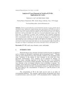
assessment (Fig. 1). These visually distinct developmental phases in microspore embryogenesis induction represent crucial stages in the acquisition of embryogenic
potential, which were elucidated in various cytological
studies [1, 7, 14]. It has been shown that microspores,
before, or immature pollen, directly after pollen mitosis
I, are most responsive for stress treatment-induced embryogenic development. The first effect after stress treatment is a rearrangement of the cytoskeleton resulting in
the re-localisation of the nucleus to the center of the
cell. The nucleus is surrounded by cytoplasmic strands
and thus a star-like structure is formed by this process,
which was suggested to be the first sign of embryogenic
induction [2, 15]. The manual sorting procedure that we
applied for RNA-seq facilitates a very high homogeneity
and thus a stage-specific analysis of the pooled microspores as well as an exclusion of injured or dead cells.
Due to higher RNA amount requirements for sRNAseq, a gradient centrifugation-based isolation was performed, which delivers the required cell numbers at
the cost of slightly reduced population homogeneity.
To control for batch-to-batch variations, donor material for all microspore isolations for RNA-seq and
sRNA-seq were cultured until plant regeneration. In
Fig. 1 Microspore development stages sampled for RNA sequencing analysis. Brightfield micrographs of representative samples from three
microspore stages. All bars represent 20 μm. Arrowheads point to cells with morphological characteristics that meet our criteria for manual cell
selection. a Untreated vacuolated microspores at uninucleate stage (S1); manually selected microspores were characterized by a large central
vacuole and a clear cytoplasm. b Microspores with star-like structure after 10-days cold stress pre-treatment (S2); microspores are slightly enlarged
after stress induction, the vegetative nucleus migrates into the center of the cell, the cytoplasm becomes structured and shows cytoplasmic strands,
the so-called “star-like structure”. c Microspores undergoing first nuclear division (S3); the vegetative nucleus is centrally located and has divided
Seifert et al. BMC Plant Biology (2016) 16:97
Page 3 of 16
either case the high regeneration frequency was
equivalent to the usually observed response for the
cultivar “Svilena”.
Transcriptome sequencing
RNA-seq allows an unrestricted and global analysis of
gene expression as well as the identification of unknown
transcripts. To facilitate a comprehensive overview of
gene expression through acquisition of embryogenic
potential of microspores, we sequenced the samples in
biological triplicates for each stage. All libraries were
indexed with unique nucleic acid identifiers and 50 bp
single end reads were sequenced on an Illumina HiSeq
2000 sequencer. In total, 608,233,335 clean RNA-seq
reads were generated, with individual libraries covering
55.6 Mio. to 75.6 Mio. reads (see Table 1).
De novo transcriptome assembly and annotation
A de novo transcriptome assembly using the Trinity de
novo assembler [16] was performed based on the RNAseq reads of all stages and replicates and resulted in
29,388 contigs with an average length of 417.87 bp. The
size distribution of the contigs is shown in Fig. 2a. Our
approach allows for an expression comparison as well as
a functional annotation for the identification of important gene functions in microspore embryogenesis induction. We did not pursue resolving the homeologs or
isoforms, this would have required a higher sequencing
depth as well as longer and paired end reads. A BLASTx
mapping resulted in 18,344 (62.42 %) contigs with homology to protein sequences in the NCBI nr database.
The majority of contigs exhibits the highest sequence
homology with Aegilops tauschii and Triticum urartu,
known to be the diploid progenitors for the wheat A and
D genome, respectively [17], followed by other grass species (Fig. 2b). This indicates wheat specific sequencing
results without contamination and an effective de novo
Table 1 Summary of RNA-seq/sRNA-seq data
RNA-seq data
Sample Trimmed
replicate reads
sRNA-seq data (18 to 28-nt)
Uniquely mapping Trimmed
reads to de novo
reads
transcriptome [%]
Distinct reads
assembly resulting in high homology to known monocot
transcripts.
We annotated the contigs by assigning gene ontology
(GO) terms via Blast2GO [18] and Trinotate [19]. This annotation resulted in 13,553 (46.12 %) contigs with a
homology-based annotation, with on average 7.41 GO
terms per contig. Mapping of the contigs to known wheat
cDNA sequences (ensembl release 26) [20] resulted in
10,919 cDNAs covered by contigs from the RNA-seq de
novo assembly. A subset of 5,139 wheat cDNAs were covered by multiple contigs (on average 2.78 contigs per
cDNA) most likely due to fragmented assembly of the
short reads. The restructuring of the contigs to transcripts
based on wheat cDNA sequences revealed 20,224 transcripts covered by our de novo assembly. The contig
assignment to transcripts is listed in Additional file 1:
Table S1. This restructured assembly contains 9,305 new
transcripts not covered by known wheat cDNAs from
ensembl release 26 [20], presumably because the specific
cell-types, developmental stages and induction conditions
used in the present study were not covered by previous
sequencing efforts. Our dataset thus provides a valuable
resource for the analysis of microspore embryogenesis.
3,206 (32.21 %) of the new transcripts could be annotated
by BLASTx mapping. The top hits from BLASTx for contigs attributed to restructured transcripts are shown in
Additional file 2: Table S2. After the restructuring of contigs to transcripts, a GO annotation could be derived for
8,527 (42.16 %) of all transcripts including 996 transcripts
not covered by wheat cDNAs (Additional file 3: Table S3).
The GO annotation resulted in a large number of transcripts with biological processes related to response to
stress and abiotic stimulus, which is most likely caused by
the cold-stress treatment for microspore induction. Other
main biological processes covered are cellular component
organization, post-embryonic development, cell cycle, cell
differentiation, embryo development and epigenetic regulation of gene expression (Table 2), which might be related
to the developmental shift from gametophytic to embryogenic cell fate. The complete list of GO terms for all
categories is shown in Additional file 4: Table S4.
Expression analysis
S1a
70,216,602 39.442
10,425,301
1,813,927
S1b
75,697,405 38.307
10,247,910
2,332,967
S1c
55,646,245 34.298
9,892,496
2,983,585
S2a
66,434,325 42.742
10,707,291
3,092,207
S2b
66,122,827 43.012
9,718,833
2,572,135
S2c
73,711,917 42.240
10,154,174
3,174,185
S3a
71,321,690 44.759
10,789,357
2,908,387
S3b
70,400,647 50.624
10,664,717
3,063,376
S3c
58,681,677 40.775
9,939,297
4,006,577
The expression levels of all transcripts were estimated
based on uniquely mapping reads to the de novo assembled transcriptome (see Table 1). To allow for a comparison of replicates and stages the expression values
were quantile normalized and scaled to one million
quantile normalized reads per library (rpmqn). Correlation based clustering revealed that the expression
values between the replicates exhibited a high similarity
for each of the three specific stages and a clear separation from the other two stages (Fig. 2c). This clearly
indicates that the manually sorted cells represent
Seifert et al. BMC Plant Biology (2016) 16:97
Page 4 of 16
Fig. 2 Results from RNA-seq transcriptome assembly and expression analysis. a Size distribution of contigs assembled from RNA-seq reads of all
replicates of the three microspore stages using the Trinity assembler. b Species distribution for BLASTx top hits of RNA-seq assembled contigs
against the NCBI nr database. c Correlation-based clustering analysis for RNA-seq transcript expression values between the replicates of all
microspore stages
uniform samples of developmentally distinct stages.
Additionally we observed a much higher overall similarity between the transcriptomes of the stages S1 and S2
than between the first two stages and S3 (Fig. 2c). This
result suggests, that the stress treatment causes few but
drastic changes that direct to a large-scale reprogramming in the following transition.
For the analysis of stage specific transcription, we
regarded transcripts with an expression of at least 1
rpmqn in all three replicates of at least one of the three
stages as expressed. These thresholds revealed 14,792
(73.14 %) transcripts to be expressed in S1, an increase
to 15,026 (74.3 %) expressed transcripts in S2 followed
by a decrease to 13,927 (68.86 %) expressed transcripts
in S3, respectively. The overlap of transcripts exclusively
expressed in the stages S1 and S2 is 2,439 (12.06 %)
transcripts, but only 455 (2.25 %) transcripts were exclusively expressed in the stages S2 and S3 (see Fig. 3). A
core set of 11,765 (58.17 %) transcripts was expressed in
all three stages. The differing sets of expressed transcripts reflect the change of developmental fate in the
transcriptome. Microspores that eventually develop into
embryos have been shown to undergo a step of dedifferentiation first, which is completed at the stage exhibiting
a star-like structure [7]. We found 24, 7, and 666 transcripts to be exclusively expressed in S1, S2, and S3,
respectively (see Fig. 3). The transcripts along with their
BLASTx top hits are listed in Additional file 5: Table S5.
Interestingly, transcripts exclusively expressed in S3
cover transcripts which are known to be involved in
acquisition of embryogenic cell fate, like transcript_14378
and transcript_18369 with similarity to RWP-RK DOMAIN
CONTAINING 1 (RKD1), a transcription factor involved in
female gametogenesis and early embryogenesis identified
from isolated wheat egg cells [21]. transcript_7306 with
similarity to AINTEGUMENTA-like 5 (AIL5), an AP2like ethylene-responsive transcription factor, which is
a homolog to BABY BOOM (BBM) and known to
confer embryonic identity to cells [22]. transcript_11677
exhibits similarity to HIGH-LEVEL EXPRESSION OF
SUGAR-INDUCABLE GENE2-LIKE1 (HSL1), which was
shown to be specifically and highly expressed in early
embryogenesis. Its interaction with the HISTONE DEACETYLASE 19 (HDA19) results in epigenetic repression
of seed maturation genes [23]. Another epigenetic component, exclusively expressed in S3, is transcript_12642 with
similarity to SHOOTLESS2 (SHL2), an orthologue of the
Arabidopsis RNA-dependent RNA polymerase 6, which
was shown to be involved in shoot apical meristem formation during embryogenesis [24]. Additionally, the specific
expression of transcript_13594 and transcript_20002 in
S3, both with homology to the DNA (cytosine-5)-methyltransferase 1A (MET1a), is in agreement with reported
DNA methylation dynamics and MET1a-like gene expression changes during stress-induced microspore reprogramming [25]. Overall, the large number of transcripts
with homologies to known embryogenesis related genes
suggests that we have identified many more not yet uncovered genes related to wheat microspore embryogenesis
induction.
Analysis of differentially expressed transcripts
The transitions between the stages S1 and S2 (in the
following denoted as T1) as well as between S2 and S3
(named T2) represent pivotal steps in induction and
reprogramming from gametophytic fate of the microspore
into embryo formation [7]. The differential expression
(DE) of transcripts was determined for all transcripts with
Seifert et al. BMC Plant Biology (2016) 16:97
Page 5 of 16
Table 2 Number of transcripts covered by GO terms of GO
category biological process (n > =100)
GO term
GO description
Number of
transcripts
GO:0009987 cellular process
3037
GO:0009058 biosynthetic process
1457
GO:0006950 response to stress
1373
GO:0016043 cellular component organization
1364
GO:0006810 transport
1232
GO:0009056 catabolic process
1197
GO:0008152 metabolic process
1147
GO:0006139 nucleobase-containing compound
metabolic process
1094
GO:0006464 cellular protein modification process
1094
GO:0009628 response to abiotic stimulus
913
GO:0005975 carbohydrate metabolic process
763
GO:0008150 biological process
757
GO:0006350 transcription, DNA-templated
701
GO:0007275 multicellular organismal development
699
GO:0019538 protein metabolic process
676
GO:0006259 DNA metabolic process
587
GO:0009791 post-embryonic development
578
GO:0000003 reproduction
541
GO:0006629 lipid metabolic process
517
GO:0007165 signal transduction
512
GO:0007049 cell cycle
498
GO:0009653 anatomical structure morphogenesis
498
GO:0009607 response to biotic stimulus
482
GO:0006412 translation
427
GO:0006519 cellular amino acid metabolic process
376
GO:0009719 response to endogenous stimulus
369
GO:0030154 cell differentiation
356
GO:0009908 flower development
329
GO:0009790 embryo development
302
GO:0006091 generation of precursor metabolites and energy 277
GO:0040029 regulation of gene expression, epigenetic
256
GO:0016049 cell growth
224
GO:0019748 secondary metabolic process
209
GO:0006355 regulation of transcription, DNA-templated
183
GO:0055114 oxidation-reduction process
180
GO:0006351 transcription, DNA-templated
170
GO:0006468 protein phosphorylation
149
GO:0006886 intracellular protein transport
118
GO:0009605 response to external stimulus
112
GO:0008219 cell death
109
GO:0055085 transmembrane transport
107
GO:0006457 protein folding
100
at least 2 reads per million quantile normalized reads
(rpmqn) in the higher expressed stage, and a two-fold
expression change in the transition between the respective
stages. The expression analysis resulted in 756 DE
transcripts for the first transition (T1) and 5,629 DE transcripts for T2 (Additional file 6: Table S6). In both transitions the majority of transcripts is downregulated, 66.67 %
in T1 and 56.96 % in T2. 301 (39.81 %) of the DE transcripts after the cold-stress treatment in T1 exhibit also
DE in T2. The proportion of the number of up- and
downregulated transcripts in T1 resembles a previous
microarray-based study for the effect of mannitoltreatment on microspore embryogenesis in barley [26].
The correlation-based cluster analysis of the expression stage specific expression values (Fig. 2c) suggested
more differences in gene expression in T2 than in T1.
These results were supported by a principal component
analysis (PCA) for all DE transcripts in at least one stage
transition, which resulted in a clear separation of the
first two microspore stages S1 and S2 from the later
stage S3, explaining 72.45 % of the variance (Additional
file 7: Figure S1). The similarity of S1 and S2 in comparison to S3 in the PCA highlights that this separation pattern is not a result from higher expression variation
between the replicates that could have been potentially
caused by the manual sampling of the microspores, but
differential expression of specific sets of transcripts.
A k-means cluster analysis for all DE transcripts was
performed to uncover expression switches throughout
the two stage transitions (see Fig. 4). In agreement with
the expression comparison (Fig. 2c) as well as with the
results from the PCA the clustering resulted predominantly in two major expression pattern clusters, with
basically either up (cluster 1, 9 and 12; see Fig. 4a, Fig. 4i
and Fig. 4l) or down (cluster 3 and 5; see Fig. 4c and
Fig. 4e) regulation of expression between the microspore
stages S2 and S3. Another expression pattern is up-/
downregulation specifically after the stress treatment in
T2 with reversion of the expression pattern towards T3
given for clusters 4, 6 and 7 (see Fig. 4d, Fig. 4f and
Fig. 4g). Interestingly only clusters exhibiting a steady
decrease (cluster 10 and 11; see Fig. 4j and Fig. 4k) but
none for steady increase of gene expression could be
observed. Changes in gene expression either up or down
in T1 is given only for a smaller number of transcripts
(cluster 2 and 8; see Fig. 4b and Fig. 4h).
The clusters were inspected for known regulatory
transcripts, which signify the transition from the gametophytic to the embryonic developmental program.
Strikingly, cluster 1 contains a transcript with homology to the embryogenesis related transcription
factor BABY BOOM 2 (BBM2, transcript_4758). Interestingly, the major clusters 1 and 3 both contain
various transcripts with homology to epigenetic
Seifert et al. BMC Plant Biology (2016) 16:97
Page 6 of 16
Table 3 Overrepresented biological processes of transcript expression clusters
Cluster
GO term
GO term description
Number of transcripts
Enrichment p-value
1
GO:0006259
DNA metabolic process
90
<10-6
1
GO:0007049
cell cycle
78
<10-6
1
GO:0007018
microtubule-based movement
12
3 · 10-6
1
GO:0007067
mitotic nuclear division
12
3 · 10-6
1
GO:0040029
regulation of gene expression, epigenetic
44
1.1 · 10-5
1
GO:0043531
ADP binding
16
1.6 · 10-5
1
GO:0006275
regulation of DNA replication
12
7.9 · 10-5
1
GO:0003677
DNA binding
111
9.6 · 10-5
1
GO:0004803
transposase activity
8
1.86 · 10-4
1
GO:0006260
DNA replication
15
2.86 · 10-4
1
GO:0003774
motor activity
12
3.81 · 10-4
1
GO:0006418
tRNA aminoacylation for protein translation
10
7.99 · 10-4
1
GO:0008017
microtubule binding
6
9.88 · 10-4
1
GO:0006313
transposition, DNA-mediated
7
1.01 · 10-3
1
GO:0007131
reciprocal meiotic recombination
9
1.21 · 10-3
1
GO:0000911
cytokinesis by cell plate formation
11
1.69 · 10-3
1
GO:0006281
DNA repair
18
2.16 · 10-3
1
GO:0010332
response to gamma radiation
7
2.95 · 10-3
1
GO:0006261
DNA-dependent DNA replication
7
3.88 · 10-3
1
GO:0009909
regulation of flower development
13
4.31 · 10-3
1
GO:0003676
intracellular protein transport
77
5.49 · 10-3
1
GO:0006886
intercellular protein transport
19
6.59 · 10-3
1
GO:0016043
cellular component organization
140
8.09 · 10-3
5
GO:0009987
cellular process
205
5.18 · 10-4
5
GO:0019538
protein metabolic process
59
5.25 · 10-4
5
GO:0051603
proteolysis involved in cellular protein catabolic process
5
1.3 · 10-3
5
GO:0005839
proteasome core complex
5
1.75 · 10-3
5
GO:0006810
transport
91
3.29 · 10-3
9
GO:0009220
pyrimidine ribonucleotide biosynthetic process
7
1.1 · 10-4
9
GO:0003735
structural constituent of ribosome
12
3.43 · 10-4
9
GO:0006412
translation
25
3.16 · 10-3
9
GO:0006094
gluconeogenesis
5
3.68 · 10-3
9
GO:0009560
embryo sac egg cell differentiation
5
5.98 · 10-3
10
GO:0009058
biosynthetic process
85
<10-6
10
GO:0006629
lipid metabolic process
42
<10-6
10
GO:0009987
cellular process
138
4.7 · 10-5
10
GO:0009056
catabolic process
65
1.17 · 10-4
10
GO:0019748
secondary metabolic process
19
1.37 · 10-4
10
GO:0008152
metabolic process
62
2.1 · 10-4
10
GO:0005975
carbohydrate metabolic process
44
5.72 · 10-4
10
GO:0019538
protein metabolic process
39
1.17 · 10-3
10
GO:0006091
generation of precursor metabolites and energy
19
3.9 · 10-3
10
GO:0006519
cellular amino acid metabolic process
23
6.15 · 10-3
11
GO:0009058
biosynthetic process
60
<10-6
Seifert et al. BMC Plant Biology (2016) 16:97
Page 7 of 16
Table 3 Overrepresented biological processes of transcript expression clusters (Continued)
11
GO:0008152
metabolic process
45
<10-6
11
GO:0006629
lipid metabolic process
35
<10-6
11
GO:0005975
carbohydrate metabolic process
35
<10-6
11
GO:0006091
generation of precursor metabolites and energy
21
<10-6
11
GO:0019748
secondary metabolic process
17
<10-6
11
GO:0009056
catabolic process
42
1.8 · 10-5
11
GO:0005488
binding
51
1.09 · 10-4
11
GO:0006096
glycolytic process
7
2 · 10-4
11
GO:0009987
cellular process
77
2.75 · 10-4
11
GO:0006519
cellular amino acid metabolic process
17
5.5 · 10-4
11
GO:0015979
photosynthesis
7
6.02 · 10-4
11
GO:0055114
oxidation-reduction process
10
1.86 · 10-3
11
GO:0009628
response to abiotic stimulus
29
2.28 · 10-3
11
GO:0006950
response to stress
39
2.8 · 10-3
components such as the Argonaute genes AGO4
(transcript_3992), AGO5 (transcript_1301) and AGO6
(transcript_2354), the dicer-like gene DCL3 (transcript_378), a large number of chromatin remodelling
factors, such as various DNA-methyltransferases
(DDM1, transcript_1568; DRM2, transcript_1831; ME
T1, transcript_3460; CMT3, transcript_6805), histone
methyltransferases (NSD3, transcript_633; SUVH1,
transcript_1921; SUVR5, transcript_1884) as well as
the histone deacetylase (HD2A, transcript_5605) in
cluster 1. The opposing cluster 3 contains DCL1 (transcript_3100), the DNA-methyltransferase (DRM1, transcript_5286), as well as various histone deacetylases
(HDA6, transcript_4537; HDA19, transcript_2818). The
histone deacetylases HDA6 and HDA19 have been shown
to be suppressors of embryonic properties [27] and thus
were rightly found in cluster 3. Likewise, changes in histone methylation and acetylation are associated with cell
totipotency during microspore reprogramming to embryogenesis [9]. In agreement with other studies on androgenesis in various species [8–10] the large number
of epigenetic components we found to be differentially expressed between the stages highlights their
importance in the reprogramming of immature microspores to embryogenic cell fate.
Interestingly, homologues of previously discussed
embryogenesis-marker genes are covered by the de novo
assembled transcripts, such as SOMATIC EMBRYOGENESIS RELATED KINASE 1 (SERK1) [7] or LATE
EMBRYO ABUNDANT (LEA) [28]. Unexpectedly, we
found SERK1 with highest expression in fresh microspores and the expression level decreases through both
transitions. This is in agreement with the finding that
SERK1 was essential for male gametophyte production
[29] and indicates that its expression pattern is not
exclusively attributed to embryogenic reprogramming.
We found LEA to be expressed at low levels in all three
stages without any significant changes in expression
levels neither after induction-treatment (T1) nor towards
induced embryogenesis (T2). Thus the transcription profiles of these known embryogenesis-marker genes do not
indicate their involvement in the reprogramming of
wheat microspores.
GO enrichment analysis
Fig. 3 Overlap of expressed transcripts in the three analyzed stages
To further functionally characterize the stage transitions
and expression clusters we performed a GO enrichment
analysis for DE transcripts. The full results are listed in
Additional file 8: Table S7 and Additional file 9: Table S8,
Seifert et al. BMC Plant Biology (2016) 16:97
Page 8 of 16
Fig. 4 Clustering of DE transcript expression profiles. Representation of DE transcript expression profiles derived from k-means clustering of expression
z-scores. The red line shows average expression z-scores to visualize the dominant expression trend of the cluster. a cluster 1, b cluster 2, c cluster 3,
d cluster 4, e cluster 5, f cluster 6, g cluster 7, h cluster 8, i cluster 9, j cluster 10, k cluster 11, l cluster 12. The number of transcripts (n) is given for
each cluster
for the transitions and the expression pattern clusters, respectively. Additionally, major enrichments of the expression clusters are shown in Table 3. In T1, GO terms were
only found to be enriched for downregulated transcripts,
namely, amongst others, “carbohydrate metabolic
process”, “vacuole”, and “response to stress”, which all
likely represent the dedifferentiation of the microspores
due to the inductive treatment. A large set of GO terms
overlaps among the downregulated transcripts in both
transitions such as “generation of precursor metabolites”
and “energy”, “lipid metabolic process”, “metabolic
process”, “catabolic process”, “biosynthetic process”, and
“response to abiotic stimulus”, which presumably represent
sustained dedifferentiation from the microgametophytic
pathway. The set of GO terms for upregulated transcripts
in T2 contains the general terms protein binding, DNA
binding and nucleic acid binding, most likely reflecting initiation of embryogenic transcription and protein machinery. Likewise, the GO terms “cell cycle”, “cellular
component organization” as well as numerous microtubule
and mitosis related terms, “histone H3K9 methylation”,
“DNA methylation” and “histone phosphorylation” were
enriched among upregulated transcripts in T2. The indicated downregulation of metabolic and biosynthetic processes in both transitions with concurrent upregulation of
chromatin modifications and organization of cellular components as well as the cell cycle in T2 is in agreement with
a cell cycle arrest, which was suggested to be required for
the reprogramming to embryogenic fate before the cell
cycle is again released [1]. The GO term “H3K9 methylation” for upregulated transcripts in T2 is in accord with
the finding of increased H3K9 methylation in embryo-like
structures as compared to microspores [9].
Cluster 1 exhibits an enrichment for various GO-terms
reflecting karyokinesis, the microspores are undergoing in
T2, such as “microtubule binding”, “cytokinesis by cell
plate formation”, “DNA-dependent DNA replication” and
“cytoskeleton”. The stress-induced rearrangement of the
cytoskeleton followed by a symmetric division of the
microspore has been described in various studies as initial
Seifert et al. BMC Plant Biology (2016) 16:97
steps towards microspore embryogenesis (see review [2]).
Although cluster 3 is the second largest cluster, it is only
enriched for the single GO-term “endoplasmic reticulum”.
That there are no other terms enriched, might reflect that
the downregulation of transcript expression in T2 covers a
multitude of functions and processes. In contrast, cluster
5 with a similar expression pattern but continuous downregulation in T1 and T2 has an enrichment for numerous
GO-terms for protein related processes, such as “protein
metabolic process”, “proteolysis involved in cellular processes”, “Golgi apparatus” and “proteasome core complex”, which might reflect the previously described
degradation of gametophytic cell fate-related proteins to
allow for a reprogramming towards embryogenesis. Especially, the enrichment for “Golgi apparatus” might resemble findings in Brassica napus where autophagy and
cytoplasmic cleaning by excretion was found to be unique
to microspores undergoing reprogramming to an embryogenic fate: In contrast to non-responding microspores,
freshly isolated microspores at the vacuolated stage, which
were optimal for induction, exhibit Golgi-stacks [30].
Surprisingly, the corresponding genes show equal expression levels in untreated isolated microspores and after the
stress-treatment. This might indicate an early transcriptional stress response to the mannitol buffer and would fit
to a similar observation by Marashin et al. [3]. Cluster 9 is
enriched for GO-terms related to transcription and translation: “structural constituent of ribosome”, “ribosome”,
“DNA binding”, “translation” and “DNA-directed RNA
polymerase activity” and is likely related to the establishment of an embryogenic program. Interestingly, cluster 9
exhibits also enrichment for the GO-term “embryo sac
egg cell differentiation”, which might be indicative for the
reprogramming of the microgametophytic pathway.
Although cluster 10 and 11 represent progressive downregulation of transcripts and both cover only a relative
small amount of DE transcripts, they exhibit enrichments
for a large number of GO-terms related to catalytic activity and various metabolic processes, which might relate to
downregulation of microgametophytic pathways. The additional enrichment for various stress-related terms, such
as “oxidation-reduction process”, “response to abiotic
stimulus”, “response to stress” and interestingly “embryo
development” in cluster 11 was unexpected, since it has
been shown, that the anther pre-treatment activates plant
defense gene expression in response to mannitol solution
and cold stress treatment [31]. Considering the decreasing
expression levels with initiated embryogenesis the latter
GO term most likely represents suppressors of embryo
development.
sRNA sequencing results
The sRNA-seq resulted in 92.54 Mio. clean reads, with
9.71 Mio. to 10.79 Mio. reads per library (see Table 1).
Page 9 of 16
In total 19.63 Mio. distinct sequences were obtained,
with 1.8 Mio. to 4 Mio. distinct sequences per library
(see Table 1). The sRNA length distribution exhibits a
peak at 24-nt for all replicates of all three stages, representing the most abundant short interfering RNAs
(siRNA). However, two of the three replicates from S1
showed a smaller fraction of 24-nt sRNAs (Fig. 5a). The
length distribution of distinct sRNA reads exhibited an
additional peak for 21-nt sRNAs (Fig. 5b), a fraction of
which most likely represents microRNAs (miRNA). The
fraction of 24-nt sRNAs exhibited a higher variability
than given for the total sRNA length distribution in
contrast to the sRNA lengths from 15-nt to 20-nt as
well as 25-nt to 28-nt, which, except of 20-nt sRNAs,
are not representing known functionally active sRNA
classes [32].
sRNA expression analysis
Distinct sRNAs were defined as expressed if their expression was equal or higher than 1 rpmqn, this criterion was satisfied for 63,880 to 70,478 sRNAs per library.
A comparison of expression values between all replicates
of the three stages revealed a high similarity for the three
replicates of each stage (Fig. 5c), again reflecting the
overall uniformity of the biological replicates beside the
differences in abundance of RNAs of specific length.
The variance between replicates for sRNAs is higher
than for transcripts. A possible explanation provides the
less specific generation of siRNAs from various genomic
loci in contrast to the defined gene loci of transcripts. In
contrast to the transcript expression, the correlation
between the replicates revealed drastic expression
changes from S1 to S2 as well as S2 to S3, as the replicates of S2 are less correlated to S1 and S3 than S1 and
S3 to each other. This drastic difference to transcript expression pattern might be explainable by stress-induced
activation of transposons resulting in the generation of
new sets of siRNAs and delayed effects on gene expression by de novo methylation of target TEs [33, 34].
Furthermore this difference might be attributed to the
different isolation procedures of microspores for mRNA
and sRNA sequencing: In contrast to individual selection
of microspores with specific morphology for mRNA
sequencing (Fig. 1), the gradient centrifugation, we used
to isolate microspores for sRNA sequencing, enrich for
living microspores only and thus more likely include
sRNAs from microspores undergoing cell fates other
than embryogenesis.
DE sRNAs were determined from all sRNAs with an
expression level of at least 2 rpmqn in the higher
expressed stage, a minimum of two-fold expression
change. The expression analysis with these thresholds
resulted in 867 DE sRNAs for T1, with 830 (95.73 %)
being upregulated and 37 (4.27 %) being downregulated
Seifert et al. BMC Plant Biology (2016) 16:97
Page 10 of 16
Fig. 5 Results from sRNA-seq expression analysis. a Total sRNA read length distribution for all replicates, b Distinct sRNA read length distribution
for all stage replicates, c Correlation based clustering analysis for sRNA-seq expression values between the replicates of all microspore stages.
d Length distribution of DE sRNAs in the first transition (T1 between stages S1 and S2, downregulated n = 37, upregulated n = 830), e Length
distribution of DE sRNAs in the second transition (T2 between stages S2 and S3, downregulated n = 4,240, upregulated n = 8,868) f Length distribution
of sRNAs negatively correlated with predicted target transcripts with DE pattern (T1 n = 5, T2 n = 243)
(Additional file 10: Table S9, Additional file 11: Table S10).
The upregulated sRNAs account primarily to 24-nt
sRNAs while the downregulated sRNAs are scattered from
19-nt to 23-nt (Fig. 5d). For T2 13,108 DE sRNAs were
identified in total, with 8,868 (67.65 %) being upregulated
and 4,240 (32.35 %) being downregulated (Additional file
10: Table S9, Additional file 11: Table S10). In T2, 24-nt
sRNAs accounted for the majority of up as well as downregulated sRNAs. Furthermore, the downregulated sRNAs
exhibited a high fraction of 21-nt sRNAs (Fig. 5e). 304 of
the DE sRNAs overlap between T1 and T2 that all are of
24-nt length.
In the various developmental stages analyzed, 66 of 119
known mature miRNAs of wheat listed in miRBase release
21 [35] were found to be expressed. Three of these (taemiR9669-5p, tae-miR397-5p, and tae-miR9658-3p) showed
upregulation, whereas one (tae-miR9672b) showed downregulation in T2. Consistent with a putative role in microspore embryogenesis, which involve the generation of
undifferentiated multicellular structures at first, miR397
was shown to be highly expressed in undifferentiated but
not in differentiated rice embryogenic calli from somatic
tissues [36]. Interestingly, miR397 is upregulated under
cold conditions [37] and overexpression resulted in higher
cold stress tolerance in Arabidopsis [38]. In wheat microspores, cold inducibility of miR397 might be reduced or
delayed, since we revealed no upregulation in S2 right after
the cold stress treatment but in the later stage S3. Another
miRNA, which might be involved in the regulation of
androgenesis, is tae-miR9658, since it was shown to be
highly expressed in developing grains but less abundant in
vegetative tissues [39].
Prediction of sRNA target transcripts
To identify potential regulatory effects of sRNAs on
mRNAs we predicted sRNA targets among the assembled transcripts for all DE sRNAs. The target prediction
resulted in 97 putative target transcripts for DE sRNAs
in T1 and 1,179 putative sRNA target transcripts in T2.
Five sRNA/target pairings in T1 exhibited DE transcripts
and a strong negative correlation between sRNA and
target expression. All these targets were downregulated
from S1 to S2. For T2, we found 251 DE target transcripts of which 133 exhibit a strong negative correlation
Seifert et al. BMC Plant Biology (2016) 16:97
with the targeting sRNA expression values, 86 of these
targets were up and 47 were downregulated from S2 to
S3 (Additional file 12: Table S11). No DE targets could
be identified among the assembled contigs for any of the
4 DE known miRNAs listed in miRBase release 21 [35].
Although we used different microspore isolation procedures for mRNA and sRNA sequencing, both datasets
are highly related by identical tissue culture conditions
and staging. Thus, a negative correlation between sRNA
and targeted transcript expression should support the
involvement of the sRNA in regulation of the target
transcript either by post-transcriptional gene silencing
(PTGS) or transcriptional gene silencing (TGS) [40].
The targeted transcripts identified include a number of
genes indicated to be involved in embryogenesis or even
androgenesis, such as HDA19 (transcript_2818), as
already discussed in the context of the DE transcripts
analysis, which is downregulated in T2. HDA19 has been
shown to contribute to the repression of embryogenesisrelated genes after germination [27]. It has been furthermore shown that histone deacetylase inhibitors promote
totipotency to microspores [41]. Chromatin modifications are suggested to be required for the accessibility of
embryogenesis-related genes [9]. Another interesting
gene is PROLIFERA (PRL), represented by transcript_9383, which is upregulated in T2. PRL has been
shown to be involved in megaspore and embryo development but not in the developing microgametophyte
[42] and thus might represent the deviation from the
gametophytic cell fate. TaCer1 (transcript_178), which is
highly increased in microspores after stress treatment
(S2) and is 528-fold downregulated in T2, represents
ECERIFERUM1 a putative decarbonylase involved in
cuticular wax biosynthesis from very long chain fatty
acids (VLCFA), which can be regulated by various
stresses [43]. It was suggested that Cer1 is involved in
stress tolerance by modulating the aldehydes/alkanes
ratio of cuticular wax in wheat as well as other plants
[43–46]. A previous study suggested the involvement of
wax biosynthesis from VLCFA in wheat microspore embryogenesis as a pathway resulting in signal molecules
leading to controlled cell division in absence of surrounding tissue [47]. Overall, only 48 of 666 DE target
genes potentially regulated by sRNAs obtained an annotation. We expect the remaining set of transcripts to
contain important regulatory genes involved in the alteration of cell fate towards embryogenic development, not
yet discovered by other studies.
Prediction of miRNA precursors
To differentiate between potential PTGS and TGS, miRNA
precursors on wheat genomic sequences were predicted.
The prediction of miRNA precursors (pre-miRNA) revealed in total 1,717 distinct candidates (Additional file 13:
Page 11 of 16
Table S12). 39 pre-miRNAs candidates showed homology
to known pre-miRNAs (Table 4). The comparison of the
predicted mature miRNAs with the identified sRNA/target
pairs revealed two putative miRNAs, namely sRNA
1167446 potentially targeting transcript_1777 with high
homology to the Aegilops tauschii predicted protein
F775_10365 of unknown function and sRNA 6202839 targeting transcript_5689 with high homology to the wheat
cDNA TRAES_3AS_647411E39.2 of unknown function.
These results with nearly no consensus between predicted
mature miRNAs and the sRNAs with predicted targets
suggests that miRNA-mediated post-transcriptional regulation does not explain the majority of the predicted sRNA/
target pairs with high negative correlation. In the light of
the high number of chromatin modifiers identified to be
expressed, it is assumed that different sRNA-guided TGS
mechanisms such as chromatin modifications and DNA
methylation are involved in the induction of microspore
embryogenesis in wheat. This assumption is supported by
the high variability of the number of distinct 24-nt sRNAs
between stages (Fig. 5b), as well as the high fraction of DE
24-nt sRNAs negatively correlated with their potential target transcript expression (Fig. 5f), as 24-nt sRNAs were
shown to be predominantly involved in TGS [32].
Conclusions
Our study provides the first large-scale transcriptome
dataset for microspore embryogenesis induction in
wheat. The de novo assembly and mapping to public
wheat cDNA sequences resulted in a high number of
novel transcripts. A major part of these might be presumably largely specific transcripts for microspore embryogenesis induction. A GO annotation revealed a large
fraction of stress as well as embryo development related
transcripts. Many transcripts were found to be specifically expressed in microspores undergoing their first
visible nuclear division, numerous with functions in embryogenesis or epigenetic mechanisms. While the inductive treatment resulted in 756 DE transcripts, the following
transition resulting in first nuclear divisions exhibits a
larger set of 5,269 DE transcripts. A GO enrichment analysis for the DE transcripts revealed metabolism and biosynthesis related transcripts to be downregulated in both
stage transitions whereas chromatin related transcripts
were found to be enriched and thus represent a dedifferentiation of the developing microspore followed by a
reprogramming towards embryogenic development. The
sRNA sequencing mirrors the transcriptome in terms of
numbers with 867 DE sRNAs in the first and 13,108 DE
sRNAs in the second stage transition. Prediction of sRNA
targets identified a large number of putative target transcripts, which contained genes previously shown to be
involved in microspore or zygotic embryogenesis. Our
results suggest epigenetic mechanisms related to TGS
Seifert et al. BMC Plant Biology (2016) 16:97
Page 12 of 16
Table 4 Numbers of pre-miRNA candidates with homology to
known pre-miRNAs in miRBase release 21 [35]
Predicted pre-miRNA
BLASTn top hit in Organism
miRBase release 21
miRCandidate1467
osa-MIR159e
Oryza sativa
miRCandidate1
ata-MIR166a
Aegilops tauschii
miRCandidate1079
ata-MIR166b
Aegilops tauschii
miRCandidate770
ata-MIR166e
Aegilops tauschii
miRCandidate1062
bdi-MIR166g
Brachipodium
distachion
miRCandidate923
bdi-MIR166g
Brachipodium
distachion
miRCandidate1425
osa-MIR168a
Oryza sativa
miRCandidate437
ata-MIR393
Aegilops tauschii
miRCandidate1319
osa-MIR396c
Oryza sativa
miRCandidate675
ata-MIR398f
Aegilops tauschii
miRCandidate90
tae-MIR1122a
Triticum aestivum
miRCandidate1040
tae-MIR1128
Triticum aestivum
miRCandidate1068
tae-MIR1128
Triticum aestivum
miRCandidate1370
tae-MIR1128
Triticum aestivum
miRCandidate30
tae-MIR1128
Triticum aestivum
miRCandidate342
tae-MIR1128
Triticum aestivum
miRCandidate45
tae-MIR1128
Triticum aestivum
miRCandidate941
tae-MIR1128
Triticum aestivum
miRCandidate1469
tae-MIR1135
Triticum aestivum
miRCandidate1585
tae-MIR1135
Triticum aestivum
miRCandidate471
tae-MIR1135
Triticum aestivum
miRCandidate720
tae-MIR1135
Triticum aestivum
miRCandidate1023
tae-MIR1136
Triticum aestivum
miRCandidate1678
tae-MIR1136
Triticum aestivum
miRCandidate1641
tae-MIR5048
Triticum aestivum
miRCandidate1033
tae-MIR5084
Triticum aestivum
miRCandidate1332
tae-MIR5084
Triticum aestivum
miRCandidate171
tae-MIR9653a
Triticum aestivum
miRCandidate166
tae-MIR9653b
Triticum aestivum
miRCandidate177
tae-MIR9653b
Triticum aestivum
miRCandidate823
tae-MIR9657c
Triticum aestivum
miRCandidate689
tae-MIR9661
Triticum aestivum
miRCandidate309
tae-MIR9671
Triticum aestivum
miRCandidate1069
tae-MIR9672a
Triticum aestivum
miRCandidate1146
ata-MIR9674a
Aegilops tauschii
miRCandidate883,
miRCandidate892
tae-MIR9674a
Triticum aestivum
miRCandidate930
ata-MIR9674a
Aegilops tauschii
miRCandidate884
tae-MIR9674b
Triticum aestivum
miRCandidate1355
tae-MIR9777
Triticum aestivum
rather than miRNA-based PTGS to be largely involved in
the reprogramming of the microspore developmental fate.
These results and the generated sequencing resources will
contribute to a deeper understanding of the molecular
mechanisms involved in the induction of wheat microspore embryogenesis.
Methods
Plant material
The Bulgarian winter wheat cultivar ”Svilena”, European
Wheat Database accession: BG 2001-TRT-AE-125 [48],
which is highly responsive to stress-induced microspore
embryogenesis [12] was used for this study. Seeds of this
cultivar were initially provided to us by the Institute of
Wheat and Sunflower "Dobrudja", General Toshevo,
Bulgaria and propagated by us thereafter. Donor plants
were grown as described in Rubtsova et al. 2013 [13].
The inductive pre-treatment of spikes for 10 days and
the following in vitro culture of anthers was performed
as described in Rubtsova et al. 2013 [13].
Microspore isolation
Two different methods were used for microspore isolations: a) manual sampling for small sample sizes and b)
purification of microspores using gradient centrifugation
for bigger sample sizes. Three stages of microspore embryogenesis were isolated by both methods.
For transcriptome analysis, microspores were manually
sampled under sterile conditions using a 10x objective
(100x magnification) and an Eppendorf CellTram vario
(Eppendorf AG, Hamburg, Germany). The first stage
(S1) represented untreated microspores at the late vacuolated uninucleate stage and were isolated from freshly
harvested spikes. At the second time point (S2), slightly
enlarged microspores with a star-like structure were
sampled directly after a 10-days cold pre-treatment of
the spikes. For the third stage (S3), enlarged microspores
showing first visible karyokinesis were isolated from invitro anther culture. Karyokinesis was generally visible
after 4-8 days in culture. During this time, the developmental stages of the microspores were checked daily.
When 5-10 % of microspores showed visible karyokineses, all anthers of one petri dish were used for isolation.
Extensive preliminary tests were performed, which
yielded in the isolation of only ten microspores from 2-8
anthers at a time to keep isolation time short. Microspores were isolated in 0.4 M mannitol, directly frozen
in 10 μl 0.4 M mannitol with liquid nitrogen, and stored
at -70 °C until further processing.
For sRNA analysis, the same in vitro culture conditions were applied and a gradient isolation of microspores was performed, which enriched for viable
microspores but do not select for specific morphology.
Also here, extensive preliminary tests were performed to
Seifert et al. BMC Plant Biology (2016) 16:97
optimize the concentration and purity of the individual
developmental microspore stages S1 – S3. Since the
amount of responding microspores is reduced with each
stage, the number of spikes had to be adapted, e.g. for
S3 up to 24 spikes were used for one isolation. For optimized microspore isolation, anthers were collected in
0.4 M mannitol, homogenized, sieved and centrifuged at
98 g for 5 min to pellet the microspores. The precipitate
was concentrated and washed 2 times in 0.4 M mannitol
at 98 g for 5 min. The resuspension was stacked on
20 % maltose and centrifuged at 98 g for 4 min. During
this step, all cell debris and non-viable microspores were
discarded. The interphase was washed with 0.4 M mannitol and centrifuged at 98 g for 10 min before resuspending in a small amount of 0.4 M mannitol. The
purity of each isolation was checked by light microscopy
and the concentration of microspores was determined
by counting microspores in 1 μl suspension in triplicate.
Page 13 of 16
Transcriptome de novo assembly and annotation
All trimmed RNA-seq reads from all replicates of all
three stages were merged and used for de novo assembly
of a wheat microspore embryogenesis transcriptome.
The assembly was performed using Trinity version
r2013-08-14 [16] with default parameters.
The contigs were aligned to the NCBI non-redundant
protein (nr) database (downloaded 12/13/2013, ftp.ncbi
.nlm.nih.gov/blast/db) using BLASTx (version 2.2.29+)
[51] with maximum e-value of 10-10 and a limitation to
the 20 most significant alignments. Based on this mapping the contigs were GO annotated using Blast2GO
[18] and Trinotate [19].
The assembled contigs were mapped to wheat cDNA
sequences from ensembl release 26 [20] using BLASTn
(version 2.2.29+) [51] to identify fragmented assemblies.
Multiple contigs mapping to one cDNA were clustered
to a single transcript identifier. GO terms obtained for
transcripts with fragmented assembly were merged.
RNA preparation and sequencing
For RNA-seq and sRNA-seq, 3 biological replicates for
each of the 3 stages were used. For RNA-seq, mRNA
was isolated from 100 hand-sorted microspores and
cDNA libraries were generated according to the protocol
described in Lê et al. 2005 [49]. RNA-seq library preparation of the sample replicates indexed with unique nucleic acid identifiers was performed using Illumina
Nextera DNA Library Kit (Illumina Inc, San Diego, CA,
USA). For sRNA-seq, total RNA was isolated using the
mirVana miRNA Isolation Kit (Life Technologies Corp.,
Carlsbad, CA, USA) from ~100,000 gradient-sorted microspores. The quality of the RNA samples was tested
and verified by BGI Tech Solutions Co., Ltd. (Hong
Kong) on a 2100 Bioanalyzer (Agilent, Santa Clara, CA,
USA) prior to sequencing library generation with the
TrueSeq Small RNA Library Preparation Kit. Illumina
sequencing of 50 bp single-end reads was performed on
Illumina Hi-Seq 2000 instruments (Illumina Inc, San
Diego, CA, USA) by BGI Tech Solutions Co., Ltd. (Hong
Kong).
Sequencing data processing
The RNA-seq data was generated with CASAVA 1.8.1
(Illumina Inc, San Diego, CA, USA) and initially processed by BGI Tech Solutions Co., Ltd. (Hong Kong) to
remove adapter sequences, contamination and lowquality reads. The preprocessed RNA-seq reads were
quality-trimmed to 99.9 % sequencing quality using a
custom Java program using the Picard API [50].
Trimmed reads with less than 40 bp were discarded.
The sRNA-seq reads were trimmed from adapter sequences and quality-trimmed to 99.9 % sequencing quality. All sRNA reads longer than 15 nt were retained for
further analysis.
sRNA annotation
The sRNA sequences were mapped to mature miRNAs
listed for wheat in miRBase release 21 [35] using the
sequence aligner Bowtie (version 1.01) [52] without
mismatches.
Expression analysis
The expression patterns of the transcripts identified by
the de novo assembly were calculated by counting all
reads uniquely mapping to the contigs of transcript clusters using the sequence aligner Bowtie (version 1.01)
[52] allowing one mismatch. The sample replicates were
hierarchically clustered based on their Pearson correlation coefficients.
The sRNA sequences were collapsed to unique sequence reads and read counts were determined.
Read count data from transcriptome as well as sRNA
were individually normalized by quantiles normalization
[53] with a modification preventing the allocation of
expression values to transcripts not expressed in the
sample. The quantile normalized expression values were
finally scaled to 1 Mio. quantiles normalized reads
(rpmqn). The sample replicates were hierarchically clustered based on their Pearson correlation coefficients
with the complete linkage method in R [54].
Differential expression analysis
The DE of transcripts and analogously of sRNAs was
tested individually for both transitions (T1 from stage S1
to S2, T2 from S2 to S3) for all transcripts or sRNAs,
respectively, with at least two-fold expression change
between the stages a minimal expression of 1 rpmqn in
the higher expressed stage and a maximal standard deviation for the stage replicates of 25 % from the average
Seifert et al. BMC Plant Biology (2016) 16:97
expression of the stage. The significance of DE was tested
by Student's t-tests with a FDR of 5 % [55] in a custom
Java program using the Java Statistical Classes API [56].
Cluster analysis of differentially expressed transcripts
A grouping of the replicates of all microspore stages was
performed by principal component analysis in R [54]
based on z-normalized expression data for all transcripts
as well as separately for all DE transcripts in at least one
transition.
A k-means cluster analysis of mean z-normalized stage
replicate transcript expression means with k set to 12
clusters was performed in Gene Cluster 3.0 using the
centered correlation as dissimilarity measure [57].
GO enrichment analysis for transcript expression pattern
clusters
The enrichment for GO terms was tested for all clusters
in a bootstrap analysis with 1 Mio. runs in a custom Java
program, all terms with at least 5 transcripts and a FDR
[55] below 5 % were considered to be enriched.
sRNA target prediction
A sRNA target prediction was performed for all DE
sRNAs on the de novo assembled contigs from the
RNA-seq using psRNATarget with default parameters
[58]. All putative sRNA/target pairs were subsequently
filtered by correlation analysis of the sRNA and transcript expression. Only sRNA/target pairs with strong
negative correlation were retained as putatively sRNA
regulated transcripts.
miRNA prediction
A prediction of miRNA precursors for the identification
of miRNAs was performed using miRDeep-P (version
1.3) [59] individually for all stage replicates using the
IWGSP1 chromosome based draft sequence [60] as genome. The pre-miRNAs were searched for homology to
known pre-miRNAs in miRBase release 21 [35] using
BLASTn (version 2.2.29+) [51]. The predicted mature
miRNAs were filtered for sRNAs with predicted targets
from the sRNA target prediction.
Ethics approval and consent to participate
Our experimental research complied with institutional,
national, or international guidelines. We did not use any
endangered species and complied with the Convention on
the Trade in Endangered Species of Wild Fauna and Flora.
Consent to publish
Not applicable
Page 14 of 16
Availability of data and materials
The data sets supporting the results of this article are
available in the NCBI BioProject repository, under accession PRJNA297977, [ />project/PRJNA297977]. This Transcriptome Shotgun
Assembly project has been deposited at DDBJ/ENA/GenBank under the accession GDTJ00000000. The version
described in this paper is the first version, GDTJ01000000.
Additional files
Additional file 1: Table S1. Transcript reconstruction. Reconstruction
of de novo assembled contigs to wheat transcripts based on known
wheat cDNAs. (XLS 1968 kb)
Additional file 2: Table S2. BLASTx hits of transcripts. BLASTx top
hit results for de novo assembled contigs of restructured transcripts.
(XLS 4120 kb)
Additional file 3: Table S3. GO annotation for transcript reconstruction.
GO annotation for restructured de novo assembled transcripts based on
functional homology using Blast2GO [16] and Trinotate [17]. (XLS 1212 kb)
Additional file 4: Table S4. GO annotation counts for the assembled
transcripts. Summary of retrieved GO annotations for de novo assembled,
restructured transcripts. (XLS 263 kb)
Additional file 5: Table S5. Stage specific transcripts. Listing of
restructured de novo assembled transcripts with stage-specific expression
(at least 0.5 qnrpm in all replicates) including BLASTx top hits. (XLS 88 kb)
Additional file 6: Table S6. DE transcripts in T1 and T2. Listing of
transcript identifiers for DE, restructured de novo transcripts up- and
downregulated in the stage transitions. (XLS 275 kb)
Additional file 7: Figure S1. PCA results for DE transcripts. PCA plot for
DE transcript expression patterns resulting in clear separation of sample
stage replicates. (PNG 55 kb)
Additional file 8: Table S7. Enriched GO terms for DE transcripts in the
stage transitions. Listing of GO terms enriched for DE transcripts in stage
transitions seperately for up- and downregulated expression patterns
with term description, number of transcripts, and p-value. (XLS 35 kb)
Additional file 9: Table S8. Enriched GO terms for DE transcripts
clustered by expression patterns. Listing of GO terms enriched for k-means
clustered expression patterns of DE transcripts with description, number
transcripts, and p-value. (XLS 27 kb)
Additional file 10: Table S9. DE sRNAs in T1 and T2. Listing of
identifiers of DE sRNA up- and downregulated in the stage transitions.
(XLS 349 kb)
Additional file 11: Table S10. Sequences of DE sRNAs and predicted
miRNAs. Listing of all DE sRNAs as well as miRNAs with their sRNA
identifier (XLS 1022 kb)
Additional file 12: Table S11. Putative sRNA/target pairs identified
from negatively correlated DE sRNAs and DE transcripts. Listing of sRNA/
target pairs based on negative correlation of DE sRNA and DE transcript
expression patterns that were identified using psRNATarget [58]. Stage
transitions of DE, correlation coefficient, and transcript BLASTx top hits
are listed. (XLS 36 kb)
Additional file 13: Table S12. miRNA prediction results. Results from
miRNA precursor predictions using miRDeep-P [59] performed from sRNA
sequencing data individually for all stage replicates. (XLS 790 kb)
Abbreviations
cDNA: complementary RNA; DE: differentially expressed; FDR: false
discovery rate; GO: gene ontology; mRNA: messenger RNA;
miRNA: microRNA; NCBI: National Center for Biotechnology Information;
PCA: principal component analysis; pre-miRNA: miRNA precursor;
PTGS: post-transcriptional gene silencing; RNA-seq: transcriptome
sequencing; rpmqn: reads per million quantile normalized reads;
Seifert et al. BMC Plant Biology (2016) 16:97
sRNA: small RNA; sRNA-seq: small RNA sequencing; siRNA: small
interfering RNA; TGS: transcriptional gene silencing; VLCFA: very
long chain fatty acids.
Competing interest
The authors declare that they have no competing interests.
Authors’ contributions
FS analyzed and interpreted the data and wrote the manuscript. SB
performed the microspore isolation. JK introduced the isolation technique
of individual microspores and provided the facilities. HG participated in the
design of the study, conducted the microspore isolation and helped writing
the paper. SS designed the study, conducted the sequencing experiments,
interpreted the data and wrote the manuscript. All authors read and
approved the final manuscript.
Acknowledgements
The authors thank Dominika Rybka for excellent technical assistance with
RNA isolation from limited sample material.
Funding
This work was funded by the German ministry for education and research
(BMBF, grant “SPEEDWHEAT” 031A108A-C). The funding body had no role in
design of the study, analysis, and interpretation of data and in writing the
manuscript.
Author details
1
Developmental Biology, Biocenter Klein Flottbek, University of Hamburg,
Ohnhorststrasse 18, 22609 Hamburg, Germany. 2Saaten-Union Biotec GmbH,
Am Schwabenplan 6, 06466 Seeland, OT Gatersleben, Germany. 3Plant
Reproductive Biology, Leibnitz Institute of Plant Genetics and Crop Plant
Research (IPK), 06466 Seeland, OT Gatersleben, Germany. 4Institute for Plant
Breeding, Seed Science and Population Genetics, University of Hohenheim,
70599 Stuttgart, Germany.
Received: 17 December 2015 Accepted: 14 April 2016
References
1. Seguí-Simarro JM, Nuez F. How microspores transform into haploid
embryos: changes associated with embryogenesis induction and
microspore-derived embryogenesis. Physiol Plant. 2008;134:1–12.
2. Soriano M, Li H, Boutilier K. Microspore embryogenesis: Establishment of
embryo identity and pattern in culture. Plant Reprod. 2013;26:181–96.
3. Germanà MA. Gametic embryogenesis and haploid technology as valuable
support to plant breeding. Plant Cell Rep. 2011;30:839–57.
4. Food and Agriculture Organisation of the United Nations. 2011.
Accessed 14 March 2016
5. Maraschin SF, Caspers M, Potokina E, Wulfert F, Graner A, Spaink HP, et al.
cDNA array analysis of stress-induced gene expression in barley
androgenesis. Physiol Plant. 2006;127:535–50.
6. Muñoz-Amatriaín M, Svensson JT, Castillo AM, Close TJ, Vallés MP.
Microspore embryogenesis: assignment of genes to embryo formation and
green vs. albino plant production. Funct Integr Genomics. 2009;9:311–23.
7. Maraschin SF. Androgenic switch: an example of plant embryogenesis from
the male gametophyte perspective. J Exp Bot. 2005;56:1711–26.
8. El-Tantawy AA, Solís M, Risueño MC. Changes in DNA methylation levels
and nuclear distribution patterns after microspore reprogramming to
embryogenesis in barley. Cytogenet Genome Res. 2014;143:200–8.
9. Rodríguez-Sanz H, Moreno-Romero J, Solís MT, Köhler C, Risueño MC,
Testillano PS. Changes in Histone Methylation and Acetylation during
Microspore Reprogramming to Embryogenesis Occur Concomitantly with
Bn HKMT and Bn HAT Expression and Are Associated with Cell Totipotency,
Proliferation, and Differentiation in Brassica napus. Cytogenet Genome Res.
2014;143:209–18.
10. Solís M-T, El-Tantawy AA, Cano V, Risueño MC, Testillano PS. 5-azacytidine
promotes microspore embryogenesis initiation by decreasing global DNA
methylation, but prevents subsequent embryo development in rapeseed
and barley. Front Plant Sci. 2015;6:1–17.
11. Bond DM, Baulcombe DC. Small RNAs and heritable epigenetic variation in
plants. Trends Cell Biol. 2014;24:100–7.
Page 15 of 16
12. Schlegel R, Belchev I, Kostov K, Atanassova M. Inheritance of high anther
culture response in hexaploid wheat, Triticum aestivum L. var. Svilena.
Bulg J Agric Sci. 2000;6:261–70.
13. Rubtsova M, Gnad H, Melzer M, Weyen J, Gils M. The auxins
centrophenoxine and 2, 4-D differ in their effects on non-directly induced
chromosome doubling in anther culture of wheat (T. aestivum L.).
Plant Biotechnol Rep. 2013;7:247–55.
14. Touraev A, Pfosser M, Heberle-Bors E. The Microspore: A Haploid
Multipurpose Cell. Adv Bot Res. 2001;35:53–109.
15. Indrianto A, Barinova I, Touraev A, Heberle-Bors E. Tracking individual wheat
microspores in vitro: Identification of embryogenic microspores and body
axis formation in the embryo. Planta. 2001;212:163–74.
16. Grabherr MG, Haas BJ, Yassour M, Levin JZ, Thompson DA, Amit I, et al.
Full-length transcriptome assembly from RNA-Seq data without a reference
genome. Nat Biotechnol. 2011;29:644–52.
17. Dvorak J. Molecular Characterization of a Diagnostic DNA Marker for
Domesticated Tetraploid Wheat Provides Evidence for Gene Flow from Wild
Tetraploid Wheat to Hexaploid Wheat. Mol Biol Evol. 2006;23:1386–96.
18. Conesa A, Götz S, García-Gómez JM, Terol J, Talón M, Robles M. Blast2GO:
a universal tool for annotation, visualization and analysis in functional
genomics research. Bioinformatics. 2005;21:3674–6.
19. Haas BJ, Papanicolaou A, Yassour M, Grabherr M, Blood PD, Bowden J, et al.
De novo transcript sequence reconstruction from RNA-seq using the Trinity
platform for reference generation and analysis. Nat Protoc. 2013;8:1494–512.
20. Ensembl Plants. Triticum aestivum cDNA sequences. 2015: ftp://ftp.
ensemblgenomes.org/pub/plants/release-26/fasta/triticum_aestivum/cdna/.
21. Kőszegi D, Johnston AJ, Rutten T, Czihal A, Altschmied L, Kumlehn J, et al.
Members of the RKD transcription factor family induce an egg cell-like gene
expression program. Plant J. 2011;67:280–91.
22. Tsuwamoto R, Yokoi S, Takahata Y. Arabidopsis Embryomaker encoding an
AP2 domain transcription factor plays a key role in developmental change
from vegetative to embryonic phase. Plant Mol Biol. 2010;73:481–92.
23. Zhou Y, Tan B, Luo M, Li Y, Liu C, Chen C, et al. Histone Deacetylase19
Interacts with HSL1 and Participates in the Repression of Seed Maturation
Genes in Arabidopsis Seedlings. Plant Cell. 2013;25:134–48.
24. Nagasaki H, Itoh J, Hayashi K, Hibara K, Satoh-Nagasawa N, Nosaka M, et al.
The small interfering RNA production pathway is required for shoot
meristem initiation in rice. PNAS. 2007;104:14867–71.
25. Solís MT, Rodríguez-Serrano M, Meijón M, Cañal MJ, Cifuentes A, Risueño
MC, et al. DNA methylation dynamics and MET1a-like gene expression
changes during stress-induced pollen reprogramming to embryogenesis.
J Exp Bot. 2012;63:6431–44.
26. Muñoz-Amatriaín M, Svensson JT, Castillo AM, Cistué L, Close TJ, Vallés MP.
Transcriptome analysis of barley anthers: Effect of mannitol treatment on
microspore embryogenesis. Physiol Plant. 2006;127:551–60.
27. Tanaka M, Kikuchi A, Kamada H. The Arabidopsis Histone Deacetylases
HDA6 and HDA19 Contribute to the Repression of Embryonic Properties
after Germination. Plant Physiol. 2007;146:149–61.
28. Wakui K, Takahata Y. Isolation and expression of Lea gene in desiccationtolerant microspore-derived embryos in Brassica spp. Physiol Plant. 2002;
116:223–30.
29. Colcombet J, Boisson-Dernier A, Ros-Palau R, Vera CE, Schroeder JI.
Arabidopsis Somatic Embryogenesis Receptor Kinases 1 and 2 are essential
for tapetum development and microspore maturation. Plant Cell. 2005;17:
3350–61.
30. Corral-Martínez P, Parra-Vega V, Seguí-Simarro JM. Novel features of Brassica
napus embryogenic microspores revealed by high pressure freezing and
freeze substitution: evidence for massive autophagy and excretion-based
cytoplasmic cleaning. J Exp Biol. 2013;64:3061–75.
31. Jacquard C, Mazeyrat-Gourbeyre F, Devaux P, Boutilier K, Baillieul F, Clement
C. Microspore embryogenesis in barley: anther pre-treatment stimulates
plant defence gene expression. Planta. 2008;229:393–402.
32. Borges F, Martienssen RA. The expanding world of small RNAs in plants.
Nat Rev Mol Cell Biol. 2015;16:727–41.
33. Matsunaga W, Ohama N, Tanabe N, Masuta Y, Masuda S, Mitani N, et al.
A small RNA mediated regulation of a stress-activated retrotransposon
and the tissue specific transposition during the reproductive period in
Arabidopsis. Front Plant Sci. 2015;6:48.
34. McCue AD, Nuthikattu S, Reeder SH, Slotkin RK. Gene Expression and Stress
Response Mediated by the Epigenetic Regulation of a Transposable
Element Small RNA. PLoS Genet. 2012;8, e1002474.
Seifert et al. BMC Plant Biology (2016) 16:97
35. Griffiths-Jones S. miRBase: microRNA sequences, targets and gene
nomenclature. Nucleic Acids Res. 2006;34:D140–4.
36. Luo YC, Zhou H, Li Y, Chen JY, Yang JH, Chen YQ, et al. Rice embryogenic
calli express a unique set of microRNAs, suggesting regulatory roles of
microRNAs in plant post-embryogenic development. FEBS Lett. 2006;580:
5111–6.
37. Zhao B, Liang R, Ge L, Li W, Xiao H, Lin H, et al. Identification of droughtinduced microRNAs in rice. Biochem Biophys Res Commun. 2007;354:585–90.
38. Dong CH, Pei H. Over-expression of miR397 improves plant tolerance to
cold stress in Arabidopsis thaliana. J Plant Biol. 2014;57:209–17.
39. Han R, Jian C, Lv J, Yan Y, Chi Q, Li Z, et al. Identification and characterization of
microRNAs in the flag leaf and developing seed of wheat (Triticum aestivum L.).
BMC Genomics. 2014;15:289.
40. Jones-Rhoades MW, Bartel DP, Bartel B. MicroRNAS and their regulatory
roles in plants. Annu Rev Plant Biol. 2006;57:19–53.
41. Li H, Soriano M, Cordewener J, Muiño JM, Riksen T, Fukuoka H, et al.
The histone deacetylase inhibitor trichostatin a promotes totipotency in
the male gametophyte. Plant Cell. 2014;26:195–209.
42. Holding DR, Springer PS. The Arabidopsis gene PROLIFERA is required for
proper cytokinesis during seed development. Planta. 2002;214:373–82.
43. Hu X, Zhang Z, Li W, Fu Z, Zhang S, Xu P. cDNA cloning and expression
analysis of a putative decarbonylase TaCer1 from wheat (Triticum aestivum
L.). Acta Physiol Plant. 2009;31:1111–8.
44. Aarts MG, Keijzer CJ, Stiekema WJ, Pereira A. Molecular characterization of
the CER1 gene of arabidopsis involved in epicuticular wax biosynthesis and
pollen fertility. Plant Cell. 1995;7:2115–27.
45. Bourdenx B, Bernard A, Domergue F, Pascal S, Leger A, Roby D, et al.
Overexpression of Arabidopsis Eceriferum1 Promotes Wax Very-Long-Chain
Alkane Biosynthesis and Influences Plant Response to Biotic and Abiotic
Stresses. Plant Physiol. 2011;156:29–45.
46. Wang W, Liu X, Gai X, Ren J, Liu X, Cai Y, et al. Cucumis sativus L. WAX2
Plays a Pivotal Role in Wax Biosynthesis, Influencing Pollen Fertility and
Plant Biotic and Abiotic Stress Responses. Plant Cell Physiol. 2015;56:
1339–54.
47. Sánchez-Díaz RA, Castillo AM, Vallés MP. Microspore embryogenesis in
wheat: New marker genes for early, middle and late stages of embryo
development. Plant Reprod. 2013;26:287–96.
48. European Wheat Database. European Cooperative Programme for Plant
Genetic Resources. 2016. />asp?accn=132973. Accessed 24 March 2016
49. Lê Q, Gutièrrez-Marcos JF, Costa LM, Meyer S, Dickinson HG, Lörz H, et al.
Construction and screening of subtracted cDNA libraries from limited
populations of plant cells: A comparative analysis of gene expression
between maize egg cells and central cells. Plant J. 2005;44:167–78.
50. Broad Institute. Picard. Cambridge (MA), USA. />picard/releases/tag/1.106. (2014).
51. Camacho C, Coulouris G, Avagyan V, Ma N, Papadopoulos J, Bealer K, et al.
BLAST+: architecture and applications. BMC Bioinform. 2009;10:421.
52. Langmead B, Trapnell C, Pop M, Salzberg SL. Ultrafast and memory-efficient
alignment of short DNA sequences to the human genome. Genome Biol.
2009;10:R25.
53. Bolstad BM, Irizarry R, Astrand M, Speed TP. A comparison of normalization
methods for high density oligonucleotide array data based on variance and
bias. Bioinformatics. 2003;19:185–93.
54. R Core Team. R: A language and environment for statistical computing. R
Foundation for Statistical Computing, Vienna, Austria. 2012. http:// www.
R-project. org/
55. Benjamini Y, Hochberg Y. Controlling the False Discovery Rate: A Practical
and Powerful Approach to Multiple Testing. J Roy Stat Soc (B). 1995;289–
300.
56. Bertie A. Java Applications for Teaching Statistics. MSOR Connect. 2005;2:78–81.
57. de Hoon MJL, Imoto S, Nolan J, Miyano S. Open Source Clustering Software.
Bioinformatics. 2004;20:1453–4.
58. Dai X, Zhao PX. psRNATarget: A plant small RNA target analysis server.
Nucleic Acids Res. 2011;39 Suppl 2:155–9.
59. Yang X, Li L. miRDeep-P: A computational tool for analyzing the microRNA
transcriptome in plants. Bioinformatics. 2011;27:2614–5.
60. IWGSC. A chromosome-based draft sequence of the hexaploid bread wheat
(Triticum aestivum) genome. Science. 2014;345:1251788–8.
Page 16 of 16
Submit your next manuscript to BioMed Central
and we will help you at every step:
• We accept pre-submission inquiries
• Our selector tool helps you to find the most relevant journal
• We provide round the clock customer support
• Convenient online submission
• Thorough peer review
• Inclusion in PubMed and all major indexing services
• Maximum visibility for your research
Submit your manuscript at
www.biomedcentral.com/submit