The effect of UV-B on Arabidopsis leaves depends on light conditions after treatment
Bạn đang xem bản rút gọn của tài liệu. Xem và tải ngay bản đầy đủ của tài liệu tại đây (3.33 MB, 16 trang )
Sztatelman et al. BMC Plant Biology (2015) 15:281
DOI 10.1186/s12870-015-0667-2
RESEARCH ARTICLE
Open Access
The effect of UV-B on Arabidopsis leaves
depends on light conditions after
treatment
Olga Sztatelman1,2, Joanna Grzyb3, Halina Gabryś1 and Agnieszka Katarzyna Banaś1,4*
Abstract
Background: Ultraviolet B (UV-B) irradiation can influence many cellular processes. Irradiation with high UV-B doses
causes chlorophyll degradation, a decrease in the expression of genes associated with photosynthesis and its
subsequent inhibition. On the other hand, sublethal doses of UV-B are used in post-harvest technology to prevent
yellowing in storage. To address this inconsistency the effect of short, high-dose UV-B irradiation on detached
Arabidopsis thaliana leaves was examined.
Results: Two different experimental models were used. After short treatment with a high dose of UV-B the
Arabidopsis leaves were either put into darkness or exposed to constant light for up to 4 days. UV-B inhibited
dark-induced chlorophyll degradation in Arabidopsis leaves in a dose-dependent manner. The expression of
photosynthesis-related genes, chlorophyll content and photosynthetic efficiency were higher in UV-B -treated
leaves left in darkness. UV-B treatment followed by constant light caused leaf yellowing and induced the expression
of senescence-related genes. Irrespective of light treatment a high UV-B dose led to clearly visible cell death 3 days
after irradiation.
Conclusions: High doses of UV-B have opposing effects on leaves depending on their light status after UV
treatment. In darkened leaves short UV-B treatment delays the appearance of senescence symptoms. When
followed by light treatment, the same doses of UV-B result in chlorophyll degradation. This restricts the potential
usability of UV treatment in postharvest technology to crops which are stored in darkness.
Keywords: Cell death, Chlorophyll degradation, Light, Photosynthesis, Senescence, UV-B
Background
Beside visible light the solar radiation which strikes the
Earth’s atmosphere also contains ultraviolet (UV) and infrared irradiation. Based on the biological effects it induces, UV is divided into UV-C (100–280 nm), UV-B
(280–320 nm) and UV-A (320–400 nm). UV-C, the
most dangerous, is completely absorbed by the ozone
layer in the atmosphere. As a consequence, UV-B is the
shortest wavelength component of the sunlight which
reaches the surface of the Earth. As an integral part of
* Correspondence:
1
Department of Plant Biotechnology, Faculty of Biochemistry, Biophysics and
Biotechnology, Jagiellonian University, Gronostajowa 7, Krakow 30-387,
Poland
4
The Malopolska Centre of Biotechnology, Jagiellonian University,
Gronostajowa 7, Krakow 30-387, Poland
Full list of author information is available at the end of the article
solar radiation, UV always accompanies visible light.
This is of special importance for plants which are both
sessile and photosynthetic organisms. The UV-B range is
absorbed by many constituents of the cell with harmful
consequences. UV-B is cytotoxic, damaging the cell at
many levels, including nucleic acids, lipids, photosynthetic pigments and proteins [1]. Higher levels of UV-B
cause the production of reactive oxygen species (ROS)
and activate general stress signaling pathways [2]. Moreover, the UV-B-dependent formation of dimers between
adjacent pyrimidines in DNA strands may be both mutagenic and genotoxic due to blocking the progress of
DNA polymerase. As a result, the exposure of plants to
high levels of UV can lead to cell death dependent on
ROS signaling (for a review see: [3]).
The depletion of the ozone layer has resulted in an increase in the level of UV-B reaching the Earth’s surface.
© 2015 Sztatelman et al. Open Access This article is distributed under the terms of the Creative Commons Attribution 4.0
International License ( which permits unrestricted use, distribution, and
reproduction in any medium, provided you give appropriate credit to the original author(s) and the source, provide a link to
the Creative Commons license, and indicate if changes were made. The Creative Commons Public Domain Dedication waiver
( applies to the data made available in this article, unless otherwise stated.
Sztatelman et al. BMC Plant Biology (2015) 15:281
That is why the impact of this wavelength range on living organisms started to be intensively investigated in
the eighties. Most experiments have been performed in
growth chambers with relatively low photosynthetically
active radiation (PAR) supplemented with a high dose of
UV-B [4]. They showed a very strong impact of UV-B on
the content of photosynthesis dependent pigments, the
activity of photosynthetic enzymes and photosynthetic
efficiency (for a review see: [5]). UV-B affects Photosystem
II (PSII) to much greater extent than Photosystem I [1].
The degradation of integral components of PSII reaction
centers, including D1 and D2 proteins, is an extensively
studied aspect of the effect of UV-B on photosynthesis.
Many compounds have been hypothesized as primary
targets of UV-B action on photosynthesis, including
the reaction centre itself, quinone acceptors and redoxactive tyrosines [1]. UV-B is also absorbed by the oxygenevolving Mn cluster and can cause its damage [6].
The reaction to UV depends both on its dose and the
irradiation scheme. Acute treatment has a more severe
effect than chronic exposure which activates acclimation
responses [7, 8]. These responses are aimed at minimizing the impact of UV-B on plant cells. They include leaf
thickening, alterations in cuticular wax layers and the
biosynthesis of UV-B-absorbing phenolic compounds,
such as flavonoids [5]. Leaf yellowing is one of the most
visible symptoms of irradiation with high doses of UV-B.
It results from chlorophyll degradation and the decreased
expression of photosynthesis-related genes. There are
similar symptoms during many abiotic and biotic stresses,
as well as during natural senescence [9–11]. Many of the
stress conditions which cause leaf yellowing also induce
the expression of senescence-associated genes (SAGs)
[12]. Indeed, UV-B treatment of mature leaves markedly
up-regulates the expression of these genes and downregulates some photosynthesis-related genes [13, 14]. The
influence of UV irradiation on plants also depends on
their age. DNA damage, measured by homologous recombination events, is clearly more severe in younger
Arabidopsis [15] and Nicotiana plants than in older
ones [7]. On the other hand, the decline in anthocyanin, chlorophyll and carotenoid contents as well as in
photosynthetic yield is higher in older plants [16, 17].
The effect of UV-B on plant functioning is also
affected by environmental conditions (for a review see:
[18]). The negative impact of UV irradiation on the
growth parameters of cucumber increased with increasing nitrogen fertilization [19]. Arabidopsis plants grown
in an elevated temperature are more sensitive to UV-B
irradiation [20].
There is often a synergistic effect between stresses induced by different factors. Pre-treatment of barley seedlings with other stressors, like a high NaCl concentration
minimized the UV-B-induced decrease in the content of
Page 2 of 16
photosynthetic pigments and in photosynthetic efficiency
[21]. After UV-B pretreatment, plant survival was enhanced under biotic and abiotic stress conditions. Plant
tolerance to cold is increased by UV-B as shown by studies both in a growth chamber and in the field [15, 22].
Drought stress also has a lesser impact on plants pretreated with UV-B [23] (for a review see: [24]). UV-B
irradiation can enhance plant resistance to pathogen attack via changing plant morphology, the production of
secondary metabolites and the expression of genes controlling pathogen viability [25]. On the other hand, rice
plants overexpressing WRKY89, a gene induced by pathogen attack, are more resistant to UV-B [26].
The interplay between PAR and UV irradiation is the
most widely studied (for a review see: [27]). High light
up-regulates the expression of the genes involved in flavonoids synthesis including PHENYLALANINE AMMONIALYASE 1 (PAL1) and CHALCONE SYNTHASE (CHS),
as well as the genes encoding ROS scavengers [28].
Flavonoids such as isoflavons and anthocyanins are
UV absorbing pigments shown to increase plant tolerance
to strong UV irradiation [29]. Arabidopsis plants with an
impaired production of ascorbate, a ROS scavenger, are
more sensitive to UV-B [30]. This suggests that enhanced
ascorbate synthesis helps plants to cope with UV-Binduced stress. The resistance of bean plants to elevated
UV-B irradiation positively correlates with light intensity
[31, 32]. A low dose of UV-B, when supplemented with
visible light, does not influence photosynthesis or the expression of photosynthesis-related genes [33]. The chlorophyll content in plants grown in a UV-B-enriched
environment may be even 25 % higher than that of the
control [34]. The recovery of photosynthesis after UV-B
damage is also faster under illumination with photosynthetically active light [35]. UV-B causes the degradation of
D1 protein to a 20 kDa fragment which is subsequently
completely degraded by proteases in a light-dependent
manner. Additionally, de novo synthesis of D1 protein occurs only under visible light [35]. Growing plants under
visible light supplemented with UV-B activates mechanisms which allow them to survive under subsequent high
light stress [36].
Although high doses of UV-B have a negative impact
on photosynthetic systems, UV-B is used in post-harvest
technology to slow down yellowing during storage
[37, 38]. To address this inconsistency we examined
the effect of short, 5 min high-dose (8 W·m−2) UV-B
irradiation on detached Arabidopsis thaliana leaves.
As light is known to alleviate the effects of UV-B on
plants, two different experimental regimes were applied
after irradiation. The irradiated samples were kept either
in darkness or under constant white light for up to 4 days.
To characterize the influence of UV-B on photosynthesis
the content of photosynthetic pigments, levels of D1
Sztatelman et al. BMC Plant Biology (2015) 15:281
protein as well as photosynthetic efficiency were analyzed.
The production of anthocyanins was examined both at the
levels of gene expression and anthocyanin accumulation.
Additionally, the expression of the senescence-associated
genes, SAG12, SAG13, SENESCENCE1 (SEN1) and
WRKY53 was tested. Finally, cell death was checked using
trypan blue staining. The results clearly showed that irradiation with a high dose of UV-B can induce two different pathways. The key controlling factor is the presence
or absence of visible light after UV-B irradiation.
Methods
Page 3 of 16
refers to samples collected 1 h after UV-B treatment. The
scheme of the experiment is summarized in Fig. 1.
Chlorophyll Fluorescence Measurements
Chlorophyll fluorescence in the leaves was imaged using
an Open FluorCam FC 800-O/1010 imaging fluorometer
(Photon Systems Instruments). Before measurements the
leaves were dark-adapted for at least 30 min. The basal
fluorescence (F0) was recorded for 5 s, followed by a 1 s
pulse of saturating white light (2000 μmol·m−2·s−1). Data
points represent the means of at least 12 leaf halves in 3
independent replicates.
Plant material
Arabidopsis thaliana Columbia-0, and uvr8-6 (uvb-resistance 8–6, SALK_033468, [39]) and mcp2d-1 (metacaspase 2d-1, SAIL_856_D05, [40]) seeds were obtained
from The Nottingham Arabidopsis Stock Centre (NASC,
Nottingham, UK). Mutant plants were identified by
PCR analysis according to standard protocol [41] using
Lba1 and gene specific primers listed in Additional file 1:
Table S1.
Seeds were sown in Jiffy-7® Peat Pellet (Jiffy International AS, Kristiansand, Norway) and stratified for
2 days at 4 °C. Plants were grown in a growth chamber
(Sanyo MLR 350H, Japan) at 23 °C, 80 % relative humidity, with a 10/14 light/dark cycle and fluorescent lamps
(FL40SS.W/37, Sanyo) as a light source with a photosynthetic photon flux density of 70 μmol·m−2 · s−1. Adult
leaves from 5–6 week old Arabidopsis plants were used
for all experiments.
Pigment extraction
Frozen leaf material was ground in a mortar with 0,5 ml
methanol on ice, the extract was collected and the mortar and pestle were washed with an additional 1 ml of
methanol. The extract was centrifuged with a table-top
centrifuge at 14 000 g for 1 min. The supernatant was
transferred to a new tube and the pellet was re-extracted
with 0,5 ml methanol twice. All supernatants were combined together and used for the HPLC analysis of photosynthetic pigments. Pellets were extracted on ice with
1 ml of 0,1 % HCl in methanol, centrifuged and reextracted twice with 0,5 ml of acidic methanol. The supernatants were combined together and their absorption
UV-B treatment
Two experimental models were used involving either
dark or continuous light treatment. Leaves meant for
dark treatment were taken from plants dark-adapted for
16 h prior to the experiment and handled in green safe
light. Leaves meant for light treatment were taken directly from the growth chamber during the light period.
The irradiation of both kind of samples started at
10 a.m. i.e. 2 h after the photoperiodic light had been
turned on. Just before irradiation the Arabidopsis thaliana
leaves were detached from the plant and put on watersoaked paper. One half of each leaf was covered with black
paper (control) and the whole leaf was exposed to 5 min
of high intensity UV-B (8 W·m- 2, under USHIO UV-B
Lamps G8T5E). After treatment the covers were removed
and leaves were transferred either to constant darkness or
to constant white light (100 μmol·m−2 ·s−1) delivered by
LEDs (Tops 10 Power Pure White Led OSW4XAHAE1E).
After the specified time period leaves were cut into halves
and the control and treated halves from 4 different leaves
were pooled together, immediately frozen in liquid nitrogen, weighed and kept at −80 °C until further analysis.
Each measurement was repeated at least 3 times. Day 0
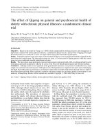
Fig. 1 The overall scheme of the experiment. The leaves from 6 week
old Arabidopsis thaliana were detached either from dark adapted
overnight plants or directly from plants growing in the growth
chamber during the light period (2 h after the photoperiodic light had
been turn on). The leaves were put on petri dishes on water-soaked
paper. Half of each leaf was covered with black paper (control)
and the leaves were irradiated with UV-B (8 W·m−2) for 5 min.
After irradiation the leaves were either left in darkness (leaves
from dark-adapted plants) or under continuous illumination with
white light (100 μmol ·m−2· s−1) for up to 4 days. Thus, 4 different kinds
of samples were analyzed, i) darkened control (CD), ii) control
illuminated with continuous light (CL), iii) UV-B irradiated leaves
left in darkness (UVD) and finally, iv) UV-B irradiated leaves kept
under continuous illumination (UVL)
Sztatelman et al. BMC Plant Biology (2015) 15:281
spectra were measured. Anthocyanin content was inferred from absorbance at 532 nm.
HPLC measurement
HPLC analysis of pigments was done by a method
modified from [42].100 μl of methanol pigment extract
was loaded with a loop onto a C-18 column (Bionacom
Velocity, 5uicrons, 4.6x250 mm), connected to an
Akta Purifier (GE Healthcare). The column was preequilibrated with 5 ml of solvent A (90 % acetonitrile,
10 % water), and elution was done with following gradient
with solvent B (100 % ethyl acetate):
1.
2.
3.
4.
5.
1–5 ml, 100 % A to 80 % A
5–20 ml, 80 % A to 50 % A
20–25 ml, 50 % A to 30 % A
25–30 ml, 30 % A (isocratic)
40–45 ml, 30 % A to 100 % A.
The flow rate was 1 ml/min. Elution was monitored
spectrophotometrically at three wavelengths simultaneously (405 nm, 436 nm and 280 nm). Pigments were
identified by retention time, compared to standards. The
chromatogram analysis and peak integration were done
using Unicorn software (GE Healthcare).
For a qualitative determination of pigments, extinction
coefficients in HPLC (Additional file 2: Table S2) solvents
were determined as follows. Fractions corresponding to
pigments of interest were collected separately in a known
volume. After recording the spectra in the HPLC solvent,
the fractions were dried and resuspended respectively
in 80 % acetone—chlorophyll a, chlorophyll b [43],
methanol—violaxanthin, lutein [44], ethanol—neoxanthin [45] and hexane—β-carotene [46].
The statistical significance of the differences between
treatments was assessed with one-way ANOVA, using
GraphPad InStat Software (Additional file 3: Table S3).
Page 4 of 16
proportion of 10 μl of extraction buffer per 1 mg of
powder mass. The samples were vortexed vigorously, incubated at 80 °C for 3 min, centrifuged for 10 min at 16
000 g at 4 °C and supernatant was mixed with an SDSPAGE loading buffer. The SDS-PAGE was performed according to [49] in a gel containing 12 % polyacrylamide
using the Mini Protean system (Bio-Rad). After separation the proteins were either stained with Coomassie
Brilliant Blue staining (for total protein visualization)
or transferred to a PVDF membrane (ImmobilonP,
Millipore) by the semi-dry transfer method (Trans-Blot
SD Semi-Dry Transfer Cell, Bio-Rad) for Western Blot
analysis. Membranes were stained with Ponceau S to ensure proper transfer, blocked with 5 % fat free dried milk
in PBS with 0,5 % Tween and incubated with an anti-D1
antibody (AS05 084, Agrisera) diluted 1:10 000 for
1 h at room temperature, followed by secondary antibody incubation (Goat anti-rabbit IgG HRP conjugated, Agrisera) under the same conditions. After that
a chemiluminescence substrate was added (Clarity
Western ECL Substrate, Bio-Rad) and the chemiluminescence was imaged using the BioSpectrum imaging
system (UVP).
Trypan Blue staining
The samples were pretreated (i.e. kept in darkness or left
for 2 h under photoperiodic light in the growth chamber), irradiated and kept in either darkness or constant
light as described in the “UV-B treatment” section. The
only exception was that prior to the irradiation, instead
of leaf halves, a middle, narrow part of the detached leaf
(perpendicular to the vasculature) was covered with
black paper. After the specified time the leaves were covered with 2,5 mg/ml Trypan Blue in lactophenol, heated
in a boiling water bath for 1 min, stained at room
temperature for an additional 2 h, and destained with a
saturated chloral hydrate solution.
RNA isolation and real-time PCR
Results
RNA isolation, cDNA synthesis and real-time RT-PCR
reactions were performed as given elsewhere [47]. All
reactions were run in triplicates. The sequence of the
primers and their annealing temperatures are listed in
Additional file 1: Table S1. A single dark-adapted
overnight control sample from day 0 was used as the
reference for calculating relative expression levels.
The normalization was performed with normalization
factors based on the reference gene levels calculated
by geNorm v3.4 [48].
Effect of UV-B on dark-induced yellowing of Arabidopsis
leaves
Protein extraction and Western Blot
The leaf material was ground in liquid nitrogen. An extraction buffer (4 % SDS, 2 % β-mercaptoethanol, 2 mM
PMSF, 100 mM TrisHCl, pH 8,8) was added in the
Different UV-B doses were applied in order to check
whether UV-B irradiation can slow down the onset of
dark-induced senescence in darkened Arabidopsis leaves.
Whereas in the non-irradiated leaf halves visible symptoms of senescence, i.e. yellowing, were easy to observe
(Fig. 2), UV-B treatment clearly influenced chlorophyll
degradation in a dose-dependent manner. Differences in
leaf color between the irradiated and non-irradiated
halves started to be visible after 3 min of the UV-B
(8 W·m−2) treatment and persisted up to the 10th minute of irradiation. Based on the results of this preliminary experiment, we decided on a 5 min treatment for
further analysis.
Sztatelman et al. BMC Plant Biology (2015) 15:281
Page 5 of 16
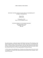
Fig. 2 Photographs of the detached leaves of 6-week old A. thaliana
with one half covered with black paper, and another half irradiated with
UV-B (8 W·m−2) for the indicated time and left in darkness for 4 days
The core idea of the study was to compare the effects
of UV-B in dark and light conditions and that was kept
in mind when setting up experimental treatments. On
the one hand, we wanted to avoid possible effects of the
circadian clock. On the other hand, we wanted to test
the influence of UVB on either the dark- or lightadapted state of the leaves. Therefore, we decided to
start both light and dark experiments at the same time
point i.e. 2 h after dawn. In consequence, the plants used
for testing the dark-adapted state were kept in darkness
for that time. To make sure that this extended night did
not result in drastic changes in the observed phenomena, leaf yellowing was observed in leaves taken from
plants which were either dark-adapted or kept in photoperiodic light for 2 h before UV-B irradiation, and transferred to darkness afterwards. In both cases chlorophyll
degradation was lower in UV-B irradiated leaf halves
(Fig. 3, compare a and b), with differences observed only
in the rate of degradation which was more prominent in
control leaf halves from dark-adapted plants. Thus, for
further experiments on the UV-inhibition of darkinduced chlorophyll degradation only dark-adapted
plants were used (see below).
Macroscopic appearance of leaves under different posttreatment light conditions
Two different experimental models were used (Fig. 1).
The first of these involved detached leaves from darkadapted plants. The leaves were UV-B irradiated and
kept in darkness for up to 4 days. In the other model
leaves were taken from plants 2 h after the start of the
light period. They were UV-B irradiated and placed in
constant light (100 μmol·m−2 ·s−1). Dark-induced leaf yellowing was observed in control leaf halves, while those
from constant light stayed green but showed reddening,
probably due to anthocyanin accumulation (Fig. 3). The
opposite effect of post-UV-treatment light conditions
was observed in leaf halves irradiated with 8 W·m−2 of
UV-B for 5 min. In irradiated leaf halves kept in
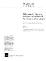
Fig. 3 Photographs of detached A. thaliana leaves with one half
covered with black paper, and another half irradiated with UV-B
(8 W·m−2) for 5 min and left in darkness (a and b) or under constant illumination (100 μmol·m−2 ·s−1 of white light, c) for the indicated time. The leaves were taken from plants dark-adapted
overnight (a), or from plants kept in a growth chamber for 2 h after
the dawn (b and c)
darkness dark-induced chlorophyll degradation was alleviated and yellowing was barely visible even after 4 days.
In contrast, leaf halves subjected to UV-B treatment and
then transferred to continuous light showed yellowing
without the appearance of red coloring. To examine the
observed effect in detail different parameters including
chlorophyll fluorescence, the expression of senescenceinduced and photosythesis-related genes as well as the
level of photosynthetic pigments and anthocyanins were
investigated.
Photosynthetic efficiency and photosynthetic pigment
content
To analyze the changes in pigment composition of the
leaves, HPLC analysis of isolated photosynthetic pigments starting from day 0 to day 4 after UV-B treatment
was carried out. The results are shown in Fig. 4a and b.
The overall changes in the levels of chlorophyll a (chl a)
and chlorophyll b (chl b) were similar. In continuous
light, starting from the second day, the chlorophyll levels
began to drop in the UV-B followed by continuous light
Sztatelman et al. BMC Plant Biology (2015) 15:281
Fig. 4 (See legend on next page.)
Page 6 of 16
Sztatelman et al. BMC Plant Biology (2015) 15:281
Page 7 of 16
(See figure on previous page.)
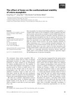
Fig. 4 Changes in photosynthetic pigments (a chlorohyll a, b chlorophyll b, c chlorophyll a/b, d violaxanthin, e neoxanthin, f lutein, g β-carotene ) in
detached Arabidopsis leaf halves either irradiated with UV-B (8 W·m−2) for 5 min or covered with black paper (control) and left either in darkness or
under constant white light (100 μmol·m−2·s−1) for the indicated time. Day 0 means 1 h after the treatment. Non-irradiated leaf halves served as a control.
Pigments were separated by HPLC with detection by absorbance at 436 nm (chlorophylls) or 405 nm (carotenoids) and their content was determined
from the area under the peak of the chromatogram using the extinction coefficients listed in Additional file 2: Table S2. Statistical significance of the
differences between treatments was assessed with one-way ANOVA and the results of this analysis are listed in Additional file 3: Table S3
(UVL) samples, resulting in a statistically relevant difference between 0UVL and 4UVL, as well as between
1UVL and 4UVL (Additional file 3: Table S3). Meanwhile, in control leaves (control continuous light—CL)
the chlorophyll content remained stable or even slightly
increased, resulting in a statistically significant difference
of p < 0.005 between 4UVL and 4CL for both chl a and
chl b. The content of both chlorophylls in dark-adapted
samples decreased both in treated (UV-B, then darkness—UVD) and un-treated (control darkness—CD) ones,
leading to a statistically significant difference of p < 0.005
for 4CD vs 4CL and 4UVD vs 4CL for chl a and chl b,
as well as lower but still statistically significant differences for the preceding days. However, the dynamic of
these processes was different. While in UV-B treated
leaves (UVD) the decrease was slow and steady from day
1 on, in the control (CD) it was pretty rapid after 3 days.
UV-B treatment slowed down chlorophyll degradation.
On day 4 in UVD samples chl a and chl b amounted to
129 % and 153 % of that observed in CD respectively.
This difference was not statistically significant. However,
whereas a difference between day 0 and day 4 was statistically significant for CD, no statistical significance was
observed for UVD. 4 days after irradiation the levels of
both chlorophylls were clearly lower in UVL than in CL
and similar to CD leaves. During treatment, the chl a/b
ratio did not change significantly for CL samples, but increased in CD samples. In both UV-B treated samples
this ratio was lower than in the corresponding controls
(Fig. 4c). Differences of p < 0.005 were noted between
day 4 in UVL leaves and its CL control, as well as between 4 UVD and 4CD. Statistically significant differences were found already on 3rd day (i.e. 3UVL vs 3CL,
and 3CD vs 3UVD).
Similar trends were observed for all carotenoids tested
(Fig. 4d-g). Again, in control samples kept in continuous
light, the contents of violaxanthin, lutein and β-carotene
increased or stayed unchanged. On the other hand, dark
treatment led to a decrease in all carotenoids tested,
what manifests as a statistically significant difference between 4CD and 1 to 4 CL. UV-B irradiation either did
not influence the effect of darkness (violaxanthin, Fig. 4d)
or slightly inhibited it (see: neoxanthin, lutein and βcarotene Fig. 4e-g), although the difference was not statistically significant. After a transient increase on day 1,
the decrease in carotenoid levels in UVL leaves on day
4th was either similar (lutein and violaxanthin), 50 %
lower (neoxanthin), or slightly higher (β -carotene) than
in the darkened control.
Bearing in mind the fact that the experimental treatment applied led to a decrease in the photosynthetic pigment content, we examined how these changes
influenced photosynthetic performance. We assessed the
yield of PSII via the measurement of chlorophyll fluorescence (Fig. 5a). The differences between maximum
quantum yield of PSII (QYmax) levels were more clearly
visible than these between levels of photosynthetic pigments. QYmax stayed unchanged in the control leaves
kept in continuous light (no statistically significant differences between subsequent days in CL leaves). Leaves
treated with UV-B prior to being transferred to continuous light showed a fast and very pronounced decrease in
QYmax, consistent with the yellowing of the samples.
These differences manifest as statistically significant between 3UVL and other leaves from this series (0UVL,
1UVL, 2UVL). In the CD leaves, the quantum yield decreased, first slowly, and from day 3 on, quite rapidly.
Leaves treated with UV-B and darkened showed a steady
decrease in QYmax, which resulted in higher values of
this parameter on day 3 and 4 than in CD leaves (statistically significant difference with p < 0.005), which corresponds to the slightly higher amounts of chlorophylls in
those samples.
The changes in pigment contents were also accompanied by changes in protein levels. Quantitatively extracted
total proteins were separated by SDS-page (Fig. 5b). The
amount of proteins decreased in all but CL leaves. The
loss of proteins in darkened samples was slower when
they were UV-B pre-treated. The amount of D1 protein
of PSII was also examined and showed similar trends to
total proteins (Fig. 5b). Interestingly, a lower mass product resulting from UV-B-induced degradation could be
observed in UV-B-treated samples. This product, present
1 h after irradiation (day 0), was no longer visible after
1 day in the light exposed sample. In darkness its degradation was very slow and the product was still clearly
visible even after 4 days.
In order to see if the influence on photosynthetic
processes was also reflected at the level of expression
of photosynthesis-related genes, quantitative real-time
PCR analysis was carried out (Fig. 5c and d). Typical,
photosynthesis-related gene transcripts, RIBULOSE
Sztatelman et al. BMC Plant Biology (2015) 15:281
Page 8 of 16
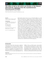
Fig. 5 Influence of 5 min UV-B (8 W·m−2) irradiation on photosynthesis in Arabidopsis leaves. After irradiation samples were kept either in darkness or
under constant light (100 μmol·m−2·s−1) for the given time. Day 0 means 1 h after the treatment. Non-irradiated leaf halves served as a control. a
Changes in PSII maximal quantum yield (Fv/Fm) during experimental treatment, measured with an imaging fluorometer. The results are the
means of measurements for at least 12 different leaves. Statistical significance of the differences between treatments was assessed with one-way
ANOVA and the results of this analysis are listed in Additional file 3: Table S3. b Total proteins (upper- Coomassie stained SDS-PAGE) and D1 protein
(lower- Western blot with anti-D1 antibodies) in examined leaves. Each well contains proteins extracted from 120 mg of tissue The degradation
product of D1 protein is marked with an arrow. c and d Relative expression levels of photosynthesis-related genes (CAB, RBSC1) measured
with real-time RT-PCR and normalized for the expression of four housekeeping genes (PDF2, UBC9, UBQ10, SAND). After the specified time
period leaves were cut into halves and control and treated halves from 4 different leaves were pooled. Each measurement was repeated
at least 3 times. A single dark-adapted overnight control sample from day 0 was used as a reference for calculating relative expression
levels. Error bars indicate the standard error
BISPHOSPHATE CARBOXYLASE SMALL CHAIN 1A
(RBCS1A) and CHLOROPHYLL A/B BINDING PROTEINS (CABs, including CAB1 and CAB2), were analyzed (Fig. 5c and d). Whereas the amount of RBCS1
mRNA stayed unchanged in the CL leaves even after
4 days, it decreased constantly in the darkened control leaves. On day 4 the transcript level of this gene
was similar in UVD and UVL samples reaching a
level almost 6 times lower than that observed in CL
samples. The dark-induced decrease in control leaves
was very rapid. After darkness exceeding 4 days
(4 days plus overnight pre-treatment) the amount of
RBCS1 reached only 0,3 % of that observed in the
leaves kept in continuous light.
The time-course of changes in the CAB transcript
level was slightly different (Fig. 5c). The steady-state
level of this gene decreased during the experiment, with
the most drastic drop in the darkened control samples.
4 days after treatment the amount of CAB was similar in
CL and UVD leaves. The decrease in UVL leaves was
clearly faster, reaching only 0,13 % of the transcript
present on day 0. Finally, in darkened control leaves,
at the end of the experiment, the CAB transcript level
was only 1,1 % of that present in leaves kept in continuous light.
Senescence and cell death
As leaf yellowing and changes in photosynthetic efficiency
often accompany senescence the level of senescenceassociated genes (SAGs) was also analyzed (Fig. 6). The
first of these was SAG13, an early senescence marker [12].
Interestingly, only 1 h after the treatment (day 0) the level
Sztatelman et al. BMC Plant Biology (2015) 15:281
Page 9 of 16
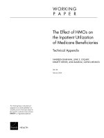
Fig. 6 Influence of 5 min UV-B (8 W·m−2) irradiation on the senescence and cell death of Arabidopsis leaves. After irradiation samples were kept
either in darkness or under constant light (100 μmol·m−2·s−1) for given time. a-d Time-course of the relative expression of senescence associated
genes: (a) SAG13, (b) SAG12, (c) SEN1 and (d) WRKY53 normalized for the expression of four housekeeping genes (PDF2, UBC9, UBQ10, SAND). Day
0 means 1 h after the treatment. Non-irradiated leaf halves served as a control. After the specified time period leaves were cut into halves and
control and treated halves from 4 different leaves were pooled. Each measurement was repeated at least 3 times. A single dark-adapted overnight
control sample from day 0 was used as a reference for calculating relative expression levels. Error bars indicate the standard error. e Trypan blue
staining for cell death of leaves irradiated with UV-B with the middle part covered and transferred either to light or to dark conditions
of this gene was slightly higher in irradiated samples as
compared to control ones (Fig. 6a, compare UVD vs CD
and UVL vs CL on day 0). The amounts of SAG13 transcripts increased very strongly in UVL leaves up to the
second day, and stayed at the same elevated level on day 3
and 4. Finally, its level was almost 20 times higher than in
CL leaves. As in CL, in darkened samples the amount of
SAG13 started to increase between the 1st and 2nd day,
but in CD leaves it continued to increase until day 4. An
interesting situation was observed for UV irradiated and
darkened samples. After strong up-regulation on days 1
and 2, the amount of SAG13 started to decline, reaching
the level similar to CL samples on the 4th day. Nevertheless, this level was still higher as compared to nonirradiated darkened leaves.
The second gene tested was SAG12, a late senescence
marker [12] (Fig. 6b). The steady-state level of SAG12
transcript increased strongly in all samples tested in a
time-dependent manner. Similarly to SAG13 the highest
level of this gene transcript was observed in UV-B
Sztatelman et al. BMC Plant Biology (2015) 15:281
irradiated samples kept in continuous light. After 4 days
the amount of this transcript increased by 442 times as
compared with CL leaves. Interestingly, the changes in
the SAG12 gene in all but UVL samples were similar,
though with slightly different kinetics.
The expression of SEN1, another senescence marker
was tested in addition to SAG12 and SAG13 [50]. Its expression depended mostly on light (Fig. 6c). It was very
strongly induced in darkened samples starting from day
0 and lower in samples illuminated with continuous
light. Prolonged night caused a very strong upregulation of SEN1, by 130 times as compared to leaves
taken directly from the photoperiod 2 h after the light
onset (day 0, compare CD and CL). Finally, on the 4th
day the amount of SEN1 transcript was over 7.300 times
higher in darkened leaves than in those kept in continuous light. The UV effect started to be visible between
the 2nd and 3rd day after irradiation. At this time, the
amount of SEN1 started to decrease in UVD leaves and
to increase in UVL ones. 4 days after irradiation the level
of this gene transcript was over 11 times higher in CD
leaves than in UVD ones. The opposite effect was observed in samples from continuous light. In this case,
the level of SEN1 transcript was 57 times higher in UVL
leaves as compared to non-irradiated ones.
The steady-state level of WRKY53, a transcription factor up-regulated during early senescence, was also examined. Both darkness and UV-B treatment caused an
increase in the level of this gene as compared to samples
from constant light (Fig. 6d). Prolonged night caused
over a 6-fold increase in the transcript level of this gene
(compare CD and CL at the day 0). UV-B acted stronger
than darkening, and the effect of UV-B and darkness
was synergistic as the strongest, by over 39 times, upregulation was observed in UVD samples. The amount
of WRKY53 changed over time, decreasing in all but the
CL leaves. In control samples from constant light it transiently decreased 1 day after irradiation, but finally
reached the same level as on day 0. On the 4th day the
highest level of WRKY53 was observed in both UV-B
irradiated samples (6 times higher than in CL ones).
Finally, the occurrence of cell death in the leaves was
studied using trypan blue staining (Fig. 6e). UV-B caused
the gradual appearance of cell death irrespective of light
conditions. Dark-treated leaf parts did not show trypan
blue staining until day 4 and even then it was faint compared to that induced by UV-B.
Anthocyanin content
It is well known that anthocyanin synthesis is strongly
up-regulated not only by visible light but also by UV-B.
However, macroscopic observation of the samples
treated under our experimental conditions did not confirm this up-regulation. Thus, we checked both the
Page 10 of 16
expression of genes involved in anthocyanin synthesis
and the content of those pigments more carefully
(Fig. 7a). Consistent with visual observations, a very
strong increase in the levels of anthocyanins was observed in leaves transferred to continuous light. At the
end of experiment, the anthocyanin content in leaves
from constant light was 36 times higher than that observed in dark-treated leaves. Interestingly, treatment
with UV-B almost completely abolished this response.
The anthocyanin level was stable in darkened leaves independent of UV-B irradiation.
We also checked the expression of the genes involved
in anthocyanin synthesis, PAL1 and CHS. The expression of PAL1 was very strongly down-regulated in darkened samples, whereas it stayed nearly unchanged in
those undergoing constant illumination (Fig. 7b; CD vs
CL). On the 4th day, the level of transcript was almost
200 times higher in leaves from constant light than in
those from dark conditions. Interestingly, the effect of
darkness was weaker in UV-B irradiated samples. Starting from the 1st day after irradiation the amount of
PAL1 transcript was from 1.5 to over 5 times higher in
UV-B treated samples than in dark controls.
A similar strong effect of darkness was observed for
CHS (Fig. 7c). Prolonged night caused a decrease in the
mRNA of this gene (day 0). Its level was 90 times higher
in illuminated samples (CL) than in darkened ones independent of UV-B treatment (compare CD and UVD).
The decrease in CHS level in control leaves left in darkness progressed during the experiment. On the 4th day
this level was 123 800 times higher in control leaves
from constant light than in darkened ones. UV-B downregulated the CHS level in leaves from the light (3–8
times as compared to CL). In dark-treated leaves UV-B
up-regulated the amount of this transcript starting from
the 2nd day after irradiation.
Macroscopic appearance of leaves of selected mutants
In order to elucidate possible mechanism(s) underlying
the observed UV-B effect, two mutants were examined:
uvr8-6, depleted of UV-B receptor [39] and mcp2d,
lacking metacaspase 2d involved in programmed cell
death [40].
The former one was used to check the involvement of
UVR8-activated signalling pathway in either inhibition
or promotion of chlorophyll degradation in darkness
and in light respectively. The mcp2d mutant served to
test the possible role of this metacaspase in chlorophyll
degradation in Arabidopsis leaves illuminated after
irradiation.
The dark-induced leaf yellowing was slowed down in
mcp2d leaves as compared with WT ones (Additional file 4:
Figure S1, dark control). Leaves of uvr8 plants were more
sensitive to UV-B-induced damage. The damage symptoms
Sztatelman et al. BMC Plant Biology (2015) 15:281
Fig. 7 (See legend on next page.)
Page 11 of 16
Sztatelman et al. BMC Plant Biology (2015) 15:281
Page 12 of 16
(See figure on previous page.)
Fig. 7 Influence of UV-B on anthocyanins in Arabidopsis leaves. After the specified time period leaves were cut into halves and control and treated
halves from 4 different leaves were pooled. Each measurement was repeated at least 3 times. Error bars indicate the standard error. a Anthocyanin
content was analyzed by measuring absorbance at 532 nm and normalized to fresh weight in examined leaves. b and c Time-course of the
relative expression of genes involved in anthocyanin biosynthesis: (b) PAL1 and (c) CHS normalized for the expression of four housekeeping
genes (PDF2, UBC9, UBQ10, SAND). A single dark-adapted overnight control sample from day 0 was used as a reference for calculating
relative expression levels
were more severe in uvr8 leaves darkened after irradiation.
The influence of UV-B on chlorophyll degradation was
comparable in WT Col and uvr8 and mcp2d mutants
independent on the light conditions (Additional file 4:
Figure S1).
Discussion
Treatment with high doses of UV-B alleviates darkeninginduced senescence symptoms in detached Arabidopsis
leaves
One of the most evident symptoms of senescence is
yellowing which originates from faster catabolism of
chlorophylls in comparison to yellow pigments. This
process may be a result of natural senescence or stress
(biotic or abiotic). Senescence is also induced in leaves
that are detached and stored in darkness or individually
darkened on a plant kept in a photoperiod [51]. Yellowing may be delayed by interference with the chlorophyll
degradation pathway, as observed in numerous mutants,
collectively known as “stay-green”. Those mutants can
be classified as either functional, i.e. those with delayed
overall senescence, or cosmetic, i.e. those where only
chlorophyll degradation is delayed while other aspects of
senescence progress normally [52].
In our model, the darkening of detached leaf halves resulted in a decrease in both chl a and chl b content and
an increase in the chl a/chl b ratio. These trends are
similar to those observed before [5, 53]. However, we
did not observe such a significant amount of chlorophyll
degradation products as previously reported [53]. Some
amount of pheophytins was found only after 4 days of
darkness (not shown).
A much higher retention of carotenoids is typical of
the senescing leaves of almost all plants. Usually, the carotenoid/chlorophyll ratio increases [54]. Changes in the
levels of neoxanthin, violaxanthin, lutein and β-carotene
occur in parallel [55, 56]. Our results are consistent with
those observations. The level of carotenoids did not
change during the first two days of darkening, and
started to decrease from the 3rd day. Taken together,
these results indicate that our system is an appropriate
experimental model of senescence.
Sub-lethal doses of UV-C, UV-B and gamma irradiation
are widely used in post-harvest technology. Beneficial,
hormetic effects of such irradiation include the sanitization
of fresh vegetables and fruits, an increase in phenolic
compounds, in the content of lycopene and other pigments,
the up-regulation of antioxidant content and antioxidant
enzyme activity (for a rewiev see: [57]). Treatment with
ultraviolet light may also influence chlorophyll content. Irradiation with a relatively high dose of UV-B (8.8 kJ ·m-2)
has been shown to delay the yellowing of broccoli florets
([37]) and lime peel during storage [38]. This yellowing results from the storage of detached plant parts in darkness.
It has been shown that dark storage induces senescence
and the expression of senescence-associated genes in broccoli starting after 3 days of storage [58].
We show that a similar dose-dependent effect can be
observed in detached Arabidopsis leaves kept in darkness
after irradiation with 8 W·m−2 UV-B (i.e. 2,4 kJ·m−2)
(Fig. 2). Our results indicate that high-doses of UV-B
interfere with chlorophyll degradation in darkness and
slow it down. The contents of all photosynthetic pigments
tested, except neoxanthin and violaxanthin, were slightly
lower in irradiated leaves during the first 2 days after treatment (Fig. 4). However, starting from day 3 chlorophyll
degradation in control leaves progressed much faster and
the balance reversed in favor of UV-B treated plants. The
same was observed for photosynthesis efficiency (Fig. 5a).
The maximal quantum yield of PSII was higher in UV
treated leaves than in control (only darkened) ones. Thus,
starting from 3 days after irradiation the positive effects of
the treatment outweighed the negative ones. Chlorophyll
a and b were degraded in darkened samples but the degradation rate of both pigments was modulated by UV irradiation. The chl a/chl b ratio increased in darkened
control leaves (Fig. 4b), in accordance with previous reports [53]. This probably results from the faster degradation of LHCII-derived (Light-harvesting complex II)
chlorophylls mediated by a complex of STAY-GREEN1
(SGR1) with chlorophyll catabolic enzymes [59], as it is
counteracted in a mutant with impaired SGR1 protein
function (nonyellowing1-1, nye1-1) during mild stress
treatment [60]. Interestingly, in UV-treated samples the
chl a/chl b ratio remained almost unchanged during the
whole time-course of the experiment. This suggests that
in the absence of visible light UV-B activated signals can
interfere with a specific pathway of LHCII degradation. In
our system the Lhcb1 protein level remained unchanged
during the experiment (Western blot data not shown). It
has been observed that the degradation of Lhc proteins
starts later than degradation of D1 protein [60]. It is likely
Sztatelman et al. BMC Plant Biology (2015) 15:281
that the components of LHCII, namely proteins and pigments, are degraded sequentially and that chlorophyll degradation precedes other processes.
The amounts of β-carotene and lutein were slightly
higher after UV irradiation (Fig. 4f and g). These pigments
are constituents of the photosynthetic protein pigment
complexes, PSII and LHCII respectively. The increased retention of β-carotene after UV-B treatment correlates with
the slower degradation of D1 protein and may result from
the increased stability of the whole complex.
Lutein and other xanthophylls which build LHCII
complexes are important in photoprotection mechanisms [61, 62]. The higher level of lutein and unchanged
level of violaxanthin after UV-B pretreatment may result
from a slower degradation of LHCII. Neoxanthin was
the only pigment that showed an immediate decrease
after UV-B treatment. It is known that neoxanthin absorbs UV and that it can photoisomerize upon excitation
[63]. Although other carotenoids also can photoisomerize, the effect for neoxanthin is the strongest and can
reach 10 % of this pigment’s content. Thus, the observed
decrease may result from photoisomerization. Light is
necessary for an effective conversion of neoxanthin isomers to the primary conformation. Since there is no
light in our system, the process is very slow and the level
of neoxanthin in UV treated samples remains lowest
during the whole time-course of the experiment.
All UV effects on photosynthesis in darkened leaves
were accompanied with a slower decrease in total protein
level, a slower decrease in the amount of D1 protein as
well as a slower decrease in the level of photosynthesisrelated transcripts (Fig. 5). This is consistent with observed changes in photosynthetic efficiency. The specific
product of UV-induced D1 protein cleavage was present
after the treatment (Fig. 5b, day 0). As in [35], we showed
the persistence of this product in the absence of light, in
our case for up to 4 days. Taken together, our results suggest that UV-B treatment alleviates the effects of darkening on the functionality of the photosynthetic apparatus.
The examination of expression levels of senescenceassociated genes showed that the UV effect observed
was not caused by a simple retardation of senescence
(Fig. 6). On the one hand, the mRNA levels of SAG12
and SEN1 were lower after short UV-B treatment in
samples darkened for 3 days, when the differences in
chlorophyll degradation started to be visible. On the
other hand, over the whole experiment the expression of
WRKY53 was elevated in irradiated samples. The
amount of SAG13 transcripts was higher for the first
2 days after the treatment and started to decrease on 3rd
day. This rather complicated pattern of senescenceassociated gene regulation suggests that UV-B interferes
with their expression; it does not supplement darkeninginduced senescence but tends to modulate it.
Page 13 of 16
It should be noted that the advantageous effects of UV
started to be visible at the same time as cell death symptoms appeared, confirmed by trypan blue staining. This
shows that in our experimental system leaves are highly
susceptible to both the damaging effects of UV-B and to
the beneficial ones. The exact mechanism of the inhibition of dark-induced leaf yellowing by high doses of UV
remains to be determined. To date, studies on the
UV-dependent inhibition of chlorophyll degradation
have been performed using broccoli florets and lime
peel [37, 38]. It has been shown that UV-B inhibits the
activity of chlorophyll peroxidase [37] and recently, the
specifically affected by UV-B chlorophyll peroxidase C has
been identified [64].
Our results demonstrated that the inhibition of
chlorophyll degradation in darkness was independent of
the UV-B photoreceptor, UVR8, since leaves of the uvr8
mutant showed the same symptoms as those of WT
plants i.e. slowed dark-induced chlorophyll degradation
(Additional file 4: Figure S1, dark). The more severe
damage observed in uvr8 mutants probably results from
the lack of UVR8-regulated protective mechanisms It is
in line with the hypothesis that UVR8 mediates responses to low UV-B doses, whereas high doses of UV-B
activate an independent pathway involving the mitogenactivated protein kinase (MAPK) cascade [3]. This cascade regulates UV-B dependent programmed cell death
(PCD) in plants. One of the regulators of cell death induced during biotic and abiotic stresses in Arabidopsis is
metacaspase 2d (MCP2d). Experiments employing the
mcp2d mutant demonstrated that the effects of UV-B
observed in this mutant were comparable to the wild
type (Additional file 4: Figure S1, dark). Thus, the inhibition of dark-induced chlorophyll degradation was not
due to the inhibition of the activity of this metacaspase.
Visible light influences UV-B action
Visible light dramatically affected the response of leaves
to high doses of UV. While darkened leaves stayed green
even 4 days after UV irradiation, yellowing was observed
in leaves transferred to continuous light starting from
the 2nd day (Fig. 3). This yellowing was a result of a decrease in the levels of all photosynthetic pigments and
was accompanied by a decrease in photosynthesis efficiency (Figs. 3 and 5). In contrast to the senescing CD
samples, in UVL ones the chl a/chl b ratio decreased.
The decrease resembled the effect observed in the nye11 mutant during mild salt stress (Sakuraba et al. 2014).
This suggests that the level of activation of different
pigment degradation pathways during UV-mediated
senescence in light was not the same as during darkinduced senescence. In particular, the specific, SGR1dependent pathway of LHCII degradation appeared
not to be activated.
Sztatelman et al. BMC Plant Biology (2015) 15:281
The cumulative dose of UV-B necessary to decrease
the chlorophyll level has been calculated for Pisum sativum as 300 kJ·m−2 [65]. In Arabidopsis grown in a 12 h
light photoperiod with photosynthetic photon flux density
(PPFD) of 300 μmol·m−2 ·s−1 plus 6 kJ·m−2·d−1 of UV-B
[34], the chlorophyll content increased [34]. Even the
addition 2,4 W·m−2 of UV-B for 5 h (i.e. 8,64 kJ·m−2 dose)
to 40 μmol·m−2 ·s−1 of PPFD did not influence the chlorophyll level in 29 day old Arabidopsis [66]. In this
experiment the content of both chlorophylls, lutein, violaxanthin and antheraxanthin remained unchanged as
long as 4 days after UV-B irradiation. The UV-B dose used
in our experiment e.g. 2,4 kJ·m−2 was comparable with
above experiments. The main differences were: using detached leaves from plants grown in a shorter (10 h of light)
photoperiod and using constant illumination after the
treatment. However, when leaves after UV-B treatment
were transferred back to the photoperiod, the same symptoms were observed, but with slower kinetics (data not
shown). The influence of PAR intensity during Arabidopsis
growth on the effect of UV-B has been shown before
[67, 68]. The light intensity during the growth was
lower in our experiment (70 μmol·m−2 ·s−1) than those
reported by [66] (130 μmol·m−2 ·s−1) and [34]
(300 μmol·m−2·s−1). Thus, it is possible that either
light intensity and/or the duration of the photoperiod
modulate the UV-B effects on photosynthesis (compare [28]). Additionally, Götz et al. [68] showed that
a low level of UV-B during growth leads to a low accumulation of protective isoflavonoids. This may lead
to a higher susceptibility of the plants to UV-B irradiation, compared to the plants grown in the presence
of higher UV-B levels. While in our growth chamber
UV-B irradiation was completely excluded, no data on
the intensity of UV-B during plant growth are provided either by Moon’s or Poulson’s groups [34, 66].
The expression of senescence-associated genes has
been shown to be up-regulated in Arabidopsis grown
under white light supplemented with UV-B [13]. Similarly, in our system the expression of all senescenceassociated genes tested was strongly up-regulated in
UVL leaves (Fig. 6). A comparison of the results obtained for darkened and illuminated samples shows that
PAR is a key factor in the induction of senescenceassociated genes by UV-B. In consequence, the observed
decrease in photosynthesis resulting from chlorophyll
degradation seems to be a result of the initiation of the
senescence process.
Interestingly, UV-B pretreatment completely inhibited
the accumulation of anthocyanins in continuous light.
The expression of anthocyanin regulatory genes as well
as anthocyanin accumulation have been shown to be
strongly increased by UV-A and by visible light, mainly
in the blue range. UV-B alone is much less effective, but
Page 14 of 16
it acts synergistically with visible light [29, 69, 70]. In our
experiments the expression levels of PAL1 and CHS were
elevated in leaves continuously illuminated with white
light as compared to darkened samples (Fig. 7). UV-B either did not influence (CHS) or slightly reduced (PAL1)
this increase. In contrast, while anthocyanin accumulation
was very strongly enhanced by white light, UV-B pretreatment counteracted this effect. The activation of the senescence program by UV-B irradiation, might eliminate the
need for the production of photoprotective pigments.
Although the impact of UV-B on photosynthesis,
chlorophyll degradation, expression of senescenceassociated genes was modulated by the subsequent light
conditions, cell death was observed in samples both
darkened and illuminated with continuous light. Visible
light proved to be necessary for the proper course of a
senescence program triggered by a high dose of UV-B.
Light is required for the onset of cell death under
nutrient-limiting conditions [71] and in Arabidopsis protoplasts after UV-C treatment [72]. Chloroplast delivered
signals, most probably ones connected with the production of reactive oxygen species, seem to be involved in
executing senescence and cell death [73–75]. Indeed, in
our system the senescence program leading to cell death
was initiated in illuminated samples. In darkened ones
other processes leading to cell death were activated. The
signaling pathways leading to cell death after UV-B irradiation and darkening do not act synergistically but
seem to be mutually exclusive to some extent.
Again, UVR8-dependent signalling pathway was not
involved in UV-B-induced chlorophyll degradation in
light (Additional file 4: Figure S1).
Conclusions
Our results show the importance of the light conditions
applied after the irradiation with high dose of UV-B. These
conditions influenced the expression of photosynthesisrelated and senescence-associated genes, chlorophyll
degradation and photosynthesic efficiency. Short UV-B
treatment promoted leaf yellowing in light and inhibited it
upon the storage of leaves in darkness. However, irrespective of light conditions, visible cell death symptoms appeared 3 days after UV-B irradiation.
Additional files
Additional file 1: Table S1. Sequences of primers used in this study.
(DOCX 15 kb)
Additional file 2: Table S2. Extinction coefficients in HPLC solvent.
(DOCX 10 kb)
Additional file 3: Table S3. Analysis of statistical relevance in changes
of photosynthetic pigments level (a: chlorophyll a, b: chlorophyll b, c:
chlorophyll a/chlorophyll b ratio, d: violaxanthin, e: neoxanthin, f: lutein,
g: β-carotene) as well as in yield of Photosystem II (h). (PDF 325 kb)
Sztatelman et al. BMC Plant Biology (2015) 15:281
Additional file 4: Figure S1. Photographs of the detached leaves of
6-week old A. thaliana WT, mcp2d and uvr8 mutants with one half covered
with black paper, and another half irradiated with UV-B (8 W · m−2)
for 5 min and (A) left in darkness or (B) illuminated with white light
(100·μmol·m−2·s−1) for 4 days. (TIF 10951 kb)
Page 15 of 16
7.
8.
9.
Abbreviations
CAB: CHLOROPHYLL A/B BINDING PROTEIN; CD: Control dark, non-treated leaves
dark adapted and left in the darkness; chl: Chlorophyll; CHS: CHALCONE
SYNTHASE; CL: Control light, non-treated leaves left under continuous light
−1
(100 μmol·ms−2
); LHC II: Light-harvesting complex II; mcp2d: metacaspase 2d;
·
nye1-1: non-yellowing1-1; PAL1: PHENYLALANINE AMMONIA-LYASE 1;
PAR: Photosynthetically active radiation; PPFD: Photosynthetic photon flux
density; PSII: Photosystem II; QYmax: Maximum quantum yield of Photosystem II;
RBCS1A: RIBULOSE BISPHOSPHATE CARBOXYLASE SMALL CHAIN 1A; ROS: Reactive
oxygen species; SAG: SENESCENCE ASSOCIATED GENE; SEN1: SENESCENCE1;
SGR1: STAY-GREEN1; UV: Ultraviolet; UVD: UV dark—dark adapted leaves
irradiated for 5 min with 8 W·m−2 of UV-B and left in darkness; UVL: UV
light—leaves irradiated for 5 min with 8 W·m−2 of UV-B and left under continuous light (100 μmol·m−2 ·s−1); uvr8: uvb-resistance 8.
10.
11.
12.
13.
14.
Competing interests
The authors declare that they have no competing interests.
15.
Authors’ contributions
OS: participated in the design of the study, conducted the experiments,
discussed and analyzed the data and improved the manuscript. JG:
participated in the HPLC analysis, interpreted the results and improved the
manuscript. AKB: conceived of the study, participated in its design and
coordination, drafted the manuscript. HG: participated in the design of the
study and in the discussion of results. All authors read and approved the
final manuscript.
16.
17.
18.
Acknowledgements
The study was supported by the Polish National Science Centre, grant no.
UMO-2011/03/D/NZ3/00210. The Faculty of Biochemistry, Biophysics and
Biotechnology of Jagiellonian University is the beneficiary of structural funds
from the European Union; grant no. POIG.02.01.00-12-064/08. The Faculty of
Biochemistry, Biophysics and Biotechnology is a partner of the Leading
National Research Center (KNOW) supported by the Ministry of Science
and Higher Education. HPLC measurements were performed in NanoFun
Laboratories, POIG.02.02.00-00-025/09.
Author details
1
Department of Plant Biotechnology, Faculty of Biochemistry, Biophysics and
Biotechnology, Jagiellonian University, Gronostajowa 7, Krakow 30-387,
Poland. 2Current address: Institute of Biochemistry and Biophysics, Polish
Academy of Sciences, Warszawa 02-106, Poland. 3Laboratory of Biological
Physics, Institute of Physics, Polish Academy of Sciences, Al. Lotników 32/46,
Warszawa 02-668, Poland. 4The Malopolska Centre of Biotechnology,
Jagiellonian University, Gronostajowa 7, Krakow 30-387, Poland.
19.
20.
21.
22.
23.
24.
25.
Received: 10 August 2015 Accepted: 17 November 2015
26.
References
1. Hollósy F. Effects of ultraviolet radiation on plant cells. Micron. 2002;33:179–97.
2. Brown BA, Jenkins GI. UV-B signaling pathways with different fluence-rate
response profiles are distinguished in mature Arabidopsis leaf tissue by
requirement for UVR8, HY5, and HYH. Plant Physiol. 2008;146:576–88.
3. Nawkar GM, Maibam P, Park JH, Sahi VP, Lee SY, Kang CH. UV-induced cell
death in plants. Int J Mol Sci. 2013;14:1608–28.
4. Teramura AH. Effects of ultraviolet‐B radiation on the growth and yield of
crop plants. Physiol Plant. 1983;58:415–27.
5. Frohnmeyer H, Staiger D. Ultraviolet-B radiation-mediated responses in
plants. Balancing damage and protection. Plant Physiol. 2003;133:1420–8.
6. Sarvikas P, Hakala M, Pätsikkä E, Tyystjärvi T, Tyystjärvi E. Action spectrum of
photoinhibition in leaves of wild type and npq1-2 and npq4-1 mutants of
Arabidopsis thaliana. Plant Cell Physiol. 2006;47:391–400.
27.
28.
29.
30.
31.
Boyko A, Greer M, Kovalchuk I. Acute exposure to UVB has a more profound
effect on plant genome stability than chronic exposure. Mut Res Fund Mol
Mech Mutagen. 2006;602:100–9.
Lake JA, Field KJ, Davey MP, Beerling DJ, Lomax BH. Metabolomic and
physiological responses reveal multi‐phasic acclimation of Arabidopsis
thaliana to chronic UV radiation. Plant Cell Env. 2009;32:1377–89.
Pourtau N, Marès M, Purdy S, Quentin N, Ruël A, Wingler A. Interactions of
abscisic acid and sugar signalling in the regulation of leaf senescence.
Planta. 2004;219:765–72.
Munné-Bosch S, Alegre L. Die and let live: Leaf senescence contributes to
plant survival under drought stress. Funct Plant Biol. 2004;31:203–16.
Diaz C, Saliba-Colombani V, Loudet O, Belluomo P, Moreau L, Daniel-Vedele
F, et al. Leaf yellowing and anthocyanin accumulation are two genetically
independent strategies in response to nitrogen limitation in Arabidopsis
thaliana. Plant Cell Physiol. 2006;47:74–83.
Weaver LM, Gan S, Quirino B, Amasino RM. A comparison of the expression
patterns of several senescence-associated genes in response to stress and
hormone treatment. Plant Mol Biol. 1998;37:455–69.
John C, Morris K, Jordan B, Thomas B, A‐H‐Mackerness S. Ultraviolet‐B
exposure leads to up‐regulation of senescence‐associated genes in
Arabidopsis thaliana. J Exp Bot. 2001;52:1367–73.
Kusano M, Tohge T, Fukushima A, Kobayashi M, Hayashi N, Otsuki H, et al.
Metabolomics reveals comprehensive reprogramming involving two
independent metabolic responses of Arabidopsis to UV‐B light. Plant J.
2011;67:354–69.
Dunning CA, Chalker‐Scott L, Scott JD. Exposure to ultraviolet‐B radiation
increases cold hardiness in Rhododendron. Physiol Plant. 1994;92:516–20.
Kakani VG, Reddy KR, Zhao D, Gao W. Senescence and hyperspectral
reflectance of cotton leaves exposed to ultraviolet‐B radiation and carbon
dioxide. Physiol Plant. 2004;121:250–7.
Pradhan M, Nayak L, Joshi P, Mohapatra P, Patro L, Biswal B, et al.
Developmental phase-dependent photosynthetic responses to ultraviolet-B
radiation: Damage, defence, and adaptation of primary leaves of wheat
seedlings. Photosynthetica. 2008;46:370–7.
Kakani V, Reddy K, Zhao D, Sailaja K. Field crop responses to ultraviolet-B
radiation: A review. Agric Forest Meteorol. 2003;120:191–218.
Hunt J, Mcneil D. Nitrogen status affects UV-B sensitivity of cucumber. Funct
Plant Biol. 1998;25:79–86.
Jenkins ME, Suzuki TC, Mount DW. Evidence that heat and ultraviolet radiation
activate a common stress-response program in plants that is alterd in the uvh6
mutant of Arabidopsis thaliana. Plant Physiol. 1997;115:1351–8.
Fedina IS, Grigorova ID, Georgieva KM. Response of barley seedlings to UV-B
radiation as affected by NaCl. J Plant Physiol. 2003;160:205–8.
Chalker‐Scott L, Scott JD. Elevated ultraviolet‐B radiation induces cross‐
protection to cold in leaves of rhododendron under field conditions.
Photochem Photobiol. 2004;79:199–204.
Teklemariam T, Blake TJ. Effects of UVB preconditioning on heat tolerance of
cucumber (Cucumis sativus L.). Env Exp Bot. 2003;50:169–82.
Bandurska H, Niedziela J, Chadzinikolau T. Separate and combined
responses to water deficit and UV-B radiation. Plant Sci. 2013;213:98–105.
Paul ND, Moore JP, Mcpherson M, Lambourne C, Croft P, Heaton JC, et al.
Ecological responses to UV radiation: Interactions between the biological
effects of UV on plants and on associated organisms. Physiol Plant.
2012;145:565–81.
Wang H, Hao J, Chen X, Hao Z, Wang X, Lou Y, et al. Overexpression of rice
WRKY89 enhances ultraviolet B tolerance and disease resistance in rice
plants. Plant Mol Biol. 2007;65:799–815.
Krizek DT. Influence of PAR and UV‐A in determining plant sensitivity and
photomorphogenic responses to UV‐B radiation. Photochem Photobiol.
2004;79:307–15.
Kimura M, Yamamoto YY, Seki M, Sakurai T, Sato M, Abe T, et al.
Identification of Arabidopsis genes regulated by high light–stress using
cDNA microarray. Photochem Photobiol. 2003;77:226–33.
Bieza K, Lois R. An Arabidopsis mutant tolerant to lethal ultraviolet-B levels
shows constitutively elevated accumulation of flavonoids and other
phenolics. Plant Physiol. 2001;126:1105–15.
Gao Q, Zhang L. Ultraviolet-B-induced oxidative stress and antioxidant
defense system responses in ascorbate-deficient vtc1 mutants of
Arabidopsis thaliana. J Plant Physiol. 2008;165:138–48.
Cen Y-P, Bornman JF. The response of bean plants to UV-B radiation under
different irradiances of background visible light. J Exp Bot. 1990;41:1489–95.
Sztatelman et al. BMC Plant Biology (2015) 15:281
32. Deckmyn G, Martens C, Impens I. The importance of the ratio UV‐B/
photosynthetic active radiation (PAR) during leaf development as
determining factor of plant sensitivity to increased UV‐B irradiance: Effects
on growth, gas exchange and pigmentation of bean plants (Phaseolus
vulgaris cv. Label). Plant Cell Env. 1994;17:295–301.
33. Hectors K, Prinsen E, De Coen W, Jansen MA, Guisez Y. Arabidopsis thaliana
plants acclimated to low dose rates of ultraviolet B radiation show specific
changes in morphology and gene expression in the absence of stress
symptoms. New Phytol. 2007;175:255–70.
34. Poulson ME, Boeger MRT, Donahue RA. Response of photosynthesis to high
light and drought for Arabidopsis thaliana grown under a UV-B enhanced
light regime. Photosynt Res. 2006;90:79–90.
35. Bergo E, Segalla A, Giacometti GM, Tarantino D, Soave C, Andreucci F,
et al. Role of visible light in the recovery of photosystem II structure
and function from ultraviolet‐B stress in higher plants. J Exp Bot.
2003;54:1665–73.
36. Bolink EM, Van Schalkwijk I, Posthumus F, Van Hasselt PR. Growth under
UV-B radiation increases tolerance to high-light stress in pea and bean
plants. Plant Ecol. 2001;154:147–56.
37. Aiamla-Or S, Kaewsuksaeng S, Shigyo M, Yamauchi N. Impact of UV-B
irradiation on chlorophyll degradation and chlorophyll-degrading enzyme
activities in stored broccoli (Brassica oleracea L. Italica Group) florets.
Food Chem. 2010;120:645–51.
38. Srilaong V, Aiamla-Or S, Soontornwat A, Shigyo M, Yamauchi N. UV-B
irradiation retards chlorophyll degradation in lime (Citrus latifolia Tan.)
fruit. Postharvestal Biol Technol. 2011;59:110–2.
39. Favory JJ, Stec A, Gruber H, Rizzini L, Oravecz A, Funk M, et al. Interaction of
COP1 and UVR8 regulates UV‐B‐induced photomorphogenesis and stress
acclimation in Arabidopsis. EMBO J. 2009;28:591–601.
40. Watanabe N, Lam E. Arabidopsis metacaspase 2d is a positive mediator of
cell death induced during biotic and abiotic stresses. Plant J. 2011;66:969–82.
41. Alonso JM, Stepanova AN, Leisse TJ, Kim CJ, Chen H, Shinn P, et al.
Genome-wide insertional mutagenesis of Arabidopsis thaliana. Science.
2003;301:653–7.
42. Van Leeuwe M, Villerius L, Roggeveld J, Visser R, Stefels J. An optimized
method for automated analysis of algal pigments by HPLC. Marin Chem.
2006;102:267–75.
43. Inskeep WP, Bloom PR. Extinction coefficients of chlorophyll a and b in N,
N-dimethylformamide and 80 % acetone. Plant Physiol. 1985;77:483–5.
44. Karrer P, Jucker E. Carotenoids. New York: Elsevier; 1950.
45. Mallams A, Waight E, Weedon B, Chapman DJ, Haxo F, Goodwin T et al.
A new class of carotenoids. Chem Commun. 1967;6:301–2.
46. Craft NE, Soares JH. Relative solubility, stability, and absorptivity of lutein
and. beta.-carotene in organic solvents. J Agricult Food Chem. 1992;40:431–4.
47. Łabuz J, Sztatelman O, Banaś AK, Gabryś H. The expression of phototropins
in Arabidopsis leaves: Developmental and light regulation. J Exp Bot. 2012;
63:1763–71.
48. Vandesompele J, De Preter K, Pattyn F, Poppe B, Van Roy N, De Paepe A et
al. Accurate normalization of real-time quantitative RT-PCR data by
geometric averaging of multiple internal control genes. Genome Biol. 2002;
3:research0034.
49. Laemmli UK. Cleavage of structural proteins during the assembly of the
head of bacteriophage T4. Nature. 1970;227:680–5.
50. Oh SA, Lee SY, Chung IK, Lee C-H, Nam HG. A senescence-associated gene
of Arabidopsis thaliana is distinctively regulated during natural and
artificially induced leaf senescence. Plant Mol Biol. 1996;30:739–54.
51. Weaver LM, Amasino RM. Senescence is induced in individually darkened
Arabidopsis leaves, but inhibited in whole darkened plants. Plant Physiol.
2001;127:876–86.
52. Grassl J, PružInská A, HöRtensteiner S, Taylor NL, Millar AH. Early events in
plastid protein degradation in stay-green Arabidopsis reveal differential
regulation beyond the retention of LHCII and chlorophyll. J Proteom Res.
2012;11:5443–52.
53. Pružinská A, Tanner G, Aubry S, Anders I, Moser S, Müller T, et al. Chlorophyll
breakdown in senescent Arabidopsis leaves. Characterization of chlorophyll
catabolites and of chlorophyll catabolic enzymes involved in the
degreening reaction. Plant Physiol. 2005;139:52–63.
54. Lu C, Lu Q, Zhang J, Kuang T. Characterization of photosynthetic pigment
composition, photosystem II photochemistry and thermal energy
dissipation during leaf senescence of wheat plants grown in the field.
J Exp Bot. 2001;52:1805–10.
Page 16 of 16
55. Whitfield DM, Rowan KS. Changes in the chlorophylls and carotenoids of
leaves of Nicotiana tabacum during senescence. Phytochemistry. 1974;13:77–83.
56. Young AJ, Wellings R, Britton G. The fate of chloroplast pigments during
senescence of primary leaves of Hordeum vulgare and Avena sativum.
J Plant Physiol. 1991;137:701–5.
57. Ribeiro C, Alvarenga N. Prospects of UV radiation for application in
postharvest technology. Emir. J. Food Agric. 2012;24(6):586-597.
58. Page T, Griffiths G, Buchanan-Wollaston V. Molecular and biochemical
characterization of postharvest senescence in broccoli. Plant Physiol. 2001;
125:718–27.
59. Sakuraba Y, Schelbert S, Park S-Y, Han S-H, Lee B-D, Andrès CB, et al.
STAY-GREEN and chlorophyll catabolic enzymes interact at light-harvesting
complex II for chlorophyll detoxification during leaf senescence in
Arabidopsis. Plant Cell. 2012;24:507–18.
60. Sakuraba Y, Lee S-H, Kim Y-S, Park OK, Hörtensteiner S, Paek N-C. Delayed
degradation of chlorophylls and photosynthetic proteins in Arabidopsis
autophagy mutants during stress-induced leaf yellowing. J Exp Bot. 2014:eru008
65(14):3915-25.
61. Jahns P, Holzwarth AR. The role of the xanthophyll cycle and of lutein in
photoprotection of photosystem II. Biochim Biophys Acta. 1817;2012:182–93.
62. Goss R, Lepetit B. Biodiversity of NPQ. J Plant Physiol. 2015;172:13–32.
63. Zubik M, Luchowski R, Grudzinski W, Gospodarek M, Gryczynski I, Gryczynski
Z, et al. Light-induced isomerization of the LHCII-bound xanthophyll
neoxanthin: Possible implications for photoprotection in plants. Biochim
Biophys Acta. 1807;2011:1237–43.
64. Aiamla-Or S, Shigyo M, Ito S-I, Yamauchi N. Involvement of chloroplast
peroxidase on chlorophyll degradation in postharvest broccoli florets and
its control by UV-B treatment. Food Chem. 2014;165:224–31.
65. Strid Å, Chow WS, Anderson JM. UV-B damage and protection at the
molecular level in plants. Photosynt Res. 1994;39:475–89.
66. Moon YR, Lee MH, Tovuu A, Lee C-H, Chung BY, Park Y-I, et al. Acute
exposure to UV-B sensitizes cucumber, tomato, and Arabidopsis plants to
photooxidative stress by inhibiting thermal energy dissipation and
antioxidant defense. J Radiat Res. 2011;52:238–48.
67. Berkelaar EJ, Ormrod DP, Hale BA. The Influence of Photosynthetically Active
Radiation on the Effects of Ultraviolet‐B Radiation on Arabidopsis thaliana.
Photochem Photobiol. 1996;64:110–6.
68. Götz M, Albert A, Stich S, Heller W, Scherb H, Krins A, et al. PAR modulation
of the UV-dependent levels of flavonoid metabolites in Arabidopsis thaliana
(L.) Heynh. leaf rosettes: Cumulative effects after a whole vegetative growth
period. Protoplasma. 2010;243:95–103.
69. Cominelli E, Gusmaroli G, Allegra D, Galbiati M, Wade HK, Jenkins GI, et al.
Expression analysis of anthocyanin regulatory genes in response to different
light qualities in Arabidopsis thaliana. J Plant Physiol. 2008;165:886–94.
70. Ryan KG, Swinny EE, Markham KR, Winefield C. Flavonoid gene expression
and UV photoprotection in transgenic and mutant Petunia leaves.
Phytochemistry. 2002;59:23–32.
71. Wingler A, Brownhill E, Pourtau N. Mechanisms of the light-dependent
induction of cell death in tobacco plants with delayed senescence. J Exp
Bot. 2005;56:2897–905.
72. Danon A, Rotari VI, Gordon A, Mailhac N, Gallois P. Ultraviolet-C
overexposure induces programmed cell death in Arabidopsis, which is
mediated by caspase-like activities and which can be suppressed by
caspase inhibitors, p35 and Defender against Apoptotic Death. J Biol Chem.
2004;279:779–87.
73. Zapata J, Guera A, Esteban-Carrasco A, Martin M, Sabater B. Chloroplasts
regulate leaf senescence: Delayed senescence in transgenic ndhF-defective
tobacco. Cell Death Differ. 2005;12:1277–84.
74. Kim C, Meskauskiene R, Zhang S, Lee KP, Ashok ML, Blajecka K, et al.
Chloroplasts of Arabidopsis are the source and a primary target of a plantspecific programmed cell death signaling pathway. Plant Cell. 2012;24:3026–39.
75. Gutiérrez J, González-Pérez S, García-García F, Daly CT, Lorenzo Ó, Revuelta
JL, et al. Programmed cell death activated by Rose Bengal in Arabidopsis
thaliana cell suspension cultures requires functional chloroplasts. J Exp Bot.
2014;65:3081–95.