Repetitive sequence analysis and karyotyping reveals centromere-associated DNA sequences in radish (Raphanus sativus L.)
Bạn đang xem bản rút gọn của tài liệu. Xem và tải ngay bản đầy đủ của tài liệu tại đây (2.08 MB, 12 trang )
He et al. BMC Plant Biology (2015) 15:105
DOI 10.1186/s12870-015-0480-y
RESEARCH ARTICLE
Open Access
Repetitive sequence analysis and karyotyping
reveals centromere-associated DNA sequences in
radish (Raphanus sativus L.)
Qunyan He1,2†, Zexi Cai2†, Tianhua Hu1, Huijun Liu2, Chonglai Bao1, Weihai Mao1* and Weiwei Jin2*
Abstract
Background: Radish (Raphanus sativus L., 2n = 2x = 18) is a major root vegetable crop especially in eastern Asia.
Radish root contains various nutritions which play an important role in strengthening immunity. Repetitive
elements are primary components of the genomic sequence and the most important factors in genome size
variations in higher eukaryotes. To date, studies about repetitive elements of radish are still limited. To better
understand genome structure of radish, we undertook a study to evaluate the proportion of repetitive elements
and their distribution in radish.
Results: We conducted genome-wide characterization of repetitive elements in radish with low coverage genome
sequencing followed by similarity-based cluster analysis. Results showed that about 31% of the genome was
composed of repetitive sequences. Satellite repeats were the most dominating elements of the genome. The
distribution pattern of three satellite repeat sequences (CL1, CL25, and CL43) on radish chromosomes was
characterized using fluorescence in situ hybridization (FISH). CL1 was predominantly located at the centromeric
region of all chromosomes, CL25 located at the subtelomeric region, and CL43 was a telomeric satellite. FISH signals
of two satellite repeats, CL1 and CL25, together with 5S rDNA and 45S rDNA, provide useful cytogenetic markers to
identify each individual somatic metaphase chromosome. The centromere-specific histone H3 (CENH3) has been
used as a marker to identify centromere DNA sequences. One putative CENH3 (RsCENH3) was characterized and
cloned from radish. Its deduced amino acid sequence shares high similarities to those of the CENH3s in Brassica
species. An antibody against B. rapa CENH3, specifically stained radish centromeres. Immunostaining and chromatin
immunoprecipitation (ChIP) tests with anti-BrCENH3 antibody demonstrated that both the centromere-specific
retrotransposon (CR-Radish) and satellite repeat (CL1) are directly associated with RsCENH3 in radish.
Conclusions: Proportions of repetitive elements in radish were estimated and satellite repeats were the most
dominating elements. Fine karyotyping analysis was established which allow us to easily identify each individual
somatic metaphase chromosome. Immunofluorescence- and ChIP-based assays demonstrated the functional
significance of satellite and centromere-specific retrotransposon at centromeres. Our study provides a valuable basis
for future genomic studies in radish.
Keywords: Radish, Repetitive DNA, Satellite, Karyotyping, CENH3, Centromere
* Correspondence: ;
†
Equal contributors
1
Institute of Vegetables, Zhejiang Academy of Agricultural Sciences,
Hangzhou 310021, China
2
National Maize Improvement Center of China, Beijing Key Laboratory of
Crop Genetic Improvement, China Agricultural University, Beijing 100193,
China
© 2015 He et al.; licensee BioMed Central. This is an Open Access article distributed under the terms of the Creative Commons
Attribution License ( which permits unrestricted use, distribution, and
reproduction in any medium, provided the original work is properly credited. The Creative Commons Public Domain
Dedication waiver ( applies to the data made available in this article,
unless otherwise stated.
He et al. BMC Plant Biology (2015) 15:105
Background
Repetitive DNAs, including transposable elements and
tandem repeats, are the major components of the genomic sequence and the most important factors in genome size variations in higher eukaryotes [1-3]. Based on
the mechanism of transposition, transposable elements
can be divided into two classes, transposons and retrotransposons. The majority of these elements in plant
genome are long terminal repeat (LTR) retrotransposons
and most of them are dispersed throughout all chromosomes [4,5]. Tandem repeats consist of large number of
repeat units and are usually found in centromeres, pericentromeres or telomeres [6]. Tandem repeats are good
cytogenetic markers for chromosome identification and
molecular karyotyping [7].
Centromeres are specialized regions on chromosomes
where centromeric protein and spindle microtubules attach via the kinetochore and typically contain large arrays of satellite repeats and/or retrotransposon-related
repetitive sequences in eukaryotes [8,9]. They are essential for proper chromosome segregation during mitosis
and meiosis. Although the function of centromeres is
conserved in organisms, centromeric repeats appear to
evolve rapidly [10]. Satellite repeats go through rapid
evolution and significant variation between closely related species or even among different chromosomes of
the same species [11-14]. Centromeric regions are comprised of repetitive sequences in most species, suggesting
that those sequences play important roles in centromere
function [15]. Centromeres are universally marked by
the presence of a centromere-specific histone H3 (CENH3,
called CENP-A in human), that replaces canonical histone
H3 in centromeric nucleosomes to form functional centromeres [16]. CENH3 is a good marker to identify the core
centromeric sequences by chromatin immunoprecipitation
(ChIP) with an anti-CENH3 antibody [11,17,18].
Radish (Raphanus sativus L., 2n = 2x = 18), belonging
to the family Cruciferae, is an important vegetable crop
especially in eastern Asia. Radish root contains various
nutritions which play a part in strengthening immunity
[19,20]. Radish is a healthy vegetable and is popular in
many dishes. Although radish is a significant vegetable
crop, it still lacks cytogenetic analysis. Location of 5S
rDNA loci and 45S rDNA loci were confirmed via FISH
mapping [21,22]. These two sequences are located at the
pericentromeric heterochromatin regions. A few studies
of the radish repetitive DNAs were previously reported.
First an alphoid-like satellite repeat in radish was found
in 1986 [23]. It was a big step to get the draft sequences
of the Japanese radish ‘Aokubi’, with a long and thick
root, for the study of repetitive elements. It has been estimated that the genome size of the radish is 530 Mb
[24] and about 26.6% of the genome is made of various
DNA repeats. The transposons and retrotransposons
Page 2 of 12
were characterized [25]. Nevertheless, up to now, understanding of the repetitive sequences of radish is still not
sufficient, especially for the tandem repeats. In this
study, 5Gb of sequence data was used to analyze the
repetitive elements of radish. We found three types
of tandem repeats (CL1, CL25, and CL43) in the
radish genome. An integrated metaphase chromosome
karyotype was established using tandem repeats (CL1
and CL25), along with rDNAs as probes. The coding
sequence of CENH3 of radish was identified. Immunostaining and chromatin immunoprecipitation tests demonstrated that both CR-Radish and CL1 are associated
with RsCENH3 proteins in radish.
Results
Composition of the repetitive sequences in the radish
genome
5Gb sequencing data, which amounts to 4.8× coverage
of the radish genome, was obtained from the HiSeq2000
platform. RepeatExplorer, a Graph-based clustering and
characterization of repetitive sequence utilities was used
for analyzing repetitive elements of the genome. 174
clusters were generated with cluster size threshold of
0.01%, and clusters which were annotated putative mitochondrial and plastid contaminations were removed.
Finally, 144 clusters were used for calculating genome
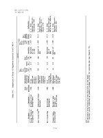
proportions (see Additional file 1). The genome proportions of each type of repetitive DNA are shown in Table 1.
About 30.73% of the genome is repetitive DNAs. According to our results, it has different repetitive DNA types:
retrotransposons (including Copia, Gypsy, and LINE/
SINE), transposons (including hAT, Mutator, DNA/CMCEnSpm, and Tc1-Mariner), rDNA and satellites. Satellite
repeats, which occupy 12.93% of the genome, make up the
Table 1 Repeat elements and their proportions in radish
Elements
Retrotransposon
Copia
GP (genome proportion, %)
11.05
5.81
Gypsy
4.88
LINE/SINE
0.33
Unclassified LTR
0.03
Transposon
1.41
hAT
0.93
Mutator
0.27
DNA/CMC-EnSpm
0.10
Tc1-Mariner
0.11
rDNA
4.32
Satellite
12.93
Unclassified
1.02
Total
30.73
He et al. BMC Plant Biology (2015) 15:105
most dominant part of the repetitive DNAs in radish. The
majority of retrotransposons are Ty1/Copia and Ty3/
Gypsy retrotransposons, with genome proportions of
5.81% and 4.88%, respectively. The genome proportion of
transposons is only 1.41% and the most abundant transposon is hAT, with a 0.93% genome proportion. Estimation of rDNA elements abundance showed that they
comprise 4.32% of the genome.
Identification of subtelomeric repeats and centromeric
repeats in radish
In addition to 5S rDNA and 45S rDNA, three tandemly
organized repeats (CL1, CL25 and CL43) were identified
by bioinformatics analysis of the sequencing data. The
CL1, CL25 and CL43 repeats were estimated to make up
12.32%, 0.44%, and 0.17% of the genome, respectively.
CL43 is a telomeric repeat, consisting of a 7 bp monomer (TTTAGGG, the same as the Arabidopsis telomere
sequence), located at both ends of chromosomes (see
Additional file 2). PCR of CL1 and CL25 resulted in a
ladder like pattern for tandemly organized repetitive
units. To ascertain the size of the monomers of CL1 and
CL25, specific primers were designed for amplifying
these two repeats and then sequenced. According to sequencing results, CL1 consists of ~177 bp monomers
(Figure 1), which is almost exactly the same as the size of
the alphoid-like satellite repeat reported by Grellet [23].
Searching GenBank and PlantSat databases revealed high
Page 3 of 12
similarities to centromeric tandem repeats centBr1 and
centBr2 from Brassica species (~80% identity over 177 bp)
[26] and satellite sequences from Sinapis alba (~78% identity over 165 bp) [27]. The CL25 repeat is characterized by
a ~348 bp monomer unit (Figure 1) and is a newly found
satellite. Similar to CL1, the CL25 sequences shared high
similarities to Brassica species (~78% identity over
348 bp). In addition, a small part (the black rectangle outlined region in Figure 1) of the CL25 sequence present in
the C. elegans.
FISH result showed that CL1 is located at the main
primary constrictions and CL25 appears at the subtelomeric regions (Figure 2a-b). On account of CL25 sharing
a high similarity to Brassica species, we speculated that
CL25 should have a specific distribution pattern in these
species. FISH mapping of CL25 repeats was performed
on metaphase chromosomes of several Brassica species,
including B. rapa (A genome), B. nigra (B genome),
B. oleracea (C genome), and B. napus (AACC), which
are close relatives to radish (Figure 2c-f). Overall, CL25
appeared at subtelomeric regions for all the detected
species, although different species have various numbers
and varied intensities of signals. Intensities of signals are
relatively weak in B. rapa, and strong in B. oleracea. A
different distribution pattern was detected in B. nigra
with strong signals on 4 pairs of chromosomes and weak
signals on 2 pairs of chromosomes. Therefore the CL25
repeat is an ancient repeat which appeared before the
Figure 1 Consensus sequence of CL1 and CL25 repeats. The black rectangle outlines the positions in the sequence that share similarities to
C. elegans.
He et al. BMC Plant Biology (2015) 15:105
Page 4 of 12
Figure 2 FISH mapping of CL1 and CL25 repeats. (a) FISH mapping of CL1 in radish; (b-f) FISH mapping of CL25; (b) radish; (c) B. rapa;
(d) B. nigra; (e) B. oleracea; (f) B. napus. Bars = 5 μm.
differentiation of tribe Brassiceae and radish. It has
maintained its subtelomeric positions in all detected
Cruciferae species.
by the position and intensity of their signals (Figure 3d).
An integrated ideogram of radish metaphase chromosomes is shown in Figure 3e.
Karyotyping analyses of radish
Cloning of CENH3
Given the lack of DNA markers for FISH analysis, detailed molecular karyotype analyses of radish have not
yet been conducted. Repeats identified in this study provide good markers for karyotyping analysis. Sequential
FISH using repetitive DNA sequences (CL1, CL25, 5S
rDNA and 45S rDNA) as probes were performed to
identify radish chromosomes (Figure 3a-c). The CL1 signals appeared at the middle of all of the chromosomes
with varied intensities. The CL25 signals were located at
one arm of chromosomes 1, 4, 5, 6, 7, 8, and 9, and both
arms of chromosome 3 with one pairs of signals. Signals
were large and strong on chromosomes 5 and 7, but
weak on chromosomes 1, 3, 4, 6, 8 and 9. Two pairs of
5S rDNA signals were detected, and one pair of strong
signals were located at the peri-centromeric region of
the short arm of chromosome 2, and the other pair of
weak signals appeared at the peri-centromeric region of
the short arm of chromosome 1. Interestingly, we detected 3 pairs of 45S rDNA signals in early generations,
which is the same as Koo’s results [22]; however, only 2
pairs of the signals were detected 3 generations later (see
Additional file 3). Seeds from the new generation were
used for karyotyping analysis (2 pairs of signals). In our
study, the signals of 45S rDNA were located at the long
arms of chromosomes 2 and 3. Using satellite repeats
(CL1 and CL25) combined with rDNAs as FISH probes,
we distinctly identified individual somatic chromosome
To identity CENH3 in radish, we searched NCBI using the
blastn program (Nucleotide collection, nr/nt) with the
BrCENH3 complementary cDNA sequence (GenBank
accession number GU166737.1) as the query. Two radish
CENP-A gene sequences (AB299183.1 and AB299184.1)
were identified. These two putative CENH3 open reading
frames share high similarity with a small gap and some
SNPs. Based on these two sequences, specific primers
were designed to isolate the complete RsCENH3 coding
region from radish plants. According to cDNA sequencing
results, three transcripts were detected: a 635 bp length of
transcript (1/20), a 513 bp length of transcript (1/20), and
the majority 537 bp length of transcript (18/20). To
analyze the intron/exon structure of RsCENH3, the full
length of genomic DNA sequence of RsCENH3 was amplified using the same primers. On the basis of genomic
DNA results, only one type of DNA sequence was found,
which has a total length of 1415 bp. This sequence shares
100% identity to the AB299183.1 and is comprised of nine
exons and eight introns. By comparison with the full
length genomic DNA sequence, a 635 bp length of transcript transformed from the third intron into an exon, a
513 bp length of the transcript has a deletion from part of
the forth exon, and the major transcript is 537 bp. Considering the translation, alignment to other plant CENH3s,
and the proportion of these transcripts, we deemed that
the small number of transcripts were produced by mis-
He et al. BMC Plant Biology (2015) 15:105
Page 5 of 12
Figure 3 The karyotype and ideograph for radish mitotic metaphase chromosomes. (a) The mitotic metaphase chromosomes (numbered from 1
to 9) were counterstained with DAPI and pseudocolored in red; (b) FISH with the probe of CL25 (red) and 5S rDNA (green); (c). The same spread
was reprobed with the probe of CL1 (green) and 45S rDNA (red); (d) Individual chromosomes were separated from Figure (a-c) and listed
according to their order; (e) Ideogram showing the position and intensity of CL25 (red) and 5S rDNA (green), CL1 (blue) and 45S rDNA
(yellow). Bars = 5 μm.
splicing from the same loci and the CENH3 comprises an
open reading frame (ORF) of length 537 bp encoding a
predicted 178-amino acid (Aa) protein.
Multiple sequence alignment revealed that RsCENH3
shares high identities with CENH3 from Brassica species, 77% identity with BrCENH3, 64% with BnCENH3,
and 74% with BoCENH3. Several prominent features of
the deduced RsCENH3 in comparison with those CENH3s
and canonical histone H3 are as follows (Figure 4). A longer and more divergent N-terminal tail is present in the deduced RsCENH3 sequence (178 amino acids in total) that
is not alignable to BrH3 (136 amino acids in total). Each of
the predicted proteins encoded a histone fold domain with
similarities to histone H3. The loop 1 region in the histone
fold domain is longer than that of canonical histone H3s
(nine amino acids as opposed to seven for BrH3). All of
these findings demonstrate that the sequence identified is
an authentic CENH3 homolog in radish.
DNA sequences associated with RsCENH3
A B. rapa -derived CENH3 antibody (anti-BrCENH3) was
previously used to confirm CENH3-associated centromeric
sequences in different Brassica species [28]. Based on the
similarities of CENH3’s sequence between radish and
Brassica species, we speculated B. rapa -derived CENH3
antibody should recognize the RsCENH3 protein at core
centromeres in radish. To confirm whether it recognizes
the RsCENH3 protein, we applied an immunofluorescence
assay on somatic cells of radish with the anti-BrCENH3
antibody. Signals appeared at the centromeric regions of
He et al. BMC Plant Biology (2015) 15:105
Page 6 of 12
Figure 4 A multiple alignment of CENH3 sequences. A multiple alignment of radish (RsCENH3), Brassica rapa (BrCENH3), Brassica oleracea
(BoCENH3), Brassica nigra (BnCENH3) homologs and Brassica rapa H3 (BrH3). A black rectangle indicates the position of loop1 region. The right
side of the vertical bar is the N-tail region and left side is the histone fold domain.
all 18 metaphase chromosomes (Figure 5d-f). In interphase cells, RsCENH3 signals were located at the edge of
the DAPI intensively stained heterochromatic regions
(Figure 5a-c). It showed that the antibody also could
recognize RsCENH3.
It has been reported that CRB (Centromere-specific
retrotransposons of Brassica) is a core centromeric sequences of Brassica species [28]. We also detected a CRBlike retrotransposon CL4, which represents 1.14% of the
genome and was named CR-Radish in radish. To verify if
the centromere-specific retrotransposon CR-Radish and
the 177-bp satellite repeat CL1 were associated with
RsCENH3 protein in radish, we performed an immunofluorescence assay followed by FISH on the same set of
cells to detect the co-localization of BrCENH3 and centromeric DNA repeats. The size of RsCENH3 immunosignals were relatively uniform among kinetochores while
the size of CL1 signals were uneven among different chromosomes (Figure 6a-d). CL1 signals were overlapped with
the RsCENH3 immuno-signals, although they were significantly larger than the RsCENH3 immuno-signals.
These results suggest that only a limited part of the CL1
sequences are associated with the kinetochore complex.
We also conducted anti-BrCENH3 immunostaining
followed by FISH of the CR-Radish retrotransposon
(Figure 6e-h). Different from that of CL1, CR-Radish
signals were smeared and weak. As expected, the FISH
signals overlapped with most of the immuno-signals.
Therefore, we propose that the RsCENH3 protein is also
associated with CR-Radish. Dual-color FISH showed
most signals of CR-Radish and CL1 were co-localized,
while the signals of CL1 were more concentrated than
CR-Radish signals (Figure 6i-l).
To further confirm our immunostaining results, ChIP
tests with the anti-BrCENH3 antibody were conducted
to assess the association of CL1 and CR-Radish with
RsCENH3. FISH using the ChIPed DNA as a probe
showed high enhanced signals in the centromere regions
of all radish chromosomes. In contrast, using mocked
DNA as a probe showed no obvious signal (see Additional
file 4) which indicates that the centromere sequences were
specifically pulled down by the anti-BrCENH3 antibody in
ChIP. The ChIP-qPCR was performed to verify the enrichment of putative centromeric repeats (Figure 7). Two
specific primers, designed from different regions of CL1,
CL25 and CR-Radish, were used to detect each fragment.
The ChIP-qPCR was repeated three times using CL25 as
extra-centromeric control. RFE value for CL25-1 was
set at 1, and the RFE value of each sequence was normalized using the CL25-1 as a reference. The RFE of the non-
He et al. BMC Plant Biology (2015) 15:105
Page 7 of 12
Figure 5 Anti-BrCENH3 antibody staining in mitotic cells of radish. RsCENH3 localization (red) on somatic interphase cell (a-c) and metaphase
chromosomes (d-f). Bars = 5 μm.
centromeric control CL25-2, 5S rDNA, and 45S rDNA
were low and similar to each other at 1.06 ± 0.04, 1.11 ±
0.03, and 1.41 ± 0.02, respectively (Figure 7). In contrast,
the RFE of the CR-Radish fragments were as high as
22.02 ± 0.94 and 18.54 ± 0.53, respectively. Similarly, the
RFE of the CL1 fragments were 13.71 ± 0.33 and 11.64 ±
0.11, respectively. These results indicate that CR-Radish
and CL1 were significantly enriched in the ChIPed DNA.
Therefore, CR-Radish and CL1 are associated with RaCENH3.
Discussion
Karyotype of radish
Up to now, studies on the radish genome were still limited and few cytogenetic and genomic studies were
carried out [21,22,25]. Comparative analysis of rDNA
and Rfk1 gene distribution in chromosomes of Brassica
species and radish were carried through using FISH
[21,22,29]. However, to our knowledge, a complete karyotype analysis that reliably distinguishes each chromosome
of radish has not been reported. Chromosome identification is critical for cytological analyses, as well as subsequent studies in genomics, taxonomy, and the evolution
of polyploidy, enabling an understanding of the relationship between visible landmarks and genetic or physical
map features [30]. The somatic metaphase chromosomes
of radish are small and lack feasible markers, which make
adequate identification of radish chromosome pairs difficult. In this study, we used RepeatExplorer to conduct
genome-wide analysis of repetitive sequences and obtained two useful cytogenetic markers (CL1 and CL25).
Together with rDNAs, one or two signals were detected
on each chromosome (Figure 3d). We are now able to easily identify all 9 somatic metaphase chromosomes by the
position and intensity of FISH signals. In addition, an integrated metaphase chromosome karyotype was established
(Figure 3e). Our study provides a valuable basis for future
genomic studies.
Dynamic nature of radish genome
Repetitive sequences contribute significantly to extraordinary genome size variation in higher plants [31,32].
Generally speaking, LTR-retrotransposons are the most
abundant element of the genome, especially in big genome species, such as maize [5], wheat [33], and coix
[34]. However, the majority of repetitive sequences are
satellites, which make up 12.932% of the radish genome
in our study. A similar high proportion of satellites were
found in C. rubella and cucumber, in which more than
20% of the genome sequences are satellite repeats [35,36].
Ordinarily, several to dozens of types of satellite repeats
are detected from a number of species [34,37-39]. In our
study, only three satellite repeats were found in radish,
including centromeric repeats, subtelomeric repeats and
telomeric repeats (Figure 3). This is a typical pattern
where the satellite DNA sequences are appear predominantly in the centromeric, pericentromeric and telomeric
regions [40,41]. The dynamic evolutionary processes of
satellite DNA may generate changes in its chromosomal
location and distribution. Some satellite DNA families
were found to be species-specific [42], while others were
He et al. BMC Plant Biology (2015) 15:105
Page 8 of 12
Figure 6 Sequential localization of the anti-BrCENH3 antibody and centromeric repeats on radish. (a) RsCENH3 localization at mitotic metaphase
chromosomes of radish; (b) The same cell was hybridized with CL1; (c) Merged fluorescence signals from a and b; (d) Merged fluorescence
signals from c and chromosomes; (e) RsCENH3 localization at mitotic metaphase chromosomes of radish; (f) The same cell was hybridized with
CR-Radish; (g) Merged fluorescence signals from d and e; (h) Merged fluorescence signals from g and chromosomes; (i) CR-Radish localization at
mitotic metaphase chromosomes of radish; (j) The same cell was probed with CL1; (k) Merged fluorescence signals from g and h; (l) Merged
fluorescence signals from h and chromosomes. Bars = 5 μm.
Figure 7 Sequences associated with RsCENH3. Relative fold enrichments of repeats obtained by ChIP with the anti-BrCENH3 antibody are shown
for radish genomes. CL25 serves as a negative control; CR-Radish and CL1 were associated with CENH3.
He et al. BMC Plant Biology (2015) 15:105
more conserved, and similar sequences may be isolated in
closely related species [26,43]. In our study, we detected 3
pairs of 45S rDNA signals in early generations of the radish, the same result obtained by Koo [22], while only 2
pairs of 45S rDNA signals were detected in later generations (see Additional file 3). It suggests that rDNA also
have a rapid evolution in the genome. Furthermore radish inbred lines from different areas might contain varied ratio of repetitive sequences. 30.73% of the 0713D
genome is repetitive DNA in our study, while repetitive
sequences occupied 26.6% of the Japanese radish
‘Aokubi’ genome [25].Compositions of each type of repetitive elements are also different between these two
radishes. Overall, these results demonstrate the highly
dynamic nature of radish genome.
Rapid evolution of centromere sequence
The centromeres of higher eukaryotes are rich in repetitive DNA sequences which include large arrays of satellite repeats and/or retrotransposon-related repetitive
sequences [8,9]. It has been shown that one single major
satellite repeat is the dominating sequence in all centromeres in most diploid species [8,9]. In our study, the
similar pattern of one type of centromeric satellite repeat
(CL1) was detected by immunostaining and the ChIP
test. However, it has been reported that some plant
and animal species contain multiple satellite repeats
associated with centromeres, such as in the common
bean [44], potato [13], and chicken [45]. Centromeric
satellite repeats diverge rapidly across species and often
do not share any sequence similarity [8]. Several centromeric repeats were identified in potato and its closely
related wide species S. verrucosum, respectively. Nevertheless, only one single homoeologous centromeric
sequence was detected between these two species. This
means centromeric regions of Solanum species show
rapid evolution.
Taxonomic studies and rDNA gene space sequence
analysis demonstrated that genus Brassica is a close relative of the genus Raphanus [46,47]. Our results also
proved this. In this study, a new satellite CL25 was detected, which is distributed in radish and all tested Brassica species and located at the subtelomeric region of all
tested species (Figure 2). Even in closely related species,
centromeric satellites go through rapid evolution. CL1,
the centromeric satellite repeat, shares high similarities
with CentBr1 and CentBr2 sequences. These CentBr
sequences appeared in the A and C genomes of Brassica
species, while the corresponding centromeric repeats
have not yet been identified in the B genome. Even in
the same species, CentBr1 and CentBr2 have different
distribution patterns on chromosomes [26]. These results
indicate that centromeric satellite repeats of Cruciferae
species evolve rapidly.
Page 9 of 12
Conclusions
In this study, we used low-coverage sequencing on
Raphanus sativus L. (2n = 18) to analyze repeat elements. We revealed the genome structure of radish and
found that satellite repeats are most dominating elements,
which is differ from most reported species, in which LTRretrotransposons are the most abundant element of the
genome. The fine karyotyping analysis using satellites and
rDNAs as markers allow us to easily identify each individual somatic metaphase chromosome. Only one putative
CENH3 (RsCENH3) gene was characterized and cloned
from radish. Its deduced amino acid sequence shares high
similarities to those of the CENH3s in Brassica species. In
addition, Immunofluorescence- and ChIP-based assays
demonstrated the functional significance of satellite and
centromere-specific retrotransposon at centromeres. Our
study provides a valuable basis for future genomic studies
in radish.
Availability of supporting data
The data sets supporting the results of this article are available in the NCBI SRA archive (accession no. SRX957720).
Methods
Plant materials
0713D (2n = 2x = 18, R genome), a Chinese Raphanus
sativus L. inbred line, was used for Solexa genome sequencing, ChIP and cytogenetic studies. Plants were
grown in the greenhouse with 16 hours in lights and
8 hours in the dark.
Genomic DNA isolation and Solexa sequencing
DNA was isolated from 5 g of fresh young plant as described previously [48]. DNA was treated with DNase-freeRNase A for 3 h at RT for removing RNA, and purified by
phenol/chloroform precipitation. Pellets were resuspended
to a final concentration of 200–300 ng/μl. The sequencing
was performed by HiSeq2000 platform (BerryGenomics.
Beijing, China). One hundred bp paired-end reads were
obtained from the results.
Data analysis
Following a removal of linker/primer contaminations and
artificially duplicated reads, a set of 5Gb whole genome
Illumina paired end reads (Average length of reads was
100 bp), representing about 4.8× genome equivalent of
radish [24] were used for similarity-based clustering analysis [38]. The clustering analysis was performed using a
read similarity cutoff of 90% over at least 70% of the
shorter sequence length. Reads within individual clusters
were assembled into contigs. Sequence-similarity searches
of assembled contigs were done for finding out which type
and family of repeats they present. Clusters containing
satellite repeats were identified based on graphs and the
He et al. BMC Plant Biology (2015) 15:105
presence of tandem repeats within assembled contig sequences. Satellite sequences were identified using the Tandem Repeat Finder [49]. Clusters corresponding to putative
mitochondrial and plastid contaminations were identified by
searching GenBank and eliminated. The genome proportion
of each cluster was calculated as the percentage of reads.
FISH and immunostaining
In the FISH procedure, mitotic chromosomes were prepared as follows: seeds were geminated on moist miracloth at 28°C in the dark for 2 days, root tips from radish
were collected and treated with pressurized nitrous oxide
for 90 min, fixed in 3:1 (100% ethanol: glacial acetic acid)
Carnoy’s solution for 2 days at room temperature (25°C)
and then stored at −20°C until used. Probes were prepared
by PCR amplification from radish genomic DNA with specific primers (see Additional file 5). The amplified DNAs
were labeled with bio-16-UTP, digoxigenin-11-dUTP or
DEAC (Roche. Basel, Switzerland) using a standard nick
translation reaction. The FISH experiments, including
slide pre-treatment, probe hybridization and signal detection were performed as reported according to published
protocols [17]. Chromosomes were counterstained with
4′, 6-diamidino-2-phenylindole (DAPI) (Vector Laboratories. Burlingame, USA). Images were captured digitally
using a Sensys CCD camera (QIMAGING, RETIGA-SRV,
FAST 1394) attached to an Olympus BX61 epifluorescence microscope (Olympus. Tokyo, Japan). Images were
adjusted with Adobe Photoshop 5.0. In order to draw an
integrated ideogram of radish metaphase chromosomes,
chromosomes in 5 metaphase cells were measured.
In the immunostaining procedure, root tips were fixed in
freshly prepared 4% (w/v) paraformaldehyde solution for
30 min on ice and then washed three times for 10 min in
1× PBS (10 mM sodium phosphate, pH 7.0, and 140 mM
NaCl) on ice. After washing with 1× PBS, the root tips
were directly squashed on slides coated with poly-L-lysine.
After removal of the cover slip, the slides were immersed
in 1× PBS. The slides were incubated for 3 h at 37°C in a
moist chamber with the mouse primary sera antibody
against brassica CENH3 diluted in 1× TNB buffer. Following three rounds of washing in 1× PBS, anti-mouse-Alexa
488 diluted in 1:100 was applied for 1 h at 37°C. After three
rounds of washing in 1× PBS, the slides were dried at room
temperature. For detection of the CENH3 proteins, the
chromosomes were counterstained with DAPI. For a combined detection of the CENH3 proteins and the satellite repeats, the slides were fixed in 4% (w/v) paraformaldehyde
solution for 5 min and washed in 1× PBS for three times,
then the FISH procedure was followed as usual.
Page 10 of 12
10 g of 10-days-old radish plants were used for isolating
nuclei. The isolated nuclei were suspended in 3 ml micrococcal nuclease (MNase) buffer (10% sucrose, 50 mM
Tris–HCl Ph 7.5, 4 mM MgCl2, and 1 mM CaCl2) and
then digested with micrococcal nuclease (Sigma) to produce a chromatin solution. The digested chromatin was
used for ChIP experiments using the BrCENH3 antibody,
and normal mouse serum was used as a mock treatment.
Chromatin with the antibody was incubated with rotation
overnight at 4°C. DNA from the ChIP and input control
samples was diluted in 1× TE.
Quantitative real-time PCR analysis of pelleted DNA
was used to determine the relative fold enrichment (RFE)
of specific sequences within anti-BrCENH3 precipitated
DNA relative to the DNA sample prepared from preblood immunoprecipitation. We used the CL25, which is
located at the chromosome ends, as a negative control to
normalize enrichment of each positive amplicon. Each
sample had three replicates. 5S rDNA and 45S rDNA,
which were not localized at centromere region, were also
used for evaluating reliability of the results. Primers CL251L, CL25-1R, CL25-2L, CL25-2R, 5SL, 5SR, 45SL, 45SR,
CL1-1L, CL1-1R, CL1-2L, CL1-2R, CR-Radish-1L, CRRadish-1R, CR-Radish-2L and CR-Radish-2R were used
for real-time PCR and are listed in Additional file 1:
Table S2. The relative expression levels were calculated according to cycle number. Quantitative PCR data were performed as described previously [28].
Cloning of CENH3 cDNA
To identify radish CENH3 orthologs sequences, the
BrCENH3 complementary cDNA sequence (GenBank accession number GU166737.1), as the query, was searched
by NCBI BLAST. Two radish CENP-A genes sequence
were identified. Total RNA was extracted from leaf tissue
of an inbred line ‘0713D’. RNA samples were treated with
RNase-free DNase (Promega. Madison, USA) and dissolved
in RNase-free double-distilled water. cDNA was synthesized using the RNA and Superscript III RT (Invitrogen,
Carlsbad, USA). The primers CENH3-L and CENH3-R
were used for amplification of full length CDS of CENH3.
The fragments were cloned and sequenced. Multiple
sequence alignment of CENH3 was performed using
MUSCLE [51].
Additional files
Additional file 1: List of the annotation and genome proportion of
clusters.
ChIP and quantitative ChIP-PCR
Additional file 2: FISH mapping of CL43 repeats in radish.
ChIP using the BrCENH3 antibody was performed on radish
nucleosomes as previously described [50]. Approximately
Additional file 3: FISH mapping of 45S rDNA in different
generation of radish. (a) Early generation; (b) Later generation.
He et al. BMC Plant Biology (2015) 15:105
Additional file 4: ChIP-FISH using BrCENH3 antibodies. (a) FISH
signals derived from ChIP using normal mouse serum; (b) FISH signals
derived from ChIP using anti-CENH3 antibodies.
Additional file 5: Primers used in this study.
Abbreviations
FISH: Fluorescent in situ hybridization; Gb: Giga base pairs; Mb: Mega base
pairs; bp: Base pair; ChIP: Chromatin immunoprecipitation.
Competing interests
The authors declare that they have no competing interests.
Authors’ contributions
QH carried out molecular, cytogenetic studies and drafted the manuscript.
ZC carried out bioinformatics analysis, participated in the ChIP assay and
provided helpful discussions. TH helped to maintain the experiment
materials. CB and HL participated in cytogenetic studies. WJ and WM
designed the experiment and revised the manuscript. All authors read
and approved the final manuscript.
Acknowledgments
This research was supported by National Science Foundation of China
(31171563) and the Grand Science and Technology Special Project of
Zhejiang Province (2012C12903-3-6). Authors also thank Dr. Kyle Q.
Hoffmann for linguistic advice in the preparation of this manuscript.
Received: 2 January 2015 Accepted: 23 March 2015
References
1. Kumar A, Bennetzen JL. Plant retrotransposons. Annu Rev Genet.
1999;33:479–532.
2. Bennetzen JL, Ma J, Devos KM. Mechanisms of recent genome size variation
in flowering plants. Ann Bot. 2005;95:127–32.
3. Lysak MA, Koch MA, Beaulieu JM, Meister A, Leitch IJ. The dynamic ups and
downs of genome size evolution in Brassicaceae. Mol Biol Evol. 2009;26:85–98.
4. Arabidopsis Genome Initiative. Analysis of the genome sequence of the
flowering plant Arabidopsis thaliana. Nature. 2000;408:796–815.
5. Maize Genome Initiative. The B73 maize genome: complexity, diversity, and
dynamics. Science. 2009;326:1112–5.
6. Sharma S, Raina SN. Organization and evolution of highly repeated satellite
DNA sequences in plant chromosomes. Cytogenet Genome Res.
2005;109:15–26.
7. Kato A, Vega JM, Han F, Lamb JC, Birchler JA. Advances in plant
chromosome identification and cytogenetic techniques. Curr Opin Plant
Biol. 2005;8:148–54.
8. Henikoff S. Chromosomes on the move. Trends Genet. 2001;17:689–90.
9. Jiang J, Birchler JA, Parrott WA, Dawe RK. A molecular view of plant
centromeres. Trends Plant Sci. 2003;8:570–5.
10. Melters DP, Bradnam KR, Young HA, Telis N, May MR, Ruby JG, et al.
Comparative analysis of tandem repeats from hundreds of species reveals
unique insights into centromere evolution. Genome Biol. 2013;14:R10.
11. Zhong CX, Marshall JB, Topp C, Mroczek R, Kato A, Nagaki K, et al.
Centromeric retroelements and satellites interact with maize kinetochore
protein CENH3. Plant Cell. 2002;14:2825–36.
12. Wang G, Zhang X, Jin W. An overview of plant centromeres. J Genet
Genomics. 2009;36:529–37.
13. Gong Z, Wu Y, Koblízková A, Torres GA, Wang K, Iovene M, et al. Repeatless
and repeat-based centromeres in potato: implications for centromere
evolution. Plant Cell. 2012;24:3559–74.
14. Zhang H, Koblížková A, Wang K, Gong Z, Oliveira L, Torres GA, et al.
Boom-bust turnovers of megabase-sized centromeric DNA in Solanum
species: rapid evolution of DNA sequences associated with centromeres.
Plant Cell. 2014;26:1436–47.
15. Fukagawa T, Earnshaw WC. The centromere: chromatin foundation for the
kinetochore machinery. Dev Cell. 2014;30:496–508.
16. Malik HS, Henikoff S. Major evolutionary transitions in centromere
complexity. Cell. 2009;138:1067–82.
Page 11 of 12
17. Jin W, Melo JR, Nagaki K, Talbert PB, Henikoff S, Dawe RK, et al. Maize
centromeres: organization and functional adaptation in the genetic
background of oat. Plant Cell. 2004;16:571–81.
18. Houben A, Schroeder-Reiter E, Nagaki K, Nasuda S, Wanner G, Murata M,
et al. CENH3 interacts with the centromeric retrotransposon cereba and
GC-rich satellites and locates to centromeric substructures in barley.
Chromosoma. 2007;116:275–83.
19. Uda Y, Hayashi H, Shimizu A. Mutagenic andanti-mutagenic property of
3-hydroxymethylene-2-thioxopyrrolidine, a major product generating from
pungent principle of radish. Lebensm Wiss Technol. 2000;33:37–43.
20. Yamasaki M, Omi Y, Fujii N, Ozaki A, Nakama A, Sakakibara Y, et al. Mustard
oil in ‘shibori daikon’ a variety of Japanese radish selectively inhibits the
proliferation of H-ras-transformed 3Y1 cells. Biosci Biotechnol Biochem.
2009;73:2217–21.
21. Hasterok R, Wolny E, Hosiawa M, Kowalczyk M, Kulak-ksiazczyk S, Ksiazyk T,
et al. Comparative analysis of rDNA distribution in chromosomes of various
species of Brassicaceae. Ann Bot. 2006;97:205–16.
22. Koo DH, Hong CP, Batley J, Chung YS, Edwards D, Bang JW, et al. Rapid
divergence of repetitive DNAs in Brassica relatives. Genomics. 2011;97:173–85.
23. Grellet F, Delcasso D, Panabieres F, Delseny M. Organization and
evolution of a higher plant alphoid-like satellite DNA sequence. J Mol Biol.
1986;187:495–507.
24. Marie D, Brown SC. A cytometric exercise in plant DNA histograms, with 2C
values for 70 species. Biol Cell. 1993;78:41–51.
25. Kitashiba H, Li F, Hirakawa H, Kawanabe T, Zou ZW, Hasegawa Y, et al. Draft
sequences of the radish (Raphanus sativus L.) genome. DNA Res.
2014;21:481–90.
26. Lim KB, Yang TJ, Hwang YJ, Kim JS, Park JY, Kwon SJ, et al. Characterization
of the centromere and peri-centromere retrotransposons in Brassica rapa
and their distribution in related Brassica species. Plant J. 2007;49:173–83.
27. Capesius I. Sequence of the cryptic satellite DNA from the plant Sinapis
alba. Biochim Biophys Acta. 1983;739:276–80.
28. Wang GX, He QY, Liu F, Cheng ZK, Talbert PB, Jin WW. Characterization of
CENH3 proteins and centromere-associated DNA sequences in diploid and
allotetraploid Brassica species. Chromosoma. 2011;120:353–65.
29. Niemelä T, Seppänen M, Badakshi F, Rokka V, Heslop-Harrison JS. Size and
location of radish chromosome regions carrying the fertility restorer Rfk1
gene in spring turnip rape. Chromosome Res. 2012;20:353–61.
30. Harper LC, Cande WZ. Mapping a new frontier; development of integrated
cytogenetic maps in plants. Funct Integr Genomics. 2000;1:89–98.
31. Piegu B, Guyot R, Picault N, Roulin A, Saniyal A, Kim H, et al. Doubling
genome size without polyploidization: Dynamics of retrotransposition
driven genomic expansions in Oryza australiensis, a wild relative of rice.
Genome Res. 2006;16:1262–9.
32. Hawkins JS, Kim H, Nason JD, Wing RA, Wendel JF. Differential lineage-specific
amplification of transposable elements is responsible for genome size variation
in Gossypium. Genome Res. 2006;16:1252–61.
33. International Wheat Genome Sequencing Consortium. Aegilops tauschii draft
genome sequence reveals a gene repertoire for wheat adaptation. Nature.
2013;496:91–5.
34. Cai ZX, Liu HJ, He QY, Pu MW, Chen J, Lai JS, et al. Differential genome
evolution and speciation of Coix lacryma-jobi L. and Coix aquatica Roxb.
hybrid guangxi revealed by repetitive sequence analysis and fine
karyotyping. BMC Genomics. 2014;15:1025.
35. Han YH, Zhang ZH, Liu JH, Lu JY, Huang SW, Jin WW. Distribution of the
tandem repeat sequences and karyotyping in cucumber (Cucumis sativus L.) by
fluorescence in situ hybridization. Cytogenet Genome Res. 2008;122:80–8.
36. Slotte T, Hazzouri KM, Ågren JA, Koenig D, Maumus F, Guo YL, et al. The
Capsella rubella genome and the genomic consequences of rapid mating
system evolution. Nat Genet. 2013;45:831–5.
37. Hnibová 1E, Neumann P, Matsumoto T, Roux N, Macas J, Dolezel J.
Repetitive part of the banana (Musa acuminata) genome investigated by
low-depth 454 sequencing. BMC Plant Biol. 2010;10:204.
38. Macas J, Neumann P, Navrátilová A. Repetitive DNA in the pea (Pisum
sativum L.) genome: comprehensive characterization using 454 sequencing
and comparison to soybean and Medicago truncatula. BMC Genomics.
2007;8:427.
39. Piednoe¨l M, Aberer AJ, Schneews GM, Macas J, Novak P, Gundlach H, et al.
Next-generation sequencing reveals the impact of repetitive DNA across
phylogenetically closely related genomes of Orobanchaceae. Mol Biol Evol.
2012;29:3601–11.
He et al. BMC Plant Biology (2015) 15:105
Page 12 of 12
40. Charlesworth B, Sniegowski P, Stephan W. The evolutionary dynamics of
repetitive DNA in eukaryotes. Nature. 1994;371:215–20.
41. López-Flores I, Ramos-Garrido MA. The repetitive DNA content of eukaryotic
genomes. Genome Dyn. 2012;7:1–28.
42. Kopecna O, Kubickova S, Cernohorska H, Cabelova K, Vahala J, Rubes J.
Isolation and comparison of tribe-specific centromeric repeats within
Bovidae. J Appl Genet. 2012;53:193–202.
43. Torres GA, Gong Z, Lovene M, Hirsch CD, Buell CR, Bryan GJ, et al.
Organization and evolution of subtelomeric satellite repeats in the potato
genome. G3 (Bethesda). 2011;1:85–92.
44. Iwata A, Tek AL, Richard MM, Abernathy B, Fonsêca A, Schmutz J, et al.
Identification and characterization of functional centromeres of the
common bean. Plant J. 2013;76:47–60.
45. Shang WH, Hori T, Toyoda A, Kato J, Popendorf K, Sakakibara Y, et al.
Chickens possess centromeres with both extended tandem repeats and
short non-tandem-repetitive sequences. Genome Res. 2010;20:1219–28.
46. Lü N, Yamane K, Ohnishi O. Genetic diversity of cultivated and wild radish
and phylogenetic relationships among Raphanus and Brassica species
revealed by the analysis of trnK/matK sequence. Breed Sci. 2008;58:15–22.
47. Mun JH, Kwon S, Yang T, Seol Y, Jin M, Kim J, et al. Genome-wide
comparative analysis of the Brassica rapa gene space reveals genome
shrinkage and differential loss of duplicated genes after whole genome
triplication. Genome Biol. 2009;10:R111.
48. Doyle JJ. A rapid DNA isolation procedure for small quantities of fresh leaf
tissue. Phytochem Bull. 1987;19:11–5.
49. Benson G. Tandem Repeat Finder: a program to analyze DNA sequence.
Nucleic Acids Res. 1999;27:573–80.
50. Nagaki K, Talbert PB, Zhong CX, Dawe RK, Henikoff S, Jiang J. Chromatin
immunoprecipitation reveals that the 180-bp satellite repeat is the key
functional DNA element of Arabidopsis thaliana centromeres. Genetics.
2003;163:1221–5.
51. Edgar RC. MUSCLE: a multiple sequence alignment method with reduced
time and space complexity. BMC Bioinformatics. 2004;5:113.
Submit your next manuscript to BioMed Central
and take full advantage of:
• Convenient online submission
• Thorough peer review
• No space constraints or color figure charges
• Immediate publication on acceptance
• Inclusion in PubMed, CAS, Scopus and Google Scholar
• Research which is freely available for redistribution
Submit your manuscript at
www.biomedcentral.com/submit