Genome-wide analysis of the gene families of resistance gene analogues in cotton and their response to Verticillium wilt
Bạn đang xem bản rút gọn của tài liệu. Xem và tải ngay bản đầy đủ của tài liệu tại đây (1.93 MB, 15 trang )
Chen et al. BMC Plant Biology (2015) 15:148
DOI 10.1186/s12870-015-0508-3
RESEARCH ARTICLE
Open Access
Genome-wide analysis of the gene families
of resistance gene analogues in cotton and
their response to Verticillium wilt
Jie-Yin Chen1†, Jin-Qun Huang2†, Nan-Yang Li1†, Xue-Feng Ma1, Jin-Long Wang1, Chuan Liu2, Yong-Feng Liu2,
Yong Liang2, Yu-Ming Bao1 and Xiao-Feng Dai1*
Abstract
Background: Gossypium raimondii is a Verticillium wilt-resistant cotton species whose genome encodes numerous
disease resistance genes that play important roles in the defence against pathogens. However, the characteristics of
resistance gene analogues (RGAs) and Verticillium dahliae response loci (VdRLs) have not been investigated on a
global scale. In this study, the characteristics of RGA genes were systematically analysed using bioinformatics-driven
methods. Moreover, the potential VdRLs involved in the defence response to Verticillium wilt were identified by
RNA-seq and correlations with known resistance QTLs.
Results: The G. raimondii genome encodes 1004 RGA genes, and most of these genes cluster in homology groups
based on high levels of similarity. Interestingly, nearly half of the RGA genes occurred in 26 RGA-gene-rich clusters
(Rgrcs). The homology analysis showed that sequence exchanges and tandem duplications frequently occurred
within Rgrcs, and segmental duplications took place among the different Rgrcs. An RNA-seq analysis showed that
the RGA genes play roles in cotton defence responses, forming 26 VdRLs inside in the Rgrcs after being inoculated
with V. dahliae. A correlation analysis found that 12 VdRLs were adjacent to the known Verticillium wilt resistance
QTLs, and that 5 were rich in NB-ARC domain-containing disease resistance genes.
Conclusions: The cotton genome contains numerous RGA genes, and nearly half of them are located in clusters,
which evolved by sequence exchanges, tandem duplications and segmental duplications. In the Rgrcs, 26 loci were
induced by the V. dahliae inoculation, and 12 are in the vicinity of known Verticillium wilt resistance QTLs.
Keywords: Cotton, Verticillium wilt-resistant, Resistance gene analogues, RGA-gene-rich clusters, Verticillium dahliae
response loci
Background
Resistance (R) genes play a central role in recognising effectors from pathogens and in triggering downstream signalling during plant disease resistance [1, 2]. To date, more
than 112 R genes and 104,310 putative R-genes present in
a wide variety of plants species and conferring resistance
to 122 pathogens [3]. The known R proteins can be
grouped into several super-families based on the presence
of a few structural motifs, including nucleotide-binding
sites (NBSs), leucine-rich repeat (LRR) domains, Toll/
* Correspondence:
†
Equal contributors
1
Laboratory of Cotton Disease, Institute of Agro-Products Processing Science &
Technology, Chinese Academy of Agricultural Sciences, Beijing 100193, China
Full list of author information is available at the end of the article
Interleukin-1 receptor (TIR) domains, coiled-coil (CC) domains and transmembrane (TM) regions [4, 5]. Generally,
the most prevalent R genes in plants are of the NBS-LRR
type, which are divided into two sub-classes based on the
presence of an N-terminal CC or TIR domain [6, 7]. For
example, 480 NBS-LRR proteins are encoded by the rice
genome [8].
Previous studies demonstrated that many R genes are
clustered in plant genomes [9]. To date, clusters of R
genes have been reported in several plant genomes, including Arabidopsis [7], rice [10], soybean [11], Lotus
japonicus [12], Medicago truncatula [13] and Phaseolus
vulgaris [14]. In Arabidopsis, the genome was found to
encode 159 NBS-LRR genes, and 113 of these genes
© 2015 Chen et al. This is an Open Access article distributed under the terms of the Creative Commons Attribution License
( which permits unrestricted use, distribution, and reproduction in any medium,
provided the original work is properly credited. The Creative Commons Public Domain Dedication waiver (http://
creativecommons.org/publicdomain/zero/1.0/) applies to the data made available in this article, unless otherwise stated.
Chen et al. BMC Plant Biology (2015) 15:148
occurred in 38 clusters [15]. A similar phenomenon was
also found in the rice genome, in which 76 % of the rice
NBS-LRR genes was arranged in 44 gene clusters, with
the others occurring as singletons [8]. The lengths of
RGA gene clusters varied from dozens of kilobases (kb)
to several megabases (Mb). For example, RGA genes
were tightly linked to the RPP5 cluster in Arabidopsis,
which covers less than 100 kb [16], while the RGA genes
were distributed over several Mb of the RGC2 locus in
lettuce [17]. Different R genes from the same cluster can
confer resistance to different pathogens or to different
variants of a single pathogen [18, 19]. For example, the
Cf-9 gene cluster contains two Cf-9 and Cf-9B homologues that recognise the Avr9 and Avr9B effectors, respectively, in Cladosporium fulvum, and contribute to
the resistance against tomato leaf mould disease. Other
homologous genes in the cluster may serve as a reservoir
of variation for the generation of R genes with new specificities [20–22].
Previous research suggested that the evolution of RGA
clusters is usually mediated by sequence exchange, tandem duplication, segmental duplication, or gene conversion [9, 23, 24]. Frequent sequence exchanges tend to
homogenize the members of a gene family, like the
RGC2 genes in lettuce [25], the R1 cluster in Solanum
demissum, and the Cf-9 cluster in tomato [26, 27]. Tandem and segmental genomic duplications are also important in the evolution of RGA genes [23], which
frequently occur in NBS-LRR genes clusters, and led to
the formation of the phylogenetic lineage of NBS-LRR
genes in the Arabidopsis genome [7, 28]. The evolution
of the HcrVf cluster in apple was primarily dependent
on gene duplication, with four HcrVf genes originating
from a single progenitor gene by two sequential duplication events [29]. RGA’s evolution by gene conversion
resulted in high levels of sequence similarity, close
physical clustering, and the local recombination rate
[15, 28, 30]. In conclusion, the plants employed a complicated mechanism on the RGA genes evolution to response the variations of pathogens.
Cotton is an important crop worldwide because of its
natural fibres and oil seeds. The cotton acreage in China
has reached 4.69 million hectares, which produced 6.83
million tons of cotton in 2012 (Data from the National
Bureau of Statistics in China). At present, Verticillium wilt
caused by Verticillium dahliae is the most destructive disease of cotton, and the survival structures produced by
pathogens may remain viable in the soil, persistently
threatening crops, for more than 20 years [31]. In some
years, more than 50 % of the cotton acreage is affected by
Verticillium wilt, significantly reducing the fibre quality
and resulting in yield losses (National Cotton Council
of America Disease Database). Because of its unique
ecological niche in the plant’s vascular, Verticillium wilt is
Page 2 of 15
difficult to control using fungicides, chemicals and cultivation measures [32]. Improving genetic resistance is considered the best method to overcome Verticillium wilt, and
at least 80 different Verticillium wilt resistance quantitative trait loci (QTLs) have been reported in cotton
[33–37]. However, Gossypium hirsutum appears to lack
genetic resistance against V. dahliae [38, 39].
Gossypium barbadense, which is a cultivated tetraploid
cotton species, showed resistance or tolerance to Verticillium wilt [40]. To date, the transcriptomes and proteomes
of this Verticillium wilt-resistant cotton’s responses to V.
dahliae have been analysed, and phytoalexin biosynthesis
and hormone signalling were found to have important
roles in pathogen defense [41–46]. Moreover, several
genes that contribute to the defence response against
Verticillium wilt have been reported, including GbCAD1,
GbSSI2 [43], GbRLK [47], GbSTK [48], GbTLP1 [49] and
GbVe/GbVe1 [50, 51].
Recently, the genome sequence of a diploid cotton,
Gossypium raimondii, which is a Verticillium wilt-resistant
wild relative of cotton, was completed [52, 53]. It is
commonly thought that the tetraploid cotton species G.
hirsutum and G. barbadense were derived from a cross between a D-genome species as the pollen-providing parent
and an A-genome species as the maternal parent, and that
G. raimondii is the putative D-genome parent [54, 55].
Previous research showed that the cotton genome encodes
numerous NBS domains and that some of these genes
formed gene clusters [53, 56]. A transcriptome analysis
showed that some RGAs are involved in the defence response against V. dahliae [42, 46]. However, there are no
systematic studies of RGA genes in the cotton genome,
and the genetic resistance to Verticillium wilt is unclear.
In this study, a global analysis, including sequence
features, gene distribution and the evolution of RGA genes
in the G. raimondii genome was performed. Highthroughput RNA-seq was used to identify the RGA genes’
transcriptome in a V. dahlia-resistant cultivar of G. barbadense and to screen for potential Verticillium dahliae response loci (VdRLs) in the gene clusters. Moreover, the
association between the VdRLs and Verticillium wilt resistance QTLs were analysed to screen the Verticillium
wilt-response loci in cotton.
Results
Analysis of RGA genes in the G. raimondii genome
In this study, we focused on the RGA genes in the G.
ramondii genome that probably participate in the disease resistance response. In total, 1004 RGA genes were
classified into 11 families (R-I – R-XI) based on the integrated annotation of conserved motifs or domains in the
G. ramondii genome [53]. The genome included 32 CCNBS-LRR genes, 60 cysteine-rich receptor-like kinase
(RLK) genes, 46 genes encoding disease resistance family
Chen et al. BMC Plant Biology (2015) 15:148
proteins/LRR family proteins, 58 genes encoding leucinerich receptor-like protein kinase family proteins, 225 genes
encoding LRR protein kinase family proteins, 44 genes encoding LRR receptor-like protein kinase family proteins, 78
genes encoding LRR transmembrane protein kinases, 79
genes encoding LRR and NB-ARC (Nucleotide-Binding
adaptor shared by APAF-1, Resistance proteins and CED4) domain-containing disease resistance proteins, 194 genes
encoding NB-ARC domain-containing disease resistance
proteins, 144 receptor-like proteins (RLP) genes and 44
TIR-NBS-LRR genes (Additional file 1: Table S1). A statistical analysis showed that more than half of the RGA genes
were located on three chromosomes, with 194, 182 and
143 on Chr09, Chr07 and Chr11, respectively (Additional
file 2: Figure S1). These results indicated that the cotton
genome contains many RGA genes and numerous of them
trend to enrich in several chromosome in cotton genome.
Page 3 of 15
Generally, RGA genes contain conserved domains or
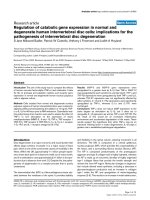
motifs, such as NBSs and LRRs. In a comparative analysis, most of the RGA genes, and their encoded proteins, showed a high identity with one another (Fig. 1A, B),
particularly RGA genes on Chr07 and Chr09, which
shared high identities (up to 80 %) with one another
(Additional file 1: Table S2). To investigate the correlation among all RGA genes, the similarity among RGA
genes were compared according to the chimeric sequence which connected the RGA gene sequences from
Chr01 to Chr13 in a series. Interestingly, the comparison of the chimeric sequence with itself showed a high
similarity apart from small similarity blocks (less than
the length of the smallest RGA gene, 216 bp) and selfmatch (Fig. 1C), indicating that many RGA genes are
similar in the cotton genome. Moreover, the chimeric
sequence segments from the same chromosome were
Fig. 1 Similarity analysis of RGA genes in the G. raimondii genome. (A) The identity matrix of all RGA genes versus all RGA genes. The RGA genes
were arranged in a series from Chr01 to Chr13. “UN” represents the RGA genes that cannot presently be mapped to chromosomes. The identity
level between each two genes was determined by BLASTN (Version 2.2.23). (B) The identity matrix of all RGAs encoding proteins versus all RGAs
encoding proteins. The identity level between each two proteins was determined using the BLASTP program (Version 2.2.23). (C) Homology
analysis between two chimeric sequences of RGA genes. The chimeric sequence was constructed by ligating the RGA sequences in a series from
Chr01 to Chr13. The similarity blocks were determined using the BLASTN program (Version 2.2.23) with chimeric sequences, ignoring self-matches
and filtering out the similarity blocks based on the length of the smallest RGA gene (216 bp)
Chen et al. BMC Plant Biology (2015) 15:148
more similar than sequence segments from different
chromosomes (Fig. 1C), indicating that RGA genes on
the same chromosome were more closely related than
genes on different chromosomes.
The homology clustering of RGA genes also indicated
that RGA genes are conserved in cotton. Of the 1004
RGA genes, 974 could be divided into 45 homology
groups (HG), with at least two genes in each HG, under
the clustering conditions of match rate and identity being
more than 33 % and 30 %, respectively. Of these, 838 were
classified into 11 HGs, with HG13 containing the minimum 23 genes and HG17 containing the maximum 242
genes (Additional file 1: Table S3). Not surprisingly, most
RGA genes in the same family could be clustered into a
single HG based on a conserved feature. For example,
five-sixths of the RGA genes in the R-II family were clustered into HG22. However, the genes of five RGA gene
families were clustered into multiple groups, including RI, R-V, R-VIII and R-IX. The RGA genes of the R-V family
were clustered into two major HGs, HG17 and HG21
(Additional file 1: Table S3), indicating that the RGA gene
families were not always clustered in one HG but could be
clustered into different HGs. Moreover, the RGA genes
could also be clustered into HGs using highly rigorous
Page 4 of 15
conditions. The 306 RGA genes were divided into 104 HGs
when the match rate and identity were more than 80 % for
each gene (Additional file 2: Figure S2). The RGA genes in
the same HGs are physically linked, such as 7 genes in the
sub-HG of HG05 (HG05-04) that are closely linked in a
small region that encodes 11 genes (Gorai.007G324100.1–
Gorai.007G325100.1) (Additional file 1: Table S4). These results suggested that many RGA genes, which are probably
multi-copy genes in cotton, are closely linked in the cotton
genome.
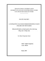
The phylogenetic relationship analysis of RGA genes
showed that most RGA genes could be arranged in
clades in accordance with RGA gene families, such as RII, R-III and R-IV (Fig. 2). These results also corresponded to the homology clustering, showing that the
major HGs in an RGA gene family were arranged in a
clade. For example, most R-II family genes were clustered into HG22, which was arranged in a single clade
(Fig. 2; Additional file 1: Table S3). Although most of the
R-V family genes could be arranged together in the
phylogenetic tree, the R-V clade was split into three
parts (Fig. 2), which indicated that variation occurred in
the R-V family. More persuasive evidence showed four
RGA gene families (R-I, R-VIII, R-IX and R-XI) which
Fig. 2 Phylogeny analyses of RGA genes in the G. raimondii genome. The phylogenetic tree of RGA genes was constructed using the protein
sequences by the neighbour-joining method, with 1000 bootstrap replicates. The branches of the mixed clade included four RGA gene families,
which are marked in purple. Other conserved clades of RGA gene families are rendered in different colours
Chen et al. BMC Plant Biology (2015) 15:148
mainly contain the NBSs and LRRs domain were arranged
in a mixed clade (Fig. 2). Together, these results indicated
that the variation in RGA genes is as important as the
conservation during the cotton genome’s evolution.
Many RGA genes are deposited in gene clusters
In the G. ramondii genome, nearly half of the RGA
genes were allocated to 26 Rgrcs (Fig. 3; Additional
file 2: Figure S3). The total length of these Rgrcs is ~
16.7 Mb, and there were 1148 genes, including 489 RGA
genes. The average proportion of RGA genes in Rgrcs is
significantly higher than in the whole genome, 42.6 %
compared with 2.7 %. The average whole gene density was
higher in Rgrcs (14.5 kb/gene) than in the whole genome
(19.7 kb/gene) (Additional file 1: Table S5). Among these
Rgrcs, Rgrc14 and Rgrc11 are the two largest clusters,
which cover ~4.2 and 3.3 Mb, respectively, and contained
82 and 103 RGA genes, respectively (Additional file 1:
Table S5). Most of the Rgrcs were located on Chr02,
Chr07, Chr09, Chr10 and Chr11 (Fig. 3; Additional file 1:
Table S5). Moreover, more than half of the RGA genes in
the eight gene families occurred in these clusters, except
those of RGA families R-IV, R-V and R-VII. Only 15.5 % of
RGA genes in the R-V family occurred in Rgrc clusters
(Additional file 1: Table S6). These results suggested that
many RGA genes occur in gene clusters in the cotton
genome.
To investigate how Rgrcs are related, all of the proteins encoded by Rgrcs were analysed using homology
clustering. Clearly, most RGA genes are homologous to
those clustered in the same HGs within the Rgrcs. This
is also true for other genes in the Rgrcs that do not encode RGA genes, such as Rgrc2, Rgrc14 and Rgrc15.
(Fig. 4). The homology of most genes within Rgrcs probably indicates that Rgrcs undergo tandem duplications
Page 5 of 15
or sequence exchanges during their evolution. Moreover,
most proteins encoded in different Rgrcs also clustered
into same HGs (Fig. 4). Thus, the genes in different
Rgrcs are homologous, indicating that some Rgrcs were
probably generated from other Rgrcs by segmental duplications in cotton.
Homology analysis of the chimeric sequence, all the
Rgrcs sequences connected in series from Chr01 to
Chr13, showed that the Rgrcs was highly similar after
apart from the small (less than the length of the smallest
RGA gene, 216 bp) and self-matching similarity blocks
(Additional file 2: Figure S4A). In total, 984 high similarity blocks in the chimeric sequence were matched to
each other (up to 3 kb, ignoring self-match), except for
the sequences of Rgrc4 and Rgrc20, and the identities
of almost all the similarity blocks were close to 80 %
(Additional file 2: Figure S4B/C). Of the similarity blocks,
589 belonged to “Rgrc-self-similarity”, including 300 blocks
within Rgrc14, and 78 blocks inside in Rgrc11 (Additional
file 2: Figure S4B), indicating that the Rgrc sequences are
similar by themselves, which could be the result of tandem
duplication or sequence exchange. However, parts of the
similarity blocks were also found among different Rgrcs,
such as 42 matching blocks between Rgrc11 and Rgrc14,
and 22 matching blocks between Rgrc11 and Rgrc24.
(Additional file 2: Figure S4B), suggesting that some Rgrcs
originated by segmental duplication in cotton.
RGA gene expression responses to V. dahliae infection
Analysis of RNA-seq data
In this study, G. barbadense cv. 7124, which is considered
to be V. dahliae-resistant (Additional file 2: Figure S5),
was inoculated with the highly aggressive defoliating V.
dahliae strain Vd991. The inoculated root samples (2, 6,
12, 24, 48 and 72 h) were collected to identify differentially
Fig. 3 The distribution of Rgrcs in the G. raimondii genome. All genes encoded by the G. raimondii genome were arranged in a series from Chr01
to Chr13. The ratio of RGA genes was calculated in the moving window (50 genes/window, walking forward 10 genes each time). RGA gene
frequencies greater than 10 % were considered Rgrcs and clusters only containing 6 RGA genes in a window, but distributed evenly, were
removed. The X-axis represents the number of genes in the cotton genome and the Y-axis represents the RGA gene ratio in the moving window
Chen et al. BMC Plant Biology (2015) 15:148
Page 6 of 15
Fig. 4 Homology clustering of proteins encoded by genes in the Rgrcs of the G. raimondii genome. The homologous relationships were
determined among proteins encoded by genes in the Rgrcs. The same homology groups of RGA genes are linked with red lines, while other
genes in the same homology groups are linked with green lines. The outer ring represents the homology groups inside in Rgrcs, and the inner
ring represents homology groups in different Rgrcs
expressed genes (DEGs) of RGAs using high-throughput
RNA-seq. For extremely deep sequencing, ~200 million
clean reads for each sample were generated, with quality
control (Q ≥ 20) (Additional file 1: Table S7). Of these
reads, ~76 % matched the reference genome of G. raimondii, including ~140 million unique matched reads and ~13
million multi-position matched reads (Additional file 1:
Table S7).
For DEG detection, the reads per exon kb per million
mapped sequence reads (RPKM) was calculated for each
gene and filtered using the false discovery rate (FDR) and
with the p-value. In total, 28,360 DEGs were detected in
the cotton genome at six inoculated time points, with
13,229 genes in common at different time points (FDR <
0.001, p < 0.001), 17,517 DEGs in all inoculated time
points and 9811 genes in common (FDR < 0.001, p <
0.001, and log2Ratio ≥ |1.0|), 8122 DEGs in all inoculated
time points and 5106 genes in common (FDR < 0.001, p <
0.001, and log2Ratio ≥ |2.0|) (Additional file 1: Table S8;
Additional file 3: Table S9). The number of up-regulated
DEGs peaked at 48 h after inoculation, and the number of
down-regulated DEGs gradually decreased from 2 to 72 h
(Additional file 2: Figure S6), which corresponded to the
important infection time point of 48 h in V. dahliae, for
the penetration of hyphae into the roots was evident about
two days [57–60].
DEGs of RGA genes
In the DEGs set, 723 RGA genes were induced in cotton
inoculated with V. dahliae, with 319 RGA genes in common at six time points (FDR < 0.001, p < 0.001) (Additional
file 1: Table S8). Real-time quantitative RT-PCR (qRTPCR) showed that the fold-change of DEGs is reliable
(Additional file 2: Figure S7). As with the DEGs in the
whole genome, the DEGs of RGA genes were also obviously induced at 48 h after inoculation (Additional file 2:
Figure S6). The statistical analysis of DEGs showed that all
11 RGA families could respond to the V. dahliae inoculation at all of the time points, although the proportion of
DEGs in the RLP family was relatively small (Additional
file 1: Table S10). These results suggested that RGA genes
are involved in the cotton response to V. dahliae. The expression pattern analysis showed that RGA gene families
that responded to V. dahliae could be classified into the
early response stage (~2–12 h) and later response stage
(~24–72 h). In the later response stage, the number of
Chen et al. BMC Plant Biology (2015) 15:148
RGA genes and their expression levels were induced more
obvious than in the early response stage (Additional file 2:
Figure S8). These results indicated that activating the later
response stage is important to the resistant cotton plant’s
response to V. dahliae.
Many genes in the plant-pathogen interaction pathway
are RGA genes, which play an important role in disease
resistance. In this study, 451 differentially expressed
RGA genes were induced in cotton inoculated with V.
dahliae, and mapped to the plant-pathogen interaction
pathway based on the Kyoto Encyclopedia of Genes and
Genomes (KEGG) annotation (Fig. 5), including eight
types of homologous genes, such as BAK1, FLS2 and
EFR (Additional file 1: Table S11). Moreover, some genes
homologous to signal factors in the plant-pathogen
interaction pathway, which are not RGA genes, were
also activated, such as protein kinases and transcription
factors (Fig. 5). In addition, genes in the phytoalexin biosynthesis pathways, including those for phenylpropanoids, flavonoids and diterpenoids, were also induced in
Page 7 of 15
cotton in response to V. dahliae (Additional file 2:
Figure S9). Overall, the transcriptome results indicated
that many RGA genes, which probably participated in
the plant-pathogen interaction pathway and regulated
the defence response, were induced in cotton.
DEGs in Rgrcs
The expression pattern analysis of DEGs in Rgrcs indicated that the RGA genes were up-regulated more often
than other genes in Rgrcs (Additional file 2: Figure S10),
which suggested that RGA genes were more sensitive to
V. dahliae inoculation than the other genes in Rgrcs. To
investigate the potential RGA gene responses to V. dahliae infection, highly rigorous conditions (log2Ratio ≥
|2.0|, with more than one up-regulated post-infection
time point) were used for screening in this study. In
total, 168 differentially expressed RGA genes were identified as potential Verticillium wilt response genes. Of
these genes, the proportion of potential Verticillium wilt
resistance genes in R-II, R-III and R-IV families was
Fig. 5 DEGs homologous to the genes of the plant-pathogen interaction pathway. The DEG genes were screened using FDR < 0.001, p < 0.001,
and log2Ratio ≥ |1.0| at all six inoculation time points. The red box represents the differentially expressed RGA genes that map to the plant-pathogen
interaction pathway, the pink box represents the other DEGs that map to the plant-pathogen interaction pathway, and the blue and white box
represents the reference KEGG pathway (map04626)
Chen et al. BMC Plant Biology (2015) 15:148
higher than in other families (Additional file 1: Table S12
and Table S13). Notably, 64 DEGs occurred in 19 Rgrcs,
and 63 of them were distributed in the 26 small regions
defined VdRL01 to VdRL26 (Fig. 6; Additional file 1:
Table S12-S14). The total length of the VdRLs is ~2.4 Mb,
and a minimum of 15 VdRLs contain at least two significantly differentially expressed RGA genes (Additional
file 1: Table S14). A total of 39 differentially expressed
RGA genes in the VdRLs belonged to the R-II, R-VII and
R-IX families (Additional file 1: Table S12), indicating that
these RGA genes were important to the cotton response
to Verticillium wilt. Moreover, most VdRLs were primarily
distributed in the small regions of a few chromosomes,
particularly Chr07 and Chr09, which included seven and
six VdRLs respectively (Additional file 1: Table S14). A further analysis showed that the RGA genes of nearly half of
the VdRLs encoded NB-ARC domain-containing disease
resistance proteins, and the RGA genes of the other
VdRLs primarily encoded cysteine-rich RLKs, leucine-rich
repeat protein kinase family proteins and RLPs (Additional
file 1: Table S15). These results indicated that some RGA
genes in the Rgrcs were strongly induced and a portion of
them formed the VdRLs that participated in Verticillium
wilt response in cotton.
VdRLs adjacent to Verticillium wilt resistance QTLs
To detect the co-localization of VdRLs and QTLs, which
had been identified to be associated with the Verticillium
Page 8 of 15
wilt resistance in cotton [33–37], the locations of these
QTLs in the diploid cotton genome were analysed based
on the information provided by their corresponding
markers. Among the 81 markers for these QTLs, 70 could
be located on the diploid cotton genome (Additional file 1:
Table S16), and 8 markers were adjacent to the VdRLs
(Fig. 7; Additional file 1: Table S14). In total, 13 VdRLs
were located on 6 chromosomes (3, 6, 7, 9, 10 and 11) with
a physical distance of less than 3 Mb to the closest QTL
marker, and 6 of them (VdRL06, VdRL07, VdRL11,
VdRL18, VdRL19 and VdRL25) were less than 1 Mb from
the closest marker (Fig. 7; Additional file 1: Table S14),
suggesting that these VdRLs were positively correlated
with the Verticillium wilt response. Moreover, the RGA
genes in five VdRLs (VdRL07, VdRL11, VdRL12, VdRL13
and VdRL18) encoded NB-ARC domain-containing disease resistance proteins, of which three (VdRL07, VdRL11
and VdRL18) were close to Verticillium wilt resistance
QTLs (Additional file 1: Table S14 and Additional file 1:
Table S15).
Interestingly, six VdRLs (VdRL07 and VdRL09-VdRL13)
located on Chr07 were found close to three Verticillium
wilt resistance QTL markers (with a physical distance of
less than 3 Mb), MUCS219, NAU5428 and CIR196 (Fig. 7;
Additional file 1: Table S14). This region, in fact, extends
about 10 Mb, which includes Rgrc10 and Rgrc11, and contains seven VdRLs (VdRL07-VdRL13). The physical distance betweenVdRL08 and the closest marker is ~3.66 Mb
Fig. 6 Analysis of RGA gene expression patterns and the screening of potential VdRLs. The RGA genes were arranged in a series from Chr01 to Chr13.
RGA genes belonging to the 26 Rgrcs are shown in red. The fold-change of log2Ratio ≥ |2.0| is marked in dotted lines. The potential VdRLs were screened
from Rgrcs using a log2Ratio ≥ |2.0|, and having more than one infection time point up-regulated. The potential VdRLs were marked with asterisks. The
numbers 2, 6, 12, 24, 48, and 72 in the boxes represent the time points (in hours) of the cotton inoculation with V. dahliae
Chen et al. BMC Plant Biology (2015) 15:148
Page 9 of 15
Fig. 7 Correlation analysis between VdRLs and Verticillium wilt resistance QTLs in cotton. The physical location of the VdRLs and disease
resistance QTLs were determined by their positions in the diploid cotton genome of G. raimondii. The VdRLs are marked in red and the QTLs
markers are labelled in blue
(Fig. 7; Additional file 1: Table S14). Of these seven VdRLs
on Chr07, five were enriched for the NB-ARC domaincontaining disease resistance genes, and two (VdRL07 and
VdRL13) were close to the Verticillium wilt resistance
QTLs (less than 1 Mb) (Fig. 7; Additional file 1: Table S14).
Overall, these results suggested that the VdRLs located on
Chr07, which mainly encoded NB-ARC domain-containing
disease resistance proteins, were closely associated with
Verticillium wilt resistance in cotton.
Discussion
Plants have evolved a complicated and effective innate immune system to recognise, or respond to, many pathogenic organisms using R genes [1, 2]. At present, many R
genes have been cloned from plants, and they can be divided into at least five classes based on conserved structural motifs, such as NBSs, LRRs and TIRs [4, 6]. In
recent years, more than 20 plant genomes have been sequenced, and ~37,000 RGA genes were predicted based
on conserved structural motifs [61]. Clearly, an analysis of
the RGA genes in the genome will be useful for speculating on R gene evolution and for applying RGAs in cotton
breeding. Recently, the genome of a diploid, G. raimondii,
which is a Verticillium wilt-resistant wild relative of cotton, was sequenced [52, 53]. In this study, all probable
RGA genes encoded by the G. raimondii genome were
systematically analysed, and potential Verticillium wilt resistance loci/genes were identified using the bioinformatics analysis of transcriptome and QTL data.
In the G. raimondii genome, at least 300 genes encode
NBS domains and most of these genes are of the CC-NBS
or CC-NBS-LRR type [53, 56]. In this research, 1004 RGA
genes were found in the G. raimondii genome based on an
integrated annotation, and they were primarily distributed
in Chr07, Chr09 and Chr11 (Additional file 2: Figure S1;
Additional file 1: Table S1). As expected, the RGA genes
showed a high similarity amongst themselves based on
their conserved structural motifs, particularly when they
occurred in small genomic regions of the same chromosome (Fig. 1, Additional file 1: Table S2). In contrast, some
RGA genes in different families also showed similarities
and were of the same phylogenetic lineage (Figs. 1 and 2).
These results may indicate that the evolution of RGA
genes in cotton had the dual characteristics of conservation and genetic variation, as did RGC2 genes in lettuce
[25]. RGA genes residing in clusters has been observed in
many plant genomes [7, 10–14]. In Arabidopsis thaliana,
more that 71 % of the NBS-LRR genes are arranged in 38
clusters [15], and the same characteristic is true of
NBS-LRR genes in the rice genome [8]. As in other
plants, the RGA genes in the G. raimondii genome reside in clusters (Fig. 3; Additional file 2: Figure S3; Additional file 1: Table S6). Previous studies have shown
that the clustering of RGA genes is usually caused by
tandem duplications [7, 62–64] or sequence exchanges
[9], which have been detected in many RGA gene clusters [17, 19, 26, 65–67]. Similar results were found in
the G. raimondii genome, where most of the RGA
Chen et al. BMC Plant Biology (2015) 15:148
genes are homologous and linked together to form the
Rgrcs (Additional file 2: Figure S2; Additional file 2:
Figure S4; Additional file 1: Table S4), indicating that
tandem duplication or sequence exchanges could have
occurred frequently in the evolution of RGA genes or
Rgrcs. Segmental duplication is another evolutionary
mechanism in RGA genes that could randomly translocate the genes in chromosomes, giving rise to a substantial number of RGA genes [9, 28, 68]. This was also
found in our analysis (Additional file 2: Figure S4B),
probably suggesting that the segmental duplication
could happen in the RGA genes evolution. Together,
these results probably indicated that tandem duplication, sequence exchange, and segmental duplication are
important to the evolution of RGA genes and Rgrcs.
Verticillium wilt is the most destructive disease in cotton, and there are no effective methods to prevent this
disease at present. Although improving genetic resistance is the direct method to combat Verticillium wilt, it
has not been successful in G. hirsutum, which accounts
for more than 90 % of the total cotton acreage in the
world, because of the lack of genetic resistance [38]. G.
barbadense is considered to be a resistant species, and
many studies regarding Verticillium wilt resistance have
been reported [36, 43, 47–51]. Recently, a transcriptome
analysis showed that some RGA genes were induced in
G. barbadense inoculated with V. dahliae [42, 46], indicating that the RGA genes contribute to the defence response in G. barbadense. In this study, the RGA genes
in the cotton response to V. dahliae were analysed using
RNA-seq. To overcome problems caused by the complicated genome and high identities between RGAs, an extremely deep RNA-seq strategy was applied in this study
to produce reliable DEG screening (Additional file 1:
Table S7). The results showed that more DEGs were
identified in this study compared with previous studies
on G. barbadense infected with V. dahliae (Additional
file 1: Table S8; Additional file 2: Figure S6) [42, 46],
which suggests that deep sequencing is useful for the
transcriptome analysis of cotton and particularly for the
analysis of homologous genes. However, it must point
out that the DEGs also possibility reflect diurnal or
developmental regulation for various times inoculated
samples compared with a single mock-inoculated sample
in our experiment. qRT-PCR validation between the inoculated samples and their corresponding mock-inoculated
controls is necessary for screening the Verticillium wilt response genes.
Plant genomes encode many RGA genes, and some of
these genes are transcriptionally activated in the plant’s
defence against pathogens [42, 46, 69–73]. Investigating
the DEGs revealed that several hundred RGA genes,
which belonged to different gene families, were induced
in our experiment (Additional file 1: Table S10), and
Page 10 of 15
many of them were homologous to genes in the plantpathogen interaction pathway (Fig. 5; Additional file 1:
Table S11), which suggests that these RGA genes could
participate in the defence response against Verticillium
wilt. Moreover, the RGA genes strongly responded from
24 to 72 h (Additional file 2: Figure S8), which is an important infection stage in V. dahliae [57–59]. These results suggest that the expression of RGA genes is
important to the defence response of Verticillium wilt
resistance.
RGA genes that are distributed in gene clusters usually
act as genetic resistance sources in plants [9, 74]. In the
G. raimondii genome, the RGA genes in the Rgrcs were
also induced, which most likely indicated that the RGA
genes formed clusters that were involved in Verticillium
wilt resistance (Fig. 6), similar to the resistance clusters
in many other plants [75–78]. In this study, at least 26
potential VdRLs, which included 63 RGA genes, were
found to be strongly induced in G. barbadense, and half
of these loci were on Chr07 and Chr09 (Fig. 6; Additional
file 1: Table S12-S14), which is consistent with a previous
finding that VdRLs were mainly distributed on Chr07 and
Chr09 in upland cotton [36]. Among these VdRLs, half
were enriched for NB-ARC domain-encoding RGAs
(Additional file 1: Table S15), which are involved in a variety of processes, including apoptosis, transcriptional regulation and effector-triggered immunity [79, 80]. Moreover,
some RGAs that clustered in several VdRLs are homologous to pattern recognition receptors (Fig. 5; Additional
file 1: Table S15), which suggests that the VdRLs, like
cysteine-rich RLKs and receptor-like proteins, participate
in PAMP-triggered immunity [2, 81, 82]. These results
suggested that the mechanisms of cotton resistance to V.
dahliae are complicated and require the participation
of multiple RGAs or loci for cotton Verticillium wilt
resistance.
To date, at least 80 different Verticillium wilt resistance
QTLs have been reported in cotton [33–37]. With the bioinformatics analysis of the RGA’s distribution and expression after V. dahliae inoculation, at least 26 VdRLs were
regarded as potential Verticillium wilt-response loci (Fig. 6).
Interestingly, a correlation analysis showed that 12 VdRLs
were less than 3 Mb (6 VdRLs were less than 1 Mb) from
the closest Verticillium wilt resistance QTL, and 5 were of
the NB-ARC gene cluster type (Fig. 7; Additional file 1:
Table S14). An association analysis between disease resistance QTLs and NBS genes found that at least 32 NBSencoding genes were adjacent to disease resistance QTLs
in cotton [56], and there were similar results in other crops
[56, 83–85]. Six of the VdRLs adjacent to Verticillium wilt
resistance QTLs were located on the short region of Chr07
(Fig. 7; Additional file 1: Table S14), which again indicated
that Verticillium wilt resistance QTLs clustered on
chromosome D7 in cotton [36]. These results will be
Chen et al. BMC Plant Biology (2015) 15:148
beneficial for understanding the VdRLs in cotton and
cloning the Verticillium wilt resistance gene.
Conclusions
In this study, the characteristics of RGA genes encoded
in the G. raimondii genome were analysed, including the
sequence structure, gene distribution and evolution. The
G. raimondii genome encodes 1004 RGA genes, of
which most are highly similar and could be clustered in
HGs. Nearly half of the RGA genes occurred in 26
Rgrcs. Interestingly, many RGA genes are homologous,
which results in most Rgrc sequences having a high
similarity, indicating that sequence exchanges and tandem duplications frequently occurred in the evolution of
RGA genes or Rgrcs. Moreover, the similarity among different Rgrcs suggests that some clusters may have
evolved by segmental duplication. The RNA-seq analysis
of the resistant cultivar G. barbadense showed that approximately half of the RGA genes were significantly induced by V. dahliae infection, and the portion of the
RGA genes that formed 26 VdRLs in the Rgrcs were
most likely involved in the Verticillium wilt response. A
correlation analysis found that 12 VdRLs were adjacent
to Verticillium wilt resistance QTLs, which strongly suggested that these loci respond during Verticillium wilt
resistance in cotton.
Methods
Bioinformatics of RGA genes
Based on the integrated annotation of the G. raimondii reference genome from the DOE Joint Genome Institute (Cotton D V2.0, />v9.0/Graimondii/) [53], there were 11 classified RGA
gene families, CC-NBS-LRR, cysteine-rich RLK, diseaseresistance family protein/LRR family protein, leucine-rich
receptor-like protein kinase family protein, LRR protein
kinase family protein, LRR receptor-like protein kinase
family protein, LRR transmembrane protein kinase, LRR
and NB-ARC domain-containing disease resistance protein, NB-ARC domain-containing disease resistance protein, RLP and TIR-NBS-LRR.
The distribution of RGA genes in the G. raimondii
genome was characterized by the number of RGA genes
in the moving window (50 genes/window, walking forward 10 genes each time). The widows with RGA gene
ratios that were greater than 10 % (considered Rgrcs)
were collected and clusters only containing 6 RGA genes
but distributed evenly were removed. Finally, the length
of the Rgrcs was manually calculated based on the distribution of the RGA genes.
BLASTN and BLASTP programs (Version 2.2.23)
were used to analyse the identities of the RGA genes
(e ≤ 1e-10), using the best hit results for each RGA gene.
The filtered results were used to construct an RGA gene
Page 11 of 15
matrix (total RGA genes versus total RGA genes) with a
Perl script.
For the similarity analysis of RGA genes, a chimeric
sequence was constructed by connecting RGA gene sequences in a series from Chr01 to Chr13. The similarities between segments of the chimeric sequences were
analysed using the BLASTN program (Version 2.2.23),
then small similarity blocks (less than the length of the
smallest RGA gene, 216 bp) and the self-matching similarity blocks were removed. The homology between segments of the chimeric sequence was displayed using the
ACT software [86]. The homology analysis of Rgrcs was
performed using the same method, except similarity
blocks less than 3 kb in length were filtered out.
In homology clustering, the reciprocal blast analysis of
the proteins encoded by RGA genes (or encoding gene in
Rgrcs) were conducted using the BLASTP program
(Version 2.2.23) (e ≤ 1e-7). The clustering of gene families
was performed as previously described [87] and the software Solar (Version 0.9.6) was used to remove redundant
members (match rate < 33 % or identities < 30 %). Three
other rigorous conditions (match rate < 70 % and identities < 70 %, match rate < 80 % and identities < 80 %, and
match rate < 90 % and identities < 90 %) were also used
for high homology analyses. The software hcluster_sg
(Version 0.5.0) was used for gene family clustering. The
homologous relationships among genes in Rgrcs were
depicted using the Circos program (Version 0.64) [88].
To construct the phylogenetic tree of RGA genes, the
MUSCLE program (Version 3.8.31) was applied to create
multiple alignments of protein sequences [89]. The
unrooted tree was generated using the TreeBeST program (Version 1.9.2) by the neighbour-joining method,
with 1000 bootstrap replicates [90].
Plant material and V. dahliae infection procedures
The resistant cultivar 7124 of G. barbadense L. was used
as the experimental material. Cotton seeds were sown on
commercial sterilised soil at 28 °C with a photoperiod of
14 h light/10 h dark for two weeks. Inoculations were performed using the high virulence V991 defoliating strain of
V. dahliae. The strain was cultured on a potato-dextrose
agar plate at 25 °C for one week. Spores were harvested
from plates by eluting with sterile distilled water, then filtering through four layers of gauze and adjusted to 5 × 10⁶
spores/ml with sterile distilled water. The cotton twoweek-old seedlings were inoculated with V. dahliae using
the root dip method. Seedlings were gently uprooted,
rinsed in sterile water, inoculated into a spore suspension
for 10 min, and then returned to new pots containing sterilised soil. Six individual seedling roots were collected at
six time points, 2, 6, 12, 24, 48 and 72 h after inoculation.
Control plants were treated with sterile distilled water in
the same way, and roots samples were immediately
Chen et al. BMC Plant Biology (2015) 15:148
Page 12 of 15
collected. All samples were immediately thrown into liquid nitrogen and stored at −80 °C until further analysis.
the phytoalexin biosynthesis pathway analyses, respectively [95, 96].
Illumina sequencing
Quantitative RT-PCR analysis
Total RNA was isolated from the root samples using an
RNA kit according to the manufacturer’s instructions
(EASYspin for plant RNA, Beijing, China). The seven
RNA samples, including the samples from the six inoculation time points and the mock-inoculated, were used
for RNA-seq. RNA samples were digested with DNase I
(Qiagen, Hilden, Germany), and the quality and quantity
were determined using a NanoDrop 2000 (Thermo Scientific, NH, USA) and an Agilent 2100 (Agilent, Santa
Clara, CA, USA) instrument. RNA of each sample was
purified using oligo(dT)-attached magnetic beads from
an mRNA-Seq Sample Prep Kit (Illumina, San Diego,
CA, USA). The purified mRNA was used for preparing a
non-directional Illumina RNA-seq library using a Small
RNA Sample Prep Kit (Illumina, San Diego, CA, USA).
The library’s quality and quantification were analysed
using an Agilent 2100 Bioanalyzer (Agilent, Santa Clara,
CA, USA) and an ABI Step One Plus Real-Time PCR
System (ABI, CA, USA). Each library was applied to an
Illumina HiSeq 2000 (Illumina, San Diego, CA, USA) for
single-end sequencing by the Beijing Genomics Institute
(Shenzhen, China). Raw sequences were transformed
into clean reads after data processing, leaving 49 nt tags.
A qRT-PCR analysis was performed using a two-step RealTime PCR system (ABI Biosystems, CA, USA). New treatment samples were collected at six time points of 2, 6, 12,
24, 48 and 72 h after inoculation and their corresponding
mock-inoculated controls. First-strand cDNA synthesis
was performed with 2.0 μg of purified total RNA using the
Superscript Reverse Transcriptase (Invitrogen, CA, USA).
Gene-specific primers for qRT-PCR were designed using
the Primer3 software ( />(Additional file 1: Table S17). The constitutively expressed
cotton 18S gene was used for normalisation. The expression levels of 15 RGA genes were analysed using qRT-PCR
with a SYBR Green PCR Master Mix according to the
manufacturer’s instructions on an ABI 7500 Real Time
PCR system (Applied Biosystems, CA, USA). The standard
PCR cycles were as follows: 40 cycles at 95 °C for 30 s,
60 °C for 30 s, and 72 °C for 15 s. Three technical replicates for each sample were performed, and the relative
quantification of gene expression levels was determined
using the comparative Ct method [97].
Mapping of Illumina reads against the G. ramondii
genome
The raw FASTQ format data sets were produced from the
software CASAVA v1.8.2, with quality controls. Reads contaminated with Illumina adapters were detected and removed, and high-quality reads (Phred score ≥ 20) were
collected for further analysis. The software SOAPaligner/
SOAP2.0 [91] was used to map reads to the reference sequence of the G. ramondii genome (DOE Joint Genome
Institute: Cotton D V2.0, />phytozome/v9.0/Graimondii/) [53], with less than two mismatches allowed in the alignment.
Analysis of DEGs
The unique mapping read counts were normalised to
RPKM, and the gene expression level was calculated
using the RPKM method [92]. A strict algorithm was
used to identify significant DEGs between mockinoculated samples and inoculated samples. The FDR
was set as 0.001 to determine the threshold of p-value
(<0.001) in multiple tests, and the absolute value of log2Ratio was 1.0 [93]. The expression patterns were clustered using Cluster software [94]. The pathways were
annotated based on the KEGG database [95] using
BLASTX (e ≤ 1e-5). KEGG mapper and iPath tools were
used for the plant-pathogen interaction pathway and
The correlation analysis between disease resistance QTL
and VdRLs
The cotton Verticillium wilt resistance QTLs were retrieved from previous studies [33–37]. The primers and
sequences of markers corresponding to these disease resistance QTLs were obtained from the Cotton Marker
Database (). The physical locations of these QTLs in the diploid genome were determined by sequence mapping using PCR [98]. The physical
distances between Verticillium wilt resistance QTLs and
VdRLs in this study were calculated using their positions
in the diploid cotton genome sequence mapping.
Availability of supporting data
All relevant supporting data can be found within the supplementary files accompanying to this article. The Raw
RNA-seq data supporting the results of this article is available through the Sequence Read Archive under accession
NO. SRP03537 at website: />sra/?term=SRP035371. Phylogenetic data supporting the results of this article are available in the TreeBASE repository,
/>
Additional files
Additional file 1: Table S1. Statistics of RGA genes in the G. raimondii
genome. R-I–R-XI represents the 11 RGA gene families. Table S2. Analysis of
the identities of RGA genes in Chr07 and Chr09. Table S3. Homology
clustering of RGA genes in the G. raimondii genome. Table S4. Information
Chen et al. BMC Plant Biology (2015) 15:148
regarding highly homologous genes screened by homology clustering. Table
S5. Information on Rgrcs in the G. raimondii genome. Table S6. Statistical
analysis of RGA genes in the Rgrcs. Table S7. Summary of sequencing yields
and alignments. Table S8. Statistical analysis of DEGs in the G. raimondii
genome and its RGA gene set. Table S10. Statistical analysis of DEGs in the 11
RGA gene families. Table S11. Information regarding differentially expressed
RGA genes involved in the plant-pathogen interaction pathway. Table S12.
Statistical analysis of potential DEGs in G. barbadense in response to V. dahliae.
Table S13. Potential DEGs and VdRLs in G. barbadense in response to
V. dahliae. Table S14. Information regarding VdRLs. Table S15. The RGA
genes family enrichment in VdRLs. Table S16. Verticillium wilt
resistance QTL information of cotton. Table S17. Primers used in this study.
Additional file 2: Supplementary figures. Figure S1. The statistics of
RGA genes in G. raimondii chromosomes. The 11 families (R-I–R-XI) of
RGA genes are cmarked in different colours. The X-axis represents the
chromosomes of the G. raimondii genome (Chr01 to Chr13), and ‘Others’
represents the RGA genes that cannot be mapped to chromosomes at
present. The Y-axis represents the number of genes. Figure S2. Homology
clustering of RGA genes in the G. raimondii genome. Homology clustering
was filtered using four conditions based on the match rates and identities
among the RGA genes. Homology groups from HG01 to HG45 are arranged
clockwise, the homology group intervals are differentiated by green and
blue in series, according to the clustering conditions of match rate ≥ 33 %
and identities ≥ 30 %. Figure S3. A sketch map of coding genes in Rgrcs.
The coding genes in the Rgrcs are marked with a red line based on the
physical map of the G. raimondii genome. For the genetic structures of the
Rgrcs, RGA genes are represented by red squares and other genes are
represented by black squares. Figure S4. Homology analysis of Rgrcs in the
G. raimondii genome. (A) Homology analysis of the Rgrcs’ chimeric
sequence. The chimeric sequence connected the Rgrc sequences in a series
from Chr01 to Chr13 and was compared using the BlastN program (Version
2.2.23), ignoring self-matches and filtering out similarity blocks less than 3 kb
in length. The forward-forward matches are marked with red lines, and the
forward-reverse matches are marked with blue lines. (B) A statistical analysis
of the similarity blocks among 26 Rgrcs. The lengths of the similarity blocks
is greater than 3 kb. (C) Distribution of the identities of homology blocks.
Figure S5. Cotton inoculated with V. dahliae. Two-week-old seedlings of
the resistant cultivar G. barbadense cv. 7124 and the susceptible cultivar G.
hirsutum cv. Jummian1 inoculated with the high virulence V991 defoliating
strain of V. dahliae (5 × 10⁶ spores/ml). The phenotypes were investigated
three weeks after inoculation in this study. Figure S6. Statistical analysis of
the DEGs in cotton inoculated with V. dahliae. The left side is the DEGs of all
the genes in the G. raimondii genome and the right side is the RGA gene
set. The X-axis represents numbers of DEGs. ‘I2–I72’ represents the six
inoculation time points (in hours). The numbers in the brackets from left to
right represent the number of DEGs with more than a two-fold and a fourfold change, respectively, compared with mock-inoculated,. Red represents
up-regulation and green represents down-regulation. Figure S7. Differentially
expressed RGA genes confirmed by qRT-PCR. In total, 15 differentially
expressed RGA genes were randomly selected for qRT-PCR validation. The left
side is the DEGs determined using RNA-seq and right side is the validation
results using qRT-PCR. Figure S8. Expression pattern analysis of the response
of 11 RGA gene families to V. dahliae. The filter conditions are FDR < 0.001 and
p < 0.001. R-I–R-XI represents the 11 RGA gene families. ‘I2–I72’ represents the
six inoculation time points. Figure S9. Phytoalexin biosynthesis pathway of
G. barbadense inoculated with V. dahliae. The DEGs used for the metabolism
pathway analysis were screened by FDR < 0.001, p < 0.001, and log2Ratio ≥ |1.0|
at all six inoculation time points. The thin lines represent the expression
change of log2Ratio ≥ |1.0|, and the thick lines represent the expression
change of log2Ratio ≥ |2.0|. Figure S10. Clustering of DEGs encoded in
Rgrcs. (A) The expression pattern analysis of RGA genes in Rgrcs. (B) The
expression pattern analysis of other genes, not encoding RGA genes in Rgrcs.
Additional file 3: Table S9. DEGs of G. barbadense inoculated with
V. dahliae.
Abbreviations
RGA: Resistance Gene Analogue; Rgrc: RGA-gene-rich cluster; VdRL: V. dahliae
response loci; HG: Homology groups; DEG: Differentially expression gene;
QTL: Quantitative trait locus.
Page 13 of 15
Competing interests
The authors declare that they have no competing interests.
Authors’ contributions
D.X.F and C.J.Y conceived and directed the project. C.J.Y performed the
genome data analysis. C.J.Y, H.J.Q, L.C, L.Y and L.Y.F performed RNA-seq data
analysis. L.N.Y and M.X.F carried out the correlation analysis between disease
resistance QTL and VdRLs. L.N.Y and W.J.L performed the qRT-PCR analysis.
L.N.Y and B.Y.M prepared the DNA and RNA sample. All authors read and
approved the final manuscript.
Acknowledgements
This work was supported by the Major State Basic Research Development
Program of China (973 Program) (2011CB100705), the China Natural Scientific
Foundation (No. 31,200,113), and the China Major Projects for Transgenic
Breeding (2011ZX08005). The G. barbadense L. variety 7124 was supplied by
National Medium-term Gene Bank of Cotton in China.
Author details
Laboratory of Cotton Disease, Institute of Agro-Products Processing Science &
Technology, Chinese Academy of Agricultural Sciences, Beijing 100193, China.
2
BGI-Shenzhen, Shenzhen, Guangdong 518083, China.
1
Received: 14 December 2014 Accepted: 27 April 2015
References
1. Jones JD, Dangl JL. The plant immune system. Nature. 2006;444:323–9.
2. Zipfel C. Pattern-recognition receptors in plant innate immunity. Curr Opin
Immunol. 2008;20:10–6.
3. Sanseverino W, Hermoso A, D’Alessandro R, Vlasova A, Andolfo G, Frusciante
L, et al. PRGdb 2.0: towards a community-based database model for the
analysis of R-genes in plants. Nucleic Acids Res. 2012;41(D1):D1167–71.
4. Martin GB, Bogdanove AJ, Sessa G. Understanding the functions of plant
disease resistance proteins. Annu Rev Plant Biol. 2003;54:23–61.
5. Joshi RK, Nayak S. Functional characterization and signal transduction ability
of nucleotide-binding site-leucine-rich repeat resistance genes in plants.
Genet Mol Res. 2011;10:2637–52.
6. Dangl JL, Jones JD. Plant pathogens and integrated defence responses to
infection. Nature. 2001;411:826–33.
7. Meyers BC, Kozik A, Griego A, Kuang H, Michelmore RW. Genome-wide
analysis of NBS-LRR encoding genes in Arabidopsis. Plant Cell. 2003;15:809–34.
8. Zhou T, Wang Y, Chen JQ, Araki H, Jing ZQ, Jiang K, et al. Genome-wide
identification of NBS genes in rice reveals significant expansion of divergent
non-TIR NBS Genes. Mol Genet Gen. 2004;406:402–15.
9. Hulbert SH, Webb CA, Smith SM, Sun Q. Resistance gene complexes:
Evolution and utilization. Annu Rev Phytopathol. 2001;39:285–312.
10. Bai J, Pennill LA, Ning J, Lee SW, Ramalingam J, Webb CR, et al. Diversity in
nucleotide binding site-leucine-rich repeat genes in cereals. Genome Res.
2002;12:1871–84.
11. Innes RW, Ameline-Torregrosa C, Ashfield T, Cannon E, Cannon SB, Chacko B,
et al. Differential accumulation of retroelements and diversification of NB-LRR
disease resistance genes in duplicated regions following polyploidy in the
ancestor of soybean. Plant Physiol. 2008;148:1740–59.
12. Sato S, Nakamura Y, Kaneko T, Asamizu E, Kato T, Nakao M, et al. Genome
structure of the legume, Lotus japonicus. DNA Res. 2008;15:227–39.
13. Ameline-Torregrosa C, Wang BB, O’Bleness MS, Deshpande S, Zhu H, Roe B,
et al. Identification and characterization of nucleotide-binding site-leucinerich repeat genes in the model plant Medicago truncatula. Plant Physiol.
2008;146:5–21.
14. David P, Chen NW, Pedrosa-Harand A, Thareau V, Sévignac M, Cannon SB,
et al. A nomadic subtelomeric disease resistance gene cluster in common
bean. Plant Physiol. 2009;151:1048–65.
15. Guo YL, Fitz J, Schneeberger K, Ossowski S, Cao J, Weigel D. Genome-wide
comparison of nucleotide-binding site-leucine-rich repeat-encoding genes
in Arabidopsis. Plant Physiol. 2011;157:757–69.
16. Noël L, Moores TL, van Der Biezen EA, Parniske M, Daniels MJ, Parker JE,
et al. Pronounced intraspecific haplotype divergence at the RPP5 complex
disease resistance locus of Arabidopsis. Plant Cell. 1999;11:2099–112.
17. Meyers BC, Chin DB, Shen KA, Sivaramakrishnan S, Lavelle DO, Zhang Z,
et al. The major resistance gene cluster in lettuce is highly duplicated and
spans several megabases. Plant Cell. 1998;10:1817–32.
Chen et al. BMC Plant Biology (2015) 15:148
18. Botella MA, Parker JE, Frost LN, Bittner-Eddy PD, Beynon JL, Daniels MJ, et al.
Three genes of the Arabidopsis RPP1 complex resistance locus recognize
distinct Peronospora parasitica avirulence determinants. Plant Cell.
1998;10:1847–60.
19. Ellis JG, Lawrence GJ, Luck JE. Dodds, PN: Identification of regions in alleles
of the flax rust resistance gene L that determine differences in gene-forgene specificity. Plant Cell. 1999;11:495–506.
20. Jones DA, Thomas CM, Hammond-Kosack KE, Balint-Kurti PJ, Jones JDG.
Isolation of the tomato Cf-9 gene for resistance to Cladosporium fulvum by
transposon tagging. Science. 1994;266:789–93.
21. Parniske M, Hammond-Kosack KE, Golstein C, Thomas CM, Jones DA,
Harrison K, et al. Novel disease resistance specificities result from sequence
exchange between tandemly repeated genes at the Cf4/9 locus of tomato.
Cell. 1997;91:821–32.
22. Laugé R, Dmitriev AP, Joosten MHAJ, De Wit PJGM. Additional resistance
genes against Cladosporium fulvum present on the Cf-9 introgression
segment are associated with strong PR protein accumulation. Mol Plant
Microbe Interact. 1998;11:301–8.
23. Leister D. Tandem and segmental gene duplication and recombination in
the evolution of plant disease resistance genes. Trends Genet. 2004;20:116–22.
24. Bent AF, Kunkel BN, Dahlbeck D, Brown KL, Schmidt R, Giraudat J, et al. RPS2
of Arabidopsis thaliana: a leucine-rich repeat class of plant disease resistance
genes. Science. 1994;265:1856–60.
25. Kuang H, Woo S-S, Meyers BC, Nevo E, Michelmore RW. Multiple genetic
processes result in heterogeneous rates of evolution within the major
cluster disease resistance genes in lettuce. Plant Cell. 2004;16:2870–94.
26. Van der Hoorn RA, Kruijt M, Roth R, Brandwagt BF, Joosten MH, De Wit PJ.
Intragenic recombination generated two distinct Cf genes that mediate
AVR9 recognition in the natural population of Lycopersicon pimpinellifolium.
Proc Natl Acad Sci U S A. 2001;98:10493–8.
27. Kuang H, Wei F, Marano MR, Wirtz U, Wang X, Liu J, et al. The R1 resistance
gene cluster contains three groups of independently evolving, type I R1
homologues and shows substantial structural variation among haplotypes
of Solanum demissum. Plant J. 2005;44:37–51.
28. Baumgarten A, Cannon S, Spangler R, May G. Genome-level evolution of
resistance genes in Arabidopsis thaliana. Genetics. 2003;165:309–19.
29. Xu M, Korban SS. Somatic variation plays a key role in the evolution of the
Vf gene family in the Vf locus that confers resistance to apple scab disease.
Mol Phylogenet Evol. 2004;32:57–65.
30. Mondragon-Palomino M, Gaut BS. Gene conversion and the evolution of
three leucine-rich repeat gene families in Arabidopsis thaliana. Mol Biol Evol.
2005;22:2444–56.
31. Klosterman SJ, Atallah ZK, Vallad GE, Subbarao KV. Diversity, pathogenicity,
and management of Verticillium species. Annu Rev Phytopathol. 2009;47:39–62.
32. Kamal ME. Integrated control of Verticillium wilt of cotton. Plant Dis.
1985;69:1025–32.
33. Wang FR, Liu RZ, Wang LM, Zhang CY, Liu GD, Liu QH, et al. Molecular
markers of Verticillium wilt resistance in upland cotton (Gossypium hirsutum L.)
cultivar and their effects on assisted phenotypic selection. Cotton Sci.
2007;19:424–30.
34. Wang HM, Lin ZX, Zhang XL, Chen W, Guo XP, Nie YC, et al. Mapping and
QTL analysis of Verticillium wilt resistance genes in cotton. J inte Pl Bio.
2008;50:174–82.
35. Yang C, Guo W, Li G, Gao F, Lin S, Zhang T. QTLs mapping for Verticillium
wilt resistance at seedling and maturity stages in Gossypium barbadense L.
Plant Sci. 2008;174:290–8.
36. Jiang F, Zhao J, Zhou L, Guo WZ, Zhang TZ. Molecular mapping of
Verticillium wilt resistance QTL clustered on chromosomes D7 and D9 in
upland cotton. Sci China C Life Sci. 2009;52:872–84.
37. Zhao Y, Wang H, Chen W, Li Y. Genetic structure, linkage disequilibrium and
association mapping of Verticillium wilt resistance in elite cotton (Gossypium
hirsutum L.) germplasm population. PLoS One. 2014;9(1):e86308.
38. Cai YF, He XH, Mo JC, Sun Q, Yang JP, Liu JG. Molecular research and
genetic engineering of resistance to Verticillium wilt in cotton: a review.
Afr J Biotechnol. 2009;8:7363–72.
39. Zhang J, Sanogo S, Flynn R, Baral JB, Bajaj S, Hughs SE, et al. Germplasm
evaluation and transfer of Verticillium wilt resistance from Pima (Gossypium
barbadense) to Upland cotton (G hirsutum). Euphytica. 2011;187:147–60.
40. Wilhelm S, Sagen JE, Tietz H. Resistance to Verticillium wilt in cotton:
sources, techniques of identification, inheritance trends, and the resistance
potential of multiple cultivars. Phytopath. 1974;64:924–31.
Page 14 of 15
41. Wang FX, Ma YP, Yang CL, Zhao PM, Yao Y, Jian GL, et al. Proteomic analysis
of the sea-island cotton roots infected by wilt pathogen Verticillium dahliae.
Proteomics. 2011;11:4296–309.
42. Xu L, Zhu LF, Tu LL, Liu LL, Yuan DJ, Jin L, et al. Lignin metabolism has a
central role in the resistance of cotton to the wilt fungus Verticillium dahliae
as revealed by RNA-Seq-dependent transcriptional analysis and histochemistry.
J Exp Bot. 2011;62:5607–21.
43. Gao W, Long L, Zhu LF, Xu L, Gao WH, Sun LQ, et al. Proteomic and virusinduced gene silencing (VIGS) analyses reveal that gossypol, brassinosteroids, and jasmonic acid contribute to the resistance of cotton to Verticillium
dahliae. Mol Cell Proteomics. 2013;12:3690–703.
44. Sun Q, Jiang HZ, Zhu XY, Wang WN, He XH, Shi YZ, et al. Analysis of seaisland cotton and upland cotton in response to Verticillium dahliae infection
by RNA sequencing. BMC Genomics. 2013;14:852.
45. Zhang XY, Yao DX, Wang QH, Xu WY, Wei Q, Wang CC, et al. mRNA-seq
analysis of the Gossypium arboreum transcriptome reveals tissue selective
signaling in response to water stress during seedling stage. PLoS One.
2013;8:e54762.
46. Zhang Y, Wang XF, Ding ZG, Ma Q, Zhang GR, Zhang SL, et al. Transcriptome
profiling of Gossypium barbadense inoculated with Verticillium dahliae provides
a resource for cotton improvement. BMC Genomics. 2013;14:637.
47. Zhao J, Gao YL, Zhang ZY, Chen TZ, Guo WZ, Zhang TZ. A receptor-like
kinase gene (GbRLK) from Gossypium barbadense enhances salinity and
drought-stress tolerance in Arabidopsis. BMC Plant Biol. 2013;13:110.
48. Zhang Y, Wang XF, Li YY, Wu LZ, Zhou HM, Zhang GY, et al. Ectopic
expression of a novel Ser/Thr protein kinase from cotton (Gossypium
barbadense), enhances resistance to Verticillium dahliae infection and
oxidative stress in Arabidopsis. Plant Cell Rep. 2013;32:1703–13.
49. Munis MF, Tu L, Deng FL, Tan JF, Xu L, Xu SC, et al. A thaumatin-like protein
gene involved in cotton fiber secondary cell wall development enhances
resistance against Verticillium dahliae and other stresses in transgenic
tobacco. Biochem Biophys Res Commun. 2010;393:38–44.
50. Zhang BL, Yang YW, Chen TZ, Yu WG, Liu TL, Li HJ, et al. Island cotton
GbVe1 gene encoding a receptor-like protein confers resistance to both
defoliating and non-defoliating isolates of Verticillium dahliae. PLoS One.
2012;7:e51091.
51. Zhang Y, Wang XF, Yang S, Chi JN, Zhang GY, Ma ZY. Cloning and
characterization of a Verticillium wilt resistance gene from Gossypium
barbadense and functional analysis in Arabidopsis thaliana. Plant Cell Rep.
2011;30:2085–96.
52. Wang KB, Wang ZW, Li FG, Ye WW, Wang JY, Song GL, et al. The draft
genome of a diploid cotton Gossypium raimondii. Nat Genet. 2012;44:1098–103.
53. Paterson AH, Wendel JF, Gundlach H, Guo H, Jenkins J, Jin D, et al.
Repeated polyploidization of Gossypium genomes and the evolution of
spinnable cotton fibres. Nature. 2012;492:423–7.
54. Sunilkumar G, Campbell LAM, Puckhaber L, Stipanovic RD, Rathore KS.
Engineering cottonseed for use in human nutrition by tissue-specific
reduction of toxic gossypol. Proc Natl Acad Sci U S A. 2006;103:18054–9.
55. Chen ZJ, Scheffler BE, Dennis E, Triplett BA, Zhang T, Guo W, et al. Toward
sequencing cotton (Gossypium) genomes. Plant Physiol. 2007;145:1303–10.
56. Wei HL, Li W, Sun XW, Zhu SJ, Zhu J. Systematic analysis and comparison of
nucleotide-binding site disease resistance genes in a diploid cotton
Gossypium raimondii. PLoS One. 2013;8:e68435.
57. Gold J, Robb J. The role of the coating response in Craigella tomatoes
infected with Verticillium dahliae, races 1 and 2. Physiol Mol Plant Pathol.
1995;47:141–57.
58. Heinz R, Lee SW, Saparno A, Nazar RN, Robb J. Cyclical systemic colonization
in Verticillium-infected tomato. Physiol Mol Plant Pathol. 1998;52:385–96.
59. Chen P, Lee B, Robb J. Tolerance to a non-host isolate of Verticillium dahliae
in tomato. Physiol Mol Plant Pathol. 2004;64:283–91.
60. Zhao P, Zhao YL, Jin Y, Zhang T, Guo HS. Colonization process of
Arabidopsis thaliana roots by a green fluorescent protein-tagged isolate of
Verticillium dahliae. Protein Cell. 2014;5(2):94–8.
61. Kim J, Lim CJ, Lee BW, Choi JP, Oh SK, Ahmad R, et al. A genome-wide
comparison of NB-LRR type of resistance gene analogs (RGA) in the plant
kingdom. Mol Cells. 2012;33:385–92.
62. Michelmore RW, Meyers BC. Clusters of resistance genes in plants evolve by
divergent selection and a birth-and-death process. Genome Res. 1998;8:1113–30.
63. Richly E, Kurth J, Leister D. Mode of amplification and reorganization of
resistance genes during recent Arabidopsis thaliana evolution. Mol Biol Evol.
2002;19:76–84.
Chen et al. BMC Plant Biology (2015) 15:148
64. Zhu H, Cannon SB, Young ND, Cook DR. Phylogeny and genomic
organization of the TIR and non-TIR NBS-LRR resistance gene family in
Medicago truncatula. Mol Plant Microbe Interact. 2002;15:529–39.
65. Song WY, Pi LY, Wang GL, Gardner J, Holsten T, Ronald PC. Evolution of the
rice Xa21 disease resistance gene family. Plant Cell. 1997;9:1279–87.
66. McDowell JM, Dhandaydham M, Long TA, Aarts MGM, Goff S, Holub EB,
et al. Intragenic recombination and diversifying selection contribute to the
evolution of downy mildew resistance at the RPP8 locus of Arabidopsis.
Plant Cell. 1998;10:1861–74.
67. Caicedo AL, Schaal BA, Kunkel BN. Diversity and molecular evolution of the
RPS2 resistance gene in Arabidopsis thaliana. Proc Natl Acad Sci U S A.
1999;96:302–6.
68. Nobuta K, Ashfield T, Kim S, Innes RW. Diversification of non-TIR class
NB-LRR genes in relation to whole-genome duplication events in
Arabidopsis. Mol Plant Microbe Interact. 2005;18:103–9.
69. Li J, Zhang QY, Gao ZH, Wang F, Duan K, Ye ZW, et al. Genome-wide
identification and comparative expression analysis of NBS-LRR-encoding
genes upon Colletotrichum gloeosporioides infection in two ecotypes of
Fragaria vesca. Gene. 2013;527:215–27.
70. Marathe R, Guan Z, Anandalakshmi R, Zhao H, Dinesh-Kumar SP. Study of
Arabidopsis thaliana resistome in response to cucumber mosaic virus
infection using whole genome microarray. Plant Mol Biol. 2004;55:501–20.
71. Coram TE, Wang M, Chen X. Transcriptome analysis of the wheat-Puccinia
striiformis f. sp. tritici interaction. Mol Plant Pathol. 2008;9:157–69.
72. Joshi RK, Kar B, Nayak S. Survey and characterization of NBS-LRR (R) genes in
Curcuma longa transcriptome. Bioinformation. 2011;6:360–3.
73. Bagnaresi P, Biselli C, Orrù L, Urso S, Crispino L, Abbruscato P, et al.
Comparative transcriptome profiling of the early response to Magnaporthe
oryzae in durable resistant vs susceptible rice (Oryza sativa L) genotypes.
PLoS One. 2012;7:e51609.
74. Kruijt M, Kock MJ DE, de Wit PJ. Receptor-like proteins involved in plant
disease resistance. Mol Plant Pathol. 2005;6:85–97.
75. Kawchuk LM, Hachey J, Lynch DR, Kulcsar F, van Rooijen G, Waterer DR,
et al. Tomato Ve disease resistance genes encode cell surface-like receptors.
Proc Natl Acad Sci U S A. 2001;98:6511–5.
76. Vinatzer BA, Patocchi A, Gianfranceschi L, Tartarini S, Zhang HB, Gessler C, et al.
Apple contains receptor-like genes homologous to the Cladosporium fulvum
resistance gene family of tomato with a cluster of genes cosegregating with Vf
apple scab resistance. Mol Plant Microbe Interact. 2001;14:508–15.
77. Smith SM, Pryor AJ, Hulbert SH. Allelic and haplotypic diversity at the rp1
rust resistance locus of maize. Genetics. 2004;167:1939–47.
78. Yue JX, Meyers BC, Chen JQ, Tian D, Yang S. Tracing the origin and
evolutionary history of plant nucleotide binding site leucine rich repeat
(NBS-LRR) genes. New Phytol. 2012;193:1049–63.
79. Takken FL, Albrecht M, Tameling WI. Resistance proteins: molecular switches
of plant defence. Curr Opin Plant Biol. 2006;9:383–90.
80. Danot O, Marquenet E, Vidal-Ingigliardi D, Richet E. Wheel of life, Wheel of
death: a mechanistic insight into signaling by STAND proteins. Structure.
2009;7:172–82.
81. Wrzaczek M, Brosché M, Salojärvi J, Kangasjärvi S, Idänheimo N, Mersmann S,
et al. Transcriptional regulation of the CRK/DUF26 group of receptor-like
protein kinases by ozone and plant hormones in Arabidopsis. BMC Plant Biol.
2010;10:95.
82. Thomma BPHJ, Nurnberger T, Joosten MHAJ. Of PAMPs and Effectors: The
Blurred PTI-ETI Dichotomy. Plant Cell. 2011;23:4–15.
83. Madsen LH, Collins NC, Rakwalska M, Backes G, Sandal N, Krusell L, et al.
Barley disease resistance gene analogs of the NBS-LRR class: identification
and mapping. Mol Genet Genomics. 2003;269:150–61.
84. Bakker E, Borm T, Prins P, van der Vossen E, Uenk G, Arens M, et al. A
genome wide genetic map of NB-LRR disease resistance loci in potato.
Theor Appl Genet. 2011;123:493–508.
85. Kang YJ, Kim KH, Shim S, Yoon MY, Sun S, Kim MY, et al. Genome-wide
mapping of NBS-LRR genes and their association with disease resistance in
soybean. BMC Plant Biol. 2012;12:139.
86. Carver TJ, Rutherford KM, Berriman M, Rajandream MA, Barrell BG, Parkhill J.
ACT: the Artemis Comparison Tool. Bioinformatics. 2005;21:3422–3.
87. Zheng W, Huang L, Huang J, Wang X, Chen X, Zhao J, et al. High genome
heterozygosity and endemic genetic recombination in the wheat stripe rust
fungus. Nat Commun. 2013;4:2673.
Page 15 of 15
88. Krzywinski M, Schein J, Birol I, Connors J, Gascoyne R, Horsman D, et al.
Circos: an information aesthetic for comparative genomics. Genome Res.
2009;19:1639–45.
89. Edgar RC. MUSCLE: multiple sequence alignment with high accuracy and
high throughput. Nucleic Acids Res. 2004;32:1792–7.
90. Vilella AJ, Severin J, Ureta-Vidal A, Heng L, Durbin R, Birney E. EnsemblCompara
GeneTrees: complete, duplication-aware phylogenetic trees in vertebrates.
Genome Res. 2009;19:327–35.
91. Li R, Yu C, Li Y, Lam TW, Yiu SM, Kristiansen K, et al. SOAP2: an improved
ultrafast tool for short read alignment. Bioinformatics. 2009;25:1966–7.
92. Mortazavi A, Williams BA, McCue K, Schaeffer L, Wold B. Mapping and
quantifying mammalian transcriptomes by RNA-Seq. Nat Methods.
2008;5:621–8.
93. Audic S, Claverie JM. The significance of digital gene expression profiles.
Genome Res. 1997;7:986–95.
94. de Hoon MJ, Imoto S, Nolan J, Miyano S. Open source clustering software.
Bioinformatics. 2004;20:1453–4.
95. Kanehisa M, Goto S, Sato Y, Furumichi M, Tanabe M. KEGG for integration
and interpretation of large-scale molecular data sets. Nucleic Acids Res.
2012;40(Database issue):D109–14.
96. Letunic I, Yamada T, Kanehisa M, Bork P. iPath: interactive exploration of
biochemical pathways and networks. Trends Biochem Sci. 2008;33:101–3.
97. Livak KJ, Schmittgen TD. Analysis of relative gene expression data using
Real-Time quantitative PCR and the 2-ΔΔCt method. Methods. 2001;25:402–8.
98. Schuler GD. Sequence mapping by electronic PCR. Genome Res.
1997;7:541–50.
Submit your next manuscript to BioMed Central
and take full advantage of:
• Convenient online submission
• Thorough peer review
• No space constraints or color figure charges
• Immediate publication on acceptance
• Inclusion in PubMed, CAS, Scopus and Google Scholar
• Research which is freely available for redistribution
Submit your manuscript at
www.biomedcentral.com/submit