The maize cytochrome P450 CYP79A61 produces phenylacetaldoxime and indole-3-acetaldoxime in heterologous systems and might contribute to plant defense and auxin formation
Bạn đang xem bản rút gọn của tài liệu. Xem và tải ngay bản đầy đủ của tài liệu tại đây (1.95 MB, 14 trang )
Irmisch et al. BMC Plant Biology (2015) 15:128
DOI 10.1186/s12870-015-0526-1
RESEARCH ARTICLE
Open Access
The maize cytochrome P450 CYP79A61 produces
phenylacetaldoxime and indole-3-acetaldoxime in
heterologous systems and might contribute to
plant defense and auxin formation
Sandra Irmisch, Philipp Zeltner, Vinzenz Handrick, Jonathan Gershenzon and Tobias G. Köllner*
Abstract
Background: Plants produce a group of aldoxime metabolites that are well known as volatiles and as
intermediates in cyanogenic glycoside and glucosinolate biosynthesis in particular plant families. Recently it has
been demonstrated that aldoximes can also accumulate as part of direct plant defense in poplar. Cytochrome P450
enzymes of the CYP79 family were shown to be responsible for the formation of aldoximes from their amino acid
precursors.
Results: Here we describe the identification and characterization of maize CYP79A61 which was heterologously
expressed in yeast and Nicotiana benthamiana and shown to catalyze the formation of (E/Z)-phenylacetaldoxime
and (E/Z)-indole-3-acetaldoxime from L-phenylalanine and L-tryptophan, respectively. Simulated herbivory on maize
leaves resulted in an increased CYP79A61 transcript accumulation and in elevated levels of L-phenylalanine and
(E/Z)-phenylacetaldoxime. Although L-tryptophan levels were also increased after the treatment, (E/Z)-indole-3acetaldoxime could not be detected in the damaged leaves. However, simulated herbivory caused a significant
increase in auxin concentration.
Conclusions: Our data suggest that CYP79A61 might contribute to the formation of (E/Z)-phenylacetaldoxime in
maize. Since aldoximes have been described as toxic compounds for insect herbivores and pathogens, the
increased accumulation of (E/Z)-phenylacetaldoxime after simulated herbivory indicates that this compound plays
a role in plant defense. In addition, it is conceivable that (E/Z)-indole-3-acetaldoxime produced by recombinant
CYP79A61 could be further converted into the plant hormone indole-3-acetic acid after herbivore feeding in
maize.
Keywords: Maize, P450, CYP79, Herbivory, Aldoxime, Auxin, Cyanogenic glycoside
Background
Aldoximes, a group of nitrogen-containing plant secondary metabolites, have been intensively studied as key intermediates in the biosynthesis of plant defense compounds
such as glucosinolates, cyanogenic glycosides, and various
phytoalexins [1–3]. Moreover, these compounds are
known to be released as volatiles from flowers and vegetative organs of a multitude of plant species [4]. In general,
aldoximes are produced from their corresponding amino
* Correspondence:
Department of Biochemistry, Max Planck Institute for Chemical Ecology,
Hans-Knöll Straße 8, 07745 Jena, Germany
acid precursors through the action of cytochrome P450
monooxygenases (CYPs) of the CYP79 family (recently
reviewed in [5]). Members of this family have been identified from several plant species and the presence of putative CYP79 genes in all angiosperm genomes sequenced
so far suggests a widespread distribution of CYP79s in
higher plants [6]. The first reported CYP79 enzyme,
CYP79A1, was isolated from sorghum (Sorghum bicolor)
and catalyzes the conversion of L-tyrosine to p-hydroxyphenylacetaldoxime which is the precursor of dhurrin, the
major cyanogenic glycoside in sorghum [7]. CYP79B2 and
CYP79B3 from Arabidopsis are two examples of CYP79
© 2015 Irmisch et al.; licensee BioMed Central. This is an Open Access article distributed under the terms of the Creative
Commons Attribution License ( which permits unrestricted use, distribution, and
reproduction in any medium, provided the original work is properly credited. The Creative Commons Public Domain
Dedication waiver ( applies to the data made available in this article,
unless otherwise stated.
Irmisch et al. BMC Plant Biology (2015) 15:128
enzymes involved in glucosinolate and phytoalexin formation. Both enzymes accept L-tryptophan as substrate and
produce indole-3-acetaldoxime which is further converted
into indole glucosinolates and camalexin in Arabidopsis
[8, 9]. The aldoxime intermediates produced by CYP79
enzymes do not accumulate in the plant but are channeled
within a large protein complex called a metabolon [10].
Recently, it has been shown that CYP79 enzymes are
also responsible for the production of volatile aldoximes.
The two enzymes CYP79D6v3 and CYP79D7v2 from
Populus trichocarpa catalyze the formation of (E/Z)-2methylbutyraldoxime, (E/Z)-3-methylbutyraldoxime, and
(E/Z)-isobutyraldoxime from L-isoleucine, L-leucine, and
L-valine, respectively [6]. The aldoximes produced are
characteristic components of the herbivore-induced volatile blend of poplar and it has been demonstrated that they
are involved in the attraction of natural enemies of herbivores [11]. In addition to the volatile aliphatic aldoximes
which are released from poplar without detectable accumulation in the plant, CYP79D6v3 and CYP79D7v2 also
produce the less volatile (E/Z)-phenylacetaldoxime. This
compound was found to accumulate in poplar leaves after
herbivore feeding and bioassays using pure (E/Z)-phenylacetaldoxime revealed a toxic effect against a generalist lepidopteran herbivore, suggesting that aldoxime accumulation
may contribute to direct plant defense against insects [6].
During the last two decades, maize (Zea mays) has
become an important model species for studying plantinsect interactions on a physiological and molecular
level. As many other plants, maize responds to caterpillar feeding by the expression of a complex arsenal of
defense reactions such as the accumulation of secondary
compounds [12, 13], the formation of defensive proteins
[14, 15], and the release of volatiles [16]. Despite the intensive research on maize, there is little information
about the occurrence of aldoximes and aldoxime-derived
defense compounds in this plant species. A few early
papers reported maize as a cyanogenic species. However,
the measured hydrogen cyanide content was rather low
in comparison to sorghum and other cyanogenic plants,
and a cyanogenic glycoside could not be identified in
maize so far [17–19]. The emission of aliphatic aldoximes from herbivore-damaged maize has been reported
for two different cultivars [20, 21] but it seems that the
majority of maize germplasm is not able to generate
such compounds [22, 23]. However, a recent survey of
all available plant genomes revealed the presence of four
putative CYP79 genes in the maize genome [6]. We have
now begun to study these enzymes and their contribution to aldoxime production in maize.
This paper reports the characterization of CYP79A61,
an enzyme able to convert L-phenylalanine and Ltryptophan into phenylacetaldoxime and indole-3acetaldoxime, respectively. Simulated herbivory on maize
Page 2 of 14
leaves resulted in the upregulation of CYP79A61 gene expression and in an increase in amino acid substrate
accumulation, corresponding to higher levels of phenylacetaldoxime in treated plants in comparison to undamaged control plants. Since indole-3-acetic acid (IAA) was
also significantly upregulated after the treatment, we
propose that CYP79A61 plays a role in herbivore-induced
auxin formation.
Results
Maize possesses four CYP79 genes
In a previous study on poplar CYP79 enzymes [6], we
performed a BLAST analysis with all available angiosperm genomes to study the distribution of CYP79 genes
in higher plants. Among others this analysis revealed the
presence of four putative CYP79 sequences in the genome of the maize inbred line B73. The open reading
frames of the four genes GRMZM2G138248, GRMZ
M2G011156, GRMZM2G105185, and GRMZM2G178
351 encode for proteins with 552, 546, 559, and 550
amino acids, respectively (Fig. 1). Motifs reported to be
conserved in CYP79 proteins such as the heme binding
site (SFSxGRRxCxA/G), the PERH motif, and the NP
motif in one of the substrate binding sites were also
found in the identified maize CYP79 sequences (Fig. 1).
A phylogenetic analysis using these sequences and
already characterized CYP79s from other plant species
showed that GRMZM2G138248 clustered together with
sorghum CYP79A1 (72 % amino acid identity) while the
other three maize proteins GRMZM2G011156, GRMZ
M2G105185, and GRMZM2G178351 formed a separate
clade in the basal part of the phylogenetic tree (Fig. 2).
A synteny analysis of the maize and sorghum genomes
revealed that GRMZM2G138248 and sorghum CYP7
9A1 seem not to represent orthologous genes since they
were found to be located in non-syntenic genomic
regions (Additional file 1: Figure S1). However, the putative sorghum CYP79 gene Sb10g022470 which encodes a
protein with 83.3 % amino acid sequence similarity to
GRMZM2G138248 could be identified as a likely orthologue of GRMZM2G138248 (Additional file 1: Figures S2
and S3).
We tried to amplify the maize CYP79 genes from cDNA
made from herbivore-damaged seedlings of the commercial
hybrid line Delprim, a cultivar commonly used in maizeinsect interaction studies. While the complete open reading
frame of GRMZM2G138248 could be isolated from
the cDNA, the amplification of GRMZM2G011156,
GRMZM2G105185, and GRMZM2G178351 failed, suggesting that these genes were not present in Delprim or not
expressed in seedlings under the experimental conditions.
The GRMZM2G138248 gene obtained was designated
CYP79A61 following the standard P450 nomenclature
(D.R. Nelson, P450 Nomenclature Committee).
Irmisch et al. BMC Plant Biology (2015) 15:128
Page 3 of 14
Fig. 1 Comparison of the amino acid sequences of putative maize CYP79s with sorghum CYP79A1. Amino acids identical in all five sequences are
marked by black boxes and amino acids with similar side chains are marked by gray boxes. Sequence motifs characteristic for CYP79 proteins
are labeled
Irmisch et al. BMC Plant Biology (2015) 15:128
Page 4 of 14
Fig. 2 Phylogenetic tree of CYP79 sequences from maize and previously characterized CYP79 enzymes from other plant species. The rooted tree
was inferred with the neighbor-joining method and n = 1000 replicates for bootstrapping. Bootstrap values are shown next to each node. As an
outgroup, CYP71E1 from Sorghum bicolor was chosen. Accession numbers are given in the Methods section
CYP79A61 produces (E)- and (Z)-isomers of
phenylacetaldoxime and indole-3-acetaldoxime after
yeast expression
For heterologous expression in yeast (Saccharomyces
cerevisiae), the complete open reading frame of
CYP79A61 was cloned into the vector pESC-Leu2d [24]
and the resulting construct was transferred into the S.
cerevisiae strain WAT11 which carries the Arabidopsis
cytochrome P450 reductase 1 (CPR1) [25]. Prepared microsomes containing recombinant CYP79A61 and CPR1
were incubated with the potential amino acid substrates
L-phenylalanine, L-tyrosine, L-tryptophan, L-isoleucine,
and L-leucine in the presence of the electron donor
NADPH. Enzyme products were detected using liquid
chromatography-tandem mass spectrometry (LC-MS/
MS) analysis and verified by the use of authentic standards prepared as described in the Methods section.
CYP79A61 accepted L-phenylalanine and L-tryptophan
as substrates and converted them into mixtures of the
(E)- and (Z)-isomers of phenylacetaldoxime and indole3-acetaldoxime, respectively (Fig. 3). No activity could
be observed with L-tyrosine, L-isoleucine, and L-leucine.
The pH optima for the formation of phenylacetaldoxime
and indole-3-acetaldoxime were 7.0 and 7.2, respectively,
and the substrate affinity for L-phenylalanine (Km = 117.2
± 6.0 μM) was slightly higher than that for L-tryptophan
(Km = 150.2 ± 9.2 μM) (Fig. 4). Since measurements of
carbon monoxide difference spectra were inconclusive, we
were not able to determine the protein concentrations in
the microsomes and thus to calculate the turnover
numbers for the different substrates. However, the large
difference between the maximal velocities (Vmax) for
1 mM L-phenylalanine (118.3 ± 3.7 ng (E/Z)-phenylacetaldoxime*h−1*assay−1) and 1 mM L-tryptophan (4.7 ± 0.1 ng
(E/Z)-indole-3-acetaldoxime*h−1*assay−1) (Fig. 4b) suggests a higher turnover number for L-phenylalanine than
for L-tryptophan.
Nicotiana benthamiana expressing CYP79A61 produces
phenylacetaldoxime, indole-3-acetaldoxime and
phenylacetaldoxime-derived metabolites
To verify the biochemical properties of the recombinant
protein in an in vivo plant system, CYP79A61 was transferred into Nicotiana benthamiana using Agrobacterium
tumefaciens and transiently expressed under control of
the 35S promoter. As a negative control, a vector carrying the 35S::eGFP fusion was used. A construct encoding
the suppressor of silencing protein p19 [26] was coinfiltrated to increase transient protein expression. The
eGFP-expressing plants showed a bright fluorescence on
the 3rd day after infiltration. Thus, CYP79A61 products
were analyzed 3 days after infiltration. To analyze potential volatile aldoxime products, a volatile collection was
performed. Plants expressing the maize CYP79A61 gene
Irmisch et al. BMC Plant Biology (2015) 15:128
Page 5 of 14
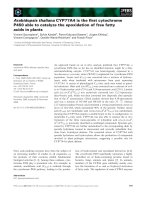
Fig. 3 Catalytic activity of CYP79A61. Yeast microsomes containing the heterologously-expressed enzyme a or an empty vector control b were
prepared and incubated with the potential substrates L-phenylalanine and L-tryptophan. Products were detected using LC-MS/MS analysis with
multiple reaction monitoring in the positive mode. Diagnostic reactions for each product: phenylacetaldoxime, m/z 136.0/119.0; indole-3-acetaldoxime,
m/z 175.0/158.0. The structures of all detected CYP79A61 products are shown in c
were found to release (E/Z)-phenylacetaldoxime in small
amounts (Fig. 5b). In addition, some structurally related
volatiles including 2-phenylacetaldehyde, 2-phenylethanol,
benzyl cyanide, and 2-phenylnitroethane could be detected in the headspace of these plants (Fig. 5b, Additional
file 1: Figure S4). In contrast, control plants expressing
eGFP released none of the above-mentioned compounds.
LC-MS/MS analysis of methanol extracts made from leaf
material harvested right after the volatile collection
revealed a strong accumulation of (E/Z)-phenylacetaldoxime and a moderate accumulation of (E/Z)-indole-3-acetaldoxime in leaves harboring the 35S::CYP79A61 construct,
while no aldoximes could be detected in leaf material harvested from eGFP-expressing control plants (Fig. 5a).
Caterpillar oral secretion induces CYP79A61 gene
expression as well as amino acid substrate accumulation
and phenylacetaldoxime formation
To test whether the expression of CYP79A61 is influenced by herbivory, young maize plants of the cultivar
Delprim were treated with oral secretion collected from
Egyptian cotton leafworm (Spodoptera littoralis) larvae
and CYP79A61 transcript accumulation was analyzed in
the leaves using quantitative (q)RT-PCR. While undamaged control plants showed a basal CYP79A61 expression, simulated herbivory led to a significant increase in
transcript accumulation (Fig. 6a). In contrast, Spi1, a
member of the YUCCA-like gene family in maize which
has been reported to be involved in indole-3-acetic acid
formation [27], was not expressed in damaged and
undamaged maize leaves (cq values >39). LC-MS/MS analysis of L-phenylalanine and L-tryptophan in methanol extracts made from the same samples revealed a significant
upregulation of both CYP79A61 substrates in response to
the oral secretion treatment (Fig. 6b and c). (E/Z)-Phenylacetaldoxime showed a similar accumulation pattern with
significantly higher amounts in damaged leaves than in undamaged controls (Fig. 6d). Indole-3-acetaldoxime, however, could not be detected in these leaf extracts.
Caterpillar secretion induces the formation of the auxins
indole-3-acetic acid and phenylacetic acid as potential
aldoxime-derived metabolites
To investigate whether the maize cultivar Delprim is
able to produce volatile aldoximes after herbivory, we
conducted a volatile collection on plants treated with
caterpillar oral secretions. Despite the accumulation of
(E/Z)-phenylacetaldoxime in leaves, no aldoximes or
aldoxime-derived nitriles or nitro compounds could be
detected as volatiles (Additional file 1: Figure S5). However, several mono- and sesquiterpenes, green leaf volatiles and esters could be identified which have already
been described in the literature [22, 23].
We then looked for potential metabolites of indole-3acetaldoxime and phenylacetaldoxime since both are
thought to be potential precursors for the biosynthesis
Irmisch et al. BMC Plant Biology (2015) 15:128
Page 6 of 14
Fig. 4 Biochemical characterization of CYP79A61. Yeast microsomes containing the heterologously-expressed enzyme were prepared and incubated
with the substrates L-phenylalanine and L-tryptophan. Time courses for the product formation in the presence of either 100 μM or 1 mM substrate
are shown in a. The Michaelis-Menten kinetics for L-phenylalanine and L-tryptophan are given in b and the pH dependency of CYP79A61 product
formation is illustrated in c. Products were detected using LC-MS/MS analysis with multiple reaction monitoring in the positive mode. Diagnostic
reactions for each product: phenylacetaldoxime, m/z 136.0/119.0; indole-3-acetaldoxime, m/z 175.0/158.0
of the auxins indole-3-acetic acid and phenylacetic acid
(PAA), respectively [28], we searched for these metabolites in leaves of undamaged and oral secretion-treated
maize plants. The accumulation of indole-3-acetic acid
as well as the accumulation of phenylacetic acid was significantly increased in treated leaves in comparison to
undamaged control leaves (Fig. 6e and f ).
Since aldoximes are intermediates in the biosynthesis
of cyanogenic glycosides, we also searched for these
compounds in maize leaves. Maize has been reported as
a cyanogenic plant species [17–19], but no cyanogenic
glycosides have been identified so far. We used LC-MS/
MS analysis to measure potential phenylacetaldoximederived cyanogenic glycosides, such as prunasin and
amygdalin, as well as the p-hydroxyphenylacetaldoximederived cyanogenic glycoside dhurrin in oral secretiontreated maize leaves and in coleoptiles of maize and
sorghum. As already reported in the literature [29, 30],
dhurrin was found in large amounts in sorghum coleoptiles. However, none of the above mentioned cyanogenic
glycosides could be detected in maize (Additional file 1:
Figure S6), suggesting that at least the tested cultivar
Delprim is not able to accumulate these compounds in
significant amounts.
Irmisch et al. BMC Plant Biology (2015) 15:128
Page 7 of 14
Fig. 5 Aldoxime accumulation a and volatile emission b of transgenic N. benthamiana plants overexpressing maize CYP79A61. Plants were
infiltrated with A. tumefaciens containing 35S:eGFP (control) or 35S:CYP79A61. Aldoximes were extracted three days after infiltration with methanol
and analyzed using LC-MS/MS. Volatiles were collected on the third day after infiltration. Identification of volatile compounds was done with
GC-MS and quantification was done with GC-FID. PAld, 2-phenylacetaldehyde; 2PE, 2-phenylethanol; BC, benzyl cyanide; PN, 2-phenylnitroethane;
(E)-PAOx, (E)-phenylacetaldoxime; (Z)-PAOx, (Z)-phenylacetaldoxime. Means and standard errors are shown (n = 5)
Discussion
Aldoximes and aldoxime-derived compounds such as nitriles and cyanogenic glycosides are widespread secondary plant metabolites. They play important roles in plant
defense against insects and pathogens [1, 3, 6, 11, 31]
and are discussed to be involved in plant-pollinator
interactions [32]. Although maize as one of the most
important crop species has been intensively investigated
during the last decades, little is known about the occurrence and role of aldoximes in this plant.
In this paper, we identified and characterized the P450
enzyme CYP79A61, one member of a small gene family
comprising four genes with similarity to plant CYP79s.
Like other CYP79 enzymes from the A- and Bsubfamilies, recombinant CYP79A61 was shown to accept
only aromatic amino acids as substrates. However, in
contrast to most other CYP79 enzymes which have very
high substrate specificity [5], both in vitro and in vivo
experiments revealed that the recombinant maize enzyme
was able to convert L-phenylalanine and L-tryptophan to
phenylacetaldoxime and indole-3-acetaldoxime, respectively (Figs. 3 and 5). The conversion of a broader range of
amino acids into aldoximes has only been reported for
two poplar CYP79D enzymes [6]. The Km values of
CYP79A61 for L-phenylalanine and L-tryptophan were
relatively high (Km (Phe) = 117.2 μM; Km (Trp) = 150.2 μM),
but in the range reported for other CYP79 enzymes. It has
been suggested that the low substrate affinity of these
enzymes has evolved to avoid possible depletion of the
free amino acid pool in plants [33].
The analysis of aldoximes in maize revealed a significant
increase in phenylacetaldoxime accumulation in leaves
treated with caterpillar oral secretion in comparison to
leaves from undamaged control plants (Fig. 6d), suggesting a role of this compound in plant defense. Phenylacetaldoxime was previously shown to accumulate in poplar
leaves after herbivory by gypsy moth (Lymantria dispar)
caterpillars and feeding of pure phenylacetaldoxime to L.
dispar larvae had negative effects on caterpillar survival,
growth, and time until pupation [6]. Although the overall
concentration of phenylacetaldoxime in maize leaves
subjected to simulated herbivory (Fig. 6d) was relatively
low compared to that found in poplar leaves, local formation of this compound giving higher concentrations around
the wound site as already reported for defensive sesquiterpenes in maize [34] is conceivable. In addition, aldoximes
have been suggested to play a role in plant defense against
pathogens [10] and the accumulation of phenylacetaldoxime in treated maize leaves might thus represent a defense
barrier against pathogen attack following insect herbivore
damage. Apart from accumulating in plant tissue, aldoximes can serve as precursors for other defensive compounds [1–3, 35]. In the Japanese apricot (Prunus mume),
for example, phenylacetaldoxime is converted into the
cyanogenic glycosides prunasin and amygdalin [36]. This is
unlikely to occur in maize since we could not detect these
compounds neither in regurgitant-treated leaves nor in
maize coleoptiles (Additional file 1: Figure S6), the developmental stage reported to possess the highest cyanogenic
potential [19]. However, we cannot rule out that phenylacetaldoxime acts as a precursor for other so far unknown
maize defense compounds.
Since CYP79A61 had similar Km values for Lphenylalanine and L-tryptophan and both amino acids
were found to accumulate in the same order of magnitude in maize leaves (Fig. 6b and c), one would expect
that the enzyme produces equal amounts of phenylacetaldoxime and indole-3-acetaldoxime in planta. However, while phenylacetaldoxime was detected in maize
leaves, no accumulation of indole-3-acetaldoxime could
be observed (Fig. 6). Local differences in amino acid
substrate concentrations caused, for example, by
specific substrate channeling processes might be an
explanation for this observation. However, it is far more
likely that the lack of indole-3-acetaldoxime detection
is due to the aldoxime being further converted into
Irmisch et al. BMC Plant Biology (2015) 15:128
Page 8 of 14
Fig. 6 The response of maize leaves to simulated herbivory. CYP79A61 gene expression a, L-phenylalanine b and L-tryptophan c accumulation,
(E/Z)-phenylacetaldoxime content d, and phenylacetic acid e and indole-3-acetic acid f levels were measured in undamaged leaves (ctr) and
leaves subjected to simulated herbivory (herb). (E/Z)-phenylacetaldoxime, L-phenylalanine, L-tryptophan, and the auxins phenylacetic acid and
indole-3-acetic acid were extracted with methanol and analyzed by LC-MS/MS. Gene expression was determined by qRT-PCR. Means and standard
errors are shown (n = 5). Asterisks indicate statistical significance in Student’s t-test. Gene expression: p < 0.001; t = −4.99; L-phenylalanine: p < 0.001,
t = 15.242; L-tryptophan: p < 0.001, t = 16.293; phenylacetaldoxime: p = < 0.001, t = 6.934; phenylacetic acid: p = < 0.001 , t = −18.259; indole-3-acetic
acid: p = < 0.001, t = −5.644
other compounds. In various plant species, including
maize, the conversion of indole-3-acetaldoxime into the
corresponding acid is thought to serve as an alternative
route for the formation of the essential plant growth
hormone indole-3-acetic acid [37–40], presumably involving indole-3-acetonitrile as an intermediate [37,
38]. The analysis of CYP79A61 transcript accumulation
in maize leaves revealed that the gene was significantly
upregulated after herbivore feeding, matching an increased accumulation of IAA in the same tissues (Fig. 6a
and f ). Moreover, overexpression of CYP79A61 in N.
benthamiana revealed that the enzyme is able to produce indole-3-acetaldoxime under natural conditions in
planta (Fig. 5a). Thus it is conceivable that CYP79A61
might produce indole-3-acetaldoxime as a specific substrate for herbivory-induced IAA formation in maize
leaves. The conversion of indole-3-acetaldoxime to
indole-3-acetonitrile is likely catalyzed by a P450 enzyme similar to the recently described poplar enzymes
CYP71B40 and CYP71B41 which were shown to produce benzyl cyanide from phenylacetaldoxime after
herbivory [35]. Indole-3-acetonitrile could then be further converted into IAA by maize nitrilase 2, an enzyme already implicated in auxin formation in maize
[41]. In future experiments, the overexpression of
maize CYP79A61 in an Arabidopsis cyp79b2 cyp79b3
double mutant which has been described to lack the
accumulation of indole-3-acetaldoxime [40] would allow
the analysis of CYP79A61-mediated formation of indole-3acetaldoxime and its metabolism in a clean and sensitive
background in planta. Since IAA can be formed via different biosynthetic pathways [28], it is possible that other
enzymes rather than CYP79A61 are responsible for the observed IAA accumulation after simulated herbivory. Thus,
a comprehensive expression analysis of candidate genes
such as TAA and YUCCA might help to understand the
biochemical origin of herbivore-induced IAA formation in
maize. However, we have already shown that Spi1, a member of the YUCCA-like gene family in maize [27], was not
expressed in damaged and undamaged maize leaves.
It is well established that herbivore feeding can cause
changes in auxin levels in plants. For example, feeding
Irmisch et al. BMC Plant Biology (2015) 15:128
of gall-inducing insects on wheat and late goldenrod
(Solidago altissima) leads to increased IAA levels in the
damaged tissues [42, 43] while simulated herbivory on
wild tobacco (Nicotiana attenuata) resulted in decreased
IAA accumulation [44]. Since auxins are potent modifiers of plant defense reactions [45], it is likely that the
elevated IAA and PAA levels in herbivore-damaged maize
also mediate defense responses. The presence of aldoximeproducing CYP79 genes in all so far sequenced angiosperm
genomes might indicate a broader occurrence of aldoximemediated auxin formation, especially under biotic stresses
such as herbivory or pathogen attack.
A sequence comparison with already characterized
CYP79s from other plants showed that CYP79A61 was
most similar to CYP79A1, an enzyme known to catalyze
the key reaction of dhurrin formation in sorghum [7].
However, despite an amino acid identity of 72 %, both
enzymes have different substrate specificities with
CYP79A1 solely converting tyrosine to p-hydroxyphenylacetaldoxime [46]. A comparative analysis of the maize
and the sorghum genome revealed that CYP79A61 and
CYP79A1 are not located on syntenic chromosomal
regions and are therefore not orthologues (Additional
file 1: Figure S1). Interestingly, no gene with orthology
to sorghum CYP79A1 could be found in the maize genome (Additional file 1: Figure S2), suggesting a recent
loss of the CYP79A1 orthologue in the maize lineage
after diversification of the common ancestor of maize
and sorghum. This gene loss might explain the absence
of dhurrin formation in maize (Additional file 1: Figure
S6). A so far uncharacterized sorghum CYP79 gene
(Sb10g022470) could be identified as the orthologue of
CYP79A61 (Additional file 1: Figures S2 and S3). However,
whether this gene encodes for a protein with the same
substrate specificity as CYP79A61 remains unknown.
Like dhurrin, we also could not detect the cyanogenic
glycosides prunasin or amygdalin in the maize cultivar
Delprim, neither in coleoptiles nor in undamaged or
damaged leaves of young plants (Additional file 1: Figure
S6). Moreover, a volatile collection experiment showed
that Delprim did not release aldoximes after herbivory
(Additional file 1: Figure S5). However, in the literature
there is evidence that maize is cyanogenic [17–19], and
a few maize lines have been reported to produce aliphatic volatile aldoximes after herbivore feeding [20, 21].
It is conceivable that the three putative CYP79 genes
GRMZM2G011156, GRMZM2G105185, and GRMZM
2G178351, which could not be amplified from Delprim
cDNA, are expressed in other maize cultivars or under
different experimental conditions and contribute to volatile aldoxime and/or cyanogenic glycoside formation.
Thus, a comprehensive characterization and gene expression analysis of different CYP79 alleles from diverse maize
cultivars will help to further understand the formation
Page 9 of 14
and function of these nitrogenous defense compounds
and their variability among maize cultivars.
Conclusions
We showed that maize produces aldoximes in response
to simulated herbivory. A P450 enzyme of the CYP79
family, CYP79A61, could be identified able to catalyze
the formation of phenylacetaldoxime and indole-3acetaldoxime in two different heterologous systems.
Since the expression of CYP79A61 was upregulated after
simulated herbivory, we hypothesize that the enzyme
contributes to herbivore-induced aldoxime formation
in maize. While phenylacetaldoxime accumulated in
herbivore-damaged leaves and might play a role in
maize defense against herbivores or pathogens, indole3-acetaldoxime could not be detected in the plant.
However, it is conceivable that this aldoxime is rapidly
converted to indole-3-acetic acid which has been described as a mediator of various plant defense responses [45].
Methods
Plant and insect material
Seeds of the maize (Zea mays L.) hybrid line Delprim
from Delley Samen und Pflanzen (Delley, Switzerland)
were grown in commercially available potting soil in a
climate-controlled chamber with a 16 h photoperiod
(1 mmol (m2)−1 s−1 of photosynthetically-active radiation,
temperature cycle 24/20 °C (day/night) and 60 % relative
humidity). Twelve day old-plants (15–25 cm high, 4
expanded leaves) were used in the experiment. Eggs of
Spodoptera littoralis Boisd. (Lepidoptera: Noctuidae) were
obtained from Aventis (Frankfurt, Germany) and were
reared on an artificial wheat germ diet (Heliothis mix,
Stonefly Industries, Bryan, TX, USA) for about 10 days at
22 °C under an illumination of 750 μmol (m2)−1 s−1.
Larvae were reared for another week on Delprim leaves
and oral secretions were collected every day with a pipette
and frozen at −20 °C until further usage. For the caterpillar secretion treatment (4 pm), 2 maize leaves per plant
were cut with a razor blade and 15 μL oral secretion (1:2
diluted in water) were applied to the wound site. This
treatment was repeated the next morning at 9 am prior to
volatile collection.
Volatile collection and analysis
For volatile collection, plants were separately placed in
airtight 3 L glass desiccators. Charcoal-filtered air was
pumped into the desiccators at a flow rate of 2 L min−1
and left the desiccators through a filter packed with
30 mg Porapaq Q (ARS, Inc., Gainesville, FL, USA).
Volatiles were collected for 5 h (10 am – 3 pm). After
collection the volatiles were desorbed by eluting the
filter twice with 100 μL dichloromethane containing
Irmisch et al. BMC Plant Biology (2015) 15:128
nonyl acetate as an internal standard (10 ng μL−1).
Qualitative and quantitative analysis of maize volatiles
was conducted using an Agilent 6890 Series gas chromatograph coupled to an Agilent 5973 quadrupole mass
selective detector (interface temp.: 270 °C; quadrupole
temp.: 150 °C, source temp.: 230 °C, electron energy:
70 eV) or a flame ionization detector (FID) operated at
300 °C, respectively. The constituents of the volatile
bouquet were separated with a DB-5MS column
(Agilent, Santa Clara, CA, USA, 30 m × 0.25 mm ×
0.25 μm) and He (MS) or H2 (FID) as carrier gas. One
microliters of the sample was injected without split at an
initial oven temperature of 40 °C. The temperature was
held for 2 min and then increased to 155 °C with a gradient of 7 °C min−1, followed by a further increase to
300 °C with 60 °C min−1 and a hold for 3 min.
Compounds were identified by comparison of retention times and mass spectra to those of authentic standards obtained from Fluka (Seelze, Germany), Roth
(Karlsruhe, Germany), Sigma (St, Louis, MO, USA) or
Bedoukian (Danbury, CT, USA), or by reference spectra
in the Wiley and National Institute of Standards and
Technology libraries and in the literature [47].
Plant tissue sampling, RNA extraction and reverse
transcription
Treated maize leaves were harvested immediately after
the volatile collection (3 pm), flash-frozen in liquid nitrogen and stored at −80 °C until further processing.
After grinding the frozen leaf material in liquid nitrogen
to a fine powder, total RNA was isolated using the
“RNeasy Plant Mini Kit” (Quiagen GmbH, Hilden,
Germany) according to manufacturer’s instructions.
RNA concentration, purity and quality were assessed
using a spectrophotometer (NanoDrop 2000c, Thermo
Scientific, Wilmington, DE, USA) and an Agilent 2100
Bioanalyzer (Agilent Technologies GmbH, Waldbronn,
Germany). Prior to cDNA synthesis, 0.75 μg RNA was
DNase-treated using 1 μL DNase (Fermentas GmbH, St.
Leon Roth, Germany). Single-stranded cDNA was prepared from the DNase-treated RNA using SuperScriptTM
III reverse transcriptase and oligo (dT12–18) primers
(Invitrogen, Carlsbad, CA, USA).
Identification and isolation of CYP79 genes
To identify putative maize CYP79 genes, a BLAST
search against the Z. maize genome database (http://
www.phytozome.net/poplar) was conducted using the
amino acid sequence of CYP79A1 from Sorghum bicolor
(L.) Moench (Genbank Q43135) as input sequence. Four
sequences representing putative P450 enzymes of the
CYP79 family were identified. One of these sequences
could be amplified from cDNA attained from herbivoreinduced leaves of Z. mays. Primer sequence information
Page 10 of 14
is available in Additional file 1: Table S1. The PCR product was cloned into the sequencing vector pCR®−Blunt
II-TOPO® (Invitrogen) and both strands were fully
sequenced.
Heterologous expression of CYP79A61 in Saccharomyces
cerevisiae
The complete open reading frame of CYP79A61 was
cloned into the pESC-Leu2d vector [24] as a NotI/BglII
fragment and the resulting construct was transferred
into the S. cerevisiae strain WAT11 [25]. For gene
expression, a single yeast colony was picked to inoculate
a starting culture which contained 30 mL SC minimal
medium lacking leucine (6.7 g L−1 yeast nitrogen base
without amino acids, but with ammonium sulfate).
Other components: 100 mg L−1 of L-adenine, L-arginine,
L-cysteine, L-lysine, L-threonine, L-tryptophan and uracil;
50 mg L−1 of the amino acids L-aspartic acid, L-histidine,
L-isoleucine, L-methionine, L-phenylalanine, L-proline, Lserine, L-tyrosine, L-valine; 20 g L−1 D-glucose. The
culture was grown overnight at 28 °C and 180 rpm. One
OD of this culture (approx. 2 × 10⁷ cells mL−1) was used
to inoculate 100 mL YPGA full medium (10 g L−1 yeast
extract, 20 g L−1 bactopeptone, 74 mg L−1 adenine hemisulfate, 20 g L−1 D-glucose) which was grown for 32–35 h
(until OD about 5), induced by the addition of galactose
and cultured for another 15–18 h. Cells were harvested
and yeast microsomes were isolated according to the procedures described by Pompon et al. [25] and Urban et al.
[48] with minor modifications. Briefly, the culture was
centrifuged (7500 g, 10 min, 4 °C), the supernatant was
decanted, the pellet was resuspended in 30 mL TEK buffer
(50 mM Tris-HCl pH 7.5, 1 mM EDTA, 100 mM KCl)
and then centrifuged again. Then the cell pellet was
carefully resuspended in 2 mL of TES buffer (50 mM
Tris-HCl pH 7.5, 1 mM EDTA, 600 mM sorbitol,
10 g L−1 bovine serum fraction V protein and 1.5 mM
β-mercaptoethanol) and transferred to a 50 mL conical
tube. Glass beads (0.45–0.50 mm diameter, SigmaAldrich Chemicals, Steinheim, Germany) were added so
that they filled the full volume of the cell suspension.
Yeast cell walls were disrupted by 5 cycles of 1 min
shaking by hand and subsequent cooling down on ice for
1 min. The crude extract was recovered by washing the
glass beads 4 times with 5 mL TES. The combined washing fractions were centrifuged (7500 g, 10 min, 4 °C), and
the supernatant was transferred into another tube and
centrifuged again (100,000 g , 60 min, 4 °C). The resulting
microsomal protein fraction was homogenized in 2 mL
TEG buffer (50 mM Tris–HCl, 1 mM EDTA, 30 % w/v
glycerol) using a glass homogenizer (Potter-Elvehjem,
Fisher Scientific, Schwerte, Germany). Aliquots were
stored at −20 °C and used for protein assays.
Irmisch et al. BMC Plant Biology (2015) 15:128
Analysis of recombinant CYP79A61
To determine the substrate specificity of CYP79A61,
yeast microsomes harboring recombinant protein were
incubated for 30 min at 25 °C and 300 rpm individually
with the potential substrates L-phenylalanine, L-valine,
L-leucine, L-isoleucine, L-tyrosine and L-tryptophan in
glass vials containing 300 μL of the reaction mixture
(75 mM sodium phosphate buffer (pH 7.0), 1 mM
substrate (concentration was variable for Km determination), 1 mM NADPH and 10 μL of the prepared microsomes). Reaction mixtures containing microsomes
prepared from WAT11 transformed with the empty
vector served as negative controls. Assays were stopped
by placing on ice after 300 μL MeOH were added. Reaction products were analyzed using LC-MS/MS as
described below. Product accumulation was measured
after different incubation times (20–40 min) and under
different pH conditions (pH 5.5–8.5). For the determination of the Km values, assays were carried out as triplicates and enzyme concentrations and incubation times
(30 min) were chosen so that the reaction velocity was
linear during the incubation time period.
qRT-PCR analysis of CYP79A61 and Spi1 expression
cDNA was prepared as described above and diluted 1:3
with water. For the amplification of the CYP79A61 gene
fragment (146 bp) and the Spi1 gene fragment (99 bp),
primer pairs were designed having a Tm ≥ 56 °C, a GC
content between 52 and 56 % and a primer length in the
range of 18–21 nt (see Additional file 1: Table S1 online
for primer information). Primer specificity was confirmed
by agarose gel electrophoresis, melting curve analysis,
standard curve analysis, and sequence verification of
cloned PCR amplicons. The transcription repressor Leunig (LUG) [49] was used as a reference gene. Samples
were run in triplicates using Brilliant® III SYBR® Green
QPCR Master Mix (Stratagene, Carlsbad, CA, USA) with
ROX as reference dye. The following PCR conditions were
applied for all reactions: Initial incubation at 95 °C for
3 min followed by 40 cycles of amplification (95 °C for
20 s, 60 °C for 20 s). Plate reads were taken during the
annealing and the extension steps of each cycle. Data for
the melting curves were recorded at the end of cycling
from 55 to 95 °C.
All samples were run on the same PCR machine
(Mx3000P, Agilent Technologies, Santa Clara, CA, USA) in
an optical 96-well plate. Five biological replicates were
analyzed as triplicates in the qRT-PCR for each of the three
treatments. Data for the relative quantity to calibrator average (dRn) were exported from the MXPro Software.
Transient expression of CYP79A61 in N. benthamiana
For gene expression in N. benthamiana, the coding region of CYP79A61 was cloned into the pCAMBiA2300U
Page 11 of 14
vector. After verification of the sequence integrity,
pCAMBiA vectors carrying the CYP79A61 or eGFP construct and the construct pBIN::p19 were separately
transferred into Agrobacterium tumefaciens strain
LBA4404. The transformation was confirmed by PCR.
Five milliliters of an overnight culture (220 rpm, 28 °C)
were used to inoculate 50 mL LB medium (50 μg mL−1
kanamycin, 25 μg mL−1 rifampicin and 25 μg mL−1 gentamicin) for overnight growth. The following day, the
cultures were centrifuged (4000 g, 5 min) and the cells
were resuspended in infiltration buffer (10 mM MES,
10 mM MgCl2, 100 μM acetosyringone, pH 5.6) to reach
a final OD of 0.5. After shaking for at least 1 h at RT,
the cultures carrying CYP79A61 or eGFP were mixed
with an equal volume of cultures carrying pBIN:p19.
Since p19 functions as a suppressor of silencing, it
enhances the expression of the desired coexpressed protein in planta [26].
For transformation, 3–4 week-old N. benthamiana
plants were dipped upside down in an A. tumefaciens
solution and vacuum was applied to infiltrate the leaves.
Infiltrated plants were shaded with cotton tissue to protect
them from direct irradiation. Volatiles were measured on
the 3rd day after transformation as described above.
LC-MS/MS analysis of aldoximes, amino acids, auxins, and
cyanogenic glycosides
For determining amino acid and aldoxime concentration,
100 mg of plant powder was extracted with 1 mL MeOH.
For the measurement of amino acids, the MeOH extract
was diluted 1:10 with water and spiked with 13C, 15 N
abeled amino acids (algal amino acids 13C,15 N, Isotec,
Miamisburg, OH, USA) at a concentration of 10 μg of the
mix per mL. The concentration of the individual labeled
amino acids in the mix had been previously determined
by classical HPLC-fluorescence detection analysis after
pre-column derivatization with o-phthaldialdehyde-mercaptoethanol using external standard curves made from
standard mixtures (amino acid standard mix and Gln, Asn
and Trp, Fluka). Amino acids in the diluted MeOH extract
were directly analyzed by LC-MS/MS. The method
described by Jander et al. [50] was used with some
modifications. Briefly, chromatography was performed
on an Agilent 1200 HPLC system (Agilent Technologies, Boeblingen, Germany). Separation was achieved
on a Zorbax Eclipse XDB-C18 column (50 × 4.6 mm,
1.8 μm, Agilent Technologies) with aqueous formic
acid (0.05 %) and acetonitrile employed as mobile
phases A and B, respectively. The elution profile was:
0–1 min, 97 % A; 1–2.7 min, 3–100 % B in A; 2.7–
3 min 100 % B and 3.1–6 min 97 % A. The mobile
phase flow rate was 1.1 mL min−1 and the column
temperature was maintained at 25 °C. The liquid chromatography was coupled to an API 5000 tandem mass
Irmisch et al. BMC Plant Biology (2015) 15:128
spectrometer (Applied Biosystems, Darmstadt, Germany)
equipped with a Turbospray ion source operated in positive ionization mode (ion spray voltage, 5500 eV; turbo
gas temp, 700 °C; nebulizing gas, 70 psi; curtain gas,
35 psi; heating gas, 70 psi; collision gas, 2 psi). Multiple reaction monitoring (MRM) was used to monitor a parent
ion → product ion reaction for each analyte. MRMs were
chosen as described in Jander et al. [50] except for Arg
(m/z 175 → 70), and Lys (m/z 147 → 84). Both Q1 and Q3
quadrupoles were maintained at unit resolution. Analyst
1.5 software (Applied Biosystems) was used for data acquisition and processing. Individual amino acids in the sample were quantified from corresponding peaks in the
13 15
C, N labeled amino acid internal standard, except for
tryptophan which was quantified using 13C,15 N-Phe applying a response factor of 0.42.
Aldoximes were measured from MeOH extracts as described in Irmisch et al. [6] using the same LC-MS/MS
system as described above. Formic acid (0.2 %) in water
and acetonitrile were employed as mobile phases A and
B, respectively, on a Zorbax Eclipse XDB-C18 column
(50 × 4.6 mm, 1.8 μm). The elution profile (gradient 1)
was: 0–0.5 min, 30 % B; 0.5–3 min, 30–66 % B; 3–
3.1 min, 66–100 % B; 3.1–4 min 100 % B and 4.1–6 min
30 % B at a flow rate of 0.8 mL min−1 at 25 °C. The API
5000 tandem mass spectrometer was operated in positive ionization mode (ion spray voltage, 5500 eV; turbo
gas temp, 700 °C; nebulizing gas, 60 psi; curtain gas,
30 psi; heating gas, 50 psi; collision gas, 6 psi). MRM
was used to monitor parent ion → product ion reactions
for each analyte as follows: m/z 136.0 → 119.0 (collision
energy (CE), 17 V; declustering potential (DP), 56 V) for
phenylacetaldoxime; m/z 102.0 → 69.0 (CE, 13 V; DP,
31 V) for 2-methylbutyraldoxime; m/z 102.0 → 46.0 (CE,
15 V; DP, 31 V) for 3-methylbutyraldoxime; m/z
175.0 → 158.0 (CE, 17 V; DP, 56 V) for indole-3acetaldoxime and m/z 152.0 → 107.0 (CE, 27 V; DP,
100 V) for p-hydroxyphenylacetaldoxime. The concentration of aldoximes was determined using external
standard curves made with authentic standards synthesized as described in Irmisch et al. [6].
The auxins IAA and PAA were analyzed based on the
protocol of Balcke et al. [51]. 100 mg of plant powder
were extracted with 300 μL MeOH. Two hundred microliters of the extract was diluted 1:10 with water containing 0.1 % formic acid and loaded to equilibrated
Chromabond® HR-X polypropylene columns (45 μm,
Macherey Nagel, Düren, Germany). The columns were
washed with acidified water. The fraction containing the
auxins was eluted with 1 mL acetonitrile, which was
then dried under a stream of nitrogen gas. The samples
were redissolved in 30 μL MeOH and subsequently analyzed by the same LC-MS/MS system as described
above. Separations were performed on an Agilent XDB-
Page 12 of 14
C18 column (50 × 4.6 mm, 1.8 μm). Eluents A and B
were water containing 0.05 % formic acid and acetonitrile, respectively. The elution profile was: 0–0.5 min,
5 % B in A; 0.5–4.0 min, 5–50 % B; 4.1–4.5 min 100 %
B and 4.6–7 min 5 % B. The flow rate was set to
1.1 mL min−1. For IAA analysis, the API 5000 tandem
mass spectrometer was operated in positive ionization
mode (ion spray voltage, 5500 eV; turbo gas temp, 700 °C;
nebulizing gas, 60 psi; curtain gas, 30 psi; heating gas,
50 psi; collision gas, 6 psi). The MRM transition and parameter settings for IAA were as follows: m/z 176 → 130
(CE, 19 V; DP, 31 V). PAA was detected separately by
mass spectrometer operated in negative ionization mode
(ion spray voltage, −4500 eV; turbo gas temp, 700 °C;
nebulizing gas, 60 psi; curtain gas, 30 psi; heating gas,
50 psi; collision gas, 6 psi). The MRM transition and
parameter settings for PAA were as follows: m/z 135 →
91 (CE, −10 V; DP, −25 V). The concentration of PAA
was determined using external standard curves made
with authentic standard (Sigma-Aldrich). IAA concentration was determined internally by spiking the plant
extracts with 2H5-IAA (OlChemIm Ltd., Olomouc,
Czech Republic).
For the analysis of cyanogenic glycosides (dhurrin,
prunasin, linamarin, and lotaustralin), 100 mg plant
powder was extracted with 300 μL MeOH and 200 μL of
the extract was diluted 1:10 with water containing 0.1 %
formic acid. Ten microliters of the extracts were directly
injected and analyzed by LC-MS/MS. The column and
eluents used for the separation were the same as already
described for the auxins. The elution profile was: 0–
0.5 min, 5 % B in A; 0.5–6.0 min, 5–50 % B; 6.1–7.5 min
100 % B and 7.6–10.5 min 5 % B. The flow rate was set
to 1.1 mL min−1. The tandem mass spectrometer was
operated in negative ionization mode (ion spray voltage,
−4500 eV; turbo gas temp, 700 °C; nebulizing gas, 60 psi;
curtain gas, 30 psi; heating gas, 50 psi; collision gas,
6 psi). MRM was used to monitor parent ion → product
ion reactions for each analyte as follows: m/z 310.0 →
179.0 for dhurrin, m/z 294.0 → 89.0 (CE, −22; DP, −15)
for prunasin, m/z 260.0 → 179.0 for lotaustralin, m/z
246.0 → 179.0 for linamarin, and m/z 456.0 → 179.0 for
amygdalin. If not stated above, the transition parameter
settings for the cyanogenic glycosides were as follows:
CE, −10 V; DP, −15 V.
Sequence analysis and phylogenetic tree reconstruction
An alignment of maize CYP79 enzymes and CYP79A1
from S. bicolor was constructed and visualized using
BioEdit ( />and the ClustalW algorithm. For the estimation of a
phylogenetic tree, we used the ClustalW algorithm (gap
open, 10; gap extend, 0.1; Gonnet; penalties, on; gap
separation, 4; cut off, 30 %) implemented in MEGA5
Irmisch et al. BMC Plant Biology (2015) 15:128
[52] to compute an amino acid alignment of the maize
CYP79 sequences and other already characterized
CYP79 enzymes. The tree was reconstructed with
MEGA5 using a neighbor-joining algorithm (Poisson
model). A bootstrap resampling analysis with 1000 replicates was performed to evaluate the tree topology.
The synteny analysis was done using the EnsemblPlants web service ().
Page 13 of 14
(prunasin, amygdalin) and MRMs for lotaustralin and linamarin were
calculated from those of dhurrin and prunasin. Amygdalin, lotaustralin
and linamarin could not be detected in maize and sorghum (data
not shown). Table S1. Oligonucleotides used in this study.
Abbreviations
CYP: Cytochrome P450 monooxygenase; LC-MS/MS: Liquid
chromatography-tandem mass spectrometry; IAA: Indole-3-acetic acid;
PAA: Phenylacetic acid; FID: Flame ionization detector; MRM: Multiple
reaction monitoring.
Statistical analysis
To test for statistical significance, data were log transformed whenever necessary and analyzed using the
Student’s t-test implemented in SigmaPlot 11.0 for
Windows (Systat Software Inc. 2008).
Accession numbers
Sequence data for genes and proteins discussed in this
article can be found in the GenBank under the following
identifiers: CYP79A61 (KP297890), CYP79D6v3 (KF56
2515), CYP79D7v2 (KF562516), CYP79D3 (AAT11920),
CYP79D4 (AAT11921), CYP79D1 (AAF27289), CYP
79D2 (AAF27290), CYP79A1 (Q43135), CYP79B3 (AEC
07294), CYP79B1 (AAD03415), CYP79B2 (AEE87143),
CYP79A2 (AAF70255), CYP79E1 (AF140609), CYP79E2
(AF140610), CYP79F2 (AAG24796), CYP79F1 (AEE29
448), CYP71E1 (AAC39318).
Additional file
Additional file 1: Figure S1. Comparative genomic analysis of Sorghum
bicolor chromosome 1 with maize chromosomes 1, 2, 5, and 9. The
analysis was done using the web server .
Figure S2. Comparative genomic analysis of Zea mays chromosome 9
with Sorghum bicolor chromosomes 1 and 10. The analysis was done
using the web server . Figure S3.
Phylogenetic tree of CYP79 sequences from maize and Sorghum bicolor.
The rooted tree was inferred with the neighbor-joining method and
n = 1000 replicates for bootstrapping. Bootstrap values are shown
next to each node. As an outgroup, CYP71A13 from Arabidopsis thaliana
was chosen. Figure S4. Volatiles released from transgenic Nicotiana
benthamiana plants transiently overexpressing either a 35S::eGFP
construct or a 35S::CYP79A61 construct. Volatiles were collected 3 days after
Agrobacterium tumefaciens infiltration and analyzed using GC-MS. 1, 5-epiaristolochene; 2, 2-phenylethanol; 3, benzyl cyanide; 4, 2-phenylnitroethane;
5, phenylacetaldoxime; IS, internal standard. Figure S5. Volatiles released
from undamaged 10 day-old Zea mays (cultivar Delprim) seedlings (control)
and seedlings treated with caterpillar oral secretion (herbivory). Volatiles
were collected and analyzed using GC-MS. 1, β-myrcene; 2, 3-hexen-1-ol
acetate; 3, limonene; 4, linalool; 5, (E)-4,8-dimethyl-1,3,7-nonatriene; 6,
phenylmethyl acetate; 7, 2-phenylethyl acetate; 8, indole; 9, geranyl acetate;
10, (E)-β-caryophyllene; 11, (E)-α-bergamotene; 12, (E)-β-farnesene; 13,
β-sesquiphellandrene; 14, 4,8,12-trimethyltrideca-1,3,7,11-tetraene; IS, internal
standard. Figure S6. Accumulation of cyanogenic glycosides in maize and
sorghum. Maize and sorghum coleoptiles were harvested 3 days after
germination. Undamaged maize leaves and caterpillar oral secretiontreated maize leaves were obtained as described in the Methods
section. Glycosylated compounds were extracted with methanol and
cyanogenic glycosides were analyzed using LC-MS/MS with multiple
reaction monitoring (MRM). MRMs for dhurrin, prunasin, and amygdalin
were established using authentic standards obtained from SIGMA-Aldrich
() (dhurrin) or prepared from bitter almonds
Competing interests
The authors declare that they have no competing interests.
Authors’ contributions
SI, PZ and VH carried out the experimental work. SI, JG, and TGK participated
in the design of the study and improved the manuscript. SI and TGK
conceived of the study and drafted the manuscript. All authors read and
approved the final manuscript.
Acknowledgments
We thank Delley Samen und Pflanzen (Delley, Switzerland) for seeds of the
maize Delprim line, Daniele Werck (Strasbourg, France) for the
pCAMBiA2300U vector and vectors carrying eGFP and p19, and David Nelson
for P450 nomenclature. This research was funded by the Max Planck Society.
Received: 13 January 2015 Accepted: 18 May 2015
References
1. Halkier BA, Gershenzon J. Biology and biochemistry of glucosinolates. Annu
Rev Plant Biol. 2006;57:303–33.
2. Bak S, Paquette SM, Morant M, Morant AV, Saito S, Bjarnholt N, et al.
Cyanogenic glycosides: a case study for evolution and application of
cytochromes P450. Phytochem Rev. 2006;5(2–3):309–29.
3. Glawischnig E. Camalexin. Phytochemistry. 2007;68(4):401–6.
4. Knudsen JT, Eriksson R, Gershenzon J, Stahl B. Diversity and distribution of
floral scent. Bot Rev. 2006;72(1):1–120.
5. Hamberger B, Bak S. Plant P450s as versatile drivers for evolution of
species-specific chemical diversity. Philos Trans R Soc Lond B Biol Sci.
2013;368(1612):20120426.
6. Irmisch S, McCormick AC, Boeckler GA, Schmidt A, Reichelt M, Schneider B,
et al. Two herbivore-induced cytochrome P450 enzymes CYP79D6 and
CYP79D7 catalyze the formation of volatile aldoximes involved in poplar
defense. Plant Cell. 2013;25(11):4737–54.
7. Sibbesen O, Koch B, Halkier BA, Moller BL. Cytochrome P-450TYR is a
multifunctional heme-thiolate enzyme catalyzing the conversion of
L-tyrosine to p-hydroxyphenylacetaldehyde oxime in the biosynthesis of
the cyanogenic glucoside dhurrin in Sorghum bicolor (L.) Moench. J Biol
Chem. 1995;270(8):3506–11.
8. Hull AK, Vij R, Celenza JL. Arabidopsis cytochrome P450s that catalyze the
first step of tryptophan-dependent indole-3-acetic acid biosynthesis. Proc
Natl Acad Sci U S A. 2000;97(5):2379–84.
9. Rauhut T, Glawischnig E. Evolution of camalexin and structurally related
indolic compounds. Phytochemistry. 2009;70(15–16):1638–44.
10. Moller BL. Plant science. Dynamic metabolons. Science. 2010;330(6009):1328–9.
11. Clavijo McCormick A, Irmisch S, Reinecke A, Boeckler GA, Veit D, Reichelt M,
et al. Herbivore-induced volatile emission in black poplar: regulation and
role in attracting herbivore enemies. Plant Cell Environ. 2014;37(8):1909–23.
12. Glauser G, Marti G, Villard N, Doyen GA, Wolfender JL, Turlings TCJ, et al.
Induction and detoxification of maize 1,4-benzoxazin-3-ones by insect
herbivores. Plant J. 2011;68(5):901–11.
13. Marti G, Erb M, Boccard J, Glauser G, Doyen GR, Villard N, et al. Metabolomics
reveals herbivore-induced metabolites of resistance and susceptibility in maize
leaves and roots. Plant Cell Environ. 2013;36(3):621–39.
14. Pechan T, Cohen A, Williams WP, Luthe DS. Insect feeding mobilizes a
unique plant defense protease that disrupts the peritrophic matrix of
caterpillars. Proc Natl Acad Sci U S A. 2002;99(20):13319–23.
Irmisch et al. BMC Plant Biology (2015) 15:128
15. Rohrmeier T, Lehle L. Wip1, a wound-inducible gene from Maize with
homology to Bowman-Birk Proteinase-Inhibitors. Plant Mol Biol.
1993;22(5):783–92.
16. Turlings TCJ, Tumlinson JH, Lewis WJ. Exploitation of herbivore-induced plant
odors by host-seeking parasitic Wasps. Science. 1990;250(4985):1251–3.
17. Brunnich JC. Hydrocyanic acid in fodder-plants. J Chem Soc. 1903;83:788–96.
18. Lehmann G, Zinsmeister HD, Erb N, Neunhoeffer O. Content of hydrocyanic
acid in corn and cereal products. Z Ernahrungswiss. 1979;18(1):16–22.
19. Erb N, Zinsmeister H, Lehmann G, Michely D. Der blausäuregehalt von
getreidearten gemässigter klimazonen. Z Lebensm Unters Forsch.
1981;173(3):176–9.
20. Takabayashi J, Takahashi S, Dicke M, Posthumus MA. Developmental stage
of herbivore Pseudaletia-Separata affects production of herbivore-induced
synomone by corn plants. J Chem Ecol. 1995;21(3):273–87.
21. Turlings TCJ, Bernasconi M, Bertossa R, Bigler F, Caloz G, Dorn S. The
induction of volatile emissions in maize by three herbivore species with
different feeding habits: possible consequences for their natural enemies.
Biol Control. 1998;11(2):122–9.
22. Degen T, Dillmann C, Marion-Poll F, Turlings TCJ. High genetic variability of
herbivore-induced volatile emission within a broad range of maize inbred
lines. Plant Physiol. 2004;135(4):1928–38.
23. Köllner TG, Schnee C, Gershenzon J, Degenhardt J. The sesquiterpene
hydrocarbons of maize (Zea mays) form five groups with distinct
developmental and organ-specific distributions. Phytochemistry.
2004;65(13):1895–902.
24. Ro DK, Ouellet M, Paradise EM, Burd H, Eng D, Paddon CJ, et al. Induction of
multiple pleiotropic drug resistance genes in yeast engineered to produce
an increased level of anti-malarial drug precursor, artemisinic acid.
BMC Biotechnol. 2008;8:83.
25. Pompon D, Louerat B, Bronine A, Urban P. Yeast expression of animal and
plant P450s in optimized redox environments. Methods Enzymol.
1996;272:51–64.
26. Voinnet O, Rivas S, Mestre P, Baulcombe D. An enhanced transient
expression system in plants based on suppression of gene silencing by the
p19 protein of tomato bushy stunt virus. Plant J. 2003;33(5):949–56.
27. Gallavotti A, Barazesh S, Malcomber S, Hall D, Jackson D, Schmidt RJ, et al.
Sparse inflorescence1 encodes a monocot-specific YUCCA-like gene required
for vegetative and reproductive development in maize. Proc Natl Acad Sci
U S A. 2008;105(39):15190–5.
28. Pollmann S, Muller A, Weiler EW. Many roads lead to “auxin”: of nitrilases,
synthases, and amidases. Plant Biol (Stuttg). 2006;8(3):326–33.
29. Akazawa T, Miljanich P, Conn EE. Studies on cyanogenic glycoside of
Sorghum vulgare. Plant Physiol. 1960;35(4):535–8.
30. Halkier BA, Olsen CE, Moller BL. The biosynthesis of cyanogenic glucosides
in higher-plants - the (E)-Isomers and (Z)-Isomers of ParaHydroxyphenylacetaldehyde Oxime as intermediates in the biosynthesis of
Dhurrin in Sorghum-Bicolor (L) Moench. J Biol Chem. 1989;264(33):19487–94.
31. Gleadow RM, Moller BL. Cyanogenic glycosides: synthesis, physiology, and
phenotypic plasticity. Annu Rev Plant Biol. 2014;65:155–85.
32. Raguso RA. Wake up and smell the roses: the ecology and evolution of
floral scent. Annu Rev Ecol Evol Syst. 2008;39:549–69.
33. Moller BL, Andersen MD, Busk PK, Svendsen I. Cytochromes P450 from
cassava (Manihot esculenta Crantz) catalyzing the first steps in the biosynthesis
of the cyanogenic glucosides linamarin and lotaustralin - Cloning, functional
expression in Pichia pastoris, and substrate specificity of the isolated
recombinant enzymes. J Biol Chem. 2000;275(3):1966–75.
34. Kollner TG, Lenk C, Schnee C, Kopke S, Lindemann P, Gershenzon J, et al.
Localization of sesquiterpene formation and emission in maize leaves after
herbivore damage. BMC Plant Biol. 2013;13:15.
35. Irmisch S, Clavijo McCormick A, Gunther J, Schmidt A, Boeckler GA,
Gershenzon J, et al. Herbivore-induced poplar cytochrome P450 enzymes of
the CYP71 family convert aldoximes to nitriles which repel a generalist
caterpillar. Plant J. 2014;80(6):1095–107.
36. Yamaguchi T, Yamamoto K, Asano Y. Identification and characterization of
CYP79D16 and CYP71AN24 catalyzing the first and second steps in
L-phenylalanine-derived cyanogenic glycoside biosynthesis in the Japanese
apricot, Prunus mume Sieb. et Zucc. Plant Mol Biol. 2014;86(1–2):215–23.
37. Rajagopal R, Larsen P. Metabolism of indole-3-acetaldoxime in plants. Planta.
1972;103(1):45–54.
Page 14 of 14
38. Bak S, Feyereisen R. The involvement of two P450 enzymes, CYP83B1 and
CYP83A1, in auxin homeostasis and glucosinolate biosynthesis. Plant
Physiol. 2001;127(1):108–18.
39. Zhao YD, Hull AK, Gupta NR, Goss KA, Alonso J, Ecker JR, et al. Trp-dependent
auxin biosynthesis in Arabidopsis: involvement of cytochrome P450s CYP79B2
and CYP79B3. Genes Dev. 2002;16(23):3100–12.
40. Sugawara S, Hishiyama S, Jikumaru Y, Hanada A, Nishimura T, Koshiba T, et
al. Biochemical analyses of indole-3-acetaldoxime-dependent auxin
biosynthesis in Arabidopsis. Proc Natl Acad Sci U S A. 2009;106(13):5430–5.
41. Park WJ, Kriechbaumer V, Muller A, Piotrowski M, Meeley RB, Gierl A, et al.
The nitrilase ZmNIT2 converts indole-3-acetonitrile to indole-3-acetic acid.
Plant Physiol. 2003;133(2):794–802.
42. Onkokesung N, Galis I, von Dahl CC, Matsuoka K, Saluz HP, Baldwin IT.
Jasmonic acid and ethylene modulate local responses to wounding and
simulated herbivory in Nicotiana attenuata leaves. Plant Physiol.
2010;153(2):785–98.
43. Tooker JF, De Moraes CM. Feeding by a gall-inducing caterpillar species
alters levels of indole-3-acetic and abscisic acid in Solidago altissima
(Asteraceae) stems. Arthropod Plant Interact. 2011;5(2):115–24.
44. Tooker J, De Moraes C. Feeding by Hessian fly (Mayetiola destructor [Say])
larvae on wheat increases levels of fatty acids and indole-3-acetic acid but
not hormones involved in plant-defense signaling. J Plant Growth Regul.
2011;30(2):158–65.
45. Erb M, Meldau S, Howe GA. Role of phytohormones in insect-specific plant
reactions. Trends Plant Sci. 2012;17(5):250–9.
46. Moller BL, Kahn RA, Fahrendorf T, Halkier BA. Substrate specificity of the
cytochrome P450 enzymes CYP79A1 and CYP71E1 involved in the
biosynthesis of the cyanogenic glucoside dhurrin in Sorghum bicolor (L.)
Moench. Arch Biochem Biophys. 1999;363(1):9–18.
47. Joulain D, König WA. The Atlas of Spectral Data of Sesquiterpene
Hydrocarbons. Hamburg, Germany: E.B.-Verlag; 1998.
48. Urban P, Werckreichhart D, Teutsch HG, Durst F, Regnier S, Kazmaier M, et
al. Characterization of recombinant plant cinnamate 4-hydroxylase
produced in yeast - kinetic and spectral properties of the major plant P450
of the Phenylpropanoid pathway. Eur J Biochem. 1994;222(3):843–50.
49. Manoli A, Sturaro A, Trevisan S, Quaggiotti S, Nonis A. Evaluation of candidate
reference genes for qPCR in maize. J Plant Physiol. 2012;169(8):807–15.
50. Jander G, Norris SR, Joshi V, Fraga M, Rugg A, Yu S, et al. Application of a
high-throughput HPLC-MS/MS assay to Arabidopsis mutant screening;
evidence that threonine aldolase plays a role in seed nutritional quality.
Plant J. 2004;39(3):465–75.
51. Balcke GU, Handrick V, Bergau N, Fichtner M, Henning A, Stellmach H, et al.
An UPLC-MS/MS method for highly sensitive high-throughput analysis of
phytohormones in plant tissues. Plant Methods. 2012;8(1):47.
52. Tamura K, Peterson D, Peterson N, Stecher G, Nei M, Kumar S. MEGA5:
molecular evolutionary genetics analysis using maximum likelihood,
evolutionary distance, and maximum parsimony methods. Mol Biol Evol.
2011;28(10):2731–9.
Submit your next manuscript to BioMed Central
and take full advantage of:
• Convenient online submission
• Thorough peer review
• No space constraints or color figure charges
• Immediate publication on acceptance
• Inclusion in PubMed, CAS, Scopus and Google Scholar
• Research which is freely available for redistribution
Submit your manuscript at
www.biomedcentral.com/submit