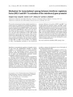
Unusual tandem expansion and positive selection in subgroups of the plant GRAS transcription factor superfamily
Bạn đang xem bản rút gọn của tài liệu. Xem và tải ngay bản đầy đủ của tài liệu tại đây (1.33 MB, 21 trang )
Wu et al. BMC Plant Biology 2014, 14:373
/>
RESEARCH ARTICLE
Open Access
Unusual tandem expansion and positive selection
in subgroups of the plant GRAS transcription
factor superfamily
Ningning Wu, Yan Zhu, Wanlu Song, Yaxuan Li, Yueming Yan and Yingkao Hu*
Abstract
Background: GRAS proteins belong to a plant transcription factor family that is involved with multifarious roles in
plants. Although previous studies of this protein family have been reported for Arabidopsis, rice, Chinese cabbage
and other species, investigation of expansion patterns and evolutionary rate on the basis of comparative genomics
in different species remains inadequate.
Results: A total of 289 GRAS genes were identified in Arabidopsis, B. distachyon, rice, soybean, S. moellendorffii,
and P. patens and were grouped into seven subfamilies, supported by the similarity of their exon? intron patterns
and structural motifs. All of tandem duplicated genes were found in group II except one cluster of rice, indicating
that tandem duplication greatly promoted the expansion of group II. Furthermore, segment duplications were
mainly found in the soybean genome, whereas no single expansion pattern dominated in other plant species
indicating that GRAS genes from these five species might be subject to a more complex evolutionary mechanism.
Interestingly, branch-site model analyses of positive selection showed that a number of sites were positively
selected under foreground branches I and V. These results strongly indicated that these groups were experiencing
higher positive selection pressure. Meanwhile, the site-specific model revealed that the GRAS genes were under
strong positive selection in P. patens. DIVERGE v2.0 was used to detect critical amino acid sites, and the results
showed that the shifted evolutionary rate was mainly attributed to the functional divergence between the GRAS
genes in the two groups. In addition, the results also demonstrated the expression divergence of the GRAS
duplicated genes in the evolution. In short, the results above provide a solid foundation for further functional
dissection of the GRAS gene superfamily.
Conclusions: In this work, differential expression, evolutionary rate, and expansion patterns of the GRAS gene
family in the six species were predicted. Especially, tandem duplication events played an important role in
expansion of group II. Together, these results contribute to further functional analysis and the molecular evolution
of the GRAS gene superfamily.
Background
Transcriptional regulation of gene expression is the
one of the most important regulatory mechanisms in
plants. Transcription factors mediate transcriptional
regulation in response to developmental and environmental changes. Generally, transcription factors can
be grouped into specific families on the basis of their
shared structural characteristics. GRAS proteins belong to a plant family of transcription factors and are
named for the three founding members: Gibberellic
Acid Insensitive (GAI), Repressor of Ga1 (RGA), and
Scarecrow (SCR) [1-5]. Recently, GRAS proteins were
also identified in bacterial [6]. Typically, GRAS proteins
are 400? 700 amino acids in length. They share a variable
N-terminus and a highly conserved C-terminus that contains five recognizable motifs, found in the following
order: leucine heptad repeat I (LHR I), VHIID, leucine
heptad repeat II (LHR II), PFYRE, and SAW [7]. Among
these, the PFYRE motif consists of three units: P, FY, and
RE and the SAW motif is characterized by three pairs of
* Correspondence:
College of Life Sciences, Capital Normal University, Beijing 100048, China
? 2014 Wu et al.; licensee BioMed Central. This is an Open Access article distributed under the terms of the Creative
Commons Attribution License ( which permits unrestricted use, distribution, and
reproduction in any medium, provided the original work is properly credited. The Creative Commons Public Domain
Dedication waiver ( applies to the data made available in this article,
unless otherwise stated.
Wu et al. BMC Plant Biology 2014, 14:373
/>
conserved residues: R-E, W-G, and W-W [5]. Significantly,
the VHIID, PFYRE, and SAW domains act as repression
domains in SLR1 protein [8]. The distinguishing domains
of GRAS proteins are two leucine-rich areas flanking a
VHIID motif, which may act as a DNA-binding domain,
analogous to the bZIP protein? DNA interaction domain
[4]. Moreover, most GRAS proteins are nuclear localized
except the PAT1 and SCL13, which are dual-localized to
cytoplasm and nucleus [9].
As transcription factors, GRAS proteins have been
shown to play critical roles in many specific biological processes related to gibberellin signal transduction [3,10,11],
axillary meristem initiation [12-14], shoot meristem maintenance [15], root radial pattering [1,16], phytochrome A
signal transduction [9], and male gametogenesis [17]. For
example, in Arabidopsis, five DELLA proteins? GAI,
RGA, RGL1, RGL2, and RGL3? act as repressors of
gibberellin-responsive plant growth. In rice, OsMOCI has
been demonstrated to control tillering [14]. In petunia,
PhHAM is essential for maintaining the shoot apical
meristem [15]. Recently, thanks to the development of
bioinformatics and novel molecular biology techniques,
comprehensive expression analyses have been carried out
by reverse transcription-PCR (RT-PCR), cDNA or oligo
microarray, and cDNA real-time PCR at the genome-wide
level. These analyses contribute to our understanding of
the function of the GRAS family [18].
After the first member of GRAS protein, Scarecrow,
being isolated from Arabidopsis [1], GRAS proteins in
different taxonomic groups have been identified, including tomato, petunia, lily, rice, grape, pine, maize, and
barley. A great diversity of GRAS genes exists, depending on the species. So far, various in silico analysis have
predicted 33, 60, and 48 GRAS genes in Arabidopsis,
rice, and Chinese cabbage [7,19], respectively. Meanwhile, the rapid development of large-scale genome
sequencing and comparative genomics would likely lead
to the discovery of GRAS proteins in other plants. Although great diversity exists among species in terms of
genome size, ploidy level and chromosome numbers, attempts have been made to reveal the existing synteny
and colinearity on the basis of comparative genomics.
The recently completed sequencing and assembly
work provide an opportunity to better understand the
evolution of the GRAS superfamily at the whole-genome
level. In present work, we identified GRAS gene families
in six plant species: Arabidopsis, B. distachyon, rice,
soybean, S. moellendorffii, and P. patens. Then we constructed a phylogenetic tree to evaluate evolutionary relationships among the GRAS genes in the six plant
species and calculated the synonymous substitution rates
(Ks) to date the duplication events. Then, we analyzed
the expression profiles of GRAS genes in different tissues, which indicated broad functional divergence within
Page 2 of 21
this family. To examine the driving force for the evolution of function, we further analyzed functional divergence and adaptive evolution at the amino acid level.
Our systematic analysis provided a solid foundation for
further functional dissection and molecular evolution of
GRAS genes in plants.
Results
Genome-wide identification of GRAS gene family
In silico analyses have predicted that 33, 44, 47, 106, 21,
and 38 GRAS genes exist in Arabidopsis, B. distachyon,
rice, soybean, S. moellendorffii, and P. patens, respectively (Additional files 1 and 2). The names of the GRAS
genes, the locus gene, the chromosome and location, the
length of the amino acid sequence, the isoelectric point
(pI), and the molecular weight (Mw) were supplied in
Additional files 3, 4, 5, 6, 7 and 8. Most of the deduced
GRAS amino acid sequence lengths varied from 400 to
700 amino acids, while more than half of proteins from
P. patens contained more than 700 amino acids. The pI
of the majority of GRAS proteins varied from 4.68 to
6.92 (faintly acidic), and a minority of GRAS proteins
were alkalescent. Of all the GRAS proteins, those from
Arabidopsis and P. patens were all faintly acid, whereas
the highest pI of the GRAS proteins, 9.57, was found in
B. distachyon. The Mw of all GRAS proteins ranged
from 39.2 kD to 111.4 kD. These results implied that the
amino acid sequence length and physicochemical properties of GRAS proteins may have changed to meet different functions.
All GRAS proteins were mapped onto the corresponding chromosomes except S. moellendorffii and P. patens
(Additional file 9). In Arabidopsis, the predicted 33
AtGRAS (Arabidopsis thaliana GRAS protein) genes were
distributed among the five chromosomes. Chromosomes
1 and 3 had a maximum of nine and seven AtGRAS
genes, respectively, whereas six AtGRAS genes were found
on each of chromosomes 2 and 5. In B. distachyon, the
predicted 44 BdGRAS (B. distachyon GRAS protein) genes
were also distributed among the five chromosomes. Chromosomes 1 and 4 had a maximum of 17 and 14 BdGRAS
genes, respectively, while chromosome 5 had a minimum
of two BdGRAS genes. In rice, the putative 47 OsGRAS
(Oryza sativa GRAS protein) genes were organized on 10
out of the 12 chromosomes. Chromosome 11 had a maximum of nine OsGRAS genes, while chromosome 10 had
a minimum of two OsGRAS genes. Chromosomes 1, 5,
and 7 contained five OsGRAS genes each, and chromosomes 2, 4, and 12 contained four OsGRAS genes each. In
soybean, the 106 GmGRAS (Glycine max GRAS protein)
genes were dispersed on the 20 chromosomes, with 14
members, the highest density of GmGRAS genes, on
chromosome 11. Five GmGRAS genes were found on
each of chromosomes 1, 2, 5, 9, 10, 16, 17, and 18, four
Wu et al. BMC Plant Biology 2014, 14:373
/>
each on chromosomes 3, 4, 6, and 7, and three each on
chromosomes 8, 14, and 20.
Phylogenetic relationships among GRAS proteins
Comparison of conserved motifs among members of the
GRAS family implied that they can be divided into different groups and subgroups. To better separate the groups
and investigate the evolutionary relationships among
GRAS proteins in Arabidopsis, B. distachyon, rice, soybean, S. moellendorffii, and P. patens, an unrooted phylogenetic tree was constructed from 289 full-length amino
acid sequences using the neighbor-joining (NJ) algorithm
(Figure 1 and Additional file 10). To confirm the tree topologies, a ML (maximum likelihood) phylogenetic tree
was also constructed, and it showed similar topology to
the NJ tree with only minor modifications (Additional file
11). A ME (Minimum-Evolution) phylogenetic tree was
also constructed, which showed the same topology to the
NJ tree (Additional file 12). Although the NJ tree was usually the same as the ME tree, when the number of taxa
was small the difference between the NJ and ME trees can
be substantial [20]. In this case if a long DNA or amino
acid sequence was used, the ME tree was preferable.
When the number of nucleotides or amino acids used was
relatively small, the NJ method generated the correct
topology more often than did the ME method [21,22]. In
this study, the average amino acid-length of 289 GRAS
proteins was ~580, so the ME tree was credible. Taken
together, the NJ phylogenetic tree was adopted for further
analysis. Based on the information from previous analyses
and from the topology of the tree and position of conserved motifs, we grouped all the GRAS genes into seven
major clusters, group I? VII [7,18]. Group V was further
divided into two subgroups, Va and Vb. The numbers
of GRAS proteins in different groups were shown in
Additional file 1. Among the groups, group II constituted
the largest clade. It contained 67 members and accounted
for 23.2% of the total GRAS genes. Meanwhile, the
number of group II genes from angiosperm also
reached the maximum in comparison with the other
subgroups, which strongly indicates that these GRAS
genes were more likely to be retained in group II. On
the contrary, the members of S. moellendorffii and
P. patens more gathered in group V. Moreover, the
identified DELLA proteins: GAI, RGA, RGL1, RGL2,
RGL3, and SLR1 (LOC_Os03g49990) were all present
in group IV [8,18]. We also deduced twelve DELLA proteins (Bradi1g11090, Glyma10g33380, Glyma08g10140,
Glyma06g23940, Glyma04g21340, Glyma05g27190, Glyma11g33720, Glyma18g04500, 139506, 122441, Pp1s12_
244V6, and Pp1s175_16V6) on the basis of the feature that
DELLA proteins contain conserved DELLA and VHYNP
motifs in their N-terminal regions and belong to group IV.
Moreover, the tree (Figure 1) also showed many putative
Page 3 of 21
orthologs (e.g., Bradi4g03867/LOC_Os12g38490, Bradi4g43680/LOC_Os03g48450) supported by the high
bootstrap values.
The comparative analyses of the complete amino acid
sequences of the GRAS proteins were in agreement with
the presented phylogenetic analysis, and showed that
several family- and subfamily-specific conserved motifs
could be determined for each of the defined groups.
GRAS proteins share a highly conserved C-terminal region containing the VHIID motif flanked by two leucine
heptad repeats (LHRI and LHRII), then the PFYRE
motif, and finally the SAW motif. The feature of five
motifs has been reported many times in previous studies
[4,5,23]. For example, LHR I and LHR II appear to consist of two repeat units (A and B). The VHIID motif is
readily recognizable in all members because of its P-NH-D-Q-L residues. Significantly, our results were quite
similar to their statements, and the multiple sequence
alignment of the six plant species? GRAS domains were
listed in Additional files 13 and 14. In short, a large
number of C-terminal homologies exist between GRAS
proteins, suggesting that these conserved residues were
required to enable the activity of the GRAS gene products. In addition, a MEME search for conserved protein
motifs outside the GRAS domain was conducted to
determine possible mechanisms for the structural evolution
of GARS genes. As a few SmGRAS (S. moellendorffii GRAS
protein) and PpGRAS (P. patens GRAS protein) genes
shared the same motif with the four other species, only the
motif data of angiosperms were presented in Additional file
15. Among them, five motif components (motifs 1, 2, 3, 5,
and 6) were only detected in group II. Interestingly, motif 5
was found only in monocots (B. distachyon and rice), suggesting that these genes diverged after the monocot? dicot
split. DELLA proteins shared the same two motif components (the DELLA and VHYNP motifs) in group IV, which
was significantly different from the other groups. Most of
the members in group I contained motif 4. A schematic
diagram of the GRAS protein motifs was shown in
Additional file 16. In short, the differences of motif
distribution in different groups or subgroups of GRAS
genes revealed that the function of the GRAS genes
may have diverged in the evolution.
The intron distribution can also provide important evidence to support phylogenetic relationships within a
gene family. To identify the gene structure evolution of
GRAS proteins, Gene Structure Display Server analysis
was applied to 289 GRAS genes. The putative gene
structure of the predicted GRAS gene family was shown
in Additional files 3, 4, 5, 6, 7 and 8. Of the 289 GRAS
genes, 53 had introns and 236 had no introns. Among
these, LOC_Os10g40390 seemed to have a complex gene
structure with nine introns. In short, a majority of GRAS
genes from angiosperm and S. moellendorffii (243 of
Wu et al. BMC Plant Biology 2014, 14:373
/>
Figure 1 (See legend on next page.)
Page 4 of 21
Wu et al. BMC Plant Biology 2014, 14:373
/>
Page 5 of 21
(See figure on previous page.)
Figure 1 Phylogenetic tree of GRAS proteins among Arabidopsis, Brachypodium distachyon, rice, soybean, Physcomitrella patens, and
Selaginella moellendorffii. A) The major clusters of orthologous genes are shown in different colors: group I = purple, group II = dark blue, group
III = yellow, group IV = light green, group V = pink, group VI = dark green, and group VII = light blue. The scale bar corresponds to 0.1 estimated
amino acid substitutions per site; B) Genes belonging to the different groups are shown. Among them, the deduced DELLA proteins are
indicated by a filled red square, and genes with similar functions clustered together are indicated by filled green circles.
251; 96.8%) either lacked introns or had only a single intron, which suggests that these GRAS genes were conserved. However, the GRAS genes from P. patens were
quite different from those of other species, 36.8% (14 of
38) genes had more than one intron, including three
PpGRAS genes with six introns, one PpGRAS gene with
five introns, seven PpGRAS genes with four introns, and
three PpGRAS genes with three introns. These results
revealed that the intron evolution of GRAS genes may
have a higher variability in P. patens. In addition, 63.2%
(24 of 38) PpGRAS genes had one or zero intron, which
was similar to that of angiosperm and S. moellendorffii.
This phenomenon indicated that the ancient PpGRAS
genes may have multiple introns but gradually lose some
introns in evolution. Finally, most PpGRAS genes lost all
introns or only retained a single intron.
Together, these results showed that GRAS proteins
can be classified into seven large groups (groups I? VII),
and this classification was supported by the position of
conserved motifs. Most GRAS proteins had a similar
exon? intron structure except P. patens, indicating that
these conserved intron structures were something like
necessary for the regulation of GRAS gene expression.
Duplication events in the GRAS gene family
It is well known that gene duplication provides the raw
material for function diversification. Gene families can
arise through tandem amplification, resulting in a clustered occurrence, or through segmental duplication of
chromosomal regions, resulting in a scattered occurrence of family members. In this analysis, we focused on
the tandem and segmental duplication modes. To identify the amplification patterns of the GRAS gene family,
we first identified the existence of tandem duplications.
Of the 289 GRAS genes, 36 (12.5%) were clustered together, with a maximum of 10 extra genes between
them, and may be considered tandemly duplicated genes
[24]. The members of tandemly duplicated genes in the
six plant species were listed in Table 1, including 4, 6, 7,
17, 0, and 2genes in Arabidopsis, B. distachyon, rice,
soybean, S. moellendorffii, and P. Patens respectively. Intriguingly, all the putative tandemly duplicated genes
were found in group II except LOC_Os02g44360 and
LOC_Os02g44370, suggesting that tandem duplication
may contribute more to the expansion of the GRAS
Table 1 Genes involved in tandem duplication
Tandem duplicated gene
Chromosome
group
AT1G07520
1
II
AT1G07530
1
II
AT2G29060
2
II
AT2G29065
2
II
Bradi4g09155
4
II
Bradi4g09160
4
II
Bradi4g09170
4
II
Bradi4g09180
4
II
Bradi4g09190
4
II
Bradi4g09197
4
II
LOC_Os02g44360
2
VI
LOC_Os02g44370
2
VI
LOC_Os11g47870
11
II
LOC_Os11g47890
11
II
LOC_Os11g47900
11
II
LOC_Os11g47910
11
II
LOC_Os11g47920
11
II
Glyma11g14670
11
II
Glyma11g14700
11
II
Glyma11g14710
11
II
Glyma11g14720
11
II
Glyma11g14740
11
II
Glyma11g14750
11
II
Glyma12g06630
12
II
Glyma12g06640
12
II
Glyma12g06655
12
II
Glyma12g06670
12
II
Glyma13g41220
13
II
Glyma13g41240
13
II
Glyma13g41261
13
II
Glyma15g04160
15
II
Glyma15g04166
15
II
Glyma15g04173
15
II
Glyma15g04190
15
II
Pp1s359_32V6
*
II
Pp1s359_34V6
*
II
Note: *represents the unknown data.
Wu et al. BMC Plant Biology 2014, 14:373
/>
genes family in group II than in other groups. An effective and efficient way to detect segmental duplication
events is to identify additional paralogous protein pairs
in the neighborhood of each of the GRAS genes [25]. As
shown in Table 2, 107 pairs (43.9%; 127 of 289genes) of
paralogous genes were detected, supported by the high
bootstrap values in the phylogenetic tree and the similar
exon? intron structures, which suggests that segmental
duplication has contributed to the expansion of the
GRAS gene family. More intriguingly, segmental duplication events appeared to be rare in the GRAS gene
family except in soybean (82 pairs), with 6, 4, 10, 0, and
4 pairs in Arabidopsis, B. distachyon, rice, S. moellendorffii, and P. patens respectively. About 79% (84 of 106)
of GmGRAS genes included segmental duplications, indicating that segmental duplication events were mainly
found in the soybean genome. In short, segmental and
tandem duplication events were involved in the expansion of the GRAS superfamily in all species except S.
moellendorffii. Among these, tandem duplication greatly
amplified group II, and segmental duplication were the
dominant pattern in the evolution of GmGRAS genes.
However, in Arabidopsis, B. distachyon, rice, S. moellendorffii, and P. patens, no single expansion pattern exhibited
dominance, indicating that GRAS genes from these species
might have been subjected to a more complex evolutionary
mechanism.
Previous studies have reported several rounds of wholegenome duplication (WGD) in Arabidopsis, B. distachyon,
rice, soybean, and P. patens. Thus, the approximate dates
of the segmental duplication events were estimated using
Ks. The mean Ks values, standard deviations, and estimated dates for all segmental duplication events corresponding to GRAS genes were listed in Table 2. In
Arabidopsis, six pairs of AtGRAS paralogous genes originated around 23.8 Mya (million years ago) to 27.9 Mya,
which was consistent with the date of the recent largescale duplications which occurred at 24? 40 Mya [26]. In
B. distachyon, three pairs of BdGRAS paralogous genes
corresponded to a WGD event that is thought to have occurred around 56? 73 Mya [27]. The other two pairs likely
resulted from a single duplication event which occurred at
about 40 Mya. In rice, nine pairs of OsGRAS paralogous
genes appeared to be derived from a WGD which occurred at 40? 50 Mya [28]. One pair (LOC_Os11g03110
and LOC_Os12g02870) of segmental duplicates were estimated to originate around 7 Mya, which was compatible
with a segmental duplication that occurred on the ends of
chromosomes 11 and 12, estimated to have been separated
in evolution for 5? 10 Mya [7]. In soybean, Schmutz et al.
have found that two large-scale duplication events occurred at approximately 59 and 13 Mya, respectively [29].
Our results focused on two periods, 9? 16 Mya and 40? 70
Mya, which were roughly consistent with the age of the
Page 6 of 21
two duplication events. In the previous study, Du et al.
[30] have identified genes which originate from WGD duplication and independent duplication in soybean genome.
To further verify the results, we compared the 84 segmentally duplicated GmGRAS genes identified in our study
with the results of Du et al. [30]. We concluded that 70 of
84 (83.3%) GmGRAS genes were originated from WGDs,
whereas 10 of 84 (11.6%) GmGRAS genes were derived
from independent duplication events (data not shown). In
P. patens, Rensing et al. found an ancient genome duplication event that was thought to have occurred between 30
and 60 Mya [31]. Later, they reported that the Ks distribution plot (i.e., the frequency classes of synonymous substitutions) among paralogs showed a clear peak at around
0.5 to 0.9 in 2008, which suggests that a large-scale duplication, possibly involving the whole genome, has occurred
[32]. Our results showed that the Ks value of four pairs
of PpGRAS paralogous genes range from 0.48 to 0.78,
which was compatible with the previous study. In S.
moellendorffii, no segmental and tandem duplication
events were detected, and this result may have some
connection with the fact that the Selaginella genome
lacks evidence of an ancient whole-genome duplication or polyploidy [33]. In addition, these results were
consistent with the analyses of Edger et al. that transcription factors were preferentially retained following
WGDs [34]. We also submitted all deduced tandemly
duplicated genes to the Plant Genome Duplication
Database to obtain tandemly duplicated pairs in six
species. However, no homologous genes were found
among species, indicating that those tandemly duplicated genes were retained after speciation of six species we studied.
In short, tandem duplication events played an important role in the expansion of group II. Segmental duplication was predominant among GRAS genes in soybean.
Moreover, a great majority of the genes involved in segmental duplication were retained after WGDs.
Functional divergence analysis of GRAS family
Two types (Type I and Type II) of functional divergence
between gene clusters of the GRAS subfamily were inferred by posterior analysis using DIVERGE2, which estimates significant changes in the site-specific shift of
evolutionary rate (Type I) or the site-specific shift of
amino acid properties (Type II) after the emergence of
two paralogous sequences [35]. The advantage of these
methods is that they use amino acid sequences and
therefore are not sensitive to the saturation of synonymous sites [36]. The estimation was based on the GRAS
protein NJ tree, in which eight major subfamilies were
clearly presented with highly significant support from
bootstrap values. The result showed that the coefficient
of Type I functional divergence (θI) between any two
Wu et al. BMC Plant Biology 2014, 14:373
/>
Page 7 of 21
Table 2 Estimates of the dates for the segmental duplication events of GRAS gene superfamily in six species
Segment pairs
Number of anchors
Ks (mean ? s.d.)
Estimated time (mya)
AT1G07520 & AT2G29065
10
0.819 ? 0.117
27.3
AT1G14920 & AT2G01570
12
0.737 ? 0.106
24.6
AT2G45160 & AT3G60630
17
0.714 ? 0.143
23.8
AT3G03450 & AT5G17490
18
0.759 ? 0.158
25.3
AT3G46600 & AT5G59450
7
0.817 ? 0.104
27.2
AT3G50650 & AT5G66770
15
0.837 ? 0.105
27.9
Bradi1g36180 & Bradi3g07160
5
0.754 ? 0.162
55.6
Bradi1g78230 & Bradi3g32890
13
0.784 ? 0.160
60.3
Bradi3g32890 & Bradi3g50930
2
0.550 ? 0.283
42.3
Bradi4g24867 & Bradi4g41880
9
0.723 ? 0.164
55.6
LOC_Os01g45860 & LOC_Os05g49930
5
0.540 ? 0.093
41.5
LOC_Os01g71970 & LOC_Os05g31380
3
0.517 ? 0.220
39.8
LOC_Os02g10360 & LOC_Os06g40780
7
0.613 ? 0.165
47.2
LOC_Os02g44360 & LOC_Os10g40390
2
0.725 ? 0.120
55.8
LOC_Os02g44360 & LOC_Os04g46860
6
0.750 ? 0.287
57.7
LOC_Os02g45760 & LOC_Os04g49110
7
0.619 ? 0.150
47.6
LOC_Os03g09280 & LOC_Os10g22430
3
0.760 ? 0.096
58.5
LOC_Os04g46860 & LOC_Os10g40390
4
0.568 ? 0.152
43.7
LOC_Os05g40710 & LOC_Os12g02870
3
0.677 ? 0.040
52.1
LOC_Os11g03110 & LOC_Os12g02870
20
0.103 ? 0.066
7.9
Glyma01g33270 & Glyma03g03760
5
0.104 ? 0.038
8.5
Glyma01g38360 & Glyma02g06530
9
0.697 ? 0.179
13.9
Glyma01g38360 & Glyma11g06980
21
0.170 ? 0.096
57.1
Glyma01g38360 & Glyma16g25570
7
0.764 ? 0.192
62.6
Glyma01g40180 & Glyma05g22460
5
0.604 ? 0.185
12.0
Glyma01g40180 & Glyma11g05110
35
0.147 ? 0.080
49.5
Glyma01g40180 & Glyma17g17400
7
0.656 ? 0.178
53.8
Glyma01g43620 & Glyma11g01850
33
0.129 ? 0.051
10.6
Glyma01g43620 & Glyma11g10170
5
0.452 ? 0.066
35.9
Glyma01g43620 & Glyma12g02490
5
0.438 ? 0.082
37.0
Glyma02g01530 & Glyma03g37851
13
0.654 ? 0.104
13.6
Glyma02g01530 & Glyma19g40440
16
0.682 ? 0.163
55.9
Glyma02g06530 & Glyma11g06980
12
0.779 ? 0.166
16.6
Glyma02g06530 & Glyma16g25570
16
0.203 ? 0.108
63.9
Glyma02g08241 & Glyma16g27310
23
0.172 ? 0.077
14.1
Glyma02g46730 & Glyma08g43780
10
0.567 ? 0.125
11.2
Glyma02g46730 & Glyma14g01960
42
0.137 ? 0.126
43.2
Glyma02g46730 & Glyma18g09030
7
0.527 ? 0.097
46.5
Glyma02g47640 & Glyma14g01020
41
0.125 ? 0.068
10.2
Glyma03g06530 & Glyma07g18934
8
0.666 ? 0.083
54.3
Glyma03g06530 & Glyma18g43580
6
0.663 ? 0.078
54.6
Glyma03g37851 & Glyma19g40440
38
0.164 ? 0.121
62.1
Glyma04g42090 & Glyma06g12701
35
0.163 ? 0.090
13.4
Glyma04g42090 & Glyma13g09220
5
0.638 ? 0.111
45.1
Wu et al. BMC Plant Biology 2014, 14:373
/>
Page 8 of 21
Table 2 Estimates of the dates for the segmental duplication events of GRAS gene superfamily in six species
(Continued)
Glyma04g42090 & Glyma14g27290
2
0.550 ? 0.085
52.3
Glyma04g43090 & Glyma06g11610
28
0.143 ? 0.074
11.7
Glyma04g43090 & Glyma13g02840
3
0.777 ? 0.135
63.7
Glyma05g03020 & Glyma17g13680
29
0.135 ? 0.067
11.1
Glyma05g03490 & Glyma17g14030
31
0.159 ? 0.065
13.0
Glyma05g22140 & Glyma17g17710
7
0.206 ? 0.114
16.7
Glyma05g22460 & Glyma11g05110
5
0.526 ? 0.062
14.3
Glyma05g22460 & Glyma17g17400
11
0.174 ? 0.093
43.1
Glyma05g27190 & Glyma08g10140
27
0.157 ? 0.107
12.9
Glyma06g11610 & Glyma13g02840
6
0.828 ? 0.141
67.9
Glyma06g12701 & Glyma13g09220
5
0.664 ? 0.090
50.4
Glyma06g12701 & Glyma14g27290
2
0.615 ? 0.106
54.4
Glyma06g41500 & Glyma12g16750
5
0.270 ? 0.280
22.1
Glyma06g41500 & Glyma12g34420
9
0.523 ? 0.070
41.8
Glyma06g41500 & Glyma13g36120
9
0.510 ? 0.150
42.9
Glyma07g04430 & Glyma16g01020
29
0.172 ? 0.144
14.1
Glyma07g15950 & Glyma18g39920
6
0.145 ? 0.092
11.9
Glyma07g18934 & Glyma18g43580
15
0.160 ? 0.048
13.1
Glyma07g39650 & Glyma09g01440
17
0.632 ? 0.156
11.4
Glyma07g39650 & Glyma15g12320
17
0.681 ? 0.173
51.8
Glyma07g39650 & Glyma17g01150
40
0.139 ? 0.123
55.8
Glyma08g43780 & Glyma14g01960
8
0.579 ? 0.138
10.2
Glyma08g43780 & Glyma18g09030
13
0.124 ? 0.028
47.5
Glyma09g01440 & Glyma15g12320
40
0.143 ? 0.070
11.7
Glyma09g01440 & Glyma17g01150
18
0.684 ? 0.162
56.1
Glyma09g40620 & Glyma18g45220
22
0.194 ? 0.140
15.9
Glyma10g04421 & Glyma13g18680
28
0.136 ? 0.065
11.1
Glyma10g33380 & Glyma20g34260
31
0.171 ? 0.115
14.0
Glyma10g35920 & Glyma20g31680
30
0.130 ? 0.062
10.7
Glyma10g37640 & Glyma16g29900
11
0.614 ? 0.125
11.6
Glyma10g37640 & Glyma20g30150
32
0.141 ? 0.068
50.3
Glyma11g01850 & Glyma11g10170
4
0.393 ? 0.025
31.0
Glyma11g01850 & Glyma12g02490
4
0.378 ? 0.029
32.2
Glyma11g05110 & Glyma17g17400
8
0.671 ? 0.175
55.0
Glyma11g06980 & Glyma16g25570
7
0.729 ? 0.149
59.8
Glyma11g10170 & Glyma12g02490
39
0.140 ? 0.076
11.5
Glyma11g10220 & Glyma12g02530
41
0.148 ? 0.076
12.1
Glyma11g14670 & Glyma12g06630
28
0.125 ? 0.048
10.2
Glyma11g14670 & Glyma15g04160
17
0.575 ? 0.157
47.1
Glyma11g14700 & Glyma12g06640
27
0.120 ? 0.034
9.8
Glyma11g14700 & Glyma13g41240
16
0.629 ? 0.182
47.1
Glyma11g14700 & Glyma15g04173
17
0.575 ? 0.158
51.6
Glyma11g33720 & Glyma18g04500
20
0.175 ? 0.177
14.3
Glyma12g06630 & Glyma13g41240
13
0.578 ? 0.148
46.8
Wu et al. BMC Plant Biology 2014, 14:373
/>
Page 9 of 21
Table 2 Estimates of the dates for the segmental duplication events of GRAS gene superfamily in six species
(Continued)
Glyma12g06630 & Glyma15g04160
16
0.571 ? 0.163
47.4
Glyma12g06640 & Glyma13g41220
13
0.578 ? 0.148
46.7
Glyma12g06640 & Glyma15g04173
16
0.570 ? 0.160
47.4
Glyma12g16750 & Glyma12g34420
4
0.513 ? 0.130
42.0
Glyma12g16750 & Glyma13g36120
4
0.543 ? 0.217
44.5
Glyma12g32350 & Glyma13g38080
28
0.189 ? 0.147
15.5
Glyma12g34420 & Glyma13g36120
27
0.149 ? 0.080
12.2
Glyma13g09220 & Glyma14g27290
2
0.115 ? 0.021
9.4
Glyma13g41220 & Glyma15g04173
43
0.149 ? 0.110
12.2
Glyma13g42100 & Glyma15g03290
38
0.149 ? 0.139
12.2
Glyma14g01960 & Glyma18g09030
6
0.548 ? 0.120
44.9
Glyma15g12320 & Glyma17g01150
16
0.682 ? 0.156
55.9
Glyma16g05751 & Glyma19g26735
9
0.132 ? 0.057
10.8
Glyma16g29900 & Glyma20g30150
8
0.633 ? 0.110
51.9
Pp1s165_77V6 & Pp1s63_181V6
2
0.480 ? 0.030
*
Pp1s130_58V6 & Pp1s31_40V6
7
0.780 ? 0.119
*
Pp1s31_35V6 & Pp1s130_63V6
8
0.749 ? 0.121
*
Pp1s72_74V6 & Pp1s117_143V6
2
0.685 ? 0.265
*
Note: *represents the unknown data.
relevant clusters was significantly greater than 0 (p <
0.05, Table 3), which indicates a highly different sitespecific altered selective constraint between them. The
coefficients of Type II functional divergence (θII) were
only significant (p < 0.05) between I/III, III/IV, and III/V,
particularly III/V. The coefficient of Type II functional
divergence (θII) between other groups was smaller than
0, while the standard errors were relatively high. These
results revealed that the functional evolution of subfamilies of the GRAS gene family might adopt Type I and
Type II functional divergence in different degrees.
To identify the critical amino acid sites (CAASs) that
may be responsible for functional divergence between
GRAS subgroups, the posterior probability (Qk) of divergence was identified using functional divergence-related
residues [35]. A large Qk value indicates a high possibility that the functional constraint or amino acid physiochemical property of a site differ between two clusters.
In this study, Qk > 0.95 was used as the cutoff to identify
CAASs between gene clusters. Our results showed distinct differences in the number of sites for which functional divergence was predicted within each pair. A total
of 66 CAASs (amino acids referring to the AT3G54220
sequence) were predicted by Type I functional divergence analysis. Of these, 24, 24, 23, and 20 Type Irelated CAASs were identified for the I/VII, II/IV, I/II,
and I/III pairs, respectively, which suggests that these
sites might act as a major evolutionary force driving the
divergence of I/VII, II/IV, I/II, and I/III. Meanwhile, 87
Type II-related CAASs were identified for I/II, I/V, I/VI,
I/VII, III/IV, and III/VII pairs. Compared with only three
CAASs for the Type I functional divergence between I/
Va, there were 57 predicted sites for Type II functional
divergence, indicating that the rapid change in amino
acid physiochemical properties was mainly attributed to
the functional divergence between the two groups of
genes, and secondarily attributed to the shift in evolution rate. The case was similar for I/II and I/VII pairs.
However, most of the pairs did not follow the above
model, indicating that site-specific shifts in evolutionary
rate and changes in amino acid property do not uniformly act on the GRAS subfamily members over evolutionary time. Finally, 44 amino acids were identified as
co-occurring amino acids for both Type I and Type II
functional divergence (Additional file 17), suggesting
that these sites were important for the subgroup-specific
functional evolution of the GRAS gene.
Positive selection in the GRAS gene family
Positive selection is one of the major forces in the emergence of new motifs and functions in proteins after gene
duplication. In this study, likelihood ratio tests were implemented in the PAML v4.4 software package [37] to
test the hypothesis of positive selection in the GRAS
gene family using a site-specific model. First, we performed independent analyses of positive selection using
full-length protein GRAS sequences from six different
species. The results (Additional files 18, 19, 20, 21, 22
Wu et al. BMC Plant Biology 2014, 14:373
/>
Page 10 of 21
Table 3 Functional divergence between subfamilies of the GRAS gene superfamily in six species
Type-I
Type-II
θI ? s.e.
LRT
group I/group II
0.646 ? 0.046
193.083
23
0.305 ? 0.693
group I/group III
0.694 ? 0.061
127.491
20
−0.144 ? 0.654
0
group I/group IV
0.530 ? 0.053
100.097
11
−0.125 ? 0.387
0
group I/group Va
0.433 ? 0.059
53.918
3
0.323 ? 0.476
group I/group Vb
0.430 ? 0.055
61.083
3
−0.042 ? 0.358
group I/group VI
0.507 ? 0.062
67.151
8
0.008 ? 0.436
8
group I/group VII
0.682 ? 0.058
139.147
24
0.175 ? 0.579
31
group II/group III
0.527 ? 0.058
82.457
9
−0.667 ? 1.661
0
group II/group IV
0.637 ? 0.052
148.226
24
−0.368 ? 0.782
0
group II/group Va
0.518 ? 0.050
108.565
13
−0.306 ? 1.302
0
group II/group Va
0.553 ? 0.061
83.348
9
−0.534 ? 0.876
0
group II/group VI
0.569 ? 0.054
110.105
13
−1.387 ? 1.364
0
group II/group VII
0.571 ? 0.050
131.122
13
−0.800 ? 1.793
0
group III/group IV
0.312 ? 0.063
24.427
2
−0.018 ? 0.502
7
group III/group Va
0.365 ? 0.068
28.522
0
−0.521 ? 1.068
0
group III/group Vb
0.155 ? 0.074
4.399
0
−0.365 ? 0.600
0
group III/group VI
0.232 ? 0.057
16.351
0
−0.167 ? 0.667
1
Qk > 0.95
θII ? s.e.
Qk > 0.95
46
57
6
group III/group VII
0.150 ? 0.066
5.175
0
−0.934 ? 1.358
0
group IV/group Va
0.218 ? 0.063
12.172
0
−0.262 ? 0.505
0
group IV/group Vb
0.072 ? 0.050
2.053
0
−0.458 ? 0.322
0
group IV/group VI
0.335 ? 0.053
40.429
5
−0.517 ? 0.402
0
group IV/group VII
0.287 ? 0.052
31.042
1
−0.579 ? 0.631
0
group Va/group VI
0.286 ? 0.058
24.61
3
−0.303 ? 0.642
0
group Va/group II
0.373 ? 0.062
36.252
group Vb/group VI
0.001 ? 0.22
1
−1.393 ? 1.367
0
0
0
−0.852 ? 0.422
0
groupVb/group VII
0.094 ? 0.043
4.769
0
−0.504 ? 0.661
0
group VI/group VII
0.190 ? 0.059
10.338
0
−1.113 ? 0.929
0
Note: θI and θII, the coefficients of Type-I and Type-II functional divergence.
LRT, Likelihood Ratio Statistic.
Qk, posterior probability.
and 23) showed that none CAASs for positive selection
were identified in Arabidopsis, rice, or soybean, B. distachyon, S. moellendorfii, while 30 (11 of them were at
the 0.05 significance level and 19 of them were at the
0.01 significance level) positive selection sites were identified in P. patens based on the Bayes empirical Bayes
(BEB) estimation method. These results implied that
PpGRAS genes were under higher positive selection
pressure, while the other five species appeared to be
more conservative. Analysis of the combined six species
was also performed, and the parameter estimates and
log-likelihood values for each model are provided in
Table 4. The LRT statistic for M3 vs. M0 comparison
was 2Δℓ = 3508.354, much greater than critical values
from aχ2distribution with d.f. = 4, indicating that one
category of ω was insufficient to describe the variability
in selection pressure across amino acid sites. However,
when M7/M8 was compared, none CAASs were identified as positively selected sites. This result suggested that
GRAS gene superfamily was relatively conserved during
evolution. In short, GRAS genes were subject to different levels of positive selection pressure, regardless of
whether the genes were intraspecific or interspecific.
To study the adaptive evolution of the GRAS subfamilies, we further analyzed the branch-site model. On the
GRAS gene tree (Figure 1), seven branches (I, II, III, IV,
V, VI, and VII) were independently defined as the foreground branch. Table 5 listed parameter estimates and
log-likelihood values under the branch-site models.
None or a few remarkably significant sites were found
under the x2 test (p < 0.05) in groups II, III, IV, VI, and
VII. However, significant positive selection was detected
Wu et al. BMC Plant Biology 2014, 14:373
/>
Page 11 of 21
Table 4 Tests for positive selection among codons of GRAS genes using site-specific model
Model
lnL
Estimates of parametera
M0(one-ratio)
−82992.756
ω = 0.12433
M3(discrete)
−81238.579
p0 = 0.20058 ω0 = 0.03406
2ΔlnL
3508.354
(M3vsM0)**
Positive selection sitesb
Not allowed
None
p1 = 0.55607 ω1 = 0.10851
p2 = 0.23655 ω2 = 0.28245
M7(beta)
−81023.838
p = 0.99909 q = 4.93337
M8(beta & ω)
−81023.839
p0 = 0.99999 p = 0.99909
0.002
(M8vsM7)
Not allowed
None
q = 4.93337 p1 = 0.00001
ω = 1.00000
Note: *p < 0.05 and **p < 0.01 (x2 test).
a
ω was estimated under model M0,M3,M7, and M8; p and q are the parameters of the beta distribution.
b
The number of amino acid sites estimated to have undergone positive selection.
when group I and V were defined as the foreground
branch. Among them, 16 sites were identified as positively selected sites when branch I was considered to be
the foreground branch and four of them (415P, 453 F,
476E, and 505 T) were significant according to the x2
test (p < 0.01). 11 sites were identified as positively selected sites when branch V was considered the foreground branch. Of these sites, one positive selection site
(418 F) was at the 0.05 significance level, while ten sites
(296Q, 303A, 412 L, 453 F, 490 W, 497D, 508 L, 511R,
513A, and 518 T) were at the 0.01 significance level.
These results suggested that groups I and V were confronted with strong positive selection pressure, as many
highly significant positive sites were present, whereas the
other groups were likely experiencing strong purifying
or neutral selection pressure.
Finally, we observed relationships between amino acid
sites under positive selection and functional divergence,
14 critical amino acid sites were under positive selection
as well as Type I and Type II functional divergence
(Additional file 17). We located them on the threedimensional GRAS structure and performed multiple sequence alignment to further investigate their function.
As the displayed sequence produced incompetence by
CPHmodels [38], only 12 sites were labeled on the
three-dimensional structure, and other amino acid sites
were labeled in multiple sequence alignment (Figure 2
and Additional file 13). Among these, two amino acids
(296Q and 368S) was located on the LHRI motif, three
amino acids (407D, 415P, and 419H) were located on the
VHIID motif, and four amino acids (446 T, 448 K, 453 F,
and 456 K) were located on the LHRII motif, and five
amino acids (490 W, 511R, 518 T, 527A, and 535 V)
were located on the PFYRE motif. In short, most of the
amino acids were located on the α - helix. These results
revealed that these amino acids may act as a major evolutionary force driving the divergence of GRAS-conserved
motifs and may further affect the divergence of GRAS subgroup functions. More experimental evidence is needed to
understand the functional importance of the identified
CAASs. In addition, Zhang et al. recovered significant hits
to several Rossmann fold methyltransferase domains in
bacterial GRAS proteins [6]. Surprisingly, we also found the
Rossmann fold (βαβαβ) in our protein (AT3G54220). These
results also showed that the structure of GRAS proteins
was conserved in lower and higher organisms.
Expression analysis of GRAS genes
To investigate the expression patterns of homologous
GRAS genes in subgroups involved in plant growth and
development, we constructed a heat map using the Gene
Pattern program. As the microarray data and RNA-Seq
atlas of B. distachyon was incomplete, we focused on the
three other species (Arabidopsis, soybean, and rice) studied in this paper. In Arabidopsis, the probeset ID of
AT2G29060 cannot be found in the ATH1 data source.
Thus, only 32 AtGRAS genes were considered in our
analysis of differential expression. In rice, eight genes
(LOC_Os11g47890, LOC_Os11g47910, LOC_Os11g47920,
LOC_Os12g04200, LOC_Os05g40710, LOC_Os12g02870,
LOC_Os12g04380, and LOC_Os06g40780) cannot be
found in their corresponding probeset. A total of 39 probesets corresponding to 39 out of 47 (83%) unigenes were
found. In soybean, the expression values of Glyma02g01530,
Glyma03g06530, Glyma10g35920, Glyma11g20980, Glyma
12g16750, Glyma15g28410, Glyma17g13680, Glyma19g
40440, and Glyma20g31680 were zero, indicating that these
9 genes were expressed in some special tissues or organs
were stress induced (i.e., induced genes). Moreover,
Glyma01g18040 lacked expression information in
SoyBase. Consequently, distinct transcript abundance
patterns for only 96 GmGRAS genes were readily
identifiable in the RNA-Seq atlas dataset.
According to the expression profiles in Additional files
24, 25 and 26, broadly, our results showed that most
GRASs had different expression levels in different tissues
or organs. Further, some of the GRAS genes were obviously
expressed in the vegetative growth stage and reproductive
Wu et al. BMC Plant Biology 2014, 14:373
/>
Page 12 of 21
Table 5 Parameters estimation and likelihood ratio tests for the branch-site models
positive selection sites (BEB)4
Foreground
branches
Estimates of parameter
Site class 0
Site class 1
Site class 2a
Site class 2b
Group I
P0 = 0.50799
P1 = 0.05716
P2a = 0.39087
P2b = 0.04398
296Q*,337A*,397 K*,407D*,
1
ω0(b) = 0.13998
ω1(b) = 1.00000
ω2a(b) = 0.13998
ω2b(b) = 1.00000
412 L*,415P**,419H*,446 T*,
ω0(f)3 = 0.13998
ω1(f) = 1.00000
ω2a(f) = 3.03087
ω2b(f) = 3.03087
453 F**,457 L*,464C*,476E**, 505 T**,510Q*,527A*,
535 V*
P0 = 0.63235
P1 = 0.07147
P2a = 0.26611
P2b = 0.03007
2
Group II
Group III
Group IV
Group V
Group VI
Group VII
ω0(b) = 0.13987
ω1(b) = 1.00000
ω2a(b) = 0.13987
ω2b(b) = 1.00000
ω0(f) = 0.13987
ω1(f) = 1.00000
ω2a(f) = 1.16777
ω2b(f) = 1.16777
P0 = 0.69273
P1 = 0.07803
P2a = 0.20604
P2b = 0.02321
ω0(b) = 0.14005
ω1(b) = 1.00000
ω2a(b) = 0.14005
ω2b(b) = 1.00000
ω0(f) = 0.14005
ω1(f) = 1.00000
ω2a(f) = 999.00000
ω2b(f) = 999.00000
P0 = 0.86848
P1 = 0.04356
P2a = 0.08376
P2b = 0.00420
ω0(b) = 0.13026
ω1(b) = 1.00000
ω2a(b) = 0.13026
ω2b(b) = 1.00000
ω0(f) = 0.13026
ω1(f) = 1.00000
ω2a(f) = 20.88429
ω2b(f) = 20.88429
P0 = 0.63670
P1 = 0.03187
P2a = 0.31563
P2b = 0.01580
ω0(b) = 0.12995
ω1(b) = 1.00000
ω2a(b) = 0.12995
ω2b(b) = 1.00000
ω0(f) = 0.12995
ω1(f) = 1.00000
ω2a(f) = 1.56269
ω2b(f) = 1.56269
P0 = 0.72413
P1 = 0.03621
P2a = 0.22825
P2b = 0.01141
ω0(b) = 0.12948
ω1(b) = 1.00000
ω2a(b) = 0.12948
ω2b(b) = 1.00000
ω0(f) = 0.12948
ω1(f) = 1.00000
ω2a(f) = 1.26601
ω2b(f) = 1.26601
P0 = 0.71371
P1 = 0.03569
P2a = 0.23866
P2b = 0.01194
ω0(b) = 0.12951
ω1(b) = 1.00000
ω2a(b) = 0.12951
ω2b(b) = 1.00000
ω0(f) = 0.12951
ω1(f) = 1.00000
ω2a(f) = 76.78801
ω2b(f) = 76.78801
644 L*
None
328Q*, 368S**
296Q**,303A**,412 L**,418 F*, 453 F*,490 W**,497D**,
508 L**, 511R**, 513A**, 518 T**,
448 K*, 456 K**, 515 K**
297C**, 335S*, 497D*, 551R*
Note: *p < 0.05 and **p < 0.01 (x2 test).
1
The sites in the sequence evolve according to the same process, the transition probability matrix is calculated only once for all sites for each branch.
2
Background ω.
3
Foreground ω.
4
The number of amino acid sites estimated to have undergone positive selection; BEB: Bayes Empirical Bayes.
growth stages, suggesting that these GRAS genes may regulate specific functions corresponding to different stages in
plant growth and development. Meanwhile, the same tissues and organs were regulated by multiple genes and the
levels of expression differed in different GRAS genes, suggesting that multiple GRAS genes were involved in regulating the growth and development of the same tissues or
organs. The GRAS genes showed different preferential expression in different species, and most GRAS genes exhibited expression profiles with marked peaks in only a single
tissue type. In particular, there were many tissue-specific
genes in soybean (Figure 3). For example, five genes were
expressed only in the root, and two genes were expressed
only in the seed. These results indicated that those GRAS
proteins function as tissue-specific regulators or were limited to a single organ or cell type. Moreover, Lee et al. have
described the expression analysis of some GRAS genes in
Arabidopsis [18]. Although the processing time was different, our results showed that many ATGRAS genes had the
similar level of expression. For example, SCL23 showed
higher levels of expression in the leaves, flowers, and seeds
than in the roots, which confirmed the previous view that
SCL23 played a role in the aerial parts. Many of the
other SCL genes showed expression in the root, including SCL4, SCL9, SCL11, SCL28, SCL30, SCL31,
and so on. In addition, there were subgroups of genes
that exhibited similar expression profiles in the same
species but were relatively phylogenetically distinct.
However, several phylogenetic clades shared the same
transcript abundance profile to a large extent. In group III,
a phylogenetic clade included nine GRAS genes from
three species (Figure 3) that were preferentially expressed
in the root. Evidently, the expression patterns of homologous gene subgroups are conserved at different degrees
among the three species we studied.
It is well known that gene duplication increases expression diversity and enables tissue or developmental
specialization to evolve. The Ohno? s classic model [39]
concerning the fate of duplicated genes and the duplication? degeneration? complementation (DDC) model, predict for each one of the duplicates the gain of a new
function (neofunctionalization), its loss (pseudogenization)
Wu et al. BMC Plant Biology 2014, 14:373
/>
Page 13 of 21
Figure 2 Model building of the three-dimensional structure of the GRAS protein. The VHIID, LHRII, PFYRE, and SAW motifs are presented in
green, yellow, blue, and pink, respectively. The figure was produced using the CPHmodels program, and amino acids refer to the
AT3G54220 sequence.
or the development of overlapping redundant functions
and expression patterns (subfunctionalization) [40,41]. To
trace expression diversification and functionality of GRAS
duplicated genes, Arabidopsis represents a model system
for which both genome structure and gene expression patterns have been extensively studied. As shown in addition
file 19, one pair of duplicated genes (AT2G45160 and
AT3G60630) had a same expression patterns. However,
AT3G4660 and AT5G17490, which exhibited the most redundant expression, develop opposite regulatory actions
as they promote/repress, respectively, germination in response to leaves and roots. This effect would be more
Figure 3 Expression profiles of Arabidopsis, rice, and soybean GRAS genes. According to the hierarchical cluster color code, the largest
values are displayed as the most red (hot), the smallest values are displayed as the most blue (cool), and the intermediate values are lighter
shades of blue or red. A, B, and C show that 9 GRAS genes clustered together in the tree have a similar preferential expression in the root.
D shows the tissue-specific genes in soybean.
Wu et al. BMC Plant Biology 2014, 14:373
/>
related to a case of neofunctionalization. The similar cases
were found in the remaining duplicated genes. In addition,
a pseudogenization process might be occurring in another
pair of duplicated genes (AT1G07520 and AT2G29065).
The former seems to have a noticeably weaker expression
than the latter in seeds. However, the fact that AT1G07520
has a certain level of expression in the seeds could mean
that the pseudogenization has not been completed.
In short, the expression profiles of the members of the
GRAS subgroups were different in various organs and
species, indicating that GRAS genes were differentially
expressed in different groups and species, and the
regulatory regions of GRAS genes may have diverged.
Significantly, the results also demonstrated the expression divergence of the GRAS duplicated genes in the
evolution.
Discussion
Comparative genomic analysis of the GRAS gene families
In this study, we identified 289 GRAS genes from six
plant species and constructed a phylogenetic tree
(Figure 1) that classified all the GRAS genes into seven
major clusters, groups I? VII supported by the positions
of conserved motifs. There was considerable bootstrapping value support for many of the defined groups and
subgroups in the tree, but poor supporting values
remained for several clusters. This was an expected consequence of performing a study like the present one
with an average about 580 amino acid-length sequences,
a constraint imposed by a large number of substitutable
residues among GRAS-conserved motifs. It is worth
mentioning that the definitions of most of the groups
were supported by the presence of common protein
motifs outside the GRAS domain. In each group, the
number of GRAS genes in soybean was two or three
times as high as the number of GRAS genes in other
species, and there were many more tandem and segmental duplication events in soybean than in other species. The main reason may be that soybean has a highly
duplicated genome (1,115 Mb) with more duplications
than Arabidopsis (145 Mb) [42], B. distachyon (272 Mb)
[27], rice (430 Mb) [43], S. moellendorffii (212.6 Mb) [33]
or P. patens (511 Mb) [44] and nearly 75% of the genes
present showed multiple copies [29].
On the other hand, most of the closely related members in the phylogenetic tree had common motif compositions, suggesting that there were functional similarities
among the GRAS proteins within the same subfamily, so
phylogenetic analysis will also facilitate functional genomics studies. For instance, the deduced twelve DELLA
proteins clustered well with the determined six DELLA
proteins (GAI, RGA, RGL1, RGL2, RGL3, and SLR1),
which mediate the regulation of gene expression by
gibberellins [45]. In the tree (Figure 1), one cluster of
Page 14 of 21
two GmGRAS proteins (Glyma02g47640 and Glyma14g
01020) was clustered well with PAT1, which functions in
the response to far-red light and appears to act early in
the phytochrome a signaling pathway. Thus, the mechanism of action of these two GmGRAS proteins may
be similar to that of the PAT1 protein. Similar cases
were found in clusters consisting of SHR (At4g37650)/
Glyma01g40180/Glyma11g05110/Glyma05g22460/Glyma
17g17400, SCL13 (AT4G17230)/Glyma17g01150/Glyma
07g39650/Glyma09g01440/Glyma15g12320, SCL3 (At1G
50420)/Glyma01g43620/Glyma11g01850/Glyma11g10170/
Glyma12g02490, and SCR (At3g54220)/Glyma18g45220/
Glyma09g40620/LOC_Os11g03110/LOC_Os12g02870.
Among these, SHR is involved in the radial organization of
the root and shoot axial organs [5], SCL13 is a positive
regulator of phytochrome-dependent red-light signaling
[46], SCL3 promotes gibberellin signaling by antagonizing
master growth repressor DELLA in Arabidopsis [47], and
the SCR gene regulates an asymmetric cell division [1].
Intron evolution is an important part of genomic evolution, as well as being an adaptive process for speciation. Our results showed that most GRAS proteins had
few introns (zero or one intron), and only a few GRAS
genes had two introns. The similar gene structure of
highly conserved introns was important to the molecular
evolution of the GRAS family. However, most GRAS
genes from P. patens had a different number of introns,
and almost half of them had a longer sequence outside
the GRAS domain than other species, suggesting that
the evolution of introns in PpGRAS genes was a diverse
and complex process.
Expansion pattern of the GRAS gene family
Edger et al. [34] stated that dosage-sensitive genes, including transcription factors, were preferentially retained
following WGDs. Recently, it was verified that some
transcription factor families, such as WRKY and DOF,
expanded through segmental duplication events, and
most of them were retained after WGDs [48,49]. Some
large multiprotein complexes also follow the same pattern. For example, Zhu et al. demonstrated that most of
the segmentally duplicated soybean expansin genes have
been retained from WGDs [50]. The present study
showed that most identified segmentally duplicated
genes in six species were also retained by WGD, which
supported the results of Edger et al. On the other hand,
in terms of groups, group II (67 genes, 23.2%) was the
largest clade within the total group of GRAS genes, and
most of the deduced tandemly duplicated genes were
found in that group. This result demonstrated that tandem duplication greatly promoted the expansion of
group II. However, the reasons for this result were unclear, and further research was needed. In terms of species, soybean had the most GRAS genes members in the
Wu et al. BMC Plant Biology 2014, 14:373
/>
six species we studied, and several factors may account
for this. One reason is that soybean is an ancient polyploid with a larger genome than many other species. Another reason is two large-scale WGDs, which occurred
at approximately 59 and 13 Mya resulted in a highly duplicated genome with nearly 75% of the genes present in
multiple copies, and most genes involved in segmental
duplication were retained after WGDs [29; 34]. Specially,
segmental duplication is the predominant expansion
pattern for GRAS genes in soybean. Among these, four
pairs of GmGRAS genes (Glyma11g14670/Glyma11g14700,
Glyma12g06630/Glyma12g06640, Glyma13g41220/Glyma
13g41240, and Glyma15g04160/Glyma15g04173) were detected in both tandem and segmental duplication events,
demonstrating that four pairs of GmGRAS genes experienced two different types of expansions. However, the estimated dates of these genes originated from segmental
duplication events were obviously different, revealing that
these GmGRAS genes first underwent tandem duplication
and secondly segmental duplication. In short, the GRAS
genes family showed different preferential the expansion
patterns in different species. These different evolutionary
patterns of the GRAS gene family in different species will
help to facilitate further gene function analysis.
As Table 2 shown, the estimated dates of all deduced
paralogous gene pairs ranged from 7.9 to 67.9 Mya, and
all deduced tandemly duplicated genes may have originated after the speciation of their respective species.
Taken together, the results clearly indicated that these
GRAS duplicated genes, including 42% (14 of 33), 32%
(14 of 44), 47% (22 of 47), 89% (94 of 106), and 26% (10
of 38) genes in Arabidopsis, B. distachyon, rice, soybean,
and P. patens respectively, postdate the monocot? dicot
split by approximately 200 Mya [51]. However, the presence of some P. patens sequences in the seven subfamilies suggests that GRAS gene family was formed before
the divergence of mosses and the seed plant ancestors.
Engstrom (2011) found that major GRAS protein subfamilies are ancient, which is consistent with results of
Nishiyama et al. that the GRAS gene family arose before
the appearance of land plants, over 400 million years
ago [52,53]. The above analysis revealed that the GRAS
gene family may originate from a common ancestor,
followed by lineage-specific expansion and divergence in
each lineage and species during its evolution. Moreover,
the change of number of introns also revealed the evolution of introns of GRAS gene family. Most GRAS genes
from angiosperm and S. moellendorffii either lacked introns or had only a single intron, while 36.8% PpGRAS
genes had multiple introns, which suggests that GRAS
gene family may initially contain multiple introns then
lost all introns or only retained a single intron in evolution. In addition, Tian et al. deduced that there were two
pairs of OsGRAS ancient duplicates, on the basis of the
Page 15 of 21
juxtaposition of LOC_Os05g42130/LOC_Os07g40020 with
At3g49950 and LOC_Os03g31880/LOC_Os07g39820 with
At4g37650 in the phylogenetic tree, and At3g49950 and
At4g37650 were ancient duplicates that appeared to be derived from a genome duplication event predating the
monocot? dicot divergence [7]. The same method was
used in this study, and we deduced that there were three
ancient BdGRAS genes, Bradi1g22907, Bradi2g20760, and
Bradi1g23060. Furthermore, all the deduced ancient
GRAS genes were from group III and contained no segmental or tandem duplication events, implying that these
ancient GRAS genes from three species, over the course
of evolution, experienced little or no amplification.
Analysis of positive selection and functional divergence
In a gene family, new genes produced by duplication either evolve a new function and are retained because of
positive selection or are lost during the course of evolution [54]. Usually, in the early stages of the evolution of
duplicated genes, the genes are not subject to selection
pressure (ka/ks ≈ 1) or display traits that subject them to
positive selection (ka/ks > 1). In specific functional evolution, every gene has a fixed function, and selection
pressure tends to purify selection (ka/ks < 1) [55,56].
Therefore, it is difficult to observe positive selection
pressure when a duplicated gene is very old. In this
study, whether the site-specific model or branch-site
model was used, no or few significant sites were found
in GRAS subfamilies except group I and group V (Tables 4
and 5). It is possible that some ancient GRAS proteins
subject to purifying selection are the dominant evolutionary type, which would partially explain the above result. Nevertheless, we detected several CAASs that were
under positive selection pressure. By contrast, PpGRAS
genes experienced a relatively higher positive selection
pressure, as they 30 positive selection sites, whereas the
other five species studied appeared to be more conservative and no positive selection sites were detected. In
addition, P. patens had a variety of exon? intron structures and longer sequence outside the GRAS domain
than other species, which strongly supported this view.
On the other hand, we detected 16 significant sites in
group I, suggesting that these amino acid sites may act
as a major evolutionary force in group I. Moreover, the
analysis of functional divergence also supported this hypothesis. The CAASs were always identified when group
I was compared with other groups in Type I (shift in
evolutionary rate), strongly suggesting that group
I-specific functional evolution of the GRAS gene is
occurring or has occurred. Meanwhile, 11 CAASs were
detected in group V. It is rather remarkable that the
number of group V genes from S. moellendorffii and
P. patens reached the maximum in comparison with
the other subgroups. Furthermore, compared with only
Wu et al. BMC Plant Biology 2014, 14:373
/>
three CAASs for the Type I functional divergence, there
were 57 and 6 Type II-related CAASs were identified
for the I/Va and I/Vb pairs, respectively, which strongly
indicated that the physiochemical properties of some
ancient amino acids may have changed in evolution,
further driving the functional divergence of group I and
group V. In addition, we identified twelve sites which
were responsible for both functional divergence and
positive selection. Typically, an amino acid residue is
highly conserved in one duplicate gene, but highly variable in the other one [57]. So these CAASs partly reflect
the coding regions of GRAS gene family may have diverged, and these CAASs may act as a major evolutionary force driving the functional divergence of GRAS
gene family. On the other hand, functional divergence
might reflect the existence of long-term selective pressures. Especially, significant differences in Type-I functional divergence between subfamily pairs indicated that
different site-specific shifts in evolutionary rate may
have occurred. In short, duplicated genes through longterm selection result in altered functional constraints
between the gene clusters of GRAS gene family.
Expression analysis of DELLA proteins
DELLA proteins constitute a subgroup of the GRAS
family of plant-specific proteins. In this paper, we predicted the existence of 14 DELLA proteins that mediate
the regulation of gene expression by gibberellins, which
are involved in the transition from vegetative to reproductive growth [58]. Previous studies showed that they
promote seed germination, leaf expansion, flowering,
stem elongation, and flower development. In our expression profiles, RGL1, RGL2, RGA, GAI were preferentially
expressed in flowers, which agrees with results from
Cao et al. that gibberellin mobilizes distinct DELLAdependent transcriptomes to regulate floral development
in Arabidopsis [59]. Meanwhile, other DELLA proteins
(LOC_Os03g4990, Glyma08g10140, and Glyma05g27190)
from rice and soybean also showed a high expression
level in flower (Additional files 24, 25 and 26). Furthermore, RGL3 was preferentially expressed in seed, as were
Glyma10g33380, Glyma06g23940, Glyma04g21340, and
Glyma18g04500. However, Glyma11g33720 was preferentially expressed in nodules. These results indicated
that the functions of DELLA proteins were relatively
conserved, but functional divergence still existed to
meet special requirements in different species. GallegoBartolome et al. reported that functional diversification of
different DELLA proteins in Arabidopsis is the result of
subfunctionalization, probably due to changes in the proteins? regulatory sequences [60]. More experiments are
needed to reveal different mechanisms of transcription by
DELLA proteins in different species.
Page 16 of 21
Conclusions
This study provides a comparative genomic analysis of
the GRAS gene family in Arabidopsis, B. distachyon,
rice, soybean, S. moellendorffii, and P. patens, assigning
the GRAS genes to seven major clusters. The results of
differential expression of the duplicated GRAS genes indicated that the proteins? functions may have diverged to
meet the special requirements of different species. The
GRAS family of genes showed different expansion patterns in different species and groups. Segmental duplication was the predominant expansion pattern of the
GRAS gene family in soybean, while tandem duplication
events played an important role in the expansion of
genes in group II. All putative duplicated genes were
identified postdate the monocot? dicot split. Furthermore, these genes from group I and group V were under
a higher positive selection pressure, which was revealed
by the branch-site model. In addition, the site-specific
model showed that GRAS genes experienced a higher
positive selection pressure in P. patens than in the other
five more conservative species. Analyses of functional divergence showed that the CAASs were always identified
when group I was compared with other groups in Type
I, strongly suggesting that the shifted evolutionary rate
may mainly attributed to group I-specific functional evolution. Finally, although the predicted 18 DELLA proteins were relatively conserved, their functions are
diverging according to the expression profiles of the
GRAS family. In short, our analysis provides a solid
foundation for further functional dissection of GRAS
genes in plants.
Methods
Identification of GRAS family members in four plant
species
In plants, the model organism Arabidopsis is commonly
used to predict the function of a gene in a newly or partially sequenced organism. Lee et al. identified 33 GRAS
members in Arabidopsis, of which we excluded one
pseudogene, At5g67411, from our analysis [18]. The 32
non-redundant GRAS gene sequences from the Arabidopsis Information Resource (TAIR) were used to blast against
the Phytozome database . A data
file containing all the information regarding the target
genes, including location on chromosomes, genomic sequences, full coding sequences, and protein sequences, was
collected from the above website. Sequences were selected
as candidate proteins if their E value was ≤ 1e-5. The
unique GRAS genes were identified by removing the redundant genes and the incomplete open reading frame
sequences. The GRAS domain for each predicted protein
was detected by searching against the SMART database
( Then, genes without a
typical GRAS domain (five recognizable motifs, LHR I,
Wu et al. BMC Plant Biology 2014, 14:373
/>
VHIID, LHR II, PFYRE, and SAW) were deleted. Moreover,
the putative GRAS proteins that contain more than one
GRAS domain were also excluded. Finally, the GRAS proteins were submitted to the ExPASY database to determine
the Mw and pI.
To avoid the interference of pseudogenes, we exclude
the pseudogenes with the following steps. Firstly, genes
without a complete domain were excluded. Secondly, to
identify the ESTs or full-length cDNA, the coding regions
of GRAS genes were searched against the non-mouse and
non-human EST databases of GenBank with BLASTN.
Thirdly, we try to find out whether these genes possess real
promoters by PlantCARE database (http://bioinformatics.
psb.ugent.be/webtools/plantcare/html/). A total of 1500-bp
nucleotide sequences upstream of the translation initiation
codon for all GRAS genes were subjected to search for insilico analysis. Those genes that contain general cis-acting
elements of eukaryotes, such as TATA-box, CAAT-box,
were not considered as pseudogenes.
Alignment, phylogenetic analysis, and gene structure
prediction
The identified GRAS proteins were aligned using the
MUSCLE program [61] with the default parameters. The
unrooted phylogenetic trees were inferred by three different analysis (neighbor-joining, maximum-likelihood,
and Minimum-Evolution) using MEGA5.0 and the reliability of interior branches was assessed with 1000bootstrap resampling [62,63]. Other motifs in the GRAS
family, except the GRAS domain, were identified statistically using MEME with default settings. The number for
the maximum number of motifs to find was 7. The analysis of the exon? intron gene structure of predicted
GRAS genes was carried out using Gene Structure Display Server and comparison with the coding sequence of
their corresponding genomic DNA sequences from Phytozome [64].
Calculating Ks to date the duplication events of the GRAS
gene family
GRAS genes showed a scattered distribution pattern on
chromosomes. Several genes were clearly adjacent to
one another based on their loci. Therefore, we focused
on the process of segmental and tandem duplication.
According to Schauser et al., an effective way to detect a
segmental duplication event was to identify additional
paralogous protein pairs in the neighborhood of each of
the family members [25]. Segmental duplication information was collected from the Plant Genome Duplication
Database (PGDD; />and we selected the 200 kb parameter model to run the
query.
Ks of duplication genes are expected to be similar over
time, so in order to date segmental duplication events,
Page 17 of 21
we used Ks as the proxy for the time to estimate the
dates of the segmental duplication events, and we chose
Ks values ranging from 0 to 1. The approximate date of
the duplication event was calculated using the mean KS
values from T = KS/2λ, assuming clocklike rates (λ) of
synonymous substitution of 6.5 ? 10 ? 9 substitutions per
synonymous site per year for monocots [65], 1.5 ? 10 ? 8
for Arabidopsis [26], and 6.1 ? 10 ? 9 for soybean [54].
However, an accurate λ for P. patens had not been
found.
Estimation of functional divergence
A maximum likelihood test of functional divergence was
performed following Gu [66], using the DIVERGE v2.0
package [67], which estimates significant changes in the
site-specific shift of evolutionary rate (Type I) or of
amino acid properties (Type II) after the emergence of
two paralogous sequences [35]. Type I designates amino
acid configurations that are highly conserved in gene 1
but highly variable in gene 2, or vice versa, implying that
these residues have experienced altered functional constraints [35,66]. Type II designates amino acid configurations that are highly conserved in both genes but whose
biochemical properties are very different, implying that
these residues may be responsible for functional specification [35]. The coefficients of Type I and Type II functional
divergence (θI and θII) between any two interesting clusters
were calculated. A value for θI or θII that was significantly
greater than 0 indicates that site-specific altered selective
constraints were present (i.e., the types are experiencing different evolutionary rates) or a radical shift in amino acid
physiochemical properties had occurred (e.g., positive versus negative charge) after gene duplication and/or speciation [35,66].
Adaptive evolution analysis
Positive selection was identified using the CODEML
program contained in the PAML v4.4 software package
[37], using the site-specific model and the branch-site
model. In the site-specific model, the non-synonymous
substitution rate (dN) is higher than the synonymous
rate (dS); when the ratio ω (dN/dS) is higher than 1, it
represents evidence for positive selection at the molecular level. In the analysis, two pairs of models were
chosen to identify positively selected sites using the BEB
[68] estimation method. Model M0 assumed a single ω
ratio for all sites. Model M3 allowed three unconstrained
ω categories (ω < 1, purifying selection; ω = 1 neutral or
positive selection) for each site. Models M0 (one ratio)
and M3 (discrete) were compared, using a test for heterogeneity between codon sites in the dN/dS ratio value,
ω. Model M7 was a null test for positive selection, assuming a Beta distribution with ω between 0 and 1.
Model M8 added an extra class with the same ratio ω
Wu et al. BMC Plant Biology 2014, 14:373
/>
[69]. The comparison of M7 (beta) with M8 (beta + ω > 1)
is the most stringent test of positive selection [70]. Finally,
the likelihood ratio test was used to determine whether the
ω ratios differed among lineages; that is, positive selection
was indicated when the models that allow for selection (M3
and M8) were significantly better than the null model (no
selection).
The branch-site method assumes that the branches of
the phylogenetic tree are divided a priori into foreground and background lineages and that the ω ratio
varies between codon sites. There are four site classes in
the sequence. The first class of sites is highly conserved
throughout the tree with 0 < ω0 < 1. The second class includes codons that are evolving neutrally throughout the
tree with ω1 = 1. In the third and fourth classes, the
background lineages are conserved or neutral, but positive selection on the foreground branches with ω2 > 1;
that is, only foreground lineages experience positive selection. The likelihood ratio test was calculated using
the BEB estimation method [68].
Extraction of microarray data or RNA-Seq atlas
The expression microarray data for the genes studied in
different developmental contexts in Arabidopsis were
obtained from the TAIR ( />experiment gene expression map of Arabidopsis development [71]. The expression values were determined
from the following tissues: young leaf, flower stage (9,
10, 12, 15), seeds stage 3 with siliques, seeds stage 4 with
siliques, seeds stage 5 with siliques, seeds stage 6 without siliques, seeds stage 7 without siliques, seeds stage 8
without siliques, seeds stage 9 without siliques, seeds
stage 10 without siliques, root. Data were normalized by
the GCOS method, TGT value of 100.
The Rice eFP Browser ( />efprice/cgi-bin/efpWeb.cgi) tool was used to search the
microarray data for rice. We also used experiment
GSE6893, which was used to analyze the spatial and
temporal gene expression in various tissues and various stages of reproductive development of rice [72].
The expression values from the following tissues and
development stages were retrieved: young leaf, various
stages of panicle (P1? P6), seedling root, and seed
(S1? S5). Data were normalized by MAS.5.0 and the
RMA method. The TGT value of 100 was used, and all
tissues were sampled in triplicate.
RNA-Seq data were introduced to analyze the expression of GmGRAS genes. Data were normalized using a
variation of the read s/Kb/Million method, and Z-score
analysis was obtained from SoyBase ( />soyseq/). The expression analyses were performed in
several organs: young leaf, flower, one-cm pod, pod shell
(10 and 14 days after flowering), seed (10, 14, 21, 25, 28,
35, and 42 days after flowering), root, and nodule.
Page 18 of 21
Meanwhile, all heat maps were generated using the Gene
Pattern program ( />software/genepattern/).
Availability of supporting data
The data sets supporting this article are included in:
Additional file 2. Protein sequences data of the GRAS
gene subfamily in Arabidopsis, Brachypodium
distachyon, rice, soybean, Selaginella moellendorffii, and
Physcomitrella patens.
Additional file 10. The phylogenetic tree data of the
GRAS gene subfamily in Arabidopsis, Brachypodium
distachyon, rice, soybean, Selaginella moellendorffii, and
Physcomitrella patens.
Additional files
Additional file 1: The number of the GRAS gene subfamily in
Arabidopsis, Brachypodium distachyon, rice, soybean, Selaginella
moellendorffii, and Physcomitrella patens.
Additional file 2: Protein sequences data of the GRAS gene
subfamily in Arabidopsis, Brachypodium distachyon, rice, soybean,
Selaginella moellendorffii, and Physcomitrella patens.
Additional file 3: Predicted AtGRAS genes and related information.
a.aa = amino acids; b. pI = isoelectric point of the deduced polypeptide;
c.Mw = molecular weight; d. the relative position of introns are indicated
by the red square.
Additional file 4: Predicted BdGRAS genes and related information.
a.aa = amino acids; b. pI = isoelectric point of the deduced polypeptide; c.
Mw = molecular weight; d. the relative position of introns are indicated
by the red square.
Additional file 5: Predicted OsGRAS genes and related information.
a.aa = amino acids; b. pI = isoelectric point of the deduced polypeptide;
c.Mw = molecular weight; d. the relative position of introns are indicated
by the red square.
Additional file 6: Predicted GmGRAS genes and related information.
a.aa = amino acids; b. pI = isoelectric point of the deduced polypeptide;
c.Mw = molecular weight; d. the relative position of introns are indicated by
the red square.
Additional file 7: Predicted SmGRAS genes and related information.
a.aa = amino acids; b. pI = isoelectric point of the deduced polypeptide;
c.Mw = molecular weight; d. the relative position of introns are indicated
by the red square.
Additional file 8: Predicted PpGRAS genes and related information.
a.aa = amino acids; b. pI = isoelectric point of the deduced polypeptide;
c.Mw = molecular weight; d. the relative position of introns are indicated
by the red square.
Additional file 9: Chromosome distribution of GRAS genes were
from Arabidopsis, Brachypodium distachyon, rice, and soybean.
The size of a chromosome is indicated by its relative length. Red genes
represent tandemly duplicated genes, and green circle represent
segmentally duplicated genes. The location information and
chromosome information were obtained from Phytozome. The figure
was produced using the MapInspector program.
Additional file 10: The phylogenetic tree data of the GRAS gene
subfamily in Arabidopsis, Brachypodium distachyon, rice, soybean,
Selaginella moellendorffii, and Physcomitrella patens.
Additional file 11: The ML Phylogenetic tree of GRAS proteins
among Arabidopsis, Brachypodium distachyon, rice, soybean,
Selaginella moellendorffii, and Physcomitrella patens. The major
Wu et al. BMC Plant Biology 2014, 14:373
/>
Page 19 of 21
clusters of orthologous genes are shown in different colors: group
I = purple, group II = dark blue, group III = yellow, group IV = light green,
group V = pink, group VI = dark green, and group VII = light blue. The
scale bar corresponds to 0.1 estimated amino acid substitutions per site.
Additional file 25: Expression of the GRAS genes in various organs
of rice. Gene names are displayed to the right of each row. The color
scheme used to represent expression level is red/blue: blue boxes
indicate a low expression, red boxes indicate a high expression.
Additional file 12: The ME Phylogenetic tree of GRAS proteins
among Arabidopsis, Brachypodium distachyon, rice, soybean,
Selaginella moellendorffii, and Physcomitrella patens. The major
clusters of orthologous genes are shown in different colors: group
I = purple, group II = dark blue, group III = yellow, group IV = light green,
group V = pink, group VI = dark green, and group VII = light blue. The
scale bar corresponds to 0.1 estimated amino acid substitutions per site.
Additional file 26: Expression of the GRAS genes in various organs
of soybean. Gene names are displayed to the right of each row. The
color scheme used to represent expression level is red/blue: blue boxes
indicate a low expression, red boxes indicate a high expression.
Additional file 13: Multiple sequence alignment of GRAS proteins
in seven groups. Multiple sequence alignment (Corpet 1988) was
applied to do complete alignment of conserved GRAS domain residues.
GRAS proteins share five conserved motifs: LHRI, VHIID, LHRII, PFYRE,
and SAW motif. Green arrow represent 16 critical amino acid residues
responsible for positive selection and two types of functional divergence.
Additional file 14: Multiple sequence alignment of GRAS proteins
in six species. Multiple sequence alignment (Corpet 1988) was applied
to do complete alignment of conserved GRAS domain residues. GRAS
proteins share five conserved motifs: LHRI, VHIID, LHRII, PFYRE, and SAW motif.
Additional file 15: Schematic distribution of conserved motifs
identified by means of MEME software among defined gene
clusters. Position of each identified motif in all GRAS proteins
represented in parenthesis. The highlighted with blue and red represents
the conserved GRAS and DELLA domain, respectively.
Additional file 16: Multilevel consensus sequences for the MEME
defined motifs observed among different GRAS proteins from
Arabidopsis, Brachypodium distachyon, rice, and soybean.
Additional file 17: The relationships between amino acid sites
under positive selection and two types functional divergence.
Additional file 18: Parameters estimation and likelihood ratio tests
for the site-specific model in Arabidopsis. Note: *p < 0.05 and **p <
0.01 (x2 test). a ω was estimated under model M0,M3,M7, and M8; p and
q are the parameters of the beta distribution. b The number of amino
acid sites estimated to have undergone positive selection.
Additional file 19: Parameters estimation and likelihood ratio tests
for the site-specific model in Brachypodium distachyon. Note:
*p < 0.05 and **p < 0.01 (x2 test). a ω was estimated under model M0,M3,
M7, and M8; p and q are the parameters of the beta distribution. b The
number of amino acid sites estimated to have undergone positive selection.
Additional file 20: Parameters estimation and likelihood ratio tests
for the site-specific model in rice. Note: *p < 0.05 and **p < 0.01
(x2 test). a ω was estimated under model M0,M3,M7, and M8; p and q are
the parameters of the beta distribution. b The number of amino acid
sites estimated to have undergone positive selection.
Additional file 21: Parameters estimation and likelihood ratio tests
for the site-specific model in soybean. Note: *p < 0.05 and **p < 0.01
(x2 test). a ω was estimated under model M0,M3,M7, and M8; p and q are
the parameters of the beta distribution. b The number of amino acid
sites estimated to have undergone positive selection.
Additional file 22: Parameters estimation and likelihood ratio tests
for the site-specific model in Selaginella moellendorffii. Note:
*p < 0.05 and **p < 0.01 (x2 test). a ω was estimated under model M0,M3,
M7, and M8; p and q are the parameters of the beta distribution. b The
number of amino acid sites estimated to have undergone positive selection.
Additional file 23: Parameters estimation and likelihood ratio tests
for the site-specific model in Physcomitrella patens. Note: *p < 0.05
and **p < 0.01 (x2 test). a ω was estimated under model M0,M3,M7, and
M8; p and q are the parameters of the beta distribution. b The number
of amino acid sites estimated to have undergone positive selection, and
amino acids refer to Pp1s84_112V6 sequence.
Additional file 24: Expression of the GRAS genes in various organs
of Arabidopsis. Gene names are displayed to the right of each row. The
color scheme used to represent expression level is red/blue: blue boxes
indicate a low expression, red boxes indicate a high expression.
Competing interests
The authors declare that they have no competing interests.
Authors? contributions
NW carried out the bioinformatic analysis and drafted the manuscript. YH
designed the study and provide guidance on the whole study. YZ and WS
participated in the study and helped to draft the manuscript. YL, YY
coordinated the study and elaborated on manuscript. All authors read and
approved the final manuscript.
Acknowledgments
Authors would like to thank the National Natural Science Foundation of
China (30971783) and the Natural Science Foundation of Beijing, China
(5132005) for financial support.
Received: 26 April 2014 Accepted: 8 December 2014
References
1. Di Laurenzio L, Wysocka-Diller J, Malamy JE, Pysh L, Helariutta Y, Freshour G,
Hahn MG, Feldman KA, Benfey PN: The SCARECROW gene regulates an
asymmetric cell division that is essential for generating the radial
organization of the Arabidopsis root. Cell 1996, 86:423? 433.
2. Peng J, Carol P, Richards DE, King KE, Cowling RJ, Murphy GP, Harberd NP:
The Arabidopsis GAI gene defines a signaling pathway that negatively
regulates gibberellin responses. Genes Dev 1997, 11:3194? 3205.
3. Silverstone AL, Ciampaglio CN, Sun TP: The Arabidopsis RGA gene
encodes a transcriptional regulator repressing the gibberellin signal
transduction pathway. Plant Cell 1998, 10:155? 169.
4. Pysh LD, Wysocka-Diller J, Camilleri C, Bouchez D, Benfey PN: The GRAS gene
family in Arabidopsis: sequence characterization and basic expression
analysis of the SCARE-CROW-LIKE genes. Plant J 1999, 18:111? 119.
5. Bolle C: The role of GRAS proteins in plant signal transduction and
development. Planta 2004, 218:683? 692.
6. Zhang D, Iyer LM, Aravind L: Bacterial GRAS domain proteins throw new
light on gibberellic acid response mechanisms. Bioinformatics 2012,
28(19):2407? 2411.
7. Tian C, Wan P, Sun S, Li J, Chen M: Genome-wide analysis of the GRAS
gene family in rice and Arabidopsis. Plant Mol Biol 2004, 54(4):519? 532.
8. Itoh H, Ueguchi-Tanaka M, Sato Y, Ashikari M, Matsuoka M: The gibberellin
signaling pathway is regulated by the appearance and disappearance of
SLENDER RICE1 in nuclei. Plant Cell 2002, 14:57? 70.
9. Bolle C, Koncz C, Chua NH: PAT1, a new member of the GRAS family, is
involved in phytochrome A signal transduction. Genes Dev 2000,
14:1269? 1278.
10. Peng J, Richards DE, Hartley NM, Murphy GP, Devos KM, Flintham JE, Beales
J, Fish LJ, Worland AJ, Pelica F, Su-dhakar D, Christou P, Snape JW, Gale MD,
Harberd NP: ? Green revolution? genes encode mutant gibberellin response
modulators. Nature 1999, 400:256? 261.
11. Ikeda A, Ueguchi-Tanaka M, Sonoda Y, Kitano H, Koshioka M, Futsuhara Y,
Matsuoka M, Yamaguchi J: Slender rice, a constitutive gibberellin response
mutant, is caused by a null mutation of the SLR1 gene, an ortholog of the
height-regulating gene GAI/RGA/RHT/D8. Plant Cell 2001, 13:999? 1010.
12. Schumacher K, Schmitt T, Rossberg M, Schmitz G, Theres K: The Lateral
suppressor (Ls) gene of tomato encodes a new member of the VHIID
protein family. ProcNat Acad Sci U S A 1999, 96:290? 295.
13. Greb T, Clarenz O, Schafer E, Muller D, Herrero R, Schmitz G, Theres K:
Molecular analysis of the LATERAL SUPPRESSOR gene in Arabidopsis
reveals a conserved control mechanism for axillary meristem formation.
Genes Dev 2003, 17:1175? 1187.
Wu et al. BMC Plant Biology 2014, 14:373
/>
14. Li X, Qian Q, Fu Z, Wang Y, Xiong G, Zeng D, Wang X, Liu X, Teng S, Hiroshi
F, Yuan M, Luo D, Han B, Li J: Control of tillering in rice. Nature 2003,
422:618? 621.
15. Stuurman J, Jaggi F, Kuhlemeier C: Shoot meristem maintenance is
controlled by a GRAS -gene mediated signal from differentiating cells.
Genes Dev 2002, 16:2213? 2218.
16. Helariutta Y, Fukaki H, Wysocka-Diller J, Nakajima K, Jung J, Sena G, Hauser
MT, Benfey PN: The SHORT-ROOT gene controls radial patterning of the
Arabidopsis root through radial signaling. Cell 2000, 101:555? 567.
17. Morohashi K, Minami M, Takase H, Hotta Y, Hiratsuka K: Isolation and
characterization of a novel GRAS gene that regulates meiosis-associated
gene expression. J Biol Chem 2003, 278:20865? 20873.
18. Lee MH, Kim B, Song SK, Heo JO, Yu NI, Lee SA, Kim M, Kim DG, Sohn SO,
Lim CE, Chang KS, Lee MM, Lim J: Large-scale analysis of the GRAS gene
family in Arabidopsis thaliana. Plant Mol Biol 2008, 67:659? 670.
19. Song XM, Liu TK, Duan WK, Ma QH, Ren J, Wang Z, Li Y, Hou XL:
Genome-wide analysis of the GRAS gene family in Chinese cabbage
(Brassica rapa ssp. Pekinensis). Genomics 2014, 103(1):135? 146.
20. Nei M, Kumar S: Molecular Evolution and Phylogenetics. New York: Oxford
University Press; 2000.
21. Nei M, Kumar S, Takahashi K: The optimization principle in phylogenetic
analysis tends to give incorrect topologies when the number of
nucleotides or amino acids used is small. Proc Natl Acad Sci U S A 1998,
95:12390? 12397.
22. Takahashi K, Nei M: Efficiencies of fast algorithms of phylogenetic
inference under the criteria of maximum parsimony, minimum
evolution, and maximum likelihood when a large number of sequences
are used. Mol Biol Evol 2000, 17:1251? 1258.
23. Heery DM, Kalkhoven E, Hoare S, Parker MG: A signature motif in
transcriptional co-activators mediates binding to nuclear receptors.
Nature 1997, 387:733? 736.
24. Ramamoorthy R, Jiang SY, Kumar N, Venkatesh PN, Ramachandran S:
A comprehensive transcriptional profiling of the WRKYgene family in
rice under various abiotic and phytohormone treatments. Plant Cell Phys
2008, 49:865? 879.
25. Schauser L, Wieloch W, Stougaard J: Evolution of NIN-like proteins in
Arabidopsis, rice and Lotus japonicus. J Mol Evol 2005, 60:229? 237.
26. Blanc G, Hokamp K, Wolfe KH: A Recent Polyploidy Superimposed on
Older Large-Scale Duplications in the Arabidopsis Genome. Genome Res
2003, 3(2):137? 144.
27. The International Brachypodium Initiative: Genome sequencing and
analysis of the model grass Brachypodium distachyon. Nature 2010,
463:763? 768.
28. Paterson AH, Bowers JE, Chapman BA: Ancient polyploidization predating
divergence of the cereals, and its consequences for comparative
genomics. Proc Natl Acad Sci 2004, 101:9903? 9908.
29. Schmutz J, Cannon SB, Chlueter J, Ma J, Mitros T, Nelson W, Hyten DL, Song
Q, Thelen JJ, Cheng J, Xu D, Hellsten U, May GD, Yu Y, Sakurai T, Umezawa
T, Bhattacharyya MK, Sandhu D, Valliyodan B, Lindquist E, Peto M, Grant D,
Shu S, Goodstein D, Barry K, Futrell-Griggs M, Abernathy B, Du J, Tian Z, Zhu
L: Genome sequence of the palaeopolyploid soybean. Nature 2010, 463
(7278):178? 183.
30. Du J, Tian Z, Sui Y, Zhao M, Song Q, Cannon SB, Cregan P, Ma J:
Pericentromeric Effects Shape the Patterns of Divergence, Retention,
and Expression of Duplicated Genes in the Paleopolyploid Soybean.
Plant Cell 2012, 24:21? 32.
31. Rensing SA, Ick J, Fawcett JA, Lang D, Zimmer A, Van de Peer Y, Reski R:
An ancient genome duplication contributed to the abundance of metabolic
genes in the moss Physcomitrella patens. BMC Evol Biol 2007, 7:130.
32. Rensing SA, Lang D, Zimmer AD, Terry A, Salamov A, Shapiro H, Nishiyama
T, Perroud PF, Lindquist EA, Kamisugi Y, Tanahashi T, Sakakibara K, Fujita T,
Oishi K, Shin-I T, Kuroki Y, Toyoda A, Suzuki Y, Hashimoto S, Yamaguchi K,
Sugano S, Kohara Y, Fujiyama A, Anterola A, Aoki S, Ashton N, Barbazuk WB,
Barker E, Bennetzen JL, Blankenship R: The Physcomitrella genome reveals
evolutionary insights into the conquest of land by plants. Science 2008,
319(5859):64? 69.
33. Banks JA, Nishiyama T, Hasebe M, Bowman JL, Gribskov M, de Pamphilis C,
Albert VA, Aono N, Aoyama T, Ambrose BA, Ashton NW, Axtell MJ, Barker E,
Barker MS, Bennetzen JL, Bonawitz ND, Chapple C, Cheng C, Correa LG,
Dacre M, DeBarry J, Dreyer I, Elias M, Engstrom EM, Estelle M, Feng L, Finet
C, Floyd SK, Frommer WB, Fujita T: The Selaginella genome identifies
Page 20 of 21
34.
35.
36.
37.
38.
39.
40.
41.
42.
43.
44.
45.
46.
47.
48.
49.
50.
51.
52.
53.
54.
55.
56.
genetic changes associated with the evolution of vascular plant. Science
2011, 332(6032):960? 963.
Edger PP, Pires JC: Gene and genome duplications: the impact of dosagesensitivity on the fate of nuclear genes. Chromosome Res 2009, 17:699? 717.
Gu X: Functional divergence in protein (family) sequence evolution.
Genetica 2003, 118:133? 141.
Liu Q, Wang H, Zhang Z, Wu J, Feng Y, Zhu Z: Divergence in function and
expression of the NOD26-like intrinsic. BMC Genomics 2009, 10:313.
Yang Z: PAML4: phylogenetic analysis by maximum likelihood. Mol Biol
Evol 2007, 24:1586? 1591.
Nielsen M, Lundegaard C, Lund O, Petersen TN: CPHmodels-3.0? remote
homology modeling using structure-guided sequence profiles. Nucleic
Acids Res 2010, 38:W576? W581.
Ohno S: Evolution by gene duplication. Berlin Heidelberg New York: Springer; 1970.
Force A, Lynch M, Pickett FB, Amores A, Yan YL, Postlethwait J: Preservation
of duplicate genes by complementary, degenerative mutations. Genetics
1999, 151:1531? 1545.
Lynch M, Force A: The probability of duplicate gene preservation by
subfunctionalization. Genetics 2000, 154:459? 473.
Arumuganathan K, Earle ED: Nuclear DNA Content of Some Important
Plant Species. Genet Res 1991, 9(3):208? 218.
Chen M, Presting G, Barbazuk WB, Goicoechea JL, Blackmon B, Fang G, Kim
H, Frisch D, Yu Y, Sun S, Higingbottom S, Phimphilai J, Phimphilai D,
Thurmond S, Gaudette B, Li P, Liu J, Hatfield J, Main D, Farrar K, Henderson
C, Barnett L, Costa R, Williams B, Walser S, Atkins M, Hall C, Budiman MA,
Tomkins JP, Luo M: An Integrated Physical and Genetic Map of the Rice
Genome. Plant Cell 2002, 14(3):537? 545.
Victoria FC, da Maia LC, de Oliveira AC: In silico comparative analysis of
SSR markers in plant. BMC Plant Biol 2011, 11:15.
Cao D, Hussain A, Cheng H, Peng J: Loss of function of four DELLA genes
leads to light- and gibberellin-independent seed germination in
Arabidopsis. Planta 2005, 223:105? 113.
Torres-Galea P, Huang LF, Chua NH, Bolle C: The GRAS protein SCL13 is a
positive regulator of phytochrome-dependent red light signaling, but
can also modulate phytochrome A responses. Mol Genet Genomics 2006,
276:13? 30.
Zhang ZL, Ogawa M, Fleet CM, Zentella R, Hu J, Heo JO, Lim J, Kamiya Y,
Yamaguchi S, Sun TP: Scarecrow-like 3 promotes gibberellin signaling by
antagonizing master growth repressor DELLA in Arabidopsis. ProcNat
Acad Sci U S A 2011, 108(5):2160? 2165.
Guo Y, Qiu LJ: Genome-wide analysis of the Dof transcription factor gene
family reveals soybean-specific duplicable and functional characteristics.
PLoS One 2013, 8(9):e76809.
Yin G, Xu H, Xiao S, Qin Y, Li Y, Yan Y, Hu Y: The large soybean (Glycine
max) WRKY TF family expanded by segmental duplication events and
subsequent divergent selection among subgroups. BMC Plant Biol 2013,
13:148.
Zhu Y, Wu N, Song W, Yin G, Qin Y, Yan Y, Hu Y: Soybean (Glycine max)
expansin gene superfamily origins: segmental and tandem duplication
events followed by divergent selection among subfamilies. BMC Plant
Biol 2014, 14(1):93.
Wolfe KH, Gouy M, Yang YW, Sharp PM, Li WH: Date of the monocot-dicot
divergence estimated from chloroplast DNA sequence data. ProcNat
Acad Sci U S A 1989, 86(16):6201? 6205.
Engstrom EM: Phylogenetic analysis of GRAS proteins from moss,
lycophyte and vascular plant lineages reveals that GRAS genes arose
and underwent substantial diversification in the ancestral lineage
common to bryophytes and vascular plants. Plant Signal Behav 2011,
6(6):850? 854.
Nishiyama T, Fujita T, Shin-I T, Seki M, Nishide H, Uchiyama I, Kamiya A,
Carninci P, Hayashizaki Y, Shinozaki K, Kohara Y, Hasebe M: Comparative
genomics of Physcomitrella patens gametophytic transcriptome and
Arabidopsis thaliana: implication for land plant evolution. ProcNat Acad
Sci U S A 2003, 100(13):8007? 8012.
Lynch M, Conery JS: The evolutionary fate and consequences of duplicate
genes. Science 2000, 290:1151? 1155.
Kondrashov FA, Rogozin IB, Wolf YI, Koonin EV: Selection in the evolution
of gene duplications. Genome Biol 2002, 3(2):0008.1? 0008.9.
Zhang JM, Dean AM, Brunet F, Long MY: Evolving protein functional
diversity in new genes of Drosophila. ProcNat Acad Sci U S A 2004,
101:16246? 16250.
Wu et al. BMC Plant Biology 2014, 14:373
/>
Page 21 of 21
57. Zheng Y, Xu D, Gu X: Functional divergence after gene duplication and
sequence? structure relationship: a case study of G-protein alpha
subunits. J Exp Zool B Mol Dev Evol 2007, 308(1):85? 96.
58. Dill A, Jung HS, Sun TP: The DELLA motif is essential for gibberellin-induced
degradation of RGA. ProcNat Acad Sci U S A 2001, 98(24):14162? 14167.
59. Cao D, Cheng H, Wu W, Soo HM, Peng J: Gibberellin mobilizes distinct
DELLA-dependent transcriptomes to regulate seed germination and
floral development in Arabidopsis. Plant Physiol 2006, 142(2):509? 525.
60. Gallego-Bartolom? J, Minguet EG, Mar?n JA, Prat S, Bl?zquez MA, Alabad? D:
Transcriptional diversification and functional conservation between
DELLA proteins in Arabidopsis. Mol Biol Evol 2010, 27:1247? 1256.
61. Edgar RC: MUSCLE: multiple sequence alignment with high accuracy and
high throughput. Nucleic Acids Res 2004, 32(5):1792? 1797.
62. Saitou N, Nei M: The neighbor-joining method? a new method for
reconstructing phylogenetic trees. Mol Biol Evol 1987, 4(40):406? 425.
63. Tamura K, Peterson D, Peterson N, Stecher G, Nei M, Kumar S: MEGA5:
molecular evolutionary genetics analysis using maximum likelihood,
evolutionary distance, and maximum parsimony methods. Mol Biol Evol
2011, 28(10):2731? 2739.
64. Guo AY, Zhu QH, Chen X, Luo JC: GSDS: a gene structure display server.
Yi Chuan 2007, 29(8):1023? 1029.
65. Nei M, Kumar S: Molecular evolution and phylogenetics. Oxford: Oxford
University Press; 2000
66. Gu X: Statistical methods for testing functional divergence after gene
duplication. Mol Biol Evol 1999, 16:1664? 1674.
67. Gu X, Velden KV: DIVERGE: phylogeny-based analysis for functional-structural
divergence of a protein family. Bioinformatics 2002, 18:500? 501.
68. Yang Z, Wong WS: Nielsen R. Bayes empirical Bayes inference of amino
acid sites under positive selection. Mol Biol Evol 2005, 22:1107? 1118.
69. Yang Z: PAML 4: phylogenetic analysis by maximum likelihood. Mol Biol
Evol 2007, 24(8):1586? 1591.
70. Anisimova M, Bielawski JP, Yang Z: Accuracy and power of the likelihood
ratio test in detecting adaptive molecular evolution. Mol Biol Evol 2001,
18:1585? 1592.
71. Schmid M, Davison TS, Henz SR, Pape UJ, Demar M, Vingron M, Sch?lkopf B,
Weigel D, Lohmann JU: A gene expression map of Arabidopsis thaliana
development. Nat Genet 2005, 37(5):501? 506.
72. Jain M, Nijhawan A, Arora R, Agarwal P, Ray S, Sharma P, Kapoor S, Tyagi AK,
Khurana JP: F-box proteins in rice. Genome-wide analysis, classification,
temporal and spatial gene expression during panicle and seed
development, and regulation by light and abiotic stress. Plant Physiol
2007, 143(4):1467? 1483.
doi:10.1186/s12870-014-0373-5
Cite this article as: Wu et al.: Unusual tandem expansion and positive
selection in subgroups of the plant GRAS transcription factor
superfamily. BMC Plant Biology 2014 14:373.
Submit your next manuscript to BioMed Central
and take full advantage of:
? Convenient online submission
? Thorough peer review
? No space constraints or color ?gure charges
? Immediate publication on acceptance
? Inclusion in PubMed, CAS, Scopus and Google Scholar
? Research which is freely available for redistribution
Submit your manuscript at
www.biomedcentral.com/submit