Developmentally distinct activities of the exocyst enable rapid cell elongation and determine meristem size during primary root growth in Arabidopsis
Bạn đang xem bản rút gọn của tài liệu. Xem và tải ngay bản đầy đủ của tài liệu tại đây (3.37 MB, 20 trang )
Cole et al. BMC Plant Biology (2014) 14:386
DOI 10.1186/s12870-014-0386-0
RESEARCH ARTICLE
Open Access
Developmentally distinct activities of the exocyst
enable rapid cell elongation and determine
meristem size during primary root growth in
Arabidopsis
Rex A Cole, Samantha A McInally and John E Fowler*
Abstract
Background: Exocytosis is integral to root growth: trafficking components of systems that control growth (e.g., PIN
auxin transport proteins) to the plasma membrane, and secreting materials that expand the cell wall to the
apoplast. Spatiotemporal regulation of exocytosis in eukaryotes often involves the exocyst, an octameric complex
that tethers selected secretory vesicles to specific sites on the plasma membrane and facilitates their exocytosis. We
evaluated Arabidopsis lines with mutations in four exocyst components (SEC5, SEC8, EXO70A1 and EXO84B) to
explore exocyst function in primary root growth.
Results: The mutants have root growth rates that are 82% to 11% of wild-type. Even in lines with the most severe
defects, the organization of the quiescent center and tissue layers at the root tips appears similar to wild-type, although
meristematic, transition, and elongation zones are shorter. Reduced cell production rates in the mutants are due to the
shorter meristems, but not to lengthened cell cycles. Additionally, mutants demonstrate reduced anisotropic cell
expansion in the elongation zone, but not the meristematic zone, resulting in shorter mature cells that are similar in
shape to wild-type. As expected, hypersensitivity to brefeldin A links the mutant root growth defect to altered vesicular
trafficking. Several experimental approaches (e.g., dose–response measurements, localization of signaling components)
failed to identify aberrant auxin or brassinosteroid signaling as a primary driver for reduced root growth in exocyst
mutants.
Conclusions: The exocyst participates in two spatially distinct developmental processes, apparently by mechanisms
not directly linked to auxin or brassinosteroid signaling pathways, to help establish root meristem size, and to facilitate
rapid cell expansion in the elongation zone.
Keywords: Exocyst, Root growth, Meristem, Cell expansion, Auxin, Brassinosteroid
Background
Roots grow into a variety of challenging local environments from which they must obtain the water, nutrients,
and anchorage essential for plant survival. The stem cells
and meristem from which all root tissues and future
growth will derive are located in a vulnerable position at
the tip of the root. Shielded in this position by a multilayered root cap, the root meristem is continuously thrust
by growth into the unknown and potentially damaging
* Correspondence:
Botany and Plant Pathology, Oregon State University, 2082 Cordley Hall,
Corvallis 97331, OR, USA
frontier of the plant’s soil environment. This root structure
allows the meristem to be in close proximity to soil conditions so that it can optimally adjust root growth and development to meet the needs of the plant, while
simultaneously responding to the specific demands of its
local environment. Such plasticity of growth is achieved
by modulating cellular development within a well-defined
and robustly controlled root tip structure. In the root tips
of Arabidopsis, cells divide regimentally to align in files;
and within those files individual cells progressively alter
their growth mechanisms, which at first support cell division, and then accelerated cell elongation, and finally cell
© 2014 Cole et al.; licensee BioMed Central. This is an Open Access article distributed under the terms of the Creative
Commons Attribution License ( which permits unrestricted use, distribution, and
reproduction in any medium, provided the original work is properly credited. The Creative Commons Public Domain
Dedication waiver ( applies to the data made available in this article,
unless otherwise stated.
Cole et al. BMC Plant Biology (2014) 14:386
differentiation and maturation, to form the root’s functionally diverse tissues. Fundamentally, growth of root cells requires controlled relaxation of their cell walls to facilitate
expansion of particular cell surfaces, balanced by strengthening that ensures the integrity of cell walls will not be
compromised during the expansion process. In response to
these complex demands, complex networks of interacting
and compensatory mechanisms have evolved to control, coordinate, and maintain primary root growth [1-7].
Within the intricate cellular infrastructure that supports
root growth is the secretory system, by which proteins,
lipid, and carbohydrates are packaged into membranebound secretory vesicles and delivered for secretion to the
growing plasma membrane and cell wall [8]. Secretion
involves exocytosis: the fusion of secretory vesicles with
the plasma membrane and the expelling of vesicle contents into the apoplast. Exocytosis delivers growth-related
membrane proteins to the plasma membrane, including
receptors (e.g. the brassinosteroid receptor, BRI1 [9]), signaling proteins (e.g. CRK5 [10]), transporters (e.g. PIN
auxin transport facilitators [11]), and proteins to build the
cell wall (e.g. components of the cellulose synthase complex [12]). Equally important to root growth is the secretion to the apoplast of hormones (e.g. brassinosteroids
[13]), proteins that modify the cell wall (e.g. expansins
[14]), and materials to build additional cell wall (e.g. pectins and hemicelluloses [15]). Consequently, the secretory
system and the process of exocytosis are essential to both
root growth and the control systems that regulate that
growth.
In eukaryotes the spatio-temporal regulation of exocytosis for a number of developmental processes has been
found to involve the exocyst, a complex of eight proteins.
The eight proteins of the exocyst complex, SEC3, SEC5,
SEC6, SEC8, SEC10, SEC15, EXO70, and EXO84, have a
coiled coil structure, and the resultant protein rods ultimately assemble to form a tether between secretory vesicles
and the plasma membrane prior to membrane fusion,
which is mediated by SNARE proteins [16-19]. The
molecular details of the exocyst’s role in exocytosis are incompletely understood, but attention has focused on two
aspects of the exocyst’s tethering function: its role as a landmark specifying the site for exocytosis, and its role as a facilitator of exocytosis. The landmark function involves
exocyst localization to specialized cortical domains at the
plasma membrane, where key membrane components are
enriched and poised for assembly into the machinery for
exocytosis [20]. In plants, the components of the exocyst
that are particularly important to this landmark function,
and how the exocyst is localized to a specific site, are uncertain. However, both the formation of the cortical domains
and tethering of vesicles by the exocyst are thought to be
under the control of small GTPases and phosphoinositides,
as they are in non-plant species [20,21]. The facilitator
Page 2 of 20
function of the exocyst provides increased rates of vesicle
docking and fusion events, and it has been speculated that
this may involve an exocyst role in localized SNARE assembly [20-23]. Supportive of such a speculation, an interaction
between an exocyst component, EXO70B2, and SNARE
protein, SNAP33, was identified in a yeast two-hybrid
screen of Arabidopsis proteins [24]; similar interactions
have been observed in S. cerevisiae [22]. The two functions
of the exocyst, i.e. as a landmark or as an exocytosis facilitator, may be separable, as suggested by the observation that
small GTPases appear to differentially regulate these two
roles of the exocyst in non-plant species [21].
The exocyst functions as a complex in plants [19,25-27],
where it is intimately associated with the process of growth.
Mutation of exocyst components is associated with aberrant tip growth in pollen tubes [27,28], decreased polarized
growth of root hairs [29], reduced elongation of hypocotyls
in dark grown seedlings [27], dwarfism [29,30], altered root
tracheary element development [31], and defects in cytokinesis [30,32,33]. Recently, the exocyst complex has been
visualized in epidermal cells of the root meristematic,
elongation, and maturation zones in Arabidopsis, demonstrating that subunits of the exocyst complex dynamically
dock and undock at the plasma membrane, potentially creating sites for vesicle tethering and exocytosis [34,35]. In
addition, the trafficking dynamics of the BRI1 brassinosteroid receptor and PIN auxin transporters in the root are altered in exocyst mutants, with the PIN trafficking defect
thought to underlie the compromised polar auxin transport
in mutant roots [36]. Another potential linkage of the exocyst and auxin is derived from characterization of a plasma
membrane-localized scaffold protein, Interactor of Constitutive active ROP 1 (ICR1), which is required to maintain
the primary root meristem [37]. ICR1 interacts with both
small ROP GTPases and the exocyst subunit, SEC3, and
also affects trafficking of PIN auxin transporters to and
from the plasma membrane in Arabidopsis roots [37,38].
Thus, it is evident that the exocyst could play an important
role in root growth, with current data pointing toward
functions in auxin and/or brassinosteroid signaling [36,38].
We therefore sought to investigate the exocyst’s role
within the integrated network of mechanisms that regulate
and produce primary root growth in Arabidopsis thaliana,
focusing on the hypothetical auxin- and brassinosteroidrelated mechanisms. Seedlings with T-DNA insert mutations
in various exocyst components that resulted in reductions,
sometimes profound, in the primary root growth rate were
evaluated. Surprisingly, previously demonstrated roles for
the exocyst in cytokinesis, as well as PIN protein and BRI1
receptor trafficking, [30,33,36], did not appear to adequately
explain the mutant root growth phenotype. However, a detailed analysis of various growth parameters revealed that
exocyst mutants exhibited defects in both root cell production rates (arising from shorter meristems) and mature cell
Cole et al. BMC Plant Biology (2014) 14:386
lengths (arising from slower rates of cell elongation), implicating the exocyst in these two distinct developmental processes, likely through distinctive mechanisms.
Results
Mutations in components of the exocyst result in
dwarfism and a primary root growth defect
In order to explore the role of the exocyst in primary root
growth, Arabidopsis seedlings with T-DNA insertion mutations in genes encoding exocyst components were evaluated, including mutations in SEC8, SEC5a, EXO70A1, and
EXO84b. A broad range of phenotypes associated with the
exo70A1-2 mutation has previously been described [29].
Many mutations in exocyst components do not result in a
discernible single mutant phenotype (e.g., sec5a), presumably because there are multiple copies of genes encoding
most of exocyst components (e.g., SEC5b) leading to functional redundancy. However, a sec5a mutation combined
with the exo70A1-2 mutation results in a synergistic defect
in hypocotyl elongation [27], and the same combination
shows a more severe root growth defect than the
exo70A1-2 mutant alone (Figure 1A). There are three
EXO84 paralogs in the Arabidopsis genome, but mutants
Page 3 of 20
of one of them, exo84b, are severely dwarfed with dramatically shorter roots [30]. SEC8 is a single copy gene, and
T-DNA insertions in the 5′ end of the gene (sec8-1 and
sec8-3) result in a severe pollen defect and complete gametophytic sterility, whereas insertions in at the 3′ end (e.g.
sec8-4 and sec8-6) produce only mild phenotypes [28]. In
order to characterize null exocyst mutations in the sporophyte, a construct containing the wild-type SEC8 gene
driven by the pollen-specific LAT52 promoter was transformed into sec8-1 and sec8-3 heterozygous seedlings. The
construct rescued the pollen defect in the sec8 mutants,
allowing generation of seedlings homozygous for the mutation, and these proved to be extremely dwarfed (Additional
file 1: Figure S1). RT-PCR (data not shown) suggests that
the LAT52 promoter can drive low-level transcription in
the sporophyte (as also shown by Van Damme, [39]), such
that these sec8-1 and sec8-3 homozygous lines probably do
not represent complete nulls for SEC8. (For brevity, these
LAT52:: SEC8 sec8 lines will be henceforth referred to
merely as sec8-1 or sec8-3 lines.) Additional lines were
generated by combining the sec8-4 or sec8-6 mutations,
which do not have an obvious phenotype in the sporophyte,
with the exo70A1-2 mutation. These combinations also
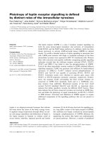
Figure 1 Slower primary root growth in exocyst mutants is associated with shorter root growth zones. (A) Root growth on vertical plates is
slower in exocyst mutants than Col-0, with defects ranging from mild (e.g. exo70A1-2) to quite severe (e.g. exo84b-1) (n = 8–19 roots for each genotype;
error bars represent standard error). (B) The number of cells in the meristem, transition, and elongation growth zones is reduced in exocyst mutants in
correlation with the root growth defect. Brassinosteroid (bri1 (SALK_003371), det2-1) and auxin transport (pin2-1, aux1-7) mutants, as well as brefeldin A
(BFA)-treated roots are shown for comparison. Error bars for meristem and elongation zone data shown in Figure 2. (C-L) The shorter growth zones in
exocyst mutants include shorter meristems that maintain a structure similar to wild-type. (C-F) Confocal images of 8 day old seedling root tips stained
with propidium iodide; white triangles and yellow triangles mark the distal and proximal ends of the meristem (i.e. the MZ) (bar = 100 microns applies
to C-F). (G-J) Confocal images of 8 day old propidium iodide-stained mutant roots (H-J) show expression of pWOX5-GFP restricted to the quiescent
center and similar to wild-type (G) (bar = 50 microns). (K-L) Confocal images of root tips expressing PLT1-YFP driven by its native promoter in sec8-3
(L) and a wild-type sibling (K) (bar = 50 microns).
Cole et al. BMC Plant Biology (2014) 14:386
synergistically inhibit hypocotyl elongation [27], and result
in a severe dwarfism of the same order of magnitude as the
sec8-3 line. Notably, the various exocyst mutants and mutant combinations reduce plant growth by differing, characteristic amounts (Additional file 1: Figure S1).
The dwarfism in seedlings with mutations in exocyst
components includes shorter roots due to slower root
growth rates, rather than premature termination of
growth (Figure 1A). Mutant lines with T-DNA insertions
in four different exocyst components demonstrate a dramatically wide range of primary root growth rates, which
vary from a low of 52 microns/hour in exo84b-1 mutants
to 391 microns/hour in exo70A1-1 mutants, compared to
478 microns/hour in Columbia 0 (wild-type) (Figure 2A).
Primary root growth in these mutants occurs at a nearly
constant rate when evaluated from five to eight days after
germination. Our focus was on this developmental period,
corresponding to the time when the early expansion of
the meristem has ceased, and after which the meristem
size remains virtually constant [7,40]. One explanation for
the observed range in severity of the root growth defects
is that the distinct mutant combinations represent an allelic series of sorts, with each reducing exocyst complex
Page 4 of 20
function in a quantitative manner, which is subsequently
manifested in quantitative effects on root growth rate.
Thus, evaluating this set of mutants provides a potentially
sensitive analysis for subtle effects of loss of exocyst function, i.e. small differences in the mutants can be considered
more credible when the magnitude of those differences
consistently correlate with the severity of the root growth
defect across all mutants evaluated.
Root growth defects in exocyst mutants are associated
with shorter growth zones
Confocal microscopic images of seven-day old roots were
evaluated to provide a detailed description of the meristematic and cell elongation root growth defects in exocyst mutants (Figure 1 and Additional file 2). Specifically, growth
parameters were determined by measuring cell lengths along
cortical cell files from the stem cell initials near the quiescent center to the beginning of the differentiation/maturation zone in seven-day old roots (see Additional file 2).
This region spans the meristematic zone (MZ) where cells
are dividing, the transition zone (TZ) where cells are not
dividing but continue to elongate at a slow rate, and the
elongation zone (EZ) where cell elongation increases
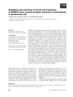
Figure 2 Primary root growth characteristics in exocyst mutants. (A-L) Characteristics of root growth in exocyst mutants are compared to
Col-0 and several brassinosteroid and auxin mutants, as well as to BFA-treated Col-0 roots of 7 day old seedlings. Data for B-L represent averages
for 14 cortical cell files (seven roots) evaluated from confocal images. Statistical comparisons to Col-0 (t-tests) are in Additional file 1: Figure S3.
Error bars = standard error.
Cole et al. BMC Plant Biology (2014) 14:386
exponentially until mature cell length is achieved, and cells
enter the differentiation zone [5,41]. The lengths of these
three distinct growth zones (MZ, TZ, and EZ) were dramatically shorter in the exocyst mutants (Figure 1B, 2C, and
2F). Notably, the overall cell file structure of the root tip was
maintained in all the exocyst mutants examined, such that
the consequences of a defect in cytokinesis, previously identified in the severe exo84b mutant [30], were not clearly evident at the tissue/organ level. Additionally, the identity and
organization of the quiescent center at the root tip was stably maintained in the mutants, as assayed by several lines of
evidence. The wild-type expression pattern of a marker for
quiescent center identity (the WOX5 promoter-driven GFP
construct, [42]) was not disturbed in even the most severe
mutant (Figure 1G-J). The preservation of the stem cell
niche, as well as the overall structure of the meristem, is associated with a gradient of the PLETHORA transcription
factors, PLT1 and PLT2 [43]. In exocyst mutants, YFPlabeled PLT1 and PLT2 proteins driven by their native
promoters showed nuclear localization and a gradient pattern of expression that was similar to that of wild-type seedlings (Figure 1K and L, and Additional file 1: Figure S2), but
compressed, coincident with the smaller size of the root
meristems of the mutants. Thus, although mutations in
components of the exocyst result in shorter growth zones,
the overall root tip structure and tissue patterning in the
mutants, which originate during embryogenesis, are similar
to wild-type. However, the sizes of the MZ, TZ and EZ are
clearly sensitive to reduced exocyst function.
The growth rate of plant roots depends upon the rate
of cell production in the meristem, and the extent of anisotropic cell expansion in the root’s EZ. To initially
evaluate whether exocyst mutations affect cell division
patterns in the root meristem, a CycB1::GUS reporter
was introduced into sec8-3, exo70A1-1, and exo70A11 sec8-6 mutant lines. Fusion of the CycB1 promoter
with GUS and a mitotic degradation signal allows this
reporter to mark only actively dividing cells [44]. As expected, GUS staining of the exocyst mutant roots revealed shorter meristems, associated with fewer dividing
cells within this zone, compared to their wild-type siblings (p < 0.001, t-test, n > 22 roots, Figure 3). Analysis of
confocal images of root cortical cell profiles from the
MZ through the EZ was then used to estimate cell production rates and cell cycle lengths in this cell layer
([45], and see Methods). The roots of five different exocyst mutant lines (7 roots per mutant line) were studied
in detail, representing a range of root growth rate defects: 11 percent of wild-type for exo84b-1 to 82 percent
of wild-type for exo70A1-1. Consistent with the pCYCB::
GUS results, the exocyst mutants demonstrated a
reduced cell production rate that correlated with the
reduced root growth rate (Figures 2A and 2B). To determine if the reduced cell production rate was due to a
Page 5 of 20
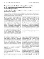
Figure 3 Meristems are shorter in exocyst mutants. pCYCB:GUS
expression is a marker for cell division activity, and the size of the
meristematic zone is designated by the span (arrows) over which GUS
staining is detected, in wild-type (A), exo70A1 (B), and sec8-3 (C) root tips.
Bar = 50 microns. (D) Average root meristem lengths measured by CYCB:
GUS are significantly shorter in exo70A1 (p < 0.01, t-test, n = 30) and sec8-3
(p < 0.001, t-test, n = 19) mutants compared to wild-type siblings (n = 30).
slower rate of cell division, the average length of the cell
cycle was estimated from the cell production rate and
the number of cells in the MZ (Additional file 2; [46]).
Brassinosteroid mutants, bri1 and det2-1, known to have
altered cell cycle progression and slower rates of cell division [47,48], served as controls and verified that our
method was capable of detecting prolonged cell cycles.
Surprisingly, the cell cycle length was not prolonged in
the exocyst mutants (p > 0.05, t-test, n = 7), and is notably shorter than wild-type (i.e., more rapid cell division)
in the most severe line, exo84b-1 (Figure 2D). Thus, the
reduced cell production rate associated with loss of exocyst function is largely due to a reduced number of dividing cells in the shorter MZ (Figure 2C), not slower cell
division.
The exocyst facilitates cell expansion in the elongation
zone
Cortical cell lengths were assessed to determine if a defect
in cell elongation also contributed to the root growth defect
in exocyst mutants. Mature cortical cell lengths, primarily
the result of cell elongation in the EZ, were indeed reduced
in the mutants with severe growth defects (Figure 2E,
p < 0.001, t-test, n = 7). The reduced cell elongation in exocyst mutants could conceivably arise because cells in exocyst
mutants have less time to elongate in their shorter
Cole et al. BMC Plant Biology (2014) 14:386
elongation zones or because they elongate at a slower rate.
To evaluate these two possibilities, data for the cortical cell
length profiles in the EZ of fourteen cell files for each mutant genotype were analyzed to estimate elongation rates, as
well as time spent in the EZ (detailed methods in Additional
file 2). Exocyst mutants with severe root growth defects have
significantly reduced rates of elongation compared to Col-0
(p < 0.001, Potthoff analysis) (Figure 4H and Additional
file 2). Furthermore, except for the mild exo70A1-1 mutant
(which did not differ from wild-type), the time cortical cells
of the exocyst mutants spent in the EZ was actually longer
than that observed for wild-type Col-0 (Figure 2G, p < 0.01,
t-test, n = 7). In other words, the reason mature cortical cells
of exocyst mutants are shorter is not because they have
spent less time elongating. Rather, loss of exocyst function is
specifically associated with a slower rate of elongation in the
EZ.
Mature cortical cell widths were also found to be significantly reduced in exocyst mutants (Figure 2I, p < 0.015,
t-test, n = 7). The width reduction correlated with the severity of the root growth defect, and not surprisingly, with
the width of the root (Figure 2J cell width measurements
in the EZ). The cell width data, combined with data for
mature cortical cell lengths, allow calculation of the average cell volume, as well as the average cell length-to-width
ratio for each genotype (Figure 2K and 2L). The mature
cortical cell volumes were dramatically reduced in the
exocyst mutants, whereas the length-to-width ratio of mature cortical cells was only minimally altered compared to
wild-type Col-0. Thus, the reductions in root growth rate
in exocyst mutants are not exclusively associated with a
reduction in mature cell lengths, but reflect a reduction in
cell expansion in the EZ, resulting in mature cortical cells
that achieve a near wild-type shape. Notably, cells of both
brassinosteroid (bri1, det2-1) and auxin mutants (pin2,
aux1-7) also alter cell expansion, but generate cells with
aberrant cortical cell length/width ratios.
We were curious to know if the elongation defect in exocyst mutants was restricted to the elongation zone, or if it
represented a more generalized defect (for example, a defect altering the basic structure and overall expansibility of
the cell wall) that also influenced cell elongation in the
meristem. To explore this question cortical cell lengths in
the meristem were evaluated, and the rates of cell elongation in the meristem were estimated (Figure 4I and
Additional file 2). The average cell elongation rates were
determined to be slightly faster, not slower in the meristems
of exocyst mutants compared to Col-0 (p < 0.05, t-test,
n = 7), with the exception of sec5a exo70A1-2, in which the
increase was not statistically significant. Thus, the reduced
rate of root cell elongation in exocyst mutants occurs specifically in the EZ. A similar conclusion was reached when
the width of cells along cell files in the MZ were measured
and plotted as a function of cell position from the quiescent
Page 6 of 20
Figure 4 Root cortical cells in exocyst mutants elongate at a slower
rate in shorter elongation zones. (A) Composite confocal image of a
Col-0 root (bar = 100 microns). (B-G) Cortical cells in elongation zones,
highlighted in white (bar = 50 μM). Col-0 (A, B,); exo70A1 (C); sec5a
exo70A1 (D); sec8-4 exo70A1 (E); sec8-3 (F); exo84b-1 (G). (H) Exponential
curves fitted to cell length data for elongation zones of 14 cell files (7
roots) of each genotype. The curves allow estimation of relative
elongation rates for each genotype (Figure 2H) (I) Reduced cell elongation
rates are not evident in the meristematic zones of exocyst mutants or
Col-0 treated with 10 μM BFA compared to Col-0; error bars represent
standard error with n = 7 roots per genotype. (see Additional file 2).
center. Cell widths expanded as the cells progressed shootward along the length of the meristem for Col-0 and all
mutants evaluated (Additional file 2). This progression of
cell width expansion in the exocyst mutants was essentially
Cole et al. BMC Plant Biology (2014) 14:386
Page 7 of 20
the same as Col-0, although there are fewer cells in the cell
files of the shorter meristem in exocyst mutants. Thus, the
role of the exocyst to enable root cell expansion (both
length and width) appears to manifest primarily in the EZ.
In summary, reduced exocyst mutant root growth
rates appear to be largely explained by a combination of
1) a reduced number of dividing cells in the meristem
and 2) a reduced cell elongation rate in the EZ. This implies that the exocyst contributes to plant growth differentially during root development, functioning to achieve
two apparently distinct outcomes: defining the sizes of
the growth zones, and enabling rapid cellular expansion
in the EZ.
Root growth in exocyst mutants is hypersensitive to BFA
treatment
To explore the hypothesis that the root growth defect in
exocyst mutants reflected an altered secretory system, the
sensitivity of root growth rate to the fungal toxin, brefeldin
A (BFA) was assessed in exocyst mutants. BFA inhibits
Golgi-based secretion and endocytic recycling, acting on
guanine nucleotide exchange factors (e.g., GNOM) to alter
the structure and function of the endomembrane system
[49,50]. Alteration of secretion in BFA-treated plant cells
is associated with altered delivery of polysaccharides and
cell wall loosening factors to the cell wall [51-54], altered
recycling of cell wall pectins in the root meristem [55],
and a reduced root growth rate [56]. Although BFA is
pleiotropic (e.g., it affects PIN protein recycling [56,57] as
well as secretion of cell wall polysaccharides, and alters
the root cell proteome accompanied by a remodeling of
the actin cytoskeleton [58]), root growth hypersensitivity
to BFA has been used as an indicator of a defect in vesicle
transport and secretion [59]. Localization of exocyst subunits themselves to the plasma membrane is BFAresistant [34,36], but root growth in exocyst mutants is
hypersensitive to BFA (Figure 5), with the normalized
growth rate reduced to a significantly greater extent in
exocyst mutants compared to Col-0 (p < 0.001, t-test,
n = 18-24) when exposed to 3.2 μM BFA. Such a result is
consistent with a predicted function for the complex in
vesicle trafficking to the PM, but does not rule out other
functions given the pleiotropic action of BFA.
Roots of Col-0 seedlings grown on plates containing
10 μM BFA were evaluated to determine if they phenocopied the root growth defects associated with exocyst
mutations. This concentration was chosen for comparison
to the most severe exocyst mutants, as its effect on growth
rate was similar to untreated sec8-3 and exo84b-1 lines.
Measurements of cortical cell lengths indicated that the
reduced growth rate in the BFA-treated Col-0 seedlings,
like that of the exocyst mutants, was due to a reduced mature cell length and, to a lesser extent than in the comparable exocyst mutants, a reduced cell production rate (and
Figure 5 Root growth rate response to brefeldin A (BFA). (A)
Exocyst mutants are hypersensitive to the root growth inhibiting effects of
BFA at a concentration of 3.2 μM. (B) Root growth rate response to BFA
normalized to the growth rate at BFA = 0. Normalized root growth rates
of exocyst mutants are significantly lower than that of Col-0, at a BFA
concentration of 3.2 μM (p < 0.001, t-test, n = 18-24). Bars = standard error.
associated shorter MZ) (Figure 2B, C, E, p < 0.001, t-test,
n = 7). As with the exocyst mutants, the reduced mature
cortical cell length was associated with a slower rate of
elongation in the shorter EZ of BFA-treated Col-0 roots
(Figure 2F, H, p < 0.001, t-test, n = 7). Remarkably, the exponential rate constant of this cortical cell elongation was
nearly identical to that of the exocyst mutants with severe
root growth defects (p > 0.05; Potthoff analysis; Additional
file 2). Ultimately, the mature cortical cell lengths were
shorter in the BFA-treated Col-0 roots than in the severe
exocyst mutants, because the cells in the BFA-treated Col0 roots spent less time in the EZ (Figure 2G). However,
the BFA-treated mature cortical cell length-to-width ratio
is quite distinct from that of the exocyst mutants
(Figure 2L), likely reflecting the more pleiotropic nature of
BFA action. The BFA results are consistent with the hypothesis that the exocyst’s function in root growth involves a role in secretory trafficking, but point toward
some distinction: the exocyst appears more important for
defining growth zone size.
Cole et al. BMC Plant Biology (2014) 14:386
Page 8 of 20
Altered auxin transport does not fully explain the root
growth defect of exocyst mutants
Polarized auxin transport and an auxin gradient with a
peak concentration at the quiescent center (QC) are key
determinants of meristem structure and function, and
thus root growth rate [42,60,61]. The size of the meristem depends in part upon an antagonistic interplay between auxin and cytokinin signaling, resulting in the
shift from cell division to cell elongation at a particular
location in the root tip [40,62-66]. Mutation of the exocyst component EXO70A1 results in a defect in acropetal (i.e., rootward) auxin transport and altered cycling
of PIN auxin transport proteins to the plasma membrane in root epidermal cells [36]. We therefore hypothesized that reduced meristem lengths were observed in
exocyst mutants because altered auxin transport shifted
the auxin-cytokinin balance to favor a shift from cell division to elongation at a position closer to the root tip.
To investigate this hypothesis, we measured primary
root growth after attempting to shift the auxin-cytokinin
balance in exocyst mutants and wild-type seedlings by
growing them on media containing a series of concentrations of the native auxin: indole acetic acid (IAA), the
synthetic auxin: 1-naphthaleneacetic acid (NAA), a cytokinin: N-6-benzyladenine, or the auxin transport inhibitor: naphthylphthalamic acid (NPA). Additionally, we
measured the root growth response of exocyst mutants
to 1-aminocyclopropanecarboxylic acid (ACC), an ethylene precursor. Ethylene promotes auxin biosynthesis
and/or auxin transport to affect epidermal cell elongation and root growth [66-69], and the root growth response to ACC is altered in auxin-related mutants
[70,71]. We reasoned that exocyst mutants should demonstrate altered sensitivity to these hormone manipulations if the reduced root growth rate in exocyst mutants
is primarily the result of a defect in auxin transport.
Contrary to expectation, the root growth dose–response
of exocyst mutants to exogenous auxins, cytokinin, or
ACC proved not to be significantly different from that of
wild-type Col-0 (Figure 6A, Additional file 1: Figure S4).
The pin2-1 mutant provided a contrasting control as, consistent with expectations, its sensitivity to IAA and NAA
was distinct from wild-type. The contrast between exocyst
and auxin mutant response was even more pronounced
with NPA treatment (Figure 6B). Normalized root growth
rates for severe exocyst mutants (i.e. sec8-3, sec8-4
exo70A1-2, and exo84b-1) were more sensitive than wildtype at NPA concentrations above 1 micromolar (p < 0.01,
t-test, n = 20-28). However, the opposite response was
exhibited by the aux1-7 and pin2-1 controls, which were
less sensitive to NPA in the same concentration range
(p < 0.01, t-test, n = 23-24). Thus, the response of exocyst
mutant roots to exogenous hormone manipulation and
auxin transport inhibition were not consistent with the
Figure 6 The root growth response of exocyst mutants to IAA
and NPA. (A) IAA dose–response of exocyst mutant root growth
was similar to Col-0, but different from auxin transport mutants
aux1-7 and pin2-1. (B) Exocyst mutants, sec8-3 and exo84b, were
slightly more sensitive to NPA at 3.16 and 10 μM compared to Col-0,
whereas two auxin transport mutants were less sensitive.
hypothesis that exocyst-dependent PIN trafficking was the
primary driver for the reduced root growth rate in exocyst
mutants.
We also examined the distribution and polar localization
of several auxin transporters in exocyst mutants. The different auxin transport proteins (PINs, AUX1, and ABC transporters) achieve their individual and polarized localizations
by delivery to the plasma membrane via functionally distinct
secretory pathways [10,72-76], a subset of which could conceivably involve the exocyst. Consequently, we examined
exocyst mutant lines containing labeled auxin transport proteins, PIN1-GFP, PIN2-GFP, PIN7-GFP, AUX1-YFP, and
ABCG36-GFP to see if mislocalizations were evident. The
polar localization within the cells and the pattern of distribution within the root tips (e.g., the MZ) of these auxin transport proteins in two exocyst mutant lines with severe root
growth defects (sec8-3, and sec8-4 exo70A1) appeared similar to wild-type, as had previously been reported for the less
severe exo70A1 mutant [36] (Figure 7, Additional file 1:
Figure S5 and Additional file 1: Figure S6). Notably, the expression of these auxin transporters in exocyst mutants was
Cole et al. BMC Plant Biology (2014) 14:386
Figure 7 Polarized localization of auxin transporters is evident in
exocyst mutants. (A-F) Polarized localization of PIN2-GFP (indicated by
white arrows) in lateral root cap (A, B), meristem (C,D), and elongation
zone (E,F) of sec8-4 exo70A1 mutants (A, C, and E) is similar to that of
wild-type siblings (B, D, and F). In C-F, cortical (left) and epidermal (right)
cell files are shown. Auxin response as indicated by pDR5:GUS (G,H) or
pDR5:GFP (I, J) appears similar in sec8-4 exo70A1 (G) and sec8-3 (I),
compared to their wild-type siblings (H, J). Bar for A-D = 20 microns;
bars for E&F, G&H, and I&J = 50 microns. Localization of additional auxin
transporters in exocyst mutant roots appear in Additional file 1: Figure
S5, and Additional file 1: Figure S6. Additional pDR5:GUS images appear
in Additional file 1: Figure S7.
observed within smaller regions than wild-type, but consistent with an interpretation of a shortened wild-type distribution pattern, correlating with their shorter meristems.
Downstream of auxin transport and development of
auxin gradients and auxin-mediated signaling is auxininduced modulation of transcription, i.e. the auxin response. To further explore the possibility that the exocyst mutants have slower root growth because of a
defect in auxin transport, we examined expression of
pDR5:GFP and pDR5:GUS, reporters of auxin response.
A subtly reduced region of auxin response peaking in
the region of the QC, and indicated by pDR5:GUS expression was previously observed in exo70A1 mutants
[36]. In four exocyst mutant lines with the most severe
root growth defects (exo84b-1, sec8-1, sec8-3, and sec8-4
exo70A1), we predicted a more profound alteration in
Page 9 of 20
the distribution/pattern of DR5 expression if the exocyst
mutant’s slower root growth was due to a defect in auxin response in the stem cell niche or MZ. However, when pDR5:
GFP or pDR5:GUS expression was observed in the roots of
these exocyst mutants, the reporters were observed in the
same region of the root as wild-type with a peak of expression in the region of the quiescent center (Figure 7G-J,
Additional file 1: Figure S7), albeit over a shorter length of
the root tip, correlating with the smaller meristem. Any
reduction in absolute expression level was not obviously correlated with the severity of the root growth defect. When
auxin response was evaluated in seedlings grown in the dark
(which reduces auxin production and acropetal auxin transport [77,78]), the significantly reduced level of pDR5:GUS
expression (compared to that of light grown seedlings) and
the pattern of that expression in the root tip were similar in
sec8-1 mutants and their wild-type siblings (Additional
file 1: Figure S7). Thus, in both light and dark-grown seedlings, the root growth defect could not be correlated with an
altered auxin response.
The maintenance and functioning of the stem cell niche
is dependent upon auxin and vital to root growth. Within
the niche, proximal stem cells next to the quiescent center
contribute cells to the MZ, whereas distal stem cells produce cells that differentiate to become the columella cells
of the root cap, identifiable by the accumulation of starch
containing amyloplasts. Mutations that alter auxin levels
or auxin transport to affect the root stem cell niche lead
to aberrant starch localization, i.e. the presence of starch
in the distal stem cells or the quiescent center, or conversely an increased number of non-differentiated (i.e.
starchless) cell layers between the quiescent center and
columella cells [42,78]. However, Lugol staining revealed
starch localization in exo70A1, sec8-3, and exo84b-1 mutants that was similar to that to Col-0 (n > 20 for each
genotype; Additional file 1: Figure S8). This contrasts with
the more diffuse presence of starch in root tips treated
with NPA (Additional file 1: Figure S8B, C; [42,79]), in
which the region of starch staining extends into the distal
stem cells and lateral root cap. The exocyst mutants also
did not demonstrate an increased number of cell layers
that were undifferentiated between the quiescent center
and the differentiated columella (Additional file 1: Figure
S8G), unlike what has been observed in PIN and auxin
biosynthesis mutants [42]. These results also distinguish
exocyst mutants from the icr1 mutant, in which altered
auxin transport is associated with the disappearance of
starch in the columella six days after germination [38].
Seven day-old exocyst mutants do have fewer layers of differentiated starch-stained columella cells compared to
Col-0 (consistent with the overall observation of shorter
developmental zones in the root meristem; Additional
file 1: Figure S8-I), but unlike the icr1 mutant the starch
staining pattern persists in the mutants after two weeks of
Cole et al. BMC Plant Biology (2014) 14:386
Page 10 of 20
growth (data not shown). Thus, an auxin-mediated alteration in the stability and functioning of the root stem cell
niche was not evident in exocyst mutants. This result is
consistent with the root tip expression patterns of WOX5
and PLETHORA transcription factors (Figure 1G-L),
which are believed to be regulated downstream of auxin
signaling to control stem cell activity [42]. In summary,
after assessing several independent characteristics linked
to auxin, we found no evidence to support the hypothesis
that the exocyst mutants’ shortened meristems have a
basis in defective auxin transport or the failure to establish
relative auxin maxima.
The root growth defect in Exocyst mutants is sensitive to
alterations in brassinosteroid signaling
One alternative to an auxin-related basis for reduced root
growth rates in the exocyst mutants would be defective
brassinosteroid signaling, which is closely linked to root cell
elongation. Similar to exocyst mutants, both BR-deficient
and BR-signaling mutants demonstrate reduced hypocotyl
elongation and reduced apical hook formation (phenotypes
associated with reduced cell elongation [27,80]), as well as
altered meristem size and mature cell lengths in the root
[47,48,81]. Furthermore, both the exocyst localization to
the plasma membrane and brassinosteroid signaling are
prominent in root epidermal cells [34,49], and the recycling
of the brassinosteroid receptor, BRI1, at the plasma membrane is disturbed in exocyst mutants [36].
A hypothesized role of the exocyst in brassinosteroid
signaling would be supported by rescue of the root growth
defect in exocyst mutants by exogenous hormone. And indeed, exocyst mutants with the most severe root growth
defects, sec8-3 and exo84b-1, demonstrated a mild rescue
when grown on media containing low concentrations (1
or 3.16 nM) epi-brassinolide, whereas wild-type Col-0
plants showed a decreased growth rate in this same concentration range, and the brassinosteroid receptor mutant,
bri1, showed no response (insensitivity) (Figure 8A). However, the rescue was small in absolute terms (Additional
file 1: Figure S9), with the partially rescued growth rate
remaining less than wild-type, and far less than the rescue
observed for the det2-1 brassinosteroid synthesis mutant.
In relative terms, exogenous brassinosteroid stimulated a
significant (p < 0.05, t-test, n = 20-44) increase in growth
rate of 27 and 39 percent over the untreated controls in
sec8-3 and exo84b-1, respectively, providing at least some
support for the hypothesis.
We next overexpressed the kinase BSK3 in sec8-3 and
exo84b-1 mutants to determine whether the observed rescue was linked to the canonical BRI1 signaling pathway.
BSK3 is phosphorylated by the BRI1 receptor to activate
downstream BR-induced transcription, and its overexpression rescues both BR biosynthetic (det2) and BR receptor
(bri1-5) mutants [82]. In both the sec8-3 and exo84b-1
Figure 8 Root growth in exocyst mutants shows a mild alteration
in response to brassinosteroids. (A) Dose–response of exocyst mutants
to exogenous epi-brassinolide shows mild rescue. (B) Combinations of
exocyst mutations (sec8-4 or exo70A1) and brassinosteroid mutations
(det2-1 or bri1) lead to more severe root growth defects; the combinations
with exo70A1 are synergistic if interpreted by a multiplicative model.
(C) qRT-PCR expression analysis indicates that the expression of
brassinosteroid synthesis genes (CPD and DWF4) are reduced in exocyst
mutants, contrary to what would be expected if the mutants had a defect
in the canonical brassinosteroid signaling pathway.
mutants, the over-expression of BSK3 resulted in only
very slight (14 percent and 9 percent, respectively) but significant (p < 0.01, t-test, n = 41-147) increases in root
growth rate (Additional file 1: Figure S10). In contrast,
there was no significant difference in the growth rates of
the wild-type siblings when seedlings with or without the
BSK3 construct were compared. Thus, consistent with the
results for treatment with exogenous epi-brassinolide,
BSK3 overexpression provided a slight rescue of the exocyst mutant phenotype.
We also tested for a genetic interaction between brassinosteroid signaling and the exocyst. A quantitative
Cole et al. BMC Plant Biology (2014) 14:386
synergistic genetic interaction is identified when a double
mutant has a more extreme phenotype than would be predicted by a neutrality function, a function that predicts the
phenotype when the two mutations do not interact [83].
Neutrality functions based upon multiplicative or additive
models have been used to study genetic interactions
[84,85], with the multiplicative model identified as being
more appropriate for predicting functional relationships
[83]. Seedlings possessing a combination of an exocyst mutation (exo70A1-1 or sec8-4, each associated with a mild
root growth defect) and a brassinosteroid-related mutation
(det2-1, affecting BR synthesis, or bri1, affecting BR signaling) were evaluated to determine if the two mutations had
a synergistic effect on reducing root growth. In each of the
four combinations, the double mutant demonstrated a
more severe root growth defect than either of the single
mutants (Figure 8B, p < 0.01, t-test, n = 20-75). The growth
rates for the double mutants were not significantly different
from what would be predicted by an additive model (p >
0.10, z-test, for all four double mutant combinations), nor
were the growth rates for sec8-4 det2 and sec8-4 bri1 mutants statistically different from prediction by multiplicative
model prediction (p > 0.05, z-test). However, the root
growth defect was significantly more severe than predicted
by the multiplicative model for exo70A1 det2 and exo70A1
bri1 mutants (p < 0.001, z-test). Thus, any synergistic interaction is not very robust, as it is only recognizable under
the multiplicative model and only in combination with the
exo70A1 mutant, suggesting that any functional interactions between exocyst-mediated events and brassinosteroid
signaling to influence root growth are limited, and perhaps
indirect.
The biosynthesis of brassinosteroids is known to be
under feedback control, such that decreased signaling
via BRI1, BSK3, and downstream transcription factors
leads to the increased synthesis of BR biosynthesis genes
[86,87]. If the exocyst mutations are causing a defect in
either brassinosteroid availability to receptors, perception, or downstream signaling through this pathway,
then one would predict that the exocyst mutants should
demonstrate an elevated expression of BR biosynthesis
genes. To test this prediction, qRT-PCR was performed
on cDNA from mutant roots to evaluate the expression
of DWF4 and CPD, two brassinosteroid biosynthesis
genes. Rather than being elevated, expression of these
genes was found to be slightly depressed in the exocyst
mutants (Figure 8C), indicating that BR signaling was
not reduced globally in the root, although this does not
rule out the possibility that signaling is reduced in some
regions or cell types of the root.
To better assess whether the root growth defects in exocyst mutants phenocopy those associated with defects in
brassinosteroid signaling, cell lengths along cortical cell
files obtained by confocal microscopy were evaluated for
Page 11 of 20
det2-1 and bri1-2 mutants and compared to those of exocyst mutants (Figure 1B, Figure 2). This revealed that, at
the cellular level, the root growth defect in the brassinosteroid mutants is distinct from that in exocyst mutants.
The shorter root meristem in brassinosteroid mutants is
primarily due to shortened cell length, whereas the shorter
meristem in exocyst mutants is due to fewer cells. A
decreased cell production rate in brassinosteroid mutant
roots is associated with a prolonged cell cycle (consistent
with previous reports: [47,48]) that is not seen in exocyst
mutants. The mature cortical cells of brassinosteroid
mutants had length-to-width ratios that were significantly
greater than Col-0 or any of the exocyst mutants
(Figure 2L, t test, p < 0.001, n = 7). On the other hand,
both the exocyst and brassinosteroid mutants demonstrate
a reduced mature cortical cell length compared to wildtype (Figure 2E, p < 0.001, t-test, n = 7) and this reduction
is partially due to slower elongation in both cases
(Additional file 2).
To determine if the partial rescue of root growth rate
with exogenous BR could be the result of an effect solely
on exocyst-dependent cell expansion in the EZ, roots
treated with and without 1 nM epibrassinolide were
studied. Root growth rates were ascertained and mature
cortical cell lengths were measured. The growth rates of
exo84b-1, sec8-4 exo70A1, and sec8-3 were 35-49%
higher in the epi-brassinolide treated groups, compared
to the untreated groups (n = 12-18 roots for each genotype/treatment group), but the corresponding mature
cortical cell lengths were only 12-19% longer in the exocyst mutants treated with epi-brassinolide, compared to
untreated controls. Thus an effect on mature cell length
could only account for about one-third of the observed
rescue, suggesting the added epi-brassinolide did not act
exclusively to increase cell elongation, but also increased
the cell production rate.
An alternative target for linking the exocyst to BR signaling is BREVIS RADIX (BRX), a plasma membraneassociated protein that is subject to endocytic recycling
[88,89], and upon auxin treatment translocates to the
nucleus [86] where it potentially mediates cross-talk between auxin and brassinosteroid signaling pathways
[90]. Mutant brx seedlings have slower growing roots
with shorter meristems and shorter mature cell lengths
[91]. Consequently, we tested whether the exocyst might
be involved in the cycling of BRX to the plasma membrane to affect root growth by attempting to rescue the
root growth defect in exocyst mutants using methods
that have been shown to rescue the brx mutant: overexpressing BRX; introducing a recessive null LONG
HYPOCOTYL 5 (hy5) mutation [88]; and growing the
mutants on a medium with a more basic pH [92]. In
each case, rescue of the exocyst mutant phenotype was
not achieved, indicating that the root growth defect in
Cole et al. BMC Plant Biology (2014) 14:386
exocyst mutants is not related to reduced BRX activity
(Additional file 1: Figure S11).
Discussion
The exocyst’s role in primary root growth occurs within
two developmental contexts
The developmental progression of root cells behind the
root cap can be visualized sequentially along cell files
beginning at the quiescent center: division in the meristem proper, slow constant elongation in the transition
zone, exponential elongation in the elongation zone,
followed by cessation of expansion and cell maturation.
As revealed by an examination of the roots of exocyst
mutants, the exocyst has two distinctive roles in root
growth that occur in two different stages of root development. First, the exocyst is involved in determining the
root cell production rate by helping to establish the size
of the root meristem, and therefore the number of cells
that are dividing. This is distinct from an effect on the
rate of cell division, which is not reduced in exocyst mutants (i.e. the cell cycle length is not prolonged in the
mutants). Second, the exocyst is involved in determining
mature cell length, primarily by enabling an exponential
rate of cell elongation in the elongation zone (EZ).
There is suggestive evidence that the specific processes
involving the exocyst in these two developmental events
(i.e. meristem size determination and cell expansion in
the EZ) are not the same. Notably, the series of exocyst
mutants differentially affect the two processes. Meristem
size is reduced in parallel with the reduction in root
growth rate over the entire range of exocyst mutants
evaluated. In contrast, rate of cell elongation is reduced
in parallel with growth rate only for the exocyst mutants
with less severe root growth defects; whereas the three
exocyst mutant lines with the most severe defects in
root growth (sec8-3, sec8-4 exo70a1, and exo84b) share
the same dramatically reduced cell elongation rate. In
other words, there is a mismatch in severity of the two
developmental defects across the range of mutants evaluated (Figure 9), with the reduction in meristem size
appearing to be a continually graded response, and cell
elongation influenced by an apparent threshold of exocyst activity.
We considered two ways the exocyst might conceivably
influence both developmental events through the same
process. First, the exocyst’s role in helping determine the
size of the growth zones might result in exocyst mutants
not only having a reduced cell production rate because of
a shortened meristem, but also a reduced mature cell
length resulting from a shortened elongation zone (allowing less time for root cells to elongate). However, such an
explanation is not supported by our data: cortical cells in
the mutants spend a longer period in the elongation zone,
but elongate at a slower rate. Second, the exocyst could
Page 12 of 20
affect cells in both the elongation zone and the meristem as
a general facilitator of cell expansion. Such a role could
conceivably slow the cell cycle and the cell production rate
in the meristem by slowing cytokinesis or entry into mitosis
based on cell size. However, again this is not the case: neither cell elongation rates nor the cell cycle is slower in the
meristems of exocyst mutants. This argues against a general
role for the exocyst in cell expansion, but rather is consistent with an interpretation in which the exocyst is required
for the remarkable cell expansion rates specifically associated with the EZ. Together, these results favor the hypothesis that the exocyst functions differentially to affect the
size of the root growth zones and, via a different process,
the rate of cell elongation in the elongation zone. Given the
exocyst’s likely role in facilitating vesicle trafficking to the
plasma membrane, one potential explanation for such
differential effects would be based on the presence of developmental stage-specific cargoes that are dependent on the
exocyst for transport and subsequent activity. In such a
scenario, the pleiotropic, multi-functional nature of the
exocyst would be due to its usage by the organism to correctly traffic different sets of developmentally-appropriate
vesicle cargoes.
The exocyst affects the size of the root growth zones by a
mechanism that could not be directly linked to auxin
Plant hormones play dominant and interacting roles in
regulating the size of the root meristem and other root
growth zones by controlling and coordinating specific processes (reviewed in [3-5,7]). For example, auxin acts on the
stem cell niche [42], promotes cell division in the meristem,
and targets the elongating epidermal cells in the root transition/elongation zone during a gravitropic response [68];
cytokinins act at the transition zone of the stele [62]; and
brassinosteroid perception in the epidermis mediates its effect on meristem size [48]. Among the many hormonemediated control pathways that affect root growth, auxin
and brassinosteroid signaling were considered the most
likely candidates for involvement of the exocyst. Plasma
membrane localization of exocyst subunits (EXO70, SEC6,
SEC8, EXO84) is prominent in the root epidermis [34], coincident with important sites for auxin and brassinosteroid
signaling. Additionally, in the root, the exocyst has been
linked to acropetal auxin transport, as well as to recycling
of the PIN1 and PIN2 auxin efflux carriers and the BRI1
brassinosteroid receptor to the plasma membrane [36].
Consequently, we hypothesized that the root growth defect
in exocyst mutants might be largely the result of a defect in
PIN-mediated auxin transport and signaling, or possibly altered brassinosteroid signaling.
We assessed exocyst mutant roots for indicators of altered
auxin signaling, including: (1) altered sensitivity to either exogenous auxins, an auxin transport inhibitor, cytokinin, or
ACC; (2) altered localization of auxin transporters, the
Cole et al. BMC Plant Biology (2014) 14:386
Page 13 of 20
Figure 9 The exocyst’s role in root growth. The exocyst is required for root growth in two developmental contexts to affect (A) cell
production; and (B) cell length. Table above graphs shows how parameters determining primary root growth are affected by exocyst mutations;
values shown are percentages of the Col-0 value for each parameter. Bars in graphs A and B represent (from white to black): Col0 (wild-type),
exo70A1, sec5a exo70A1, sec8-4 exo70A1, sec8-3 and exo84b-1. In the meristem exocyst mutations reduce the cell production rate (A), but not the
length of cells in the cortical cell file (B). However, in the transition and elongation zones, where new cells are rarely produced in either wild-type
or exocyst mutant roots, mutation of the exocyst leads to severely reduced cortical cell elongation (B).
pWOX5::GFP marker for quiescent center identity, auxinregulated PLT transcription factors, or DR5-driven
markers of auxin response, (3) rescue with a hy5 mutation,
which is associated with elevated auxin-responsive transcription, and (4) the aberrant presence of starch in the
columella initials. Exocyst mutants treated with exogenous
hormones had dose–response curves that were similar to
wild-type. Localization of markers for auxin responsiveness; PIN, AUX1, and ABCG36 transporters; quiescent
center identity; and PLT expression occurred over shorter
root regions coincident with the shorter root growth
zones, but, otherwise, the localization patterns were not
remarkably different in exocyst mutants compared to
wild-type. The hy5 mutation failed to exert any detectable
rescue of the exocyst mutant phenotype. Increased staining of starch in the columella initials, which is indicative
of altered auxin signaling in the vicinity of the stem cell
niche, was not observed in the exocyst mutants. Although
we did not conduct experiments addressing a possible link
between the exocyst and ABCB auxin transporters, mutation of ABCB transporters leads to phenotypes not
consistent with our observations of exocyst mutants (e.g.,
longer root hairs [91,93], enhanced gravitropism [94,95],
sporadic root curvature [94], prominent reduction of the
number and growth of lateral roots [94,96,97]). It must be
acknowledged that direct assessment of auxin signaling by
the TIR complex was not addressed in the range of
experiments performed on exocyst mutants. However, the
severe root growth defect observed in exocyst mutants
could not be convincingly associated with any of several
indirect indicators of altered auxin transport or signaling.
Drdová, et al. [36] demonstrated that the recycling of
PIN1 and PIN2 proteins to the plasma membrane from
brefeldin-A compartments is delayed in both exo70A1 and
sec8-1 mutants. Our results do not necessarily contradict
these data, but do argue that a role for exocyst activity in
PIN trafficking is not a major driver influencing root
growth rate. The lack of a clear connection to altered
auxin signaling in exocyst mutants also contrasts with the
mutant phenotypes of the Rop GTPase- and SEC3interacting scaffold protein ICR1 [37], which dramatically
perturbs root meristem pattern (e.g., disorganized WOX5
and DR5::GUS expression) and auxin transport (e.g., altered PIN1 and PIN2 subcellular distribution) [38]. Thus,
our observations also argue against the exocyst as the primary link between ICR1 and auxin. However, auxin regulates a broad range of developmental and physiological
processes, and auxin transport and signaling is correspondingly complex [98,99]. Thus, we cannot rule out altered auxin transport or signaling in exocyst mutants as a
contributor to their root growth defect; but we conclude
that the exocyst is unlikely to be directly linked to root
growth via a currently understood process in auxin transport and signaling.
Cole et al. BMC Plant Biology (2014) 14:386
Enhanced brassinosteroid signaling can partially
compensate for, but not fully rescue, the root growth
defect in exocyst mutants
We also investigated the possibility that the exocyst is involved in the transport of brassinosteroids to the plasma
membrane where they bind BRI1-type receptors [13], or in
the plasma membrane placement of those receptors, to
affect brassinosteroid signaling [81]. Consistent with this
possibility, exocyst mutants with severe root growth defects
did demonstrate a small, but statistically-significant, rescue
of the mutant phenotype with the application of exogenous
epi-brassinolide, and an altered dose–response curve. Overexpression of the BR signaling kinase, BSK3, to activate BR
signaling downstream of BRI1 receptor also caused a slight
increase in root growth of exocyst mutants that was not
seen in their wild-type siblings. Evaluation of double
exocyst-brassinosteroid mutants for a genetic interaction
provided a somewhat ambiguous result: the double
mutants, exo70A1 det2-1 and exo70A1 bri1, had growth
defects that were more severe than would be predicted
from the growth rates of single mutants by a multiplicative
model, but not by an additive model. These data support a
functional interaction between the exocyst and brassinosteroid signaling, but are consistent with the possibility that
this interaction is indirect.
Three lines of evidence suggest the exocyst does not directly interact with brassinosteroid signaling to affect root
growth. First, qRT-PCR analysis indicates that the expression of brassinosteroid synthesis genes is not elevated in
exocyst mutant roots. This is contrary to the expectation
for defects in brassinosteroid signaling, because feedback
control dictates increased expression of the BR synthesis
genes when the BR signal is attenuated (86). Second, although the dynamics of BRI1 cycling is altered from
wild-type in exocyst mutants treated with BFA [36], the
ultimate localization of BRI1-GFP to the plasma membrane is not significantly altered in exocyst mutants
with the most severe root growth defects (Additional
file 1: Figure S9). Third, a detailed comparison of the
cortical cell length profiles reveals important differences
between exocyst and brassinosteroid mutants. Shortened meristems in exocyst mutants are due to fewer
cells (not an altered cell cycle), whereas shortened meristems in brassinosteroid mutants (det2-1 and bri1-2) are
primarily due to shortened cell lengths (and are also associated with a prolonged cell cycle). Both exocyst and
brassinosteroid mutants demonstrate a reduced mature
cortical cell length and a slower rate of cell elongation in the
elongation zone, but the rate of cell elongation is much
more dramatically reduced in exocyst mutants, and the final
cell length-to-width ratio is also distinct. A specific role for
the exocyst in trafficking of the upstream regulatory protein,
BRX, was discounted when several experimental manipulations known to rescue the brx mutant failed to rescue the
Page 14 of 20
exocyst mutant root growth phenotype. Together these observations argue that the exocyst root growth phenotype is
not primarily driven by defects in brassinosteroid signaling.
The partial rescue of the growth rate defect by application of low concentrations of epi-brassinolide deserves
further consideration. Recent evidence suggests that in
addition to the canonical intracellular BR signaling via a
kinase cascade [9,81], there is also a non-cell autonomous
BR-induced signal from the root epidermis to the steele
[48], a BR-induced fast response involving activation of a
plasma membrane P-ATPase [100], and a BR induced cyclic GMP-mediated Ca2+ signaling cascade [101]. A direct
role for the exocyst in any of these pathways could explain
the observation that epi-brassinolide treatment provides a
stronger rescue than the specific induction of the kinase
cascade via BSK3 overexpression. Alternatively, rescue of
the exocyst phenotype could be accomplished indirectly,
by BR induction of a process that can partially compensate
for loss of exocyst function. As an example, in S. cerevisiae, overexpression of SRO7 (an Lgl family protein that
interacts with t-SNAREs) can suppress the growth defects
of multiple exocyst mutants [102]. Induction of a similar
gene function in Arabidopsis by BR would have a larger
effect if exocyst activity is reduced, explaining both the
partial epi-BR and BSK3 rescue.
Potential alternative roles for the exocyst in root growth
Overall, despite a role for the exocyst in PIN and BRI1
trafficking in root epidermal cells (36), the root growth
defect in exocyst mutants does not appear to be explained by a simple inhibition of the known auxin/cytokinin- and brassinosteroid-based mechanisms that help
define the developmental activities of the root meristem
and elongation zone [40,48]. However, observation of
the constellation of root growth characteristics seen in
exocyst mutants, occurring with an apparent independence from these phytohormones pathways, is not unprecedented. The exocyst mutant phenotype is mimicked
in seedlings overexpressing the UPBEAT transcription factor [103]. Overexpressing UPBEAT down-regulates expression of class III peroxidases in the root’s elongation
zone, leading to an altered balance of reactive oxygen species (ROS), a resultant shift in the root’s transition zone
(i.e. between proliferation and differentiation) and reduced
root growth. UPBEAT is hypothesized to act both directly
(via expression of peroxidases secreted to the apoplast)
and indirectly (via ROS signaling) to modify cell walls.
The possibility of exocyst involvement in a ROS-mediated
mechanism to affect root growth, for example, a role in
the secretion of peroxidases in the root’s elongation zone,
deserves exploration.
The exocyst mutant root phenotype (including reduction in size of growth zones and reduced rate of cell elongation, occurring without prolonged cell cycle duration,
Cole et al. BMC Plant Biology (2014) 14:386
altered stem cell niche, or a prominent defect in auxin
transport) is also strikingly similar to that reported for seedlings stressed by growth on medium containing elevated
concentrations of ammonium [104]. There is no obvious
link between the exocyst and ammonium metabolism, but
there could be a connection between the exocyst and a
plant’s response to abiotic stress. Repression of root cell
elongation occurs in response to a variety of environmental
stresses [104-106], and setting the size of the growth zones
is considered to be the key regulatory act for root growth
acclimation to environmental conditions [107]. Thus, the
primary characteristics of root growth that are affected
upon inhibition of exocyst function are coincident with
those root growth characteristics that are adjusted in response to environmental stress. The potential involvement
of the exocyst in the root’s growth response to abiotic environmental stress is an interesting possibility.
Environmental stressors elicit a host of varied signaling pathways, involving hormone-modulated systems
[2,108,109], cell-type developmentally-specific transcriptional modules [1,103], and ROS [110,111]. However, similarities in the root growth response suggest
these pathways may ultimately converge to a common
set of downstream mechanisms that alter growth. Central to the downstream mechanisms, particularly in the
root’s transition and elongation zones, are secretory
processes that deliver material to form the cell wall
matrix (e.g., secreted pectins and hemi-celluloses), and/or
proteins that modify the matrix to promote cell wall loosening and expansion (e.g. expansins, xyloglucan endotransglycolase/hydrolases, endo-(1,4)-β-D-glucanases, and
peroxidases) [14,112-117]. Modulating the activity of the
exocyst to affect these downstream secretory events and
cell wall expansion could be integral to the mechanisms
that ultimately allow for growth that is both developmentally coordinated and environmentally responsive. The
framework for exocyst function in the growing root established here will help better define future work to address
this and the aforementioned possibilities.
Conclusions
A complex network of interacting, overlapping, feedbackcontrolled, and often hormone-mediated mechanisms
have evolved to control and maintain primary root growth
[3-5,118]. In the midst of this complex system, perhaps at
multiple sites, lies the exocyst, a putative molecular tether
that facilitates secretory vesicle delivery for fusion to the
plasma membrane in both plant [19,34,35] and non-plant
species [16-18,23]. Mutate a component of the exocyst
and a dramatic reduction in root growth rate results, in
certain cases down to a mere 11% of the growth rate
observed in wild-type roots. At the same time, the resilience of the system is revealed in exocyst mutants: they
maintain the overall meristematic, growth zone, and tissue
Page 15 of 20
layer structure of wild-type roots, although the number of
cells in these regions is reduced in the mutants. Evaluation
of cortical cell files reveals that the slower growth rates in
exocyst mutants arise because their meristematic cell
production rates are lower, and their mature cells are
shorter compared to wild-type roots. This analysis provides evidence that the exocyst functions in two different
developmental contexts to affect root growth, seemingly
independent of auxin and brassinosteroids, influencing
both the size of the growth zones and the rate of cell
elongation.
Methods
Plant material and growth conditions
Lines of Columbia-0 ecotype of Arabidopsis thaliana
with T-DNA insertions and other mutants were obtained
from the SALK Institute [119]: exo70A1-2 (At5g03540)
SALK 135462; sec5a-1 (At1g76850) SALK 010127; sec81 (At3g10380) SALK 057409; sec8-3 (At3g10380) SALK
026204; sec8-4 (At3g10380) SALK 118129; sec8-6 (At3g
10380) SALK 091118; bri1 (At4g39400) SALK 003371;
det2-1 (At2g38050) CS6159; pgm-1 (At5g51820) CS210;
arg-1 (At3g68370) SALK 024542C. The exo84b-1 line
was a GABI-Kat line [120], provided from the Zarsky
Lab (30). Exocyst T-DNA insertion sites for exocyst related SALK lines were verified by sequencing. PCR genotyping for exocyst mutant alleles was performed as
previously described [27,28]. Homozygous exocyst mutants with severe root growth defects (i.e. sec8-1, sec8-3,
exo84b-1) are virtually sterile and were obtained in the
progeny of self-crossed heterozygous plants. The homozygotes could be readily identified by root growth and
root hair phenotypes and were consistently verified by
spot-checking with PCR). Mutant lines aux1-7 and pin21 were provided by M. Ivanchencko. Marker lines
pPLT1:gPLT1-YFP, pPLT2:PLT2-YFP and pWOX5:GFP
were provided by Y. Du and B. Scheres; pPIN7:PIN7GFP was provided by Wendy Peer, Purdue University;
PIN1-GFP, PIN2-GFP and AUX1-YFP were provided by
S. Robert and N. Raikhel; ABCG36/PEN3-GFP was provided by B. Underwood and S. Somerville; pDR5:GFP
and pDR5:GUS were from M. Ivanchenko. Marker
BRI1-GFP was provided by J. Chory; p35S:BSK3-YFP
was provided by Z. Wang; and p35S:BRX-GFP was provided by A. Amiquet.
Arabidopsis seeds were surface-sterilized, stratified at
4°C for 3–5 days, and planted on growth media (1x MS,
2% (w/v) sucrose, and vitamins) or soil as previously
described [28]. Serial dilutions of hormones were prepared and added to media cooled to 50°C prior to pouring plates. Phytohormones: 3-indoleacetic acid (IAA),
naphalene acetic acid (NAA), N-6-benzyladenine,
24-epibrassinolide, 1-aminocyclopropanecarboxylic acid
(ACC), and naphthylphthalamic acid (NPA), as well as
Cole et al. BMC Plant Biology (2014) 14:386
brefeldin A (BFA) were all obtained from Sigma-Aldrich.
Plants were grown in a climate chamber at 22°C under
long-day conditions (16 hr. of light per day).
Microscopy
Root growth rates at 7 days of growth were calculated from
the differences in root lengths observed on days 6 and 8,
and captured with a Canon Power Shot A710IS digital
camera, or with a Moticam 1000 camera attached to a Zeiss
Stemi SV 11 dissecting microscope. For the analysis of root
tips stained for GUS activity or starch, seedlings were fixed
in 0.3% formaldehyde in 0.33 M phosphate buffer pH 7.2
for 30 min at room temperature, and then rinsed three
times in 50 mM phosphate buffer pH 7.2. GUS activity was
analyzed after staining the fixed seedlings overnight at 37°C
and then cleared as described by Malamy and Benfey [121]
and modified by Ivanchenko, et al. [122]. Starch was stained
by placing fixed root tips in Lugol stain [0.34% (w/v) I2 with
0.68% (w/v) KI in H2O] for 15 minutes. Light microscopy
of root tips stained for GUS activity or starch was
performed on a Zeiss Axiovert microscope with differential
interference contrast optics. Live roots stained with propidium iodide (10 μg/ml for at least 15 minutes) or containing fluorescent markers were imaged at the Confocal
Microscopy Facility of the Center for Genome Research
and Biocomputing and the Environmental and Health
Sciences Center at Oregon State University using a Zeiss
LSM510 META with Axiovert 200 motorized microscope
with version 3.2 LSM software (National Institute of Health
grant number 1S10RR107903-01). Root lengths or cell dimensions in digital images were measured using ImagePro
6.2 software (MediaCybernetics, www.mediacy.com).
Cortical cell file analysis
Four distinguishable developmental zones - the meristem,
and the transition, elongation, and maturation zones
[5,41] - were identified by examining cortical cell lengths
along a cell file. Analyzed from the tip, cells lengths observed within a two-fold range were considered to be dividing and were identified as meristematic cells. Following
the meristem, a transition zone (some have called the distal elongation zone) was identified as the region where cell
lengths no longer appeared to divide to produce two
smaller cells, but instead showed a slow steady increase in
length as one proceeded up the cell file. The transition
zone ended and the beginning of an accelerated elongation zone was identified by a series of cells that increased
exponentially in length. This elongation zone ended and
the maturation zone began when there was no longer an
exponential increase in cell size, but instead cell lengths
varied around an average mature cell length, whereas in
neighboring cell layers root hairs or lateral root formation
were often observed. Analysis of growth parameters from
cortical cell length profiles is described in detail in
Page 16 of 20
Additional file 2. Briefly, the cell production rate was calculated by dividing the root growth rate by the average
mature cell length [123,124]. An estimate of the length of
the cell cycle was obtained by dividing the cell production
rate by the number of dividing cells (i.e. the number of
cells in the meristem) and multiplying by the ln(2) [46].
The average time interval between each cortical cell leaving the meristem to enter the transition and elongation
zones was estimated as the inverse of the cell production
rate [123,125,126]. Linear regression analysis of the cell
length data after logarithmic transformation then provided
a basis for determining the exponential rate constant for
elongation in the elongation zone, and a means for comparison of the elongation curves between genotypes.
qRT-PCR
Whole roots were harvested from 8 day old seedlings
growing on MS plus 2% sucrose medium on vertically
oriented plates, and immediately frozen in liquid nitrogen, and stored in −80 degree C freezer. Three biological
replicates (50–100 mg tissue each = up to ~100 roots)
were collected for each genotype, with each biological
replicate composed of the pooled root samples from a
particular planting of seeds from a unique parent plant.
RNA was collected from the frozen tissue by grinding it
under liquid nitrogen using mortar and pestle, adding
TRIzol reagent (Invitrogen Cat No. 15596–018), and collecting the aqueous phase of a chloroform extraction.
The RNA was precipitated from the aqueous phase with
the addition of isopropyl alcohol, pelleted by centrifugation, washed with 75% ethanol, and briefly dried. Each
pellet was redissolved in RNase-free water, and purified
with QIAGEN RNeasy MinElute Cleanup Kit, Cat No.
74204 following manufacturer’s instructions. Quality and
quantity of the purified RNA was verified using an Agilent Bioanalyzer 2100 (RIN’s: 9.8-10.0). RNA was treated
with DNaseI (Invitrogen, Cat No 18068–015) prior to
synthesis of cDNA using the Superscript III First-Strand
Synthesis System (Invitrogen, Cat No. 18080–051) per
manufacturers protocol.
The qRT-PCR was performed using the BioRad CFX96
cycler. Primers (listed in Additional file 3) were designed to
amplify selected sequences using the following criteria: Tm
of 59–61 C, primer lengths of 20–25 nucleotides, guaninecyctosine contents of 40-55%, amplifying near the 3′ end of
the transcripts to produce an PCR amplicon length of 55–
150 bp, and covering an exon-exon junction if possible.
Quantification cycle values from qPCR were obtained for
three technical replicates for each of the 3 biological replicates for each gene evaluated per plant genotype. Efficiency
for each qPCR reaction was determined by evaluation of a
dilution series run on the same plate as the samples (Efficiencies are included in Additional file 3). TIP41 (At4g3
4270), SAND (At2g28390), and a protein coding gene
Cole et al. BMC Plant Biology (2014) 14:386
(At4g33380) were selected as reference genes based upon
their documented stability of expression and successful use
in qRT-PCR studies of roots [127-131]. Cq values were
converted to relative quantities by comparing Cq values
from each exocyst mutant line to the corresponding Cq
value for Col 0. Calculation of these relative quantities took
into account qPCR efficiencies, utilizing the qBase framework [132].
Additional files
Additional file 1: Supplemental Figures. Figure S1 - Exocyst
mutant seedling images; Figure S2 - Plethora-YFP in mutant root tips;
Figure S3 - T-test comparisons of root growth characteristics; Figure S4
- Phytohormone dose-response curves of exocyst mutants; Figure S5 Fluorescently labeled auxin transporters in sec8-4 exo70A1 root tips;
Figure S6 - Fluorescently labeled auxin transporters in sec8-3 root tips;
Figure S7 - pDR5-GUS in dark and light grown seedling root tips;
Figure S8 - Lugol-stained starch in root tips of exocyst mutants;
Figure S9 - BRI1-GFP in mutant root tips and epibrassinolide dose-response;
Figure S10 - Effect of BSK3 overexpression on root growth in exocyst
mutants; Figure S11 - Effect of BFA on root growth in exocyst mutants.
Additional file 2: Using cortical cell length profiles to compare root
growth characteristics.
Additional file 3: qRT-PCR primers.
Abbreviations
MZ: Meristematic zone; TZ: Transition zone; EZ: Elongation zone;
ABFA: Brefeldin.
Competing interests
The authors declare that they have no competing interests.
Authors’ contributions
RC participated in the study design, implemented all experiments and wrote
the manuscript. SM performed Lugol staining of root tips and analysis of the
number of columella cell layers as a function of root age and in response to
NPA treatment. JF helped conceive the study, participated in its design, and
edited the manuscript. All authors read and approved the final manuscript.
Acknowledgments
We acknowledge the high school Apprenticeships in Science and
Engineering (ASE) program for internship guidance (S.A. McInally); and also
thank E. Harvey and M. Moreno, whose work in the lab through ASE also
helped advance this project. We thank C. Chan, B. Park, H. Pham, and A.
Sughrua for assistance with genotyping and plant maintenance; Z.
Velupkova for frequent assistance and patient laboratory support; and the
Oregon State University (OSU) Central Services Lab for sequencing. We thank
the members of the lab of V. Žárský, particularly L. Synek, E. Drdová, and M.
Fendrych, for their practical contributions and discussions regarding exocyst
investigations. We also appreciate the collaboration and support of V. Dolja
at OSU, and wish to acknowledge the Confocal Microscopy Facility of the
Center for Genome Research and Biocomputing, and the Environmental and
Health Sciences Center at Oregon State University as a contributing resource
for this work.
This work was supported by U.S. National Science Foundation grants IOS-0920747
and MCB- 1244633. The confocal microscopy data in this publication were made
possible in part by grant number 1S10RR107903-01 from the National Institutes of
Health.
Received: 29 July 2014 Accepted: 15 December 2014
Page 17 of 20
References
1. Dinneny J, Long T, Wang J, Jung J, Mace D, Pointer S, Barron C, Brady S,
Schiefelbein J, Benfey P: Cell identity mediates the response of
Arabidopsis roots to abiotic stress. Science 2008, 320:942–945.
2. Wolters H, Jürgens G: Survival of the flexible: hormonal growth control
and adaptation in plant development. Nat Rev Genet 2009, 10:305–317.
3. Garay-Arroyo A, Sánchez M, García-Ponce B, Azepeitia E, Alvarez-Buylla E:
Hormone symphony during root growth and development. Dev Dyn
2012, 241:1867–1885.
4. Perilli S, Di Mambro R, Sabatini S: Growth and development of the root
apical meristem. Curr Opin Plant Biol 2012, 15:17–23.
5. Petricka J, Winter C, Benfey P: Control of Arabidopsis root development.
Annu Rev Plant Biol 2012, 63:563–590.
6. Rymen B, Sugimoto K: Tuning growth to the environmental demands.
Curr Opin Plant Biol 2012, 15(6):683–690.
7. Ubeda-Tomás S, Bennett M: Plant development: size matters, and it’s all
down to hormones. Curr Biol 2010, 20(12):R511–R513.
8. Worden N, Park E, Drakakaki G: Trans-Golgi network – an intersection of
trafficking cell wall components. J Integr Plant Biol 2012, 54(11):875–886.
9. Clouse S: Brassinosteroid signal transduction: from receptor kinase
activation to transcriptional networks regulating plant development.
Plant Cell 2011, 23(4):1219–1230.
10. Rigó G, Ayaydin F, Tietz O, Zsigmond L, Kovács H, Páy A, Salchert K, Darula
Z, Medzihradszky K, Szabados L, Palme K, Koncz C, Cséplo A: Inactivation of
plasma membrane-localized CDPK-RELATED KINASE5 decelerates PIN2
exocytosis and root gravitropic response in Arabidopsis. Plant Cell 2013,
25:1592–1608.
11. Kleine-Vehn J, Friml J: Polar targeting and endocytic recycling in auxindependent plant development. Annu Rev Cell Dev Biol 2008, 24:447–473.
12. Wightman R, Turner S: Trafficking of plant cellulose synthase complex.
Plant Physiol 2010, 153:427–432.
13. Symons G, Ross J, Jager C, Reid J: Brassinosteroid transport. J Exp Bot 2008,
59(1):17–24.
14. Cosgrove D: Growth of the plant cell wall. Nat Rev Mol Cell Biol 2005,
6:850–861.
15. Driouich A, Follet-Gueye M, Bernard S, Kousar S, Chevalier L, Vicré-Gibouin M,
Lerouxel O: Golgi-mediated synthesis and secretion of matrix polysaccharides of the primary cell wall of higher plants. Front Plant Sci 2012, 3:1–15.
Article 79.
16. He B, Guo W: The exocyst complex in polarized exocytosis. Curr Opin Cell
Biol 2009, 21:537–542.
17. Liu J, Guo W: The exocyst complex in exocytosis and cell migration.
Protoplasma 2011, 249(3):587–597.
18. Heider M, Munson M: Exorcising the exocyst complex. Traffic 2012,
13(7):898–907.
19. Zárský V, Kulich I, Fendrych M, Pecenková T: Exocyst complexes multiple
functions in plant cells secretory pathways. Curr Opin Plant Biol 2013,
16:726–733.
20. Zárský V, Cvrčková F, Potocký M, Hála M: Exocytosis and cell polarity in
plants – exocyst and recycling domains. New Phytol 2009, 183:255–272.
21. Wu H, Rossi G, Brennwald P: The ghost in the machine: small GTPases as
spatial regulators of exocytosis. Trends in Cell Biol 2008, 18(9):397–404.
22. Sivaram M, Saporita J, Furgason M, Boettcher A, Munson M: Dimerization of
the exocyst protein Sec6p and its interaction with the t-SNARE Sec9p.
Biochemistry 2005, 44:6302–6311.
23. Rivera-Molina F, Toomre D: Live-cell imaging of exocyst links its spatiotemporal
dynamics to various stages of vesicle fusion. J Cell Biol 2013, 201(5):673–680.
24. Pečenková T, Hála M, Kulich I, Kocourková D, Drdová E, Fendrych M, Toupalová
H, Žárský V: The role for the exocyst complex subunits Exo70B2 and
Exo70H1 in the plant-pathogen interaction. J Exp Bot 2011, 62(6):2107–2116.
25. Cvrčková F, Grunt M, Bezvoda R, Hála H, Kulich I, Rawat S, Žárský V:
Evolution of the land plant exocyst complexes. Front Plant Sci 2012, 3:159.
26. Zhang Y, Liu C, Emons A, Ketelaar T: The Plant Exocyst. J Integr Plant Biol
2010, 52(2):138–146.
27. Hála M, Cole R, Synek L, Drdová E, Pečenková T, Nordheim A, Lamkemeyer
T, Madlung J, Hochholdinger F, Fowler J, Žárský V: An exocyst complex
functions in plant cell growth in arabidopsis and tobacco. Plant Cell 2008,
20:1330–1345.
28. Cole R, Synek L, Žárský V, Fowler J: SEC8, a subunit of the putative
Arabidopsis exocyst complex, facilitates pollen germination and
competitive pollen tube growth. Plant Physiol 2005, 138:2005–2018.
Cole et al. BMC Plant Biology (2014) 14:386
29. Synek L, Schlager N, Eliáš M, Quentin M, Hauser M, Žárský V: AtEXO70A1, a
member of a family of putative exocyst subunits specifically expanded
in land plants, is important for polar growth and plant development.
Plant J 2006, 48:54–72.
30. Fendrych M, Synek L, Pečenková T, Toupalová H, Cole R, Drdová E,
Nebesářová J, Šedinová M, Hála M, Fowler J, Žárský V: The Arabidopsis
exocyst complex is involved in cytokinesis and cell plate maturation.
Plant Cell 2010, 22:3053–3065.
31. Li S, Chen C, Yu D, Ren S, Sun S, Liu L, Ketelaar T, Emons A, Liu C: EXO70A1mediated vesicle trafficking is critical for tracheary element
development in Arabidopsis. Plant Cell 2013, 25:1774–1786.
32. Wu J, Tan X, Wu C, Cao K, Li Y, Bao Y: Regulation of cytokinesis by exocyst
subunit SEC6 and KEULE in Arabidopsis thaliana. Mol Plant 2013,
6(6):1863–1876.
33. Rybak K, Steiner A, Synek L, Klaeger S, Kulich I, Facher E, Wanner G, Kuster B,
Zarsky V, Persson S, Assaad F: Plant cytokinesis is orchestrated by the
sequential action of the TRAPPII and Exocyst tethering complexes.
Dev Cell 2014, 29:607–620.
34. Fendrych M, Synek L, Pečenková T, Drdová E, Sekereš J, de Rycke R, Moritz
K, Nowack M, Žárský V: Visualization of the exocyst complex dynamics at
the plasma membrane of Arabidopsis thaliana. Mole Biol Cell 2013,
24:510–520.
35. Zhang Y, Immink R, Liu C, Emons A, Ketelaar T: The Arabidopsis exocyst
subunit SEC3A is essential for embryo development and accumulates in
transient puncta at the plasma membrane. New Phytol 2013, 199:74–88.
36. Drdová E, Synek L, Pečenková T, Hála M, Kulich I, Fowler J, Murphy A,
Žárský V: The exocyst complex contributes to PIN auxin efflux carrier
recycling and polar auxin transport in Arabidopsis. Plant J 2012,
73:709–713.
37. Lavy M, Bloch D, Hazak O, Gutman I, Poraty L, Sorek N, Sternberg H,
Yalovsky S: A novel ROP/RAC effector links cell polarity, root-meristem
maintenance, and vesicle trafficking. Curr Biol 2007, 17:947–952.
38. Hazak O, Bloch D, Poraty L, Sternberg H, Zhang J, Friml J, Yalovsky S: A Rho
scaffold integrates the secretory sytem with feedback mechanisms in
regulation of auxin distribution. PLoS Biol 2010, 8(1):e1000282.
39. Van Damme D, Coutuer S, De Rycke R, Bouget F, Inzé D, Geelen D: Somatic
cytokinesis and pollen maturation in Arabidopsis depend on TPLATE,
which has domains similar to coat proteins. Plant Cell 2006, 18:3502–3518.
40. Moubayidin L, Perilli S, Ioio R, Di Mambro R, Costantino P, Sabatini S: The
rate of cell differentiation controls the Arabidopsis root meristem
growth phase. Curr Biol 2010, 20:1138–1143.
41. Verbelen J, De Cnodder T, Le J, Vissenberg K, Baluška F: The root apex of
Arabidopsis thaliana consists of four distinct zones of growth activities.
Plant Signal Behav 2006, 1(6):296–304.
42. Ding Z, Friml J: Auxin regulates distal stem cell differentiation in
Arabidopsis roots. Proc Natl Acad Sci U S A 2010, 107(26):12046–12051.
43. Galinha C, Hofhuis H, Luijten M, Willemsen V, Blilou I, Heidstra R, Scheres B:
PLETHORA proteins as dose-dependent master regulators of Arabidopsis
root development. Nature 2007, 449:1053–1057.
44. Colón-Carmona A, You R, Haimovitch-Gal, Doerner P: Spatio-temoral
analysis of mitotic activity with a labile cyclin-GUS fusion protein. Plant J
1999, 20(4):503–508.
45. Rahman A, Bannigan A, Sulaman W, Pechter P, Blancaflor E, Baskin T: Auxin,
actin, and growth of the Arabidopsis thaliana primary root. Plant J 2007,
50:514–528.
46. Ivanov V, Dubrovsky J: Estimation of the cell-cycle duration in the root apical
meristem: a model of linkage between cell-cycle duration, rate of cell
production, and rate of root growth. Int J Plant Sci 1997, 158(6):757–763.
47. González-García M, Vilarrasa-Blasi J, Zhiponova M, Divol F, Mora-García S,
Russinova E, Caño-Delgado A: Brassinosteroids control meristem size by
promoting cell cycle progression in Arabidopsis roots. Development 2011,
138:849–859.
48. Hacham Y, Holland N, Butterfield C, Ubeda-Tomás S, Bennett M, Chory J,
Savaldi-Goldstein S: Brassinosteroid perception in the epidermis controls
root meristem size. Development 2011, 138:839–848.
49. Ritzenthaler C, Nebenführ A, Movafeghl A, Stussi-Garaud C, Behnia L, Pimpl
P, Staehelin L, Robinson D: Reevaluation of the effects of brefeldin A on
plant cells using tobacco bright yellow 2 cells expressing Golgi-targeted
green fluorescent protein and COPI antisera. Plant Cell 2002, 14:237–261.
50. Robinson D, Langhans M, Saint-Jore-Dupas C, Hawes C: BFA effects are
tissue and not just plant specific. Trends Plant Sci 2008, 13(8):405–408.
Page 18 of 20
51. Driouich A, Zhang G, Staehelin L: Effect of Brefeldin A on the structure of
the Golgi apparatus and on the synthesis and secretion of proteins and
polysaccharides in sycamore maple (Acer pseudoplatanus) suspensioncultured cells. Plant Physiol 1993, 101:1363–1373.
52. Schindler T, Bergfeld R, Hohl M, Schopfer P: Inhibition of Golgi-apparatus
function by brefeldin A in maize coleoptiles and its consequences on
auxin-mediated growth, cell-wall extensibility and secretion of cell-wall
proteins. Planta 1994, 192:404–413.
53. Lanubile R, Piro G, Dalessandro G: Effect of Brefeldin A on the synthesis
and transport of cell wall polysaccharides and proteins in pea root
seedlings. J Exp Botany 1997, 48(316):1925–1933.
54. Piro G, Montefussco A, Pacoda D, Dalessandro G: Brefeldin A: a specific
inhibitor of cell wall polysaccharide biosynthesis in oat coleoptiles
segments. Plant Physiol Biochem 1999, 37(1):33–40.
55. Baluška F, Hlavacka A, Šamaj J, Palme K, Robinson D, Matoh T, McCurdy D,
Menzel D, Volkmann D: F-actin dependent endocytosis of cell wall
pectins in meristematic root cells. Insights from brefeldin A-induced
compartments. Plant Physiol 2002, 130(1):422–431.
56. Geldner N, Friml J, Stierhof Y, Jurgens G, Palme K: Auxin transport
inhibitors block PIN1 cycling and vesicle trafficking. Nature 2001,
413:425–428.
57. Grebe M, Friml J, Swarup R, Ljung K, Sandberg G, Terlou M, Palme K,
Bennett M, Scheres B: Cell polarity signaling in Arabidopsis involves a
BFA-sensitive auxin influx pathway. Current Biol 2002, 12:329–334.
58. Takáč T, Pechan T, Richter H, Müller J, Eck C, Böhm N, Obert B, Ren H, Niehaus
K, Šamaj J: Proteomics on brefeldin a-treated Arabidopsis roots reveals
profilin 2 as a new protein involved in the cross-talk between vesicular
trafficking and the actin cytoskeleton. J Proteome Res 2011, 10(2):488–501.
59. Feraru E, Feraru M, Asaoka R, Paciorek T, De Rycke R, Tanaka H, Nakano A, Friml J:
BEX5/RabA1b regulates trans-Golgi network-to-plasma membrane protein
trafficking in Arabidopsis. Plant Cell 2012, 24:3074–3086.
60. Blilou I, Xu J, Wildwater M, Willemsen V, Paponov I, Friml J, Heidstra R, Aida
M, Plame K, Scheres B: The PIN auxin efflux facilitator network controls
growth and patterning in Arabidopsis roots. Nature 2005, 433:39–44.
61. Saini S, Sharma I, Kaur N, Pati P: Auxin: a master regulator in plant root
development. Plant Cell Rep 2013, 32:741–757.
62. Ioio R, Nakamura K, Moubayidin L, Perilli S, Taniguchi M, Morita M, Aoyama
T, Costantino P, Sabatini S: A genetic framework for the control of cell
division and differentiation in the root meristem. Science 2008,
322:1380–1384.
63. Chapman E, Estelle M: Cytokinin and auxin intersection in root meristems.
Genome Biol 2009, 10:210.
64. Růžička K, Šimášková M, Duclercq J, Petrášek J, Zažímalová E, Simon S, Friml
J, Van Montagu M, Benková E: Cytokinin regulates root meristem activity
via modulation of the polar auxin transport. Proc Natl Acad Sci U S A 2009,
106(11):4284–4289.
65. Scacchi E, Salinas P, Gujas B, Santuari L, Krogan N, Ragni L, Berleth T,
Hardtke C: Spatio-temporal sequence of cross-regulatory events in root
meristem growth. Proc Natl Acad Sci U S A 2010, 107(52):22734–22739.
66. Moubayidin L, Mambro R, Sozzani R, Pacifici E, Salvi E, Terpstra I, Bao D, van
Dijken A, Ioio R, Perilli S, Ljung K, Benfey P, Heidstra R, Costantino P, Sabatini
S: Spatial coordination between stem cell activity and cell differentiation
in the root meristem. Dev Cell 2013, 26:405–415.
67. Růžička K, Ljung K, Vanneste S, Podhorská R, Beeckman T, Friml J, Benková E:
Ethylene regulates root growth through effects on auxin biosynthesis and
transport-dependent auxin distribution. Plant Cell 2007, 19:2197–2212.
68. Swarup R, Perry P, Hagenbeek D, Van Der Straeten D, Beemster G, Sandberg
G, Bhalerao R, Ljung K, Bennet M: Ethylene upregulates auxin biosynthesis
in Arabidopsis seedlings to enhance inhibition of root cell elongation.
Plant Cell 2007, 19:2186–2196.
69. Strader L, Chen G, Bartel B: Ethylene directs auxin to control root cell
expansion. Plant J 2010, 64:874–884.
70. Lewis D, Negi S, Sukumar P, Muday G: Ethylene inhibits lateral root
development, increases IAA transport and expression of PIN3 and PIN7
auxin efflux carriers. Development 2011, 138:3485–3495.
71. Stepanova A, Yun J, Likhacheva A, Alonso J: Multilevel interactions
between ethylene and auxin in Arabidopsis roots. Plant Cell 2007,
19:2169–2185.
72. Jaillais Y, Fobis-Loisy I, Miège C, Rollin C, Gaude T: AtSNX1 defines an
endosome for auxin-carrier trafficking in Arabidopsis. Nature 2006,
443:106–109.
Cole et al. BMC Plant Biology (2014) 14:386
73. Kleine-Vehn J, Dhonukshe P, Swarup R, Bennett M, Friml J: Subcellular
trafficking of the Arabidopsis auxin influx carrier uses a novel pathway
distinct from PIN1. Plant Cell 2006, 18:3171–3181.
74. Robert S, Narasimha Chary S, Drakakaki G, Li S, Yang Z, Raikhel N, Hicks G:
Endosidin1 defines a compartment involved in endocytosis of the
brassinosteroid receptor BRI1 and the auxin transporters PIN2 and AUX1.
Proc Natl Acad Sci U S A 2008, 105(24):8464–8469.
75. Qi X, Kaeda M, Chen J, Geitmann A, Zheng H: A specific role for
Arabidopsis TRAPPII in post-Golgi trafficking that is crucial for cytokinesis
and cell polarity. Plant J 2011, 68:234–248.
76. Langowski L, Růžička K, Naramoto S, Kleine-Vehn J, Friml J: Trafficking to
the outer polar domain defines the root-soil interface. Curr Biol 2010,
20:904–908.
77. Sassi M, Lu Y, Zhang Y, Wang J, Dhonukshe P, Blilou I, Dai M, Li J, Ximing G,
Jaillais Y, Yu X, Traas J, Ruberti I, Wang H, Scheres B, Vernoux T, Xu J: COP1
mediates the coordination of root and shoot growth by light through
modulation of PIN1- and PIN2-dependent auxin transport in Arabidopsis.
Development 2012, 139:3402–3412.
78. Wan Y, Jasik J, Wang L, Hao H, Volkmann D, Menzel D, Mancuso S, Baluška
F, Lin J: The signal transducer NPH3 integrates the Phototropin1
photosensor with PIN2-based polar auxin transport in Arabidopsis root
phototropism. Plant Cell 2012, 24:551–565.
79. Stahl Y, Wink R, Ingram G, Simon R: A signaling module controlling the
stem cell niche in Arabidopsis root meristems. Curr Biol 2009, 19:909–914.
80. Wang Z, Bai M, Oh E, Zhu J: Brassinosteroid signaling network and
regulation of photomorphogenesis. Annu Rev Genet 2012, 46:701–724.
81. Fridman Y, Savaldi-Goldstein S: Brassinosteroids in growth control: how,
when, and where. Plant Sci 2013, 209:24–31.
82. Tang B, Kim T, Oses-Prieto J, Sun Y, Deng Z, Zhu S, Wang R, Burlingame A,
Wang Z: BSKs mediate signal transduction from the receptor kinase BRI1
in Arabidopsis. Science 2008, 321:557–560.
83. Mani R, St Onge R, Harman J, Glaever G, Roth F: Defining genetic
interaction. Proc Natl Acad Sci U S A 2008, 105(9):3461–3466.
84. Phillips P: Epistasis – the essential role of gene interactions in the structure
and evolution of genetic systems. Nat Rev Genet 2008, 9:855–867.
85. Buschmann H, Lloyd C: Arabidopsis mutants and the network of
microtubule-associated functions. Mol Plant 2008, 1(6):888–898.
86. Wang Z, Nakano T, Gendron J, He J, Chen M, Vafaeados D, Yang Y, Fujioka
S, Yoshida S, Asami T, Chory J: Nuclear-localized BZR1 mediates
brassinosteroid-induced growth and feedback suppression of brassinosteroid biosynthesis. Dev Cell 2002, 2:505–513.
87. Tanaka K, Asami T, Yoshida S, Nakamura Y, Matsuo T, Okamoto S:
Brassinosteroid homeostasis in Arabidopsis is ensured by feedback
expressions of multiple genes involved in its metabolism. Plant Physiol
2005, 138(2):1117–1125.
88. Scacchi E, Osmont K, Beauchat J, Salinas P, Navarrete-Gómez M, Trigueros
M, Ferrándiz C, Hardtke C: Dynamic, auxin-responsive plasma membraneto-nucleus movement of Arabidopsis BRX. Development 2009,
136:2059–2067.
89. Santuari L, Scacchi E, Rodriguez-Villalon A, Salinas P, Dohmann E, Grunoud
G, Vemoux T, Smith R, Hardtke C: Positional information by differential
endocytosis splits auxin response to drive Arabidopsis root meristem
growth. Curr Biol 2011, 21:1918–1923.
90. Mouchel C, Osmont K, Hardtke C: BRX mediates feedback between
brassinosteroid levels and auxin signaling in root growth. Nature 2006,
443(28):458–461.
91. Mouchel C, Briggs G, Hardtke C: Natural genetic variation in Arabidopsis
identifies BREVIS RADIX, a novel regulator of cell proliferation and
elongation in the root. Genes Dev 2004, 18:700–714.
92. Gujas B, Alonso-Blanco C, Hardtke C: Natural Arabidopsis brx loss-offunction alleles confer root adaptation to acidic soil. Curr Biol 2012,
22:1952–1968.
93. Kubesˇ M, Yang H, Richter G, Cheng Y, Młodzińska E, Wang X, Blakeslee J,
Carraro N, Petrášek J, Zažímalová E, Hoyerová K, Peer W, Murphy A: The
Arabidopsis concentration-dependent influx/efflux transporter ABCB4
regulates cellular auxin levels in the root epidermis. Plant J 2011,
69(4):640–654.
94. Lin R, Wang H: Two homologous ATP-binding cassette transporter
proteins, AtMDR1 and AtPGP1, regulate Arabidopsis photomorphogenesis and root development by mediating polar auxin transport.
Plant Physiol 2005, 138:949–964.
Page 19 of 20
95. Lewis D, Miller N, Splitt B, Wu G, Spalding E: Separating the roles of acropetal
and basipetal auxin transport on gravitropism with mutations in two
Arabidopsis multidrug resistance-like ABC transporter genes. Plant Cell 2007,
19:1838–1850.
96. Terasaka K, Blakeslee J, Titapiwatanakun B, Peer W, Bandyopadhyay A,
Makam S, Lee O, Richards E, Murphy A, Sato F, Yazaki K: PGP4, an ATP
binding cassette P-glycoprotein, catalyzes auxin transport in Arabidopsis
thaliana roots. Plant Cell 2005, 17:2922–2939.
97. Wu G, Lewis D, Spalding E: Mutations in Arbidopsis multidrug resistancelike ABC transporters separate the roles of acropetal and basipetal auxin
transport in lateral root development. Plant Cell 2007, 19:1826–1837.
98. Zažímalová E, Murphy A, Yang H, Hoyerová K, Hošek P: Auxin transporters
– why so many? Cold Spring Harb Perspect Biology 2010, 2:a001552.
99. Sauer M, Robert S, Klein-Vehn J: Auxin: simply complicated. J Exp Bot 2013,
64(9):2565–2577.
100. Caesar K, Elgass K, Chen Z, Huppenberger P, Witthoft J, Schleifenbaum F,
Blatt M, Oecking C, Harter K: A fast brassinolide-regulated response pathway in the plasma membrane of Arabidopsis thaliana. Plant J 2011, 2011
(66):528–540.
101. Zhao Y, Qi Z, Berkowitz G: Teaching an old hormone new tricks: cytosolic
Ca2+ elevation involvement in plant brassinosteroid signal transduction
cascades. Plant Physiol 2013, 163:555–565.
102. Zhang X, Wang P, Akanksha G, Zhang J, Brennwald P, TerBush D, Guo W:
Lethal giant larvae proteins interact with the exocyst complex and are
involved in polarized exocytosis. J Cell Biol 2005, 170(2):273–283.
103. Tsukagoshi H, Busch W, Benfey P: Transcriptional regulation of ROS
controls transition from proliferation to differentiation in the root.
Cell 2010, 143:606–616.
104. Liu Y, Lai N, Gao K, Chen F, Yuan L, Mi G: Ammonium inhibits primary root
growth by reducing the length of meristem and elongation zone and
decreasing elemental expansion rate in the root apex in Arabidopsis
thaliana. PLoS One 2013, 8(4):e61031.
105. Markakis M, Cnodder T, Lewandowski M, Simon D, Boron A, Balcerowicz D,
Doubbo T, Taconnat L, Renou J, Hofte H, Verbelen J, Vissenberg K:
Identification of genes involved in the ACC-mediated control of root cell
elongation in Arabidopsis. BMC Plant Biol 2012, 12:208.
106. Niu Y, Chai R, Jin G, Wang H, Tang C, Zhang Y: Responses of root
architecture development to low phosphorus availability: a review.
Ann Bot 2013, 112:391–408.
107. Baskin T: Patterns of root growth acclimation: constant processes,
changing boundaries. WIRE Dev Biol 2013, 2:65–73.
108. Harberd N, Belfield E, Yasumura Y: The angiosperm gibberelling-GID1-DELLA
growth regulatory mechanism: how an “inhibitor of an inhibitor” enables
flexible response to fluctuating environments. Plant Cell 2009, 21:1328–1339.
109. Vanneste S, Friml J: Auxin: a trigger for change in plant development.
Cell 2009, 136:1005–1016.
110. Suzuki N, Koussevitzky S, Mittler R, Miller G: ROS and redox signaling in the
response of plants to abiotic stress. Plant Cell Environ 2012, 35:259–270.
111. Baxter A, Mittler R, Suzuki N: ROS as key players in plant stress signaling.
J Exp Botany 2014, 65(5):1229–1240.
112. Robert S, Bichet A, Grandjean O, Kierzkowski D, Satiat-Jeunemaitre B,
Pelletier S, Hauser M, Hofte H, Vernhettes S: An Arabidopsis endo1,4-βgluconase involved in cellulose synthesis undergoes regulated
intracellular cycling. Plant Cell 2005, 17:3378–3389.
113. Vissenberg K, Oyama M, Osato Y, Yokoyama R, Verbelen J, Nishitani K:
Differential expression of AtXTH17, AtXTH18, AtXTH19, and AtXTH20,
genes in Arabidopsis roots. Physiological roles in specification in cell
wall construction. Plant Cell Physiol 2005, 46(1):192–200.
114. Osato Y, Yokoyama R, Nishitani K: A principal role for AtXTH18 in
Arabidopsis thaliana root growth: a functional analysis using RNAi
plants. J Plant Res 2006, 119:153–162.
115. Passardi F, Tognolli M, De Meyer M, Penel C, Dunand C: Two cell wall
associated peroxidases from Arabidopsis influence root elongation.
Planta 2006, 2006(223):965–974.
116. Guo W, Zhao J, Li X, Qin L, Yan X, Liao H: A soybean b-expansin gene
GmEXPB2 intrinsically involved in root system architecture responses to
abiotic stresses. Plant J 2011, 66:541–552.
117. Zhang J, Xu L, Wu Y, Chen X, Liu Y, Zhu S, Ding W, Wu P, Yi K: OsGLU3, a
putative membrane-bound endo-1,4-beta-glucanase, is required for root
cell elongation and division in rice (Oryza sativa L.). Mol Plant 2012,
5(1):176–186.
Cole et al. BMC Plant Biology (2014) 14:386
Page 20 of 20
118. Ubeda-Tomás S, Beemster G, Bennett M: Hormonal regulation of root
growth: integrating local activities into global behavior. Trends Plant Sci
2012, 17(6):326–331.
119. Alonso JM, Stepanova AN, Leisse TJ, Kim CJ, Chen H, Shinn P, Stevenson DK,
Zimmerman J, Barajas P, Cheuk R, Gadrinab C, Heller C, Jeske A, Koesema E,
Meyers CC, Parker H, Prednis L, Ansari Y, Choy N, Deen H, Geralt M, Hazari
N, Hom E, Karnes M, Mulholland C, Ndubaku R, Schmidt I, Guzman P,
Aguilar-Henonin L, Schmid M et al.: Genome-wide insertional mutagenesis
of Arabidopsis thaliana. Science 2003, 301:653–657.
120. Rosso MG, Li Y, Strizhov N, Reiss B, Dekker K, Weisshaar B: An Arabidopsis
thaliana T-DNA mutagenized population (GABI-Kat) for flanking
sequence tag-based reverse genetics. Plant Mol Biol 2003, 53:247–259.
121. Malamy J, Benfey P: Organization and cell differentiation in lateral roots
of Arabidopsis thaliana. Development 1997, 124:33–44.
122. Ivanchenko M, Coffeen W, Lomax T, Dubrovsky J: Mutations in the
Digeotropica (Dgt) gene uncouple patterned cell division during lateral
root initiation form proliferative cell division in the pericycle. Plant J
2006, 46:436–447.
123. Silk W, Lord E, Eckard K: Growth patterns inferred from anatomical
records: empirical tests using longisections of roots in Zea mays L.
Plant Physiol 1989, 90(2):708–713.
124. Rymen B, Coppens F, Dhondt S, Fiorani F, Beemster G: Kinematic Analysis
of Cell Division and Expansion. In Plant Developmental Biology, Methods in
Molecular Biology, Volume 655. Edited by Hennic L, Kohler C. NewYork:
Humana Press; 2010:203–227.
125. Band L, Ubeda-Tomás S, Dyson R, Middleton A, Hodgman T, Owen M,
Jensen O, Bennett M, King J: Growth-induced hormone dilution can
explain the dynamics of plant root cell elongation. Proc Natl Acad Sci
U S A 2012, 109(19):7577–7582.
126. Ron M, Dorrity M, de Lucas M, Toal T, Hernandez R, Little S, Maloof J,
Kliebensten D, Brady S: Identification of novel loci regulating interspecific
variation in root morphology and cellular development in Tomato.
Plant Physiol 2013, 162:755–768.
127. Czechowski T, Stitt M, Altmann T, Udvardi M, Scheible W: Genome-wide
identification and testing of superior reference genes for transcript
normalization in Arabidopsis. Plant Physiol 2005, 139:5–17.
128. Expósito-Rodríguez M, Borges A, Borges-Pérez A, Pérez J: Selection of
internal control genes for quantitative real-time RT-PCR studies during
tomato development process. BMC Plant Biol 2008, 8:131.
129. Remans T, Smeets K, Opdenakker K, Mathijsen D, Vangronsveld J, Cuypers A:
Normalization of real-time RT-PCR gene expression measurements in
Arabidopsis thaliana exposed to increased metal concentrations.
Planta 2008, 227:1343–1349.
130. Rieu I, Eriksson S, Powers S, Gong F, Griffiths J, Woolley L, Benlloch R,
Nilsson O, Thomas S, Hedden P, Phillips A: Genetic analysis reveals that
C19-GA 2-oxidation is a major gibberllin inactivation pathway in Arabidopsis. Plant Cell 2008, 20:2420–2436.
131. Tromas A, Braun N, Muller P, Khodus T, Paponov I, Palme K, Ljung K, Lee J,
Benfey P, Murray J, Scheres B, Perrot-Rechenmann C: The AUXIN BINDING
PROTEIN 1 is required for differential auxin responses mediating root
growth. PLoS One 2009, 4(9):e6648.
132. Hellemans J, Mortier G, De Paepe A, Speleman F, Vandesompele J: qBase
relative quantification framework and software for management and
automated analysis of real-time quantitative PCR data. Genome Biol 2007,
8:R19.
Submit your next manuscript to BioMed Central
and take full advantage of:
• Convenient online submission
• Thorough peer review
• No space constraints or color figure charges
• Immediate publication on acceptance
• Inclusion in PubMed, CAS, Scopus and Google Scholar
• Research which is freely available for redistribution
Submit your manuscript at
www.biomedcentral.com/submit