The GRAS gene family in pine: Transcript expression patterns associated with the maturation-related decline of competence to form adventitious roots
Bạn đang xem bản rút gọn của tài liệu. Xem và tải ngay bản đầy đủ của tài liệu tại đây (2.61 MB, 19 trang )
Abarca et al. BMC Plant Biology (2014) 14:354
DOI 10.1186/s12870-014-0354-8
RESEARCH ARTICLE
Open Access
The GRAS gene family in pine: transcript expression
patterns associated with the maturation-related
decline of competence to form adventitious roots
Dolores Abarca1, Alberto Pizarro1, Inmaculada Hernández1, Conchi Sánchez2, Silvia P Solana1, Alicia del Amo1,
Elena Carneros1 and Carmen Díaz-Sala1*
Abstract
Background: Adventitious rooting is an organogenic process by which roots are induced from differentiated cells
other than those specified to develop roots. In forest tree species, age and maturation are barriers to adventitious
root formation by stem cuttings. The mechanisms behind the respecification of fully differentiated progenitor cells,
which underlies adventitious root formation, are unknown.
Results: Here, the GRAS gene family in pine is characterized and the expression of a subset of these genes during
adventitious rooting is reported. Comparative analyses of protein structures showed that pine GRAS members are
conserved compared with their relatives in angiosperms. Relatively high GRAS mRNA levels were measured in
non-differentiated proliferating embryogenic cultures and during embryo development. The mRNA levels of putative
GRAS family transcription factors, including Pinus radiata’s SCARECROW (SCR), PrSCR, and SCARECROW-LIKE (SCL) 6,
PrSCL6, were significantly reduced or non-existent in adult tissues that no longer had the capacity to form adventitious
roots, but were maintained or induced after the reprogramming of adult cells in rooting-competent tissues. A subset of
genes, SHORT-ROOT (PrSHR), PrSCL1, PrSCL2, PrSCL10 and PrSCL12, was also expressed in an auxin-, age- or
developmental-dependent manner during adventitious root formation.
Conclusions: The GRAS family of pine has been characterized by analyzing protein structures, phylogenetic
relationships, conserved motifs and gene expression patterns. Individual genes within each group have acquired
different and specialized functions, some of which could be related to the competence and reprogramming of
adult cells to form adventitious roots.
Keywords: Age, Cell fate, Conifer, Developmental plasticity, Intrinsically disordered proteins, Pluripotency, Root
meristem, Vegetative propagation
Background
Adventitious root formation is an organogenic process
induced in stem cuttings, or in intact plants, by which
roots are induced from differentiated cells other than
those specified to develop roots. In forest tree species, a
decline in the capacity to regenerate shoots, roots or
embryos from somatic differentiated cells in an ectopic
location is associated with tree age and maturation [1].
Maturation is an age-related developmental process
* Correspondence:
1
Department of Life Sciences, University of Alcalá, Ctra. de Barcelona Km
33.600, 28805 Alcalá de Henares, Madrid, Spain
Full list of author information is available at the end of the article
described in vascular plants that affects morphology,
growth rate and other physiological and developmental
traits [2-6]. Four phases of maturation have been recognized: (1) the embryonic phase, (2) the post-embryonic
juvenile vegetative phase, (3) the adult vegetative phase,
and (4) the adult reproductive phase [1,7]. The decline in
the ability to form adventitious roots from stem cuttings is
a maturational trait that limits the successful vegetative
propagation of adult trees. Regeneration efficiency is much
higher in tissues at earlier stages of development. However, the mechanisms behind the respecification of fully
differentiated progenitor cells to induce a root meristem
in an ectopic location, especially in relation to the cell’s
© 2014 Abarca et al.; licensee BioMed Central. This is an Open Access article distributed under the terms of the Creative
Commons Attribution License ( which permits unrestricted use, distribution, and
reproduction in any medium, provided the original work is properly credited. The Creative Commons Public Domain
Dedication waiver ( applies to the data made available in this article,
unless otherwise stated.
Abarca et al. BMC Plant Biology (2014) 14:354
developmental age, are unknown [8-14]. Experimental
systems based on the differential rooting capacities in
response to auxin in hypocotyl and epicotyl cuttings
from young seedlings of pine have revealed clues to the
underlying mechanisms [10,11,15-18]. Hypocotyl cuttings from 21-day-old seedlings rapidly form adventitious roots, while hypocotyl or epicotyl cuttings from
90-day-old Pinus radiata seedlings do not root or root
poorly (Figures 1 E, F, G). A continuous ring of mature
and active cambium, and a complete ring of secondary
xylem were developed in non-competent hypocotyls
and epicotyls from 90-day-old seedlings, with interruptions at the primary leaf-axillary bud traces in epicotyls.
However, while the cambium was beginning to form, it
was not yet differentiated or active in competent hypocotyls from 21-day-old seedlings [10,17,19,20]. Cells
competent to form adventitious roots are confined to
the cambial region, which is mostly located centrifugal
to the resin canal at the xylem poles of the hypocotyl
Page 2 of 19
from 21-day-old seedlings. These cells exhibit rapid division and the re-orientation of divisional planes to directly
organize a root meristem in response to exogenous auxin,
without becoming a developmentally non-identified callus
cell. Hypocotyl or epicotyl cambial cells from 90-day-old
seedlings respond to the presence of exogenous auxin by
dividing, but the re-orientation of the divisional planes
needed for the direct organization of a root meristem does
not occur or occurs infrequently. Therefore, auxininduced adventitious root meristem organization appears
to occur independently of cell reorganization and division,
and the capacity to re-enter the cell division cycle alone
[21,22] is not sufficient to reset the previous cellular state
in non-competent cells [10,15,18]. De Almeida et al. [23]
described the procambial cells as niches of pluripotent
and totipotent stem-like cells for organogenesis and
somatic embryogenesis, and Hutchison et al. [11] proposed that the maturation-related decline of adventitious
root formation could result from the suppression of gene
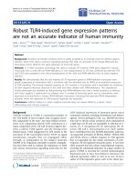
Figure 1 Experimental system used for analysis. A, B) Embryogenic masses of Pinus radiata after 7 (P7) and 14 (P14) days of proliferation.
Embryogenic tissue (in red) was stained with 1% acetocarmine. Bar: 2 mm. C) Early-maturation embryo at polarization stage (M1). Bar: 0.5 mm.
D) Late-maturation embryo at tissue differentiation stage (M3). Bar: 0.8 mm. E) Hypocotyls from 21-day-old seedlings treated with 10 μM indole-3-butyric
acid (IBA) after 28 days of culture. F, G) Hypocotyls (F) and epicotyls (G) from 90-day-old seedlings treated with 10 μM IBA.
Abarca et al. BMC Plant Biology (2014) 14:354
expression levels that are needed for adult cells to
re-enter the embryonic root formation pathway. The
mechanisms that enable a somatic differentiated cell
to become a pluripotent or totipotent cell, which can develop a root, shoot, or embryo, or repair damaged tissues,
are unknown.
While auxins do not seem to be the limiting factor at
the rooting site in the ability to form adventitious roots
at the mature stage [10,24-26], the capacity to recruit
root meristem or embryonic programs, and the effects
of auxin and cytokinin signaling pathways on the regulation of genes involved in the organization of stem cell
niches seem to be key factors in the de novo regeneration of several plant species [27-36]. The capacity of
cells to generate polar changes in the local distribution
of auxin can also influence cell fate [37]; alternatively,
transcriptional regulatory networks can function as
developmental signals underlying changes in a cell’s
fate [33,34,38,39]. The establishment of an embryonic
root meristem involves members of the GRAS family of putative transcription factors, which includes SCARECROW
(SCR), SCARECROW-LIKE (SCL) and SHORT-ROOT
(SHR) proteins. These genes are also involved in the
radial patterning of roots, hypocotyls and aerial organs.
Their expression is associated with auxin distribution in the
root apical meristem [40-45]. A P. radiata SCARECROWLIKE (PrSCL1) gene and a Castanea sativa SCARECROWLIKE (CsSCL1) gene, which are expressed in roots and root
primordia, and are induced in rooting-competent cells at
the earliest stages of adventitious root formation in the
presence of exogenous auxin, have been previously reported [16,17,46]. Additionally, Solé et al. [17] described
a P. radiata SHORT-ROOT (PrSHR) gene that is also
expressed in roots and root primordia, and is induced in
rooting-competent cells at the earliest stages of adventitious root formation in the absence of exogenous auxin.
These authors concluded that these genes and, perhaps, a
GRAS cascade of transcription factors play roles during
the earliest stages of adventitious root induction via
auxin-dependent and auxin-independent pathways [18].
To investigate if GRAS transcription factors could be
associated with the maturation-related decline in adventitious rooting, the GRAS family in pine was characterized.
Additionally, the transcript profiles of 13 GRAS genes in
rooting-competent and rooting-non-competent cuttings
in response to auxin were compared at the earliest stages
of adventitious root formation, the cell reorganization
state, prior to the onset of cell divisions leading to the formation of an adventitious root meristem. The expression
analysis was also performed until after the initiation of the
rapid cell divisions that organize the root meristem. Auxin
distribution was analyzed over the same time course. We
also examined the transcript profiles of GRAS genes during somatic embryogenesis [47], at the stages of initial-cell
Page 3 of 19
formation, embryo polarization and embryo differentiation
(Figures 1 A, B, C, D).
Results
The pine GRAS gene family: in silico identification of GRAS
genes, motif prediction and phylogenetic analysis of
GRAS proteins
To further our previous work on pine GRAS genes and
their roles in the maturation-related decline of adventitious root formation [16,17,46], an in silico search was
conducted to identify new members of the pine GRAS
family. An initial BLAST search of Pinus and Picea sequences in the Genbank database [48], using a conserved
sequence of the GRAS motif, led to the identification of
31 EST sequences that were classified into 13 groups
representing putative unigene sequences. P. radiata sequences obtained in our lab were used to design primers
for expression analyses (see below).
After a second round of searching using the Europine
database [49], a total of 90 ESTs and genomic sequences
from Picea glauca, Picea sitchensis, Pinus albaucalis,
Pinus ayacahuite, Pinus banksiana, Pinus bungeana,
Pinus cembra, Pinus contorta, Pinus densiflora, Pinus
flexilis, Pinus gerardiana, Pinus korainensis, Pinus lambertiana, Pinus monticola, Pinus morrisonicola, Pinus pinaster,
Pinus pinea, Pinus radiate, Pinus strobiformis, Pinus sibirica, Pinus squamata, Pinus sylvestris, Pinus taeda, Pinus
thumbergii, and Pinus wallichiana were obtained. Additionally, three full-length cDNAs from P. radiata [16,17]
and five 3′end cDNAs from P. radiata, P. pinea and P.
pinaster that were available in our databases were included, for a total of 98 cDNA sequences. The in silico
comparison of these sequences resulted in the identification of 21 unique members of the GRAS gene family in
pine.
After the release of the Picea abies and P. taeda
genomic sequences, a third round of searching using
the Congenie and Dendrome databases [50,51] was performed. A total of 36 P. abies and 65 P. taeda genes
models were classified and, together with the previously
identified ones, led to the identification of 32 unique
members of the pine GRAS gene family (Additional file 1).
In addition to the SCR and SHR genes, the predicted
genes were named following the nomenclature of our previous work [16], SCL1 to SCL30 (Additional file 1).
For 25 of the 32 GRAS genes, at least one predicted
gene was identified in both pine and spruce (Additional
file 1). Seven additional predicted genes were found in P.
taeda that had no putative orthologs in P. abies or other
pine species (Additional file 1). Pairwise comparisons
among the predicted amino acid sequences of the 25
members for which more than one complete sequence
was found revealed a high degree of conservation. Sequence identities ranged from 89.7% to 99.5% between
Abarca et al. BMC Plant Biology (2014) 14:354
pine sequences and from 84.7% to 97.2% between pine
and spruce sequences, except for sequences related to
AtSCL26 (see below), which showed a higher divergence between pine and spruce, ranging from 72.0 to
88.4%.
To classify the conifer GRAS proteins, a phylogenetic
analysis of 52 pine and spruce predicted GRAS protein
sequences was performed using a 493 amino acid fragment
that included the conserved GRAS C-terminal motif. To
avoid possible pseudogenes, only sequences of complete
predicted GRAS proteins were included. At least one sequence per conifer GRAS family member, either from
pine or from spruce, was included in the analysis. The tree
grouped the sequences according to their homology with
the classical GRAS protein subfamilies [52] and revealed
the existence of an additional group, containing mostly
pine sequences, with homology to AtSCL26 (Figure 2,
Additional file 2).
The evolutionary relationship of the conifer and
angiosperm GRAS proteins was phylogenetically analyzed
using 400 amino acid fragments from 100 sequences, including the 52 conifer and 47 angiosperm sequences belonging to the GRAS protein subfamilies [52]. A sequence
from Physcomitrella patens was used as the outgroup
(Additional file 2). The phylogenetic tree showed that the
predicted pine GRAS proteins do not cluster into a separate branch, but are distributed among the angiosperm
GRAS subfamilies (Additional file 2). The distribution of
the conifer sequences was similar to that obtained from
the conifer tree, and showed that the AtSCL26 branch is
indeed a subfamily that includes 12 pine, two spruce and
one Arabidopsis sequences (Additional file 2).
In addition to the 52 complete putative GRAS sequences,
a total of 37 (P. taeda) and 22 (P. abies) hypothetical genes
encoding partial GRAS proteins were identified (Additional
file 1). These could represent pseudogenes resulting from
gene duplication, and were more frequent in the SCR,
SHR, PAT and AtSCL26 subfamilies of P. taeda and in the
PAT and DELLA subfamilies of P. abies (Additional file 1).
Conserved motifs and intrinsically disordered N-terminal
domains of the pine GRAS proteins
Comparisons of the putative GRAS sequences with previously described proteins revealed that they contain
domains characteristic of the GRAS proteins. An analysis of the predicted sequences revealed the presence of
the highly conserved VHIID motif, with changes in the
valine, leucine and isoleucine residues among members, as well as the PFYRE and SAW motifs in the Cterminal region of the proteins (Additional file 3). Two
leucine repeats (LHRI and LHRII) were also identified
in the C- terminus. In addition, the LXXLL motif and
several additional amino acid residues conserved in
other known GRAS members of the protein family,
Page 4 of 19
such as the RVER or the LRITG motifs, were identified.
The SAW motif contains pairs of the conserved residues
RX4E, WX7G and WX10W. Full-length sequences were
obtained for 32 members of the multigene family. The Nterminal region of GRAS proteins is variable; however,
acidic-residue-rich regions flanking repeated hydrophobic/aromatic residues, similar to those found in PrSCL1
and PrSHR [16,17], were also found in other GRAS
proteins from pine (Additional file 4). Homopolymeric
stretches of proline and asparagine were only found in
SCL5 and SCL12, respectively, while a glycine stretch
was found in the GRAS region of the SCL21.
A common feature of the N-terminal region of the analyzed proteins was the enrichment in disorder-promoting
residues such as proline, glutamic acid, serine, glutamine,
lysine, or in amino acids that are indifferent to disorder or
structure, such as alanine, arginine or aspartic acid [53]. A
comparison of the disorder profiles of these proteins and
the corresponding proteins from angiosperms belonging
to the same subfamily shows that the N-terminal region is
intrinsically disordered. The intrinsically disordered profile
is conserved among members of the same subfamily
(Additional file 5).
The structure of the GRAS multigene family in pine
suggests different roles of individual GRAS members in
constitutive or induced processes. To extend our previous
analysis of the gene expression patterns of GRAS genes
[16,17], and to show possible differences in spatial and induced expression patterns associated with the maturationrelated decline of adventitious root formation, the relative
transcript abundance of 13 of the 32 GRAS genes was
measured by qRT-PCR in organs during vegetative development, during somatic embryo development, at the developmental transition from embryonic to postembryonic
development, and during the early stages of adventitious
root induction in response to auxin in rooting-competent
and non-competent cuttings from P. radiata. Genes selected for expression analysis were those initially identified
from the EST collection in Genbank, which included
members of all of the subfamilies except AtSCL3. PrSCL1
and PrSHR expression levels had already been measured
in organs during vegetative development and in hypocotyl
cuttings from 21-day-old seedlings during adventitious
root formation [16,17].
Constitutive transcript profiles of GRAS genes in organs
and changes in the GRAS mRNA levels during somatic
embryo development and at the embryonic-postembryonic
developmental transition
To characterize the expression patterns of GRAS genes
in different organs during vegetative development, RNAs
isolated from roots, hypocotyls, shoot apex nodal segments
(including the apical meristem, young needles and shoot
segments) and cotyledons from 35-day-old pine seedlings
Abarca et al. BMC Plant Biology (2014) 14:354
Page 5 of 19
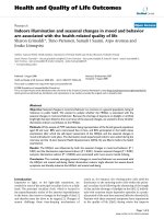
Figure 2 Phylogenetic tree of GRAS proteins SCARECROW-LIKE (SCL), SCARECROW (SCR), and SHORT-ROOT (SHR) from conifer species.
Accession no. or gene references in parentheses. Picea abies SCR (MA_1793p0010), P. abies SCL1 (MA_45656p0030), P. abies SCL2 (MA_10435790p0010),
P. abies SCL3 (MA_140003p0010), P. abies SCL4 (MA_18234p0010), P. abies SCL5 (MA_73870p0010), P. abies SCL6 (MA_94287p0010), P. abies SCL8
(MA_52903p0010), P.abies SCL9 (MA_10426489p0020), P.abies SCL10 (MA_10432093p0010), P. abies SCL11 (MA_19310p0010), P. abies SCL13
(MA_96029p0010), P. abies SCL17 (MA_10255p0010), P. abies SCL18 (MA_10430319p0010), P. abies SCL23 (MA_73173p0010); Pinus pinaster SCL7
(sp_v2.0_unigene8594), P. pinaster SCL8 (sp_v2.0_unigene8378), P. pinaster SCL9 (sp_v2.0_unigene4531), P. pinaster SCL13 (sp_v2.0_unigene1634), P.
pinaster SCL14 (sp_v2.0_unigene1578), P. pinaster SCL15 (sp_v2.0_unigene10599); Pinus radiata SCR (KM264388), P. radiata SHR (EU044786), P. radiata
SCL1 (DQ683567), P. radiata SCL2 (KM264389), P. radiata SCL10 (KM264395), P. radiata SCL12 (KM264397); Pinus taeda SCR (PITA_000043499-RA), P.
taeda SHR (PITA_000092405-RA), P. taeda SCL1 (PITA_000021589-RA), P. taeda SCL5 (PITA_000017225-RA), P. taeda SCL6 (PITA_000022609-RA), P. taeda
SCL8 (PITA_000040137-RA), P. taeda SCL9 (PITA_000009055-RA), P. taeda SCL10 (PITA_000009053-RA), P. taeda SCL11 (PITA_000068827-RA), P. taeda
SCL12 (PITA_000010887-RA), P. taeda SCL15 (PITA_000016257-RA), P.taeda SCL16 (PITA_000056676-RA), P.taeda SCL18 (PITA_000086415-RA), P. taeda
SCL19 (PITA_000075302-RA), P. taeda SCL20 (PITA_000051405-RA), P. taeda SCL21 (PITA_000056428-RA), P. taeda SCL22 (PITA_000080766-RA), P. taeda
SCL23 (PITA_000072928-RA), P.taeda SCL24 (PITA_000072831-RA), P. taeda SCL25 (PITA_000041536-RA), P. taeda SCL26 (PITA_000026833-RA), P. taeda
SCL27 (PITA_000049193-RA), P. taeda SCL28 (PITA_000066307-RA), P. taeda SCL29 (PITA_000051712-RA) and P. taeda SCL30 (PITA_000035221-RA).
PtSCL25 was used as the outgroup. Branches with bootstrap values lower than 500 were collapsed.
were used. Results were expressed as values relative to
the expression levels in roots (Figure 3A). Additionally,
changes in GRAS mRNA levels were also studied
during somatic embryo development (Figure 3B) and
at the embryonic-postembryonic developmental transition
(Figure 3C). To that end, mRNA levels were analyzed in
Abarca et al. BMC Plant Biology (2014) 14:354
Page 6 of 19
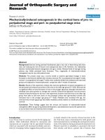
Figure 3 Expression of GRAS genes in vegetative Pinus radiata organs and at the embryonic-postembryonic developmental transition.
A) Organs from 35-day-old pine seedlings. qRT-PCR was performed using RNAs from roots (R), hypocotyls (H), cotyledons (C) or shoot apex nodal
segments (A). B) Embryo development. qRT-PCR was performed using RNAs from embryogenic masses at 7 (P7) and 14 (P14) days of proliferation,
early-maturation embryo (M1) and late-maturation embryo (M3). C) Embryonic-postembryonic development. qRT-PCR was performed using RNAs from
embryogenic masses at 7 (P7) days of proliferation, rooting-competent hypocotyls (H21) and non-competent hypocotyls (H90) or epicotyls (E90) from
seedlings of 21- and 90-day-old seedlings, respectively. A total of 1 μg RNA was reverse transcribed, and 12.5 ng of cDNA was amplified with 400 nM
of specific primers. Pine Ri18S was used as the control. Results are expressed as mean values of the relative expression to roots (A) or P7 (B and C) ± SE
from at least three biological replicates. Insets in B show details of early developmental stages. Results of PrSHR expression in C are expressed as mean
values of relative expression to H21. Expression levels of PrSCL1 and PrSHR had already been measured in organs during vegetative development
[16,17]. Expression of PrSCL16 was not detected in any of the RNA samples tested. SCL, SCARECROW-LIKE; SHR, SHORT-ROOT.
developing somatic embryos and in organs of embryonic
and postembryonic origin from seedlings of different ages.
Zygotic embryos are very difficult to isolate at specific developmental stages, but P. radiata somatic embryos show
a very similar developmental pattern; therefore, specific
developmental stages can be defined and isolated. RNAs
isolated from embryogenic masses in the proliferation
stage, from somatic embryos at the early and late maturation stage, and from rooting-competent and noncompetent hypocotyl or epicotyl cuttings from 21- and
90-day old seedlings were used to analyze the expression patterns during embryo development and at the
Abarca et al. BMC Plant Biology (2014) 14:354
embryonic-postembryonic developmental transition. Results were expressed as values relative to the expression in
embryogenic masses after 7 days in proliferation medium.
The expression of PrSCL16 was not detected in any of the
RNA samples tested.
Most GRAS genes showed relatively high mRNA levels
in roots, except PrSCL6, which showed relatively high
levels in hypocotyls and in the shoot apices of young
seedlings. PrSCL13 and PrSCL14 also showed relatively
higher expression levels in cotyledons. The relative abundances in other tissues depended on the individual GRAS
genes (Figure 3A).
The analysis of GRAS transcript profiles during somatic
embryo development (Figure 3B) showed that the transcript levels of all GRAS genes, except PrSCL10, which
showed relatively high levels in embryogenic masses, were
significantly higher in the embryos at the late maturation
stage than in other stages. mRNA levels of PrSCR, PrSHR,
PrSCL1, PrSCL6, PrSCL8 and PrSCL12 increased between
two and four times in the embryo during the early maturation stage (Figure 3B). The analysis of GRAS transcript
profiles at the developmental transition from embryonic
to postembryonic development (Figure 3C) showed that
PrSCR and PrSCL6 maintained relative high levels in
rooting-competent hypocotyls from 21-day-old seedlings,
whereas the other GRAS genes also maintained relatively
high levels in rooting-non-competent hypocotyls and epicotyls from older seedlings (Figure 3C).
Transcript profiles of GRAS genes during adventitious
rooting in competent and non-competent stem cuttings
A possible role of GRAS genes in the loss of rooting
capacity was analyzed by assessing their temporal expression patterns in response to auxin in hypocotyl
and epicotyl cuttings from 21- and 90-day-old seedlings (Figure 4). Cuttings were treated with 10 μM
indole-3-butyric acid (IBA) [16,17]. Then, transcript
profiles were analyzed in the basal ends of cuttings
during the initial 24 h, at 48 h and 5 d after the onset
of the treatment, and compared with control tissues at
their time of excision (time 0). Data are presented as
mRNA levels normalized to ribosomal 18S [16,17] and
as fold inductions relative to their time of excision
(time 0). The expression of PrSCL16 was not detected
in any of the RNA samples tested.
Several patterns of expression were observed in hypocotyl cuttings from 21-day-old seedlings during adventitious rooting (Figure 4A). PrSCL2, PrSCL6, PrSCL7,
PrSCL10 and PrSCL12 mRNA levels increased in the
presence of exogenous auxin, similar to PrSCL1’s expression pattern [16]. PrSCL2 and PrSCL12 mRNA levels
were even increased in the absence of exogenous auxin
similar to PrSHR’s expression pattern [17]. PrSCR,
PrSCL5, PrSCL8, PrSCL13 and PrSCL14 mRNA levels
Page 7 of 19
did not show any change in their expression level during
the root-induction process. No GRAS genes showed increases in mRNA levels in the absence or presence of
exogenous auxin in the rooting-non-competent hypocotyl cuttings from 90-day-old seedlings (Figure 4A).
PrSCR and PrSCL6 mRNAs were not detected in noncompetent hypocotyls under root-induction conditions
(Figure 4A).
Several expression patterns were also observed in epicotyl cuttings from 90-day-old seedlings during adventitious rooting (Figure 4B). PrSCL2, PrSCL10 and PrSCL12
mRNA levels increased in the presence of exogenous
auxin, while PrSHR, PrSCL1 and PrSCL2 mRNA levels
even increased in the absence of exogenous auxin. PrSCR
and PrSCL6 mRNAs were not detected in the presence or
absence of auxin. PrSCL5, PrSCL7, PrSCL8, PrSCL13,
PrSCL14 and PrSCL16 were not tested for in epicotyls.
PrSCL2 mRNA levels increased in the absence of exogenous auxin in both rooting-competent hypocotyl cuttings
from 21-day-old seedlings and rooting-non-competent
epicotyl cuttings from 90-day-old seedlings. However, the
increase in transcript levels was significantly higher in the
presence of exogenous auxin (Figures 4A, B).
The expression of two genes, PrSCL1 and PrSHR,
which are associated with auxin-dependent and auxinindependent signaling pathways, respectively, in rootingcompetent cuttings [16,17] were also analyzed by in situ
hybridization in non-competent cuttings. In our previous
work, it was shown that increased transcript levels of both
genes accumulated in the rooting-competent tissues of
hypocotyls from 21-day-old seedlings after 24 h of root
induction [17]. These genes were not predominantly
expressed in the cambial region of non-competent
hypocotyls or epicotyls at the time of excision, nor
under rooting conditions (Figures 5A, B, C, D). No
specific tissue-localization was observed in any samples during adventitious rooting. No signal was
observed when tissues were hybridized sense-oriented
probes (Figures 5E, F).
Auxin distribution in rooting-competent and non-competent
cuttings in the presence of exogenous auxin and polar auxin
transport inhibitors
Auxin-dependent adventitious root formation in pine is
associated with a directional flow of auxin in combination
with the competition of neighboring cells for free auxin
[10]. Tissue-specific auxin gradients can elicit specific
cellular responses. The role of the endogenous auxin
distribution in rooting-competent and non-competent
tissues during adventitious root formation was addressed
by analyzing the indole-3-acetic acid (IAA) distribution.
Experiments were performed at the time of excision and
after 24 h with or without exogenous auxin. The IAA
distribution was analyzed by an immune-cytochemical
Abarca et al. BMC Plant Biology (2014) 14:354
Page 8 of 19
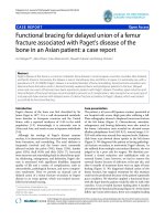
Figure 4 Expression of GRAS genes during adventitious root formation in Pinus radiata. A) qRT-PCR was performed using RNAs from
rooting-competent hypocotyls (H21) and non-competent hypocotyls (H90) from 21- and 90-day-old seedlings, respectively. B) qRT-PCR was
performed using RNAs from non-competent epicotyls (E90) from 90-day-old seedlings. RNA was extracted from the base of hypocotyl (H) or
epicotyl (E) cuttings treated with 10 μM indole-3-butyric acid at the indicated times. Hypocotyl or epicotyl cuttings maintained in water were
used as controls. A total of 1 μg RNA was reverse transcribed, and 12.5 ng of cDNA was amplified with 400 nM of specific primers. Pine Ri18S was
used as the control. Results are expressed as mean values of relative expression to time 0 ± SE from at least three biological replicates. Expression
levels of PrSCL1 and PrSHR had already been measured in competent hypocotyls from 21-day-old seedlings during adventitious rooting [16,17].
Expression of PrSCL16 was not detected in any of the RNA samples tested. SCL, SCARECROW-LIKE; SHR, SHORT-ROOT.
approach using antibodies raised against IAA (Figure 6,
Figure 7). IAA was mostly located in the cambial
region of rooting-competent hypocotyls, including the
cells positioned centrifugal to the resin canals after
excision, and during the initial 24 h of root induction
(Figures 6 A, B, C, D). Treating rooting-competent
hypocotyls with 1-N-naphthylphthalamic acid (NPA), a
polar auxin transport inhibitor, resulted in the mislocalization of endogenous auxin, which was also distributed in the pith, in the vascular cylinder and in the
cortex (Figures 6 E, F, G, H). No auxin accumulation
was detected in the cambial cells in non-competent hypocotyls or epicotyls. Auxin was mainly located in the xylem
parenchyma of hypocotyls (Figures 7 A, B, C, D), and in
the cortex of epicotyls (Figures 7 E, F, G, H). No signal
was observed when tissues were hybridized in the absence
of the antibody (Additional file 6).
Discussion
Plants do not lose their developmental potentialities during differentiation and retain a certain level of plasticity
[54], either by maintaining pro-embryonic or meristematic
Abarca et al. BMC Plant Biology (2014) 14:354
Page 9 of 19
Figure 5 In situ localization of Pinus radiata SHORT-ROOT (PrSHR) mRNA. A, B) Transverse sections of hypocotyls from 90-day-old seedlings
at time 0 (A), and after 24 h of culture in the presence of 10 μM indole-3-butyric acid (IBA) (B). C, D) Transverse sections of epicotyls from
90-day-old seedlings at time 0 (C), and after 24 h of culture in the presence of 10 μM IBA (D). The sections were hybridized with an RNA probe
obtained by in vitro transcription of PrSHR in either the antisense (A, B, C, D) or sense (E, F) orientation. Note the absence of hybridization in the
controls. Similar results were obtained using an RNA probe obtained by in vitro transcription of PrSCL1 in either the antisense or sense orientation.
ab, axillary bud; c, cambial region; co, cortex; r, resin canal; x, xylem. In situ localization of PrSCL1 and PrSHR had already been described in competent
hypocotyls from 21-day-old seedlings during adventitious rooting [17]. SCL, SCARECROW-LIKE.
cells in the adult tissues or by a major developmental reprogramming to acquire the embryonic or meristematic
status [55]. The plasticity of plant tissues results in the regenerative capacity of cells other than those of meristem,
lateral root initials or zygotes.
A decline in the regenerative capacity of somatically
differentiated cells in an ectopic location is associated with
age and maturation in forest tree species [13]. Efforts have
been made to identify genes associated with plant cell fate
switches [34,38]; however, pluripotency or indeterminacy
genes, with high expression levels in non-differentiated
embryonic cells or at the very early stages of development,
significantly reduced or even no expression levels in adult
tissues that have lost their regenerative capacities, but
maintained in tissues with regenerative capacities or
induced after the reprogramming of adult cells during
regeneration [56], have not been described. We have
made use of embryogenic cultures maintained under
non-differentiated proliferating conditions or subjected to
differentiation, as well as adult tissues from plants of different ages showing different adventitious rooting capacities in response to auxin, to identify genes, changes in
gene expression levels and regulatory mechanisms associated with the competence and reprogramming of adult
tissues to form adventitious roots in pine (Figure 1).
In our previous work, two members of the GRAS gene
family of P. radiata, PrSCL1 and PrSHR, were associated
with the adventitious root formation in rooting-competent
cuttings [16,17]. GRAS proteins are involved in a diverse
suite of physiological and developmental processes ranging
from light and hormone signal transduction to organ
identity and tissue differentiation [57,58]. Among them,
SCR and SHR are involved in root patterning, establishing the quiescent center’s identity and in maintaining
Abarca et al. BMC Plant Biology (2014) 14:354
Page 10 of 19
Figure 6 Endogenous distribution of indole-3-acetic acid (IAA) in hypocotyl cuttings from 21-day-old Pinus radiata seedlings. Transverse
sections from the base of hypocotyls after 24 h of culture in the presence of 10 μM indole-3-butyric acid (IBA) (A, B, C, D) or in the presence of
10 μM IBA + 10 μM 1-N-naphthylphthalamic acid (E, F, G, H). A, E) Differential interference contrast (DIC) image, B, F) Immunodetection of IAA,
C, G) DAPI nuclear staining, D, H) merged immunodetection of IAA and DAPI staining. c, cambial region; co, cortex; r, resin canal; x, xylem.
the stem cell status of the initial cells in the root meristem [43,44]. Additionally, they have been involved in
root tip regeneration [59] and in cell reprogramming
[38]. GRAS proteins have been identified as homologous
proteins to the STAT proteins in animals [60], which have
also been associated with differentiation, reprogramming
and regeneration [61,62].
A large gene family encodes GRAS proteins in pine.
Supporting cDNAs were identified for at least 32 unique
members in P. taeda (Additional file 1), a number
close to that described in P. abies (Additional file 1)
and Arabidopsis [63-65], higher than the number described
in P. pinaster and P. glauca (Additional file 1) [65,66], and
lower than the number described in Oryza sativa, Populus
trichocarpa and Brassica rapa [63,65,67]. Eighteen members were identified in P. radiata (Additional file 1) [16,17].
Pairwise sequence similarities among predicted polypeptides for each GRAS member of the different pine and
spruce species confirmed that they may represent intraor inter-specific alleles of the same genes, similar to those
described for other gene families in conifer species
[68,69]. The proteins belonging to the AtSCL26 group
showed a lower degree of identity, which could be related
to a high number of duplication events, perhaps to acquire
new functions (Figure 2 and Additional file 2).
A phylogenetic analysis showed that conifer GRAS
proteins do not form a separate cluster (Figure 2 and
Additional file 2) and most are included in the major
Figure 7 Endogenous distribution of indole-3-acetic acid (IAA) in hypocotyl and epicotyl cuttings from 90-day-old Pinus radiata
seedlings. Transverse sections of the base of hypocotyls (A, B, C, D) and epicotyls (E, F, G, H) after 24 of culture in the presence of 10 μM
indole-3-butyric acid. A, E) Differential interference contrast (DIC) image, B, F) Immunodetection of IAA, C, G) DAPI nuclear staining, D, H) merged
immunodetection of IAA and DAPI staining. c, cambial region; co, cortex; r, resin canal; x, xylem.
Abarca et al. BMC Plant Biology (2014) 14:354
GRAS subfamilies [16,17,52,57,58]. The HAM family
contains the AtSCL26 subfamily, which may be the result
of a high number of duplication events for conifer sequences compared with their angiosperm counterparts.
Conifers diverged from angiosperms 300 million years ago
[70]. The phylogenetic relationship between conifers and
angiosperms highlights the ancient diversification of this
family, which may precede the transition to terrestrial environments, as suggested by Engstrom [71] based on comparisons among GRAS proteins from angiosperms, bryophytes
and lycophytes, but not gymnosperms. The ancient diversification and the non-clustering of conifer sequences suggests functions or modes of action for these proteins in
primary constitutive or induced processes [72-77].
An analysis of the polypeptide sequences shows a high
degree of conservation in the representative GRAS core
motifs (Additional file 3) [52,57,58], which are involved in
transcriptional regulation, indicating that the transcriptional regulatory machinery is also conserved in conifers.
The N-terminal domain of the predicted GRAS proteins
is highly variable in pine (Additional file 4), similar to the
N-terminal domain of angiosperm GRAS proteins [57,58].
Homopolymeric stretches, such as those characterizing
angiosperm GRAS proteins [63,64,78], were not found in
conifer GRAS proteins, except for the proline and asparagine stretches found in PtSCL5 and PtSCL12. The amino
acid compositional profile of the N-terminus of GRAS
proteins from pine is very similar to that of the intrinsically disordered proteins and contains an enrichment in
disorder-promoting residues (Additional files 4 and 5).
However, the C-domain shows a compositional profile
similar to that of fully structured proteins, as described for
other GRAS proteins [57,58]. Disordered proteins lack
a well-defined three dimensional structure, resulting in
an extreme structural flexibility that enables them to
form highly specific complexes with different proteins
or nucleic acids in a reversible and transient low-affinity
interaction, depending on the changing physiological, developmental or environmental conditions [53,79]. Intrinsic
disorder has been described for several families of plant
transcription factors, and intrinsically disordered proteins
have been associated with key cellular and signaling
processes [80-82]. The intrinsic disorder could be a
way to increase functional diversity and the complexity
of biological networks without increasing the size of
the families, or even, the size of the genome, and it was
proposed as the mechanism involved in the functional
divergence within GRAS subfamilies [57,58]. Despite
the highly variable sequence of the N-terminus, GRAS
proteins in pine show conserved disordered profiles
when compared with GRAS proteins from angiosperm
species of the same subfamily (Additional file 5) [57,58].
This is in agreement with previous suggestions [83,84], indicating that the pattern of protein disorder could be
Page 11 of 19
more conserved through evolution than the amino acid
sequence in the N-terminus. Similar results have been described for the mammalian Myc proteins [85]. Consequently, mutations that do not affect the general disorder
pattern would allow the conservation of specific protein
interactions and, hence, functions.
The conservation of the protein motifs and structures,
the absence of a particular conifer subfamily, and the intrinsically disordered N-terminal domain can account for
the versatile roles of these proteins in tree biology and for
the molecular mechanisms regulating their expression
levels and functions. The dynamic ability of intrinsically
disordered proteins to recognize multiple molecular partners reveals the need for a synchronous spatio-temporal
connection between the functionally appropriate GRAS
genes and proteins participating in specific functions.
The expression of GRAS genes in the different organs,
at the embryonic-postembryonic developmental transition,
as well as during adventitious rooting, in response to
auxin showed unique and overlapping patterns, indicating a differential regulation and tissue-specific functions
(Figure 3 and Figure 4). Individual genes within each
group may have acquired different and specialized functions, some of which may relate to competence and the
reprogramming of adult cells to form adventitious roots.
Many pine GRAS genes show relatively high levels of
mRNA during the transition from the polarization stage
(M1) to the late maturation stage (M3), indicating that
they play roles in embryo development (Figure 3B). A
subset of these genes, PrSCR, PrSHR, PrSCL1, PrSCL6,
PrSCL8 and PrSCL12, increase their mRNA levels during the early maturation stage. At this stage, embryo
polarization occurs, but tissue differentiation has not
been yet completed; therefore, these tissues, along with
the proliferating embryogenic masses, may be sources of
non-determined or pluripotent cells associated with the
establishment of tissue domains [47]. However, PrSCL10
shows a relatively high level of mRNA after 7 days of proliferation, when initials are developed. Consequently, these
genes play key roles in the initial establishment of embryo
tissue domains or hormone gradients [86]. Among them,
PrSCR and PrSCL6 are highly expressed in organs of embryonic origin, such as hypocotyls, cotyledons and shoot
apices. Additionally, PrSCR, along with PrSHR, PrSCL1
[16,17], PrSCL5, PrSCL7, PrSCL8 and PrSCL12 showed
relatively higher levels of mRNAs in roots than in any
other organs tested during vegetative development, indicating a role in the roots (Figure 3A). These results
suggest that the expression of these genes is not only
restricted to embryonic development but extended to
other processes. We then analyzed if the expression levels
of genes associated with the early stages of embryo formation could be significantly reduced or even non-existent in
cuttings that have lost their rooting capacity, but
Abarca et al. BMC Plant Biology (2014) 14:354
maintained in rooting-competent tissues or induced after
the reprogramming of adult competent cells to form adventitious roots.
PrSCL6 and PrSCR maintain relatively high levels of
mRNA in rooting-competent hypocotyls, while other
GRAS genes are expressed in both rooting-competent
hypocotyls and rooting-non-competent hypocotyls or
epicotyls (Figure 3C). These results indicate that PrSCL6
and PrSCR, in addition to their functions in embryo development, are associated with an embryonic characteristic that could result in the competence for adventitious
organogenesis in cuttings. An analysis of these genes
during adventitious root formation in competent and
non-competent tissues indicated that PrSCL6 is auxininduced in rooting-competent hypocotyls only, and the
expression increases during the initial 48 h, which is the
time required for auxin action and for the reorganization
or dedifferentiation of cambial cells [10,11,16,17]. PrSCL6
is not detectable in rooting-non-competent hypocotyls or
epicotyls (Figures 4 A, B). Similar results are also obtained
when PrSCR expression is analyzed; however, PrSCR is
not induced in rooting-competent hypocotyls, but its
mRNA levels are maintained at higher levels in these tissues than in non-competent hypocotyls or epicotyls, in
which PrSCR is not detectable during the initial stages of
rooting (Figures 4A, B). Therefore, both genes could be
associated with embryonic cells or with the very early
stages of development. Their mRNA levels were significantly reduced or even lost in older and more mature
rooting cuttings that had lost their rooting capacities, but
were maintained in competent hypocotyls or increased after
the reprogramming of adult competent cells during adventitious root formation. This would make them candidate
genes for rooting competence and cell reprogramming.
The mRNA levels of other genes, such as PrSHR
PrSCL1, PrSCL2, PrSCL10 and PrSCL12, changed in an
auxin-, age- or developmental-dependent manner during
adventitious rooting in competent and non-competent
cuttings (Figures 4A, B) [16,17]. The localized increases of
PrSHR and PrSCL1 mRNAs in competent tissues [17],
which were not detected in non-competent hypocotyl or
epicotyl cuttings (Figure 5), suggests their involvement in
adventitious rooting. The expression profiles in epicotyls
could be associated with the presence of meristematic
tissues in these cuttings, such as the shoot axillary
meristem or cambium [46,87]. The tissue localization
of PrSCL2, PrSCL10 and PrSCL12 mRNAs would help
to show the roles of their mRNA variations in adventitious rooting. Other pine GRAS genes do not seem
to be related to the adventitious rooting response
(Figure 4A).
These results indicate that high levels of PrSCR and
PrSCL6 may be related to the degree of determination,
competence, and/or the reprograming capacity of tissues
Page 12 of 19
to form adventitious roots, while other genes that are also
expressed or induced, such as PrSHR, PrSCL1, PrSCL2,
PrSCL10 or PrSCL12, could be involved in transcriptional
regulatory networks associated with auxin-dependent and
auxin-independent pathways in an age- or developmentaldependent manner. Therefore, the participation of these
genes in determining whether cells become roots in
competent tissues cannot be discarded. The low expression levels of PrSCR and PrSCL6 could make these
rate-limiting steps in competence and in auxin-induced
processes. Additionally, all these genes are expressed
or induced at the very early stages of adventitious root
formation before the onset of cell divisions leading to
the formation of a root meristem. A set of 26 of the
500 transcription factors expressed during the early
events, which occur in the initial 24 h, leading to the
regeneration of Arabidopsis plants from protoplasts,
were not expressed during senescence [38].
The functional analysis of genes based on their subfamilies indicates a possible role in determination and
patterning. SCR and SHR are involved in root meristem
determination [43,44] and, along with other transcription factors, have been involved in reprogramming in
Arabidopsis [38]. Additionally, PrSCL1, which may be
related to the rooting process, has been associated with
the adventitious and lateral root meristem of pine and
chestnut [16,46], and with the shoot axillary meristem in
chestnut [46]. Also, PrSCL2 and PrSCL12 are members
of two GRAS subfamilies (the Ls and HAM families,
respectively), which have been associated with the determination of lateral meristem [88,89]. Although PrSCL10
is included in the PAT family of GRAS proteins, which
is associated with light responses [90], members of this
subfamily have also been associated with cell defense
[91,92]. Therefore, this subfamily is also functionally
diverse. Overall, a role in adventitious root competence,
reprogramming and determination could be envisaged
for a subset of the pine GRAS genes.
The asymmetrical increases of PrSCL1 and PrSHR
transcript levels previously described in the cambial region and rooting-competent cells [17] were not detected
in non-competent cuttings (Figures 5 A, B, C, D). In
these cuttings, expression spread into the cortex and
dividing cells. The asymmetrical increase in mRNA
during the earliest stages of adventitious root formation
in similar cell types at different developmental stages
suggests the presence of specific cellular signaling pathways or specific factors in pine, perhaps distributed in celltype- and developmental-stage-specific contexts in the tissues involved in rooting, which could be crucial for rooting
capacity [18,46]. The nature of these signaling pathways
or factors is unknown. De novo organ formation and
cell specification are processes involving rearrangements of tissue polarity, with the temporal and spatial
Abarca et al. BMC Plant Biology (2014) 14:354
distribution of auxin being a very important player, contributing to tissue polarization and patterning [93]. No
differences in auxin uptake, accumulation or metabolism were found between rooting-competent and noncompetent hypocotyls and epicotyls at the base of the
cuttings [10]. However, an asymmetric auxin distribution was detected in rooting-competent tissues after
excision and was maintained during the initial 24 h of
root induction (Figures 6 A, B, C, D) at locations where
PrSHR and PrSCL1 are expressed [17]. An asymmetrical distribution was not observed in non-competent
hypocotyls or epicotyls (Figure 7). Treatments with NPA,
which inhibits rooting [10] and does not change the
number of cell layers in the vascular cylinder, cortex or
pith, changed the auxin distribution pattern (Figures 6 E,
F, G, H), indicating that polar auxin transport, which resulted in an accumulation of auxin at the base of the cutting [10], as well as auxin localization and distribution at
the tissue or cellular levels. This result indicated that
rooting-competent tissues could retain an intrinsic capacity to maintain or accumulate auxin after excision,
which could be crucial for rooting. The cellular capacity of
initial cells to produce auxin gradients may be a mechanism involved in the determination and maintenance of
meristem, the induction of lateral primordia at the shoot
meristem, and the formation of lateral roots or
adventitious roots [20,94-96]. Auxin distribution largely
depends on the dynamic expression and subcellular
localization of the PIN auxin-carrier proteins [97]. However, PIN activity can be modulated by endogenous or
exogenous signals, such as other hormones, stress or
tissue-specific factors, to trigger developmental decisions
that could initiate regeneration by triggering cell fates or
other local changes [37,87,98-101]. No differences in the
wounding stress response were observed between competent and non-competent cuttings [102]; therefore, other
tissue-dependent signals could also trigger re-patterning
either by inducing cell-fate respecification or by reestablishing the auxin distribution. Transcription factors
are main players in regulatory modules controlling
auxin gradients, positional information and the development of polarity fields, resulting in a cross regulatory
network involved in organ formation [103-107]. The
differential expression of genes, such as PrSCR and PrSCL6,
in rooting-competent and non-competent cuttings, as well
as the differential responses of genes, such as PrSCL1 or
PrSHR [Figure 4, [16-18] to exogenous auxin during adventitious rooting may indicate the local involvement of specific GRAS transcription factors in the rooting capacity
by participating in the auxin distribution, control of celltype divisions, or other mechanisms. The auxin-related
increase of PrSCL1 mRNA in competent tissues after
24 h of root induction [17] could be associated with
auxin localization in these tissues at the same time
Page 13 of 19
(Figures 6A, B, C, D). The overlap in the temporal and
spatial distribution of auxin (Figures 6A, B, C, D), and
the increase of the auxin-independent PrSHR mRNA
[17] could indicate a possible crosstalk between the
signaling pathways, perhaps establishing response
domains that activate a cascade of other GRAS genes or
root determining factors before the resumption of cell
divisions. Sabatini et al. [44] proposed that SCR- and SHRexpressing cells are competent to acquire quiescent center
identity, with auxin distribution being the cue that specifies a subset of cells within the SCR or SHR expression
domains. However, the SHR pathway regulates root development through a transcriptional regulatory network and
also by affecting the expression of genes involved in
cytokinin and auxin signaling in Arabidopsis, resulting
in the fine-tuning of hormonal responses [87,99,108].
Additionally, formative divisions that generate the root’s
ground tissue are controlled by SHR in Arabidopsis,
which specifically regulates the spatiotemporal activation
of specific genes involved in cell division, and by SCR,
both activating a D-type cyclin involved in formative
divisions [109].
Conclusions
Adventitious root forming treatments induce root meristem patterning genes, such as GRAS genes, before the
onset of cell division in competent cells. The same GRAS
genes also may play a role during the earliest stages of
embryogenesis, initial-forming and polarization. The capacity to maintain or recruit root meristem or embryonic
programs in response to a specific stimulus seems to be
key in switching cells into different developmental programs, both in herbaceous and woody plants, including
forest tree species [34-36,38,110]. However, whether this
pattern of expression represents a maintenance, a dedifferentiation or a transdifferentiation to an embryonic or root
identity, or it represents a different adult developmental
program unique to regeneration, as was described in
Arabidopsis [111], remains unknown.
Methods
Plant material, root induction and somatic embryogenesis
Pine (P. radiata D. Don) seeds were germinated and
seedlings were grown as previously described [16]. The
seedlings were treated daily with water, and, after
21 days, weekly with 2 g/l of a commercial soluble
fertilizer (NPK 20-7-19 [w/w/w]). Cuttings for adventitious root induction were prepared according to [16].
Briefly, hypocotyl cuttings from 21-day-old seedlings,
including the intact epicotyl, and hypocotyl or epicotyl
cuttings from 90-day-old pine seedlings were prepared
by severing the hypocotyl or epicotyl at its base, and
trimming it to a length of 2.5 cm from the cotyledons
(hypocotyls) or from the apical buds (epicotyls). All
Abarca et al. BMC Plant Biology (2014) 14:354
but one apical tuft of needles were removed from the
epicotyls to obtain a foliar surface similar to that of the
hypocotyls. Root induction was conducted by exposing the
cuttings to 10 μM IBA continuously (Figures 1 E, F, G).
Cuttings without IBA treatment were used as controls. IBA
was obtained from Sigma (St. Louis, MO, USA) as IBA-K
and dissolved in distilled water. For experiments on auxin
immunolocalization, hypocotyls from 21-day-old seedlings
were also treated with NPA in the presence and absence of
auxin for 24 h. NPA was obtained from Duchefa (Haarlem,
Netherlands) and dissolved in DMSO. Hypocotyls treated
with DMSO were also used as controls in these experiments. Conditions for root induction were the same as described for seedling growth [16]. Embryonal suspensor
masses and somatic embryos were also used for analyses
(Figures 1 A, B, C, D). Embryogenic line M95, provided by
Dr. Christian Walter (Scion, Rotorua, New Zealand), was
proliferated and maintained by bi-weekly subcultures of individual clumps onto EDM6 medium [112]. For somatic
embryo maturation, 500 mg of embryogenic tissue was
suspended in 25 ml of EDM6 liquid medium. The tissue
suspension was collected pouring 5 ml aliquots onto a filter paper disk (80 g/m2 43–48 μm; Filter Lab, ANOIA;
Barcelona, Spain) in a Büchner funnel. A vacuum pulse
was applied to drain the liquid, and the filter paper with
the attached cells was placed into a 90 mm diameter Petri
dish with maturation medium, which was based on the
formulation of EMM1 medium [112] supplemented with
15 mg · L−1 abscisic acid, 30 g · L−1 sucrose and 6 g · L−1
Gelrite®. Cultures were maintained in darkness at 23 ± 1°C.
The pH of the media was adjusted to 5.8 before autoclaving. Solutions of amino acids and abscisic acid were filter
sterilized and added to the cooled autoclaved medium.
RNA extraction, quantification and cDNA synthesis
For analysis of gene expression during adventitious rooting, 30 basal segments, 1 cm long, of the hypocotyl or
epicotyl cuttings were pooled from each treatment and
time point as specified in each experiment, immediately
frozen in liquid nitrogen and stored at −70°C until used
for RNA isolation. Total RNA isolation and quantification from cuttings have been previously described [16].
RNA was also extracted from different organs of plant
seedlings as specified in each experiment. Samples of
embryogenic tissues were used for expression analysis
experiments at different stages of development: proliferative tissues 7 and 14 days after the last subculturing
to fresh proliferation medium (Figures 1 A-B), somatic
embryos at the early maturation stage of development
(Figure 1C) and somatic embryos at the late maturation
stage (Figure 1D). Tissues were frozen in liquid nitrogen
and stored at −70°C until used for RNA isolation. Total
RNA was extracted using the RNeasy® Plant mini kit
(Qiagen, Hilden, Germany), following manufacturer’s
Page 14 of 19
instructions. Between 50 and 100 mg of embryogenic
tissue or embryos in extraction buffer, were ground
with a pestle in 1.5-ml Eppendorf tubes. RNAs were
digested with RQ1 DNase (Promega, Madison, WI, USA)
following the manufacturer’s instructions, and then purified using the Amicon® Ultra columns (Merck Millipore,
Darmstadt, Germany). The RNA concentration and quality were determined using a ND-1000 Spectrophotometer
(NanoDrop Technologies Inc., USA). RNA was prepared
from at least two biological replicates. cDNA synthesis
was performed using 1 μg of total RNA. For quantitative
RT-PCR, RT reactions were performed using 200 ng random primers with SuperScript™III reverse transcriptase
(Invitrogen Corporation, Carlsbad, CA, USA) according to
the manufacturer’s instructions.
Phylogenetic analysis
The conserved C-terminal region of the GRAS proteins,
plus as much of the N-terminal region as the shortest protein sequence allowed, were used for the phylogenetic
analysis as previously described [16,17]. The polypeptides
were aligned with Clustal Omega and subsequently
analyzed with programs from the PHYLIP package
(Phylogeny Interference Package, version 3.67, Department
of Genetics, University of Washington, Seattle, WA, USA)
at the Mobyle portal ( [113]. A
bootstrap analysis was performed with SEQBOOT and
generated 1000 replicates that yielded a set of distance
matrices with PROTDIST using the Dayhoff PAM matrix
algorithm. A set of un-rooted trees was generated by the
neighbor-joining method using NEIGHBOR, and a consensus tree was obtained with CONSENSE. A putative
SCL encoded by a Physcomitrella patens EST [114] was
used as the outgroup. The tree was drawn using TreeDyn
at the Phylogenie portal ( [115].
Pattern of protein intrinsic disorder
Natively disordered regions of GRAS proteins were
predicted using both the Protein Disorder Prediction
System server ( [116]
and the IUPRED method (.
de/quick2_d) [117].
Quantitative RT-PCR (qRT-PCR)
RNA extraction, quantification and cDNA synthesis
were previously described [16]. Primer design, efficiency
analyses, and polymerase chain reactions were carried
out as previously described [16]. An 18S rRNA gene
(Ri18S) was used as a control [16]. Pine GRAS specific
primers were designed based on P. radiata sequences
obtained in our laboratory (see list of primers below).
Expression ratios were obtained from the equation 2^-ΔΔCT
(Applied Biosystems, Technical Bulletin #2, P/N4303859B).
Abarca et al. BMC Plant Biology (2014) 14:354
Results are expressed as mean values ± standard error from
at least three biological replicates.
Primers for amplification of P. radiata GRAS genes are
as follows: PrSCR F: TGTCACGGGCTCAGACACAA,
PrSCR R: GGAAGGAACCTCCATGGCTC, PrSCL1 F:
TCAATGTCTGGCAAATCGTCC, PrSCL1 R: GCGCCC
AGTCTCTTCAATTCT, PrSCL2 F: TCAGTGGCGTAT
TGTGATGGA, PrSCL2 R: AGAGAGAAACCCCGACG
ATTC, PrSHR F: GAACCAGTGCAAGGAGCATTG,
PrSHR R: AAATCCTGCCTCCTTGAGCCT, PrSCL5 F:
TCTAAACCCTTGCGCAGTAGC, PrSCL5 R: CCCAT
GTGCTGCAAGCCTA, PrSCL6 F:ACCCAGAGAATG
AGAAAGGCC, PrSCL6 R: TCTTTCTTCAGACCCC
ATCCA, PrSCL7 F: CCTTGCCCGAGACATAGTGAA,
PrSCL7 R: AAGCCTGCCATGGTCATTCTA, PrSCL8
F: GCTGGCTTTACCGTATACCCC, PrSCL8 R: CCC
CCTTTTCTGCCTTCAGT, PrSCL10 F: AGAATGGA
GTTTGGAGGCGTT, PrSCL10 R: GCACCCTGGAGC
TATCTGCA, PrSCL12 F: ACCTCCTCTGCCTCTTT
CGTT, PrSCL12 R: ACGGCGTCCATGTTGATGT,
PrSCL13 F: CCTTGAGGCTGTCCACATGA, PrSCL13
R: TGCCTTCTATAGGCCGCTTCT, PrSCL14 F: GGC
CAATCACAATGGACCTG, PrSCL14 R: TTGGAAGC
ACATTGCATGCT, PrSCL16 F: TTATGAGTAGTGCG
CCCGG and PrSCL16 R: GTTGCTTACGCTGCATT
CCTC.
In situ hybridization
For analysis, 1-cm basal segments of hypocotyl and
epicotyl cuttings from 90-day-old seedlings treated
with 10 μM IBA for 24 h, as well as corresponding controls were used. The basal 1 cm of the cuttings were
embedded and frozen in Jung Tissue Freezing medium
(Leyca Microsystems, Heildelberg, Germany) in dry ice.
The basal 5 mm of samples were cut into 10-μm transverse
sections and collected on 3-aminopropyl-triethoxisilan glass
slides. Cryostat sections were dried on a hot plate at 40°C
and fixed in 3:1 (v/v) ethanol:glacial acetic acid for 10 min
followed by 5 min in 70% ethanol. To generate PrSHR
specific probes, a 350 bp fragment corresponding to the
3′-untranslated region of PrSHR [lacking the poly(A)
tail] was cloned into the PCR® II vector (Invitrogen
Corporation, Carlsbad, CA, USA) and amplified. The
PCR fragment, flanked by the SP6 and T7 promoters,
was used as the template for synthesis of both sense and
antisense DIG-labeled probes, with T7 or SP6 polymerase,
respectively, according to the manufacturer’s instructions
(DIG RNA Labelling Kit SP6/T7, Roche Biochemicals,
Indianapolis, IN, USA). The probes were partially hydrolyzed to an average length of 200 nucleotides by alkali
treatment. The in situ hybridization was performed as described by Sánchez et al. [118]. Sections were treated with
Proteinase K at 1 μg · mL−1 for 30 min at 37°C. After
Proteinase K pre-treatment, sections were incubated
Page 15 of 19
overnight at 43°C with the RNA probes in a hybridization
solution containing 40% deionized formamide. After washing four times in 2XSSC (1XSSC 150 mM sodium chloride
and 15 mM sodium citrate) at 37°C, slides were treated
with RNase A (5 μg · mL−1) at 37°C for 30 min, and washed
twice with 0.1XSSC at 37°C. The hybridization signal was
detected by using the DIG Nucleic Acid Detection Kit
(Roche Biochemicals, Indianapolis, IN, USA) for 12 h in the
dark following the manufacturer’s instructions. Sections
were dehydrated through an ethanol series (v/v) (50% and
70% for 30 s each, and 99% for 1 min twice), air dried and
mounted in Eukitt (O. Kindler, GmbH & Co., Freiburg,
Germany). Photographs were taken with an Olympus
digital camera on a Nikon microscope under bright-field
illumination.
Auxin immunolocalization
The 1-cm basal segments of hypocotyls or epicotyls
from 21- and 90-day-old seedlings treated with 10 μM
IBA for 24 h, and the corresponding controls, were excised and fixed in 4% paraformaldehyde in phosphatebuffered saline (PBS) at 4°C overnight. The 1-cm basal
segments of hypocotyls from 21-day-old seedlings treated
with 10 μM IBA + 10 μM NPA for 24 h and the corresponding controls were also excised and fixed. The segments were then washed three times, 10 min each, in PBS,
and post-fixed in 0.1% paraformaldehyde in PBS at 4°C
until use. Cryosections were incubated with 5% bovine
serum albumin (BSA) in PBS for 5 min and then, with an
anti-IAA mouse monoclonal antibody (Sigma-Aldrich, St.
Louis, MO, USA) diluted 1/100 in 1% BSA overnight at
4°C in a wet chamber. After washing in 1% BSA five
times, 5 min each, the signal was revealed with ALEXA
568 conjugated anti-mouse antibodies (Molecular Probes,
Eugene, OR, USA), diluted 1:25 in PBS for 45 min in the
dark. The sections were counterstained with DAPI after
washing in PBS, mounted in Mowiol and observed in a
Leica SP5 confocal microscope. Confocal optical sections
were collected using LAS AF confocal scanning. Controls
were performed by replacing the first antibody with PBS.
Availability of supporting data
The data sets supporting the results of this article are
included within the article and its additional files. The
nucleotide sequences of P. radiata GRAS genes have
been deposited in the GenBank database under the following accession numbers: PrSCR, KM264388; PrSCL2,
KM264389; PrSCL3, KM264390; PrSCL4, KM264391;
PrSCL5, KP244290; PrSCL6, KM264392; PrSCL7,
KM264393; PrSCL8, KP244291; PrSCL9, KM264394;
PrSCL10, KM264395; PrSCL11, KM264396; PrSCL12,
KM264397; PrSCL13, KM264398; PrSCL14, KM264399;
PrSCL16, KP244292; and PrSCL18, KM264400.
Abarca et al. BMC Plant Biology (2014) 14:354
Additional files
Additional file 1: GRAS genes of Pinus radiata, Pinus taeda, Pinus
pinaster and Picea abies. Genes were grouped according to the
different GRAS families.
Additional file 2: Phylogenetic tree of GRAS proteins from conifer
and angiosperm species. Accession no. in parentheses; accession no. or
gene references of conifer sequences from Figure 2. Arabidopsis thaliana
SCR (U62798), A. thaliana SHR (AF233752), A. thaliana SCL1 (AF210731), A.
thaliana SCL3 (NM_103925), A. thaliana SCL4 (NM_126075), A. thaliana
SCL5 (NM_103942), A. thaliana SCL6 (NM_116232), A. thaliana SCL7
(NM_114925), A. thaliana SCL8 (NM_1246), A. thaliana SCL9 (NM_129321),
A. thaliana GAI (Y15193), A. thaliana GRS (CAA75493), A. thaliana RGL1
(AY048749), A. thaliana RGL3 (AL391150), A. thaliana SCL11 (NM_125336),
A. thaliana SCL13 (AF419570), A. thaliana SCL14 (NM_100627), A. thaliana
SCL18 (NM_104434), A. thaliana SCL19 (AC009895), A. thaliana SCL21
(AF210732), A. thaliana SCL22 (NM_115927), A. thaliana SCL23
(NM_123557), A. thaliana PAT1 (AF153443), A. thaliana SCL26
(NM_116894), A. thaliana SCL27 (NM_130079), A. thaliana SCL28
(NM_104988), A. thaliana SCL29 (NM_112237), A. thaliana SCL30
(NM_114527), A. thaliana SCL31 (NM_100626), A. thaliana SCL32
(NM_114855), Brasica napus SCL1 (AY664405), Castanea sativa SCL1
(DQ683579), Cucumis sativus SCR (AJ870306), Lilium longiflorum SCL
(AB106274), Lycopersicom esculentum LS (AF098674), Oryza sativa MOCI
(AY242058), O. sativa SHR1 (XM_468819), O. sativa SHR2 (NP_911918), O.
sativa GAI (NM_001057567), O. sativa CIGR1 (AY062209), O. sativa CIGR2
(AY062210), O. sativa SCR (BAD22576), O. sativa SCR1 (NP_001065617), O.
sativa SCR2 (NP_001066027), Petunia hybrida HAM (AF481952), Pisum
sativum SCR (AB048713) and Zea mays SCR (AF263457). Physcomitrella
patens PpSCL (BJ976460) was used as the outgroup. Branches with
bootstrap values lower than 500 were collapsed. SCL, SCARECROW-LIKE;
SCR, SCARECROW; SHR, SHORT-ROOT.
Additional file 3: Alignment of pine GRAS amino acid-deduced
sequence from the C-terminal region in each GRAS subfamily. Pine
members from each subfamily and representative members from other
species were aligned. Conserved amino acids are displayed in dark grey.
Similar amino acids are displayed in light grey. Specific conserved
domains are underlined. Specific pairs of conserved residues are
indicated with asterisks.
Additional file 4: Deduced amino acid sequence of GRAS proteins
from pine. Basic and acidic amino acids, as well as stretches of different
amino acids are highlighted in the N-terminal region.
Additional file 5: Prediction of intrinsic disorder for the N-terminal
region of pine GRAS proteins in each GRAS subfamily. Pine members
from each subfamily and representative members from other species
were compiled using Clustal. Predicted disordered domains are outlined.
Conserved amino acids are displayed in dark grey. Similar amino acids
are displayed in light grey.
Additional file 6: Endogenous distribution of indole-3-acetic acid
(IAA) in hypocotyl cuttings from 21-day-old seedlings, and hypocotyls
or epicotyls cuttings from 90-day-old seedlings. Transverse sections of
the base of hypocotyls (A, B) from 21-day-old seedlings, and hypocotyls
(C, D) or epicotyls (E, F) from 90-day-old seedlings after 24 h of culture in
the presence of 10 μM indole-3-butyric acid in the presence (A, C, E) or
absence (B, D, F) of an antibody raised against IAA.
Abbreviations
IAA: Indol-3-acetic acid; IBA: Indol-3-butyric acid; NPA: 1-Nnaphthylphthalamic acid; qRT-PCR: Quantitative reverse transcription-PCR;
SCL: SCARECROW-LIKE; SCR: SCARECROW; SHR: SHORT-ROOT.
Competing interests
The authors declare that they have no competing interests.
Authors’ contributions
DA performed the in silico identification of the pine GRAS multigene family,
analysis of protein structures, phylogenetic analysis, cloning and
characterization of the GRAS genes from Pinus radiata, coordinated the gene
Page 16 of 19
expression experimental work and analyzed the expression data; AP
performed the rooting experiments, the cloning and sequencing of GRAS
genes from P. radiata, the expression experiments in organs and during
adventitious rooting and auxin immunolocalization; IH performed the
maintenance of embryogenic cultures, the cloning and sequencing of GRAS
genes from P. radiata, and the expression experiments during embryogenesis
and at the embryonic-postembryonic developmental transition; CS performed
the in situ hybridization experiments; SP-S performed the cloning and
characterization of P. pinea and specific P. pinaster GRAS genes for comparative
analyses; AM contributed to the cloning of GRAS genes from P. radiata, EC
contributed to the maintenance of embryogenic cultures and RNA extractions
during embryogenesis; CD-S designed the experiments, analyzed the results
and wrote the manuscript. The authors have read and approved the final
version of the manuscript.
Acknowledgements
This work was supported by a grant from the Spanish Ministry of Economy and
Competitiveness (AGL2011-30462 RootPine to C.D.-S.). The Pinus pinea GRAS
sequences used for comparison analysis were identified in a project funded by
the Regional Government of Madrid (S2009AMB-1668 REGENFOR-CM to C.D.-S).
The embryogenic line M95 was provided by Dr. Christian Walter (Scion, Rotorua,
New Zealand).
Author details
1
Department of Life Sciences, University of Alcalá, Ctra. de Barcelona Km
33.600, 28805 Alcalá de Henares, Madrid, Spain. 2Department of Plant
Physiology, Instituto de Investigaciones Agrobiológicas de Galicia (CSIC),
Apartado 122, 15080 Santiago de Compostela, Spain.
Received: 16 August 2014 Accepted: 27 November 2014
References
1. Greenwood MS: Rejuvenation of forest trees. Plant Growth Regul 1987,
6:1–12.
2. Greenwood MS, Hutchison KW: Maturation as a developmental process. In
Clonal Forestry: Genetics, Biotechnology and Application. Edited by Ahuja MR,
Libby WJ. New York: Springer Verlag; 1993:14–33.
3. Greenwood MS: Juvenility and maturation in conifers: current concepts.
Tree Physiol 1995, 15:433–438.
4. Day M, Greenwood MS, Díaz-Sala C: Age-and size-related trends in woody
plant shoot development: regulatory pathways. Tree Physiol 2002,
22:507–513.
5. Poethig SR: Phase change and the regulation of developmental timing in
plants. Science 2003, 301:334–336.
6. Day ME, Greenwood MS: Regulation of Ontogeny in Temperate Conifers.
In Size- and Age-Related Changes in Tree Structure and Function, Tree
Physiology 4. Edited by Meinzer FC. Dordrecht, The Netherlands: Springer
Science+Business Media BV; 2011:91–119.
7. Poethig RS: Phase change and the regulation of shoot morphogenesis in
plants. Science 1990, 250:923–930.
8. Hacket WP: Donor plant maturation and adventitious root formation. In
Adventitious Root Formation in Cuttings. Advances in Plant Sciences Series.
Edited by Davis TD, Hassig BE, Sankhla N. Portland, OR: Dioscorides Press;
1988:11–28.
9. Greenwood MS, Weir RJ: Genetic variation in rooting ability of loblolly
pine cuttings: effect of auxin and family on rooting by hypocotyl
cuttings. Tree Physiol 1995, 15:41–45.
10. Díaz-Sala C, Hutchison KW, Golfbarb B, Greenwood MS: Maturation-related
loss in rooting competence by loblolly pine stem cuttings: role of polar
auxin transport and tissue sensitivity. Physiol Plant 1996, 97:481–490.
11. Hutchison KW, Singer PB, McInnis S, Díaz-Sala C, Greenwood MS: Expansins
are conserved in conifers and expressed in hypocotyls in response to
exogenous auxin. Plant Physiol 1999, 120:827–831.
12. Díaz-Sala C, Garrido G, Sabater B: Age-related loss of rooting capability in
Arabidopsis thaliana and its reversal by peptides containing the RGD
motif. Physiol Plant 2002, 114:601–607.
13. Abarca D, Díaz-Sala C: Adventitious root formation in conifers. In
Adventitious Root Formation of Forest Trees and Horticultural Plants - from
Genes to Applications. Edited by Niemi K, Scagel C. India: Research Signpost
Publishers; 2009:227–257.
Abarca et al. BMC Plant Biology (2014) 14:354
14. George EF, Hall MA, De Klerk GJ: Plant Propagation by Tissue Culture. Volume
1. The Background. Basingstoke, UK: Exegetics; 2008.
15. Greenwood MS, Cui X, Xu F: Response to auxin changes during
maturation-related loss of adventitious rooting competence in loblolly
pine (Pinus taeda) stem cuttings. Physiol Plant 2001, 111:373–380.
16. Sánchez C, Vielba JM, Ferro E, Covelo G, Solé A, Abarca D, de Mier BS,
Díaz-Sala C: Two SCARECROW-LIKE genes are induced in response to
exogenous auxin in rooting-competent cuttings of distantly related
forest species. Tree Physiol 2007, 27:1459–1470.
17. Solé A, Sánchez C, Vielba JM, Valladares S, Abarca D, Díaz-Sala C:
Characterization and expression of a Pinus radiata putative ortholog to
the Arabidopsis SHORT-ROOT gene. Tree Physiol 2008, 28:1629–1639.
18. Abarca D, Díaz-Sala C: Reprogramming adult cells during organ
regeneration in forest species. Plant Signal Behav 2009, 4:1–3.
19. Ricci A, Rolli E, Dramis L, Díaz-Sala C: N, N´-bis-(2,3-methylenedioxyphenyl)
urea and N, N´-bis-(3,4-methylenedioxyphenyl)urea enhance
adventitious rooting in Pinus and affect the expression of genes
induced during adventitious root formation. Plant Sci 2008,
175:356–363.
20. Brunoni F, Rolli E, Dramis L, Incerti M, Abarca D, Pizarro A, Díaz-Sala C, Ricci
A: Adventitious rooting adjuvant activity of 1,3-di(benzo[d]oxazol-5-yl)
urea and 1,3-di(benzo[d]oxazol-6-yl)urea: new insights and perspectives.
Plant Cell Tiss Organ Cult 2014, 118:111–124.
21. Boucheron E, Guivarc’h A, Azmi A, Dewitte W, Van Onckelen H, Chriqui D:
Competency of Nicotiana tabacum L stem tissues to dedifferentiate is
associated with differential levels of cell cycle gene expression and
endogenous cytokinins. Planta 2002, 215:267–278.
22. Ishikawa M, Murata T, Sato Y, Nishiyama T, Hiwatashi Y, Imai A, Kimura M,
Sugimoto N, Akita A, Oguri Y, Friedman WE, Hasebe M, Kubo M:
Physcomitrella cyclin-dependent kinase A links cell cycle reactivation to
other cellular changes during reprogramming of leaf cells. Plant Cell
2011, 23:2924–2938.
23. De Almeida M, Vieira de Almeida C, Mendes Graner E, Ebling Brondani G,
Fiori de Abreu-Tarazi M: Pre-procambial cells are niches for pluripotent
and totipotent stem-like cells for organogenesis and somatic
embryogenesis in the peach palm: a histological study. Plant Cell Rep
2012, 31:1495–1515.
24. Grönroos R, Von Arnold S: Initiation of roots on hypocotyl cuttings of
Pinus sylvestris, with emphasis on direct rooting, root elongation and
auxin uptake. Can J For Res 1988, 18:1457–1462.
25. Ballester A, San-José MC, Vidal N, Fernández-Lorenzo JL, Vieitez AM:
Anatomical and biochemical events during in vitro rooting of
microcuttings from juvenile and mature plants of chestnut. Ann Bot
1999, 83:619–629.
26. Vidal N, Arellano G, San-Jose MC, Vieitez AM, Ballester B: Development
stages during the rooting of in vitro-cultured Quercus robur shoots from
material of juvenile and mature origin. Tree Physiol 2003, 23:1247–1254.
27. Gordon SP, Heisler MG, Reddy GV, Ohno C, Das P, Meyerowitz EM: Pattern
formation during de novo assembly of the Arabidopsis shoot meristem.
Development 2007, 134:3539–3548.
28. Chen SK, Kurdyukov S, Kereszt A, Wang XD, Gressho PM, Rose RJ: The
association of homeobox gene expression with stem cell formation and
morphogenesis in cultured Medicago truncatula. Planta 2009,
230:827–840.
29. Su YH, Zhao XY, Liu YB, Zhang CL, O’Neill SD, Zhang XS: Auxin-induced
WUS expression is essential for embryonic stem cell renewal during
somatic embryogenesis in Arabidopsis. Plant J 2009, 59:448–460.
30. Kakani A, Li G, Peng Z: Role of AUX1 in the control of organ identity
during in vitro organogenesis and in mediating tissue specific auxin and
cytokinin interaction in arabidopsis. Planta 2009, 229:645–657.
31. Ledwon A, Gaj MD: LEAFY COTYLEDON2 gene expression and auxin
treatment in relation to embryogenic capacity of Arabidopsis somatic
cells. Plant Cell Rep 2009, 28:1677–1688.
32. Elhiti M, Tahir M, Gulden RH, Khamiss K, Stasolla C: Modulation of
embryo-forming capacity in culture through the expression of
Brassica genes involved in the regulation of the shoot apical
meristem. J Exp Bot 2010, 61:4069–4085.
33. Yang X, Zhang X, Yuan D, Jin F, Zhang Y, Xu J: Transcript profiling reveals
complex auxin signalling pathway and transcription regulation involved
in dedifferentiation and redifferentiation during somatic embryogenesis
in cotton. BMC Plant Biol 2012, 12:110. doi:10.1186/1471-2229-12-110.
Page 17 of 19
34. Feeney M, Frigerio L, Cui Y, Menassa R: Following vegetative to embryonic
cellular changes in leaves of Arabidopsis overexpressing LEAFY
COTYLEDON. Plant Phys 2013, 162:1881–1896.
35. Liu J, Sheng L, Xu Y, Li J, Yang Z, Huang H, Xua L: WOX11 and 12 are
involved in the first-step cell fate transition during de novo root
organogenesis in Arabidopsis. Plant Cell 2014, 26:1081–1093.
36. Garces HMP, Koenig D, Townsley BT, Kim M, Sinha NR: Truncation of LEAFY
COTYLEDON1 protein is required for asexual reproduction in Kalanchoë
daigremontiana. Plant Phys 2014, 165:196–206.
37. Grunewald W, Friml J: The march of the PINs: developmental plasticity by
dynamic polar targeting in plant cells. EMBO J 2010, 29:2700–2714.
38. Chupeau MC, Granier F, Pichon O, Renou JP, Gaudin V, Chupeau Y:
Characterization of the early events leading to totipotency in an
Arabidopsis protoplast liquid culture by temporal transcript profiling.
Plant Cell 2013, 25:2444–2463.
39. Guo F, Liu C, Xia H, Bi Y, Zhao C, Zhao S, Hou L, Li F, Wang X: Induced
expression of AtLEC1 and AtLEC2 differentially promotes somatic
embryogenesis in transgenic tobacco plants. PLoS One 2013, 8:e71714.
doi: 10.1371/journal.pone.0071714.
40. Di Laurenzio L, Wysocka-Diller J, Malamy JE, Pysh L, Helariutta Y, Freshour G,
Hahn MG, Feldman KA, Benfey PN: The SCARECROW gene regulates an
asymmetric cell division that is essential for generating the radial
organization of Arabidopsis root. Cell 1996, 86:423–433.
41. Sabatini S, Beis D, Wolkenfelt H, Murfett J, Guilfoyle T, Malamy J, Benfey P,
Leyser O, Bechtold N, Weisbeek P, Scheres B: An auxin-dependent distal
organizer of pattern and polarity in the Arabidopsis root. Cell 1999,
99:463–472.
42. Wysocka-Diller J, Helariutta Y, Hukaki H, Malamy J, Benfey PN: Molecular
analysis of SCARECROW functions reveals a radial patterning mechanism
common to root and shoot. Development 2000, 127:595–603.
43. Helariutta Y, Fukaki H, Wysocka-Diller J, Nakajima K, Jung J, Sena G, Hauser
MT, Benfey P: The SHORT-ROOT gene controls radial patterning of the
Arabidopsis root through radial signalling. Cell 2000, 101:555–567.
44. Sabatini S, Heidstra R, Wildwater M, Scheres B: SCARECROW is involved in
positioning the stem cell niche in the Arabidopsis root meristem. Gen
Dev 2003, 17:354–358.
45. Heidstra R, Welch D, Scheres B: Mosaic analysis using marked activation
and deletion clones dissect Arabidopsis SCARECROW action in
asymmetric cell division. Gen Dev 2004, 18:1964–1969.
46. Vielba JM, Díaz-Sala C, Ferro E, Rico S, Lamprecht M, Abarca D, Ballester A,
Sánchez C: CsSCL1 is differentially regulated upon maturation in chestnut
microshoots, and is specifically expressed in rooting-competent cells.
Tree Physiol 2011, 31:1152–1160.
47. Verdeil JL, Alemanno L, Niemenak N, Tranbarger TJ: Pluripotent versus
totipotent plant stem cells: dependence versus autonomy? Trends Plant
Sci 2007, 12:245–252.
48. Genbank database [http:// www.ncbi.nlm.nih.gov/genbank/]
49. Europine database [ />50. Congenie database []
51. Dendrome database []
52. Bolle C: The role of GRAS proteins in plant signal transduction and
development. Planta 2004, 218:683–692.
53. Oldfield CJ, Dunker K: Intrinsically disordered proteins and intrinsically
disordered protein regions. Annu Rev Biochem 2014, 83:553–584.
54. Steeves TA, Sussex IM: Patterns in Plant Development. Cambridge, UK:
Cambridge University Press; 1989.
55. Karami O, Aghavaisi B, Pour AM: Molecular aspects of somatic-toembryogenic transition in plants. J Chem Biol 2009, 2:177–190.
56. Wang D, Manali D, Wang T, Bhat N, Hong N, Li Z, Wang L, Yan Y, Liu R,
Hong Y: Identification of pluripotency genes in the Fish Medaka. Int J Biol
Sci 2011, 7:440–451.
57. Sun X, Xue B, Jones WT, Rikkerink EHA, Dunker AK, Uversky VN: A functionally
required unfoldome from the plant kingdom: intrinsically disordered
N-terminal domains of GRAS proteins are involved in molecular recognition
during plant development. Plant Mol Biol 2011, 77:205–223.
58. Sun X, Jones WT, Rikkerink EHA: GRAS proteins: the versatile roles of
intrinsically disordered proteins in plant signalling. Biochem J 2012,
442:1–12.
59. Sena G, Wang X, Liu HY, Hofhuis H, Birnbaum KD: Organ regeneration
does not require a functional stem cell niche in plants. Nature 2009,
457:1150–1154.
Abarca et al. BMC Plant Biology (2014) 14:354
60. Richards DE, Peng JR, Harberd NP: Plant GRAS and metazoan STATs: one
family? Bioessays 2000, 22:573–577.
61. Trenerry MK, Della Gatta PA, Cameron-Smith D: JAK/STAT signaling and
human in vitro myogenesis. BMC Physiol 2011, 11:6. doi:10.1186/14726793-11-6.
62. Liang J, Wang D, Renaud G, Wolfsberg TG, Wilson AF, Burgess SM: The
stat3/socs3a pathway is a key regulator of hair cell regeneration in
zebrafish. J Neurosci 2012, 32:10662–10673.
63. Tian C, Wan P, Sun S, Li J, Chen M: Genome-wide analysis of the GRAS
gene family in rice and Arabidopsis. Plant Mol Biol 2004, 54:519–532.
64. Lee MH, Kim B, Song SK, Heo JO, Yu NI, Lee SA, Kim M, Kim DG, Sohn SO,
Lim CE, Chang KS, Lee MM, Lim J: Large-scale analysis of the GRAS gene
family in Arabidopsis thaliana. Plant Mol Biol 2008, 67:659–670.
65. Canales J, Bautista R, Label P, Gomez-Maldonado J, Lesur I, Fernandez-Pozo
N, Rueda-López M, Guerrero-Fernández D, Castro-Rodríguez V, Benzekri H,
Cañas RA, Guevara MA, Rodrigues A, Seoane P, Teyssier C, Morel A, Ehrenmann
F, Le Provost G, Lalanne C, Noirot C, Klopp C, Reymond I, García-Gutiérrez A,
Trontin JF, Lelu-Walter MA, Miguel C, Cervera MT, Cantón FR, Plomion C,
Harvengt L et al: De novo assembly of maritime pine transcriptome:
implications for forest breeding and biotechnology. Plant Biotechnol J
2014, 12:286–299.
66. Duval I, Lachance D, Giguère I, Bomal C, Morency MJ, Pelletier G, Boyle B,
MacKay JJ, Séguin A: Large-scale screening of transcription factor–promoter
interactions in spruce reveals a transcriptional network involved in vascular
development. J Exp Bot 2014, 65:2319–2333.
67. Song XM, Liu TK, Duan WK, Ma QH, Ren J, Wang Z, Li Y, Hou XL: Genome-wide
analysis of the GRAS gene family in Chinese cabbage (Brassica rapa ssp.
pekinensis). Genomics 2014, 103:135–146.
68. Ralph SG, Hudgins JW, Jancsik S, Franceschi VR, Bohlmann J:
Aminocyclopropane carboxylic acid synthase is a regulated step in
ethylene-dependent induced conifer defense. full-length cDNA cloning
of a multigene family, differential constitutive, and wound- and
insect-induced expression, and cellular and subcellular localization in
spruce and douglas-fir. Plant Physiol 2007, 143:410–424.
69. Ralph SG, Jancsik S, Bohlmann J: Dirigent proteins in conifer defense II:
extended gene discovery, phylogeny, and constitutive and stress-induced
gene expression in spruce (Picea spp.). Phytochemistry 2007, 68:1975–1991.
70. Pavy N, Pelgas B, Laroche J, Rigault P, Isabel N, Bousquet J: A spruce gene
map infers ancient plant genome reshuffling and subsequent slow
evolution in the gymnosperm lineage leading to extant conifers. BMC
Biol 2012, 10:84. doi: 10.1186/1741-7007-10-84.
71. Engstrom EM: Phylogenetic analysis of GRAS proteins from moss,
lycophyte and vascular plant lineages reveals that GRAS genes arose and
underwent substantial diversification in the ancestral lineage common
to bryophytes and vascular plants. Plant Signal Behav 2011, 6:850–854.
72. Larsson E, Sundström JF, Sitbon F, von Arnold S: Expression of PaNAC01, a
Picea abies CUP-SHAPED COTYLEDON orthologue, is regulated by polar
auxin transport and associated with differentiation of the shoot apical
meristem and formation of separated cotyledons. Ann Bot 2012,
110:923–934.
73. Hedman H, Zhu T, von Arnold S, Sohlberg JJ: Analysis of the WUSCHEL-RELATED
HOMEOBOX gene family in the conifer Picea abies reveals extensive
conservation as well as dynamic patterns. BMC Plant Biol 2013, 13:89.
doi:10.1186/1471-2229-13-89.
74. Guillet-Claude C, Isabel N, Pelgas B, Bousquet J: The evolutionary
implications of knox-I gene duplications in conifers: correlated evidence
from phylogeny, gene mapping, and analysis of functional divergence.
Mol Biol Evol 2004, 21:2232–2245.
75. Bedon F, Bomal C, Caron S, Levasseur C, Boyle B, Mansfield SD, Schmidt A,
Gershenzon J, Grima-Pettenati J, Séguin A, MacKay J: Subgroup 4
R2R3-MYBs in conifer trees: gene family expansion and contribution to
the isoprenoid- and flavonoid oriented responses. J Exp Bot 2010,
61:3847–3864.
76. Klintenäs M, Pin PA, Benlloch R, Ingvarsson PK, Nilsson O: Analysis of
conifer FLOWERING LOCUS T/TERMINAL FLOWER1-like genes provides
evidence for dramatic biochemical evolution in the angiosperm FT
lineage. New Phytol 2012, 196:1260–1273.
77. Uddenberg D, Reimegård J, Clapham D, Almqvist C, von Arnold S,
Emanuelsson O, Sundström JF: Early cone setting in Picea abies acrocona
is associated with increased transcriptional activity of a MADS Box
transcription factor. Plant Physiol 2013, 161:813–823.
Page 18 of 19
78. Lim J, Jung JW, Lim CE, Lee MH, Jun Kim BJ, Kim M, Bruce WB, Benfey PN:
Conservation and diversification of SCARECROW in maize. Plant Mol Biol
2005, 59:619–630.
79. Sun X, Rikkerink EAH, Jones WT, Uversky VN: Multifarious roles of intrinsic
disorder in proteins illustrate its broad impact on plant biology. Plant Cell
2013, 25:38–55.
80. Kragelund BB, Jensen MK, Skriver K: Order by disorder in plant signaling.
Trends Plant Sci 2012, 17:625–632.
81. Costantini S, Sharma A, Raucci R, Costantini M, Autiero I, Colonna G:
Genealogy of an ancient protein family: the Sirtuins, a family of
disordered members. BMC Evol Biol 2013, 13:60. doi:10.1186/1471-214813-60.
82. Pazos F, Pietrosemoli N, García-Martín JA, Solano R: Protein intrinsic
disorder in plants. Front Plant Sci 2013, 4:1. doi:10.3389/fpls.2013.00363.
83. Toth-Petroczy A, Oldfield CJ, Simon I, Takagi Y, Dunker AK, Uversky VN,
Fuxreiter M: Malleable machines in transcription regulation: the mediator
complex. PLoS Comput Biol 2008, 4:e1000243.
84. Fuxreiter M, Simon I, Bondos S: Dynamic protein-DNA recognition: beyond
what can be seen. Trends Biochem Sci 2011, 36:415–423.
85. Mahani A, Henriksson J, Wright APH: Origins of Myc proteins – using
intrinsic protein disorder to trace distant relatives. PLoS One 2013,
8:e75057. doi: 10.1371/journal.pone.0075057.
86. Jeong S, Bayer M, Lukowitz W: Taking the very first steps: from polarity to
axial domains in the early Arabidopsis embryo. J Exp Bot 2011,
62:1687–1697.
87. Cui H, Hao Y, Kovtun M, Stolc V, Deng XW, Sakakibara H, Kojima M:
Genome-wide direct target analysis reveals a role for SHORT-ROOT in
root vascular patterning through cytokinin homeostasis. Plant Physiol
2011, 157:1221–1231.
88. Greb T, Clarenz O, Schäfer E, Müller D, Herrero R, Schmitz G, Theres K:
Molecular analysis of the LATERAL SUPPRESSOR gene in Arabidopsis
reveals a conserved control mechanism for axillary meristem formation.
Gen Dev 2003, 17:1175–1187.
89. Engstrom EM: HAM proteins promote organ indeterminacy. but how?
Plant Signal Behav 2012, 7:227–234.
90. Bolle C, Koncz C, Chua NH: PAT1, a new member of the GRAS family, is
involved in phytochrome A signal transduction. Gen Dev 2000,
14:1269–1278.
91. Day RB, Shibuya N, Minami E: Identification and characterization of two
new members of the GRAS gene family in rice responsive to
N-acetylchitooligosaccharide elicitor. Biochim Biophys Acta 2003,
1625:261–268.
92. Day RB, Tanabe S, Koshioka M, Mitsui T, Itoh H, Ueguchi-Tanaka M, Matsuoka
M, Kaku H, Shibuya N, Minami E: Two rice GRAS family genes responsive
to N -acetylchitooligosaccharide elicitor are induced by phytoactive
gibberellins: evidence for cross-talk between elicitor and gibberellin
signaling in rice cells. Plant Mol Biol 2004, 54:261–272.
93. Xu X, Hofhuis H, Heidstra R, Sauer M, Friml J, Scheres B: A molecular
framework for plant regeneration. Science 2006, 311:385–388.
94. Bohn-Courseau I: Auxin: a major regulator of organogenesis. C R Biologies
2010, 333:290–296.
95. Ahkami AH, Melzer M, Ghaffari MR, Pollmann S, Javid MG, Shahinnia F,
Hajirezaei MR, Druege U: Distribution of indole-3-acetic acid in Petunia
hybrida shoot tip cuttings and relationship between auxin transport,
carbohydrate metabolism and adventitious root formation. Planta 2013,
238:499–517.
96. Sukumar P, Maloney GS, Muday GK: Localized induction of the ATP-Binding
Cassette B19 auxin transporter enhances adventitious root Formation in
Arabidopsis. Plant Physiol 2013, 162:1392–1405.
97. Friml J: Subcellular trafficking of PIN auxin efflux carriers in auxin
transport. Eur J Cell Biol 2010, 89:231–235.
98. Grafi G, Florentin A, Ransbotyn V, Morgenstern Y: The stem cell state in
plant development and in response to stress. Front Plant Sci 2011, 2:53.
doi:10.3389/fpls.2011.00053.
99. Hao Y, Cui H: SHORT-ROOT regulates vascular patterning, but not apical
meristematic activity in the Arabidopsis root through cytokinin
homeostasis. Plant Signal Behav 2012, 7:1–4.
100. Da Costa CT, de Almeida MR, Ruedell CM, Schwambach J, Maraschin FS,
Fett-Neto AG: When stress and development go hand in hand: main
hormonal controls of adventitious rooting in cuttings. Front Plant Sci
2013, 4:133. doi:10.3389/fpls.2013.00133.
Abarca et al. BMC Plant Biology (2014) 14:354
Page 19 of 19
101. Mauriat M, Petterle A, Bellini C, Moritz T: Gibberellins inhibit adventitious
rooting in hybrid aspen and Arabidopsis by affecting auxin transport.
Plant J 2014, 78:372–384.
102. Greenwood MS, Díaz-Sala C, Singer PB, Decker A, Hutchison KW: Differential
gene expression during maturation-caused decline in adventitious rooting
ability in loblolly pine. In Biology of Root Formation and Development. Edited
by Altman A, Weisel W. New York: Plenum Press; 1997:203–207.
103. Liu H, Wang S, Yu X, Yu J, He X, Zhang S, Shou H, Wu P: ARL1, a LOB-domain
protein required for adventitious root formation in rice. Plant J 2005,
43:47–56.
104. Izhakia A, Bowman JL: KANADI and Class III HD-Zip gene families regulate
embryo patterning and modulate auxin flow during embryogenesis in
Arabidopsis. Plant Cell 2007, 19:495–508.
105. Bureau M, Rast MI, Illmer J, Simon R: JAGGED LATERAL ORGAN (JLO)
controls auxin dependent patterning during development of the
Arabidopsis embryo and root. Plant Mol Biol 2010, 74:479–491.
106. Feng Z, Sun X, Wang G, Liu H, Zhu J: LBD29 regulates the cell cycle
progression in response to auxin during lateral root formation in
Arabidopsis thaliana. Ann Bot 2012, 110:1–10.
107. Rasta MI, Simon R: Arabidopsis JAGGED LATERAL ORGANS Acts with
ASYMMETRIC LEAVES2 to Coordinate KNOX and PIN Expression in Shoot
and Root Meristems. Plant Cell 2012, 24:2917–2933.
108. Levesque MP, Vernoux T, Busch W, Cui H, Wang YJ, Blilou I, Hassan H, Nakajima
K, Matsumoto N, Lohmann JU, Scheres B, Benfey PN: Whole-genome analysis
of the SHORT-ROOT developmental pathway in Arabidopsis. PLoS Biol 2006,
4:e143. DOI: 10.1371/journal.pbio.0040143.
109. Sozzani R, Cui H, Moreno-Risueno MA, Busch W, Van Norman JM, Vernoux T,
Brady SM, Dewitte W, Murray JAH, Benfey PN: Spatiotemporal regulation of
cell-cycle genes by SHORTROOT links patterning and growth. Nature
2010, 466:128–132.
110. Legué V, Rigald A, Bhalerao RP: Adventitious root formation in tree
species: involvement of transcription factors. Physiol Plant 2014,
151:192–198.
111. Sena G, Birnbaum KD: Built to rebuild: in search of organizing principles
in plant regeneration. Curr Opin Genet Dev 2010, 20:460–465.
112. Smith D: Growth medium. In US Patent. ; 1996. No. 5.565.355.
113. Néron B, Ménager H, Maufrais C, Joly N, Maupetit J, Letort S, Carrere S,
Tuffery P, Letondal C: Mobyle: a new full web bioinformatics framework.
Bioinformatics 2009, 25:3005–3011.
114. Nishiyama T, Fujita T, Shin-I T, Seki M, Nishide H, Uchiyama I, Kamiya A,
Carninci P, Hayashizaki Y, Shinozaki K, Kohara Y, Hasebe M: Comparative
genomics of Physcomitrella patens gametophytic transcriptome and
Arabidopsis thaliana: implication for land plant evolution. Proc Natl
Acad Sci U S A 2003, 100:8007–8012.
115. Dereeper A, Guignon V, Blanc G, Audic S, Buffet S, Chevenet F, Dufayard JF,
Guindon S, Lefort V, Lescot M, Claverie JM, Gascuel O: Phylogeny.fr: robust
phylogenetic analysis for the non-specialist. Nucleic Acids Res 2008,
36(suppl 2):W465–W469.
116. Ishida T, Kinoshita K: PrDOS: prediction of disordered protein regions from
amino acid sequence. Nucleic Acids Res 2007, 35(suppl 2):W460–W464.
117. Dosztá S, Csizmók V, Tompa P, Simon I: The pairwise energy content
estimated from amino acid composition discriminates between folded
and intrinsically unstructured proteins. J Mol Biol 2005, 347:827–839.
118. Sánchez MC, Smith AG, Hackett WP: Localized expression of a proline-rich
protein gene in juvenile and mature ivy petioles in relation to rooting
potential. Physiol Plant 1995, 93:207–216.
Submit your next manuscript to BioMed Central
and take full advantage of:
• Convenient online submission
• Thorough peer review
• No space constraints or color figure charges
• Immediate publication on acceptance
• Inclusion in PubMed, CAS, Scopus and Google Scholar
• Research which is freely available for redistribution
Submit your manuscript at
www.biomedcentral.com/submit