Differential proteomic analysis of grapevine leaves by iTRAQ reveals responses to heat stress and subsequent recovery
Bạn đang xem bản rút gọn của tài liệu. Xem và tải ngay bản đầy đủ của tài liệu tại đây (1.23 MB, 17 trang )
Liu et al. BMC Plant Biology 2014, 14:110
/>
RESEARCH ARTICLE
Open Access
Differential proteomic analysis of grapevine leaves
by iTRAQ reveals responses to heat stress and
subsequent recovery
Guo-Tian Liu1,2†, Ling Ma1,2†, Wei Duan1, Bai-Chen Wang3, Ji-Hu Li1,2, Hong-Guo Xu1, Xue-Qing Yan4,
Bo-Fang Yan1,2, Shao-Hua Li1,5 and Li-Jun Wang1*
Abstract
Background: High temperature is a major environmental factor limiting grape yield and affecting berry quality.
Thermotolerance includes the direct response to heat stress and the ability to recover from heat stress. To better
understand the mechanism of the thermotolerance of Vitis, we combined a physiological analysis with iTRAQ-based
proteomics of Vitis vinifera cv Cabernet Sauvignon, subjected to 43°C for 6 h, and then followed by recovery at
25/18°C.
Results: High temperature increased the concentrations of TBARS and inhibited electronic transport in photosynthesis
apparatus, indicating that grape leaves were damaged by heat stress. However, these physiological changes rapidly
returned to control levels during the subsequent recovery phase from heat stress. One hundred and seventy-four
proteins were differentially expressed under heat stress and/or during the recovery phase, in comparison to
unstressed controls, respectively. Stress and recovery conditions shared 42 proteins, while 113 and 103 proteins
were respectively identified under heat stress and recovery conditions alone. Based on MapMan ontology, functional
categories for these dysregulated proteins included mainly photosynthesis (about 20%), proteins (13%), and stress
(8%). The subcellular localization using TargetP showed most proteins were located in the chloroplasts (34%),
secretory pathways (8%) and mitochondrion (3%).
Conclusion: On the basis of these findings, we proposed that some proteins related to electron transport chain of
photosynthesis, antioxidant enzymes, HSPs and other stress response proteins, and glycolysis may play key roles in
enhancing grapevine adaptation to and recovery capacity from heat stress. These results provide a better understanding
of the proteins involved in, and mechanisms of thermotolerance in grapevines.
Keywords: Cabernet sauvignon, Heat stress, iTRAQ, Photosynthesis, Proteomics, Recovery
Background
Temperature is one of the pivotal factors influencing
plant growth and development. Both yield and quality
are reduced when the temperature is above or below optimal levels [1]. The IPCC (Intergovernmental Panel on
Climate Change) forecasts that the extreme annual
daily maximum temperature (i.e., return value) will
likely increase by about 1-3°C by mid-twenty-first century
* Correspondence:
†
Equal contributors
1
Key laboratory of Plant Resources and Beijing Key Laboratory of Grape
Science and Enology, Institute of Botany, Chinese Academy of Sciences,
Beijing 100093, P. R., China
Full list of author information is available at the end of the article
and by about 2-5°C by the late twenty-first centry
(), and direct grape yield losses in
the range of 2.5-16% for every 1°C increase in seasonal
temperatures have been observed [2]. Therefore, a better
understanding of the mechanisms involved in thermotolerance would be greatly significant and would lay the
theoretical foundation for formulating the strategies of
adaptation to high temperatures.
Direct injuries associated with high temperatures include protein denaturation, aggregation, and increased
fluidity of membrane lipids. Indirect or slower heat injuries include inactivation of enzymes in chloroplasts
and mitochondria, inhibition of protein synthesis, protein
© 2014 Liu et al.; licensee BioMed Central Ltd. This is an Open Access article distributed under the terms of the Creative
Commons Attribution License ( which permits unrestricted use, distribution, and
reproduction in any medium, provided the original work is properly credited. The Creative Commons Public Domain
Dedication waiver ( applies to the data made available in this article,
unless otherwise stated.
Liu et al. BMC Plant Biology 2014, 14:110
/>
degradation and loss of membrane integrity [3,4]. Photosynthesis is a very sensitive process to heat stress. The
inhibition of photosystem (PS) II leads to a change in
variable chlorophyll a fluorescence, and in vivo chlorophyll may be used to detect changes in the photosynthetic apparatus, for example, with an O-J-I-P test [5,6].
Heat stress also affects the organization of microtubules
by splitting and/or elongating the spindles, forming
microtubule asters in mitotic cells, and elongating the
phragmoplast microtubules [7]. These injuries eventually
lead to starvation, inhibition of growth, reduced ion flux,
and the production of toxic compounds and reactive oxygen species (ROS) [3,8]. To counter the effects of heat
stress on cellular metabolism, plants and other organisms
respond to temperature changes by reprogramming their
transcriptome, proteome, metabolome and lipidome; that
is, by altering their composition of certain transcripts,
proteins, metabolites and lipids. Such changes are aimed
at establishing a new steady-state balance of metabolic
processes that can enable the organism to function, survive and even reproduce at a higher temperature [4]. In
general, most of the previous studies about heat stress focused on physiological or transcriptomic approaches. As
protein metabolic processes, including synthesis and degradation, are most sensitive to heat stress, proteomics research on heat stress could have a large impact on the
understanding of its consequences.
Proteomics became popular in the 1990s and has
greatly evolved to a mature stage today. The most frequently used proteomic technique is the two-dimensional
(D) gel technique, where differentially expressed spots are
excised and analyzed by mass spectrometry (MS). Proteomic responses to heat stress have been widely studied in
many species, including rice [9,10], wheat [11,12], barley
[13], Populus euphratica [14], Norway spruce [15], bitter
gourd [16]. However, not all types of proteins are amenable to gel-based electrophoresis and the dynamic range is
somewhat limited [17]. Additionally, the co-migration and
partial co-migration of proteins can compromise the accuracy of the quantification, and interfere with protein
identification [17,18]. In recent years, a new technique
termed iTRAQ (isobaric tags for relative and absolute
quantitation) has been applied for proteomic quantitation.
iTRAQ labeling overcomes some of the limitations of 2Dgel-based techniques, and also improves the throughput of
proteomic studies. This technique has a high degree of
sensitivity, and the amine specific isobaric reagents of
iTRAQ allow the identification and quantitation of up to
eight different samples simultaneously [17,19,20].
Grapevines are widely cultivated fruit vines around the
world, and are mainly used for juice, liquor and wine
production [21]. Heat stress is known to retard the
growth and development of grapes, resulting in the decline of the yield and quality of the berry [22]. Similar
Page 2 of 17
to other plants, the previous studies on the response
of grapevines to high temperatures have mainly focused on physiological changes including photosynthesis,
respiration, cell membrane stability, hormone changes
and antioxidant systems [22-29]. However, the underlying mechanisms of heat stress are still unclear. Transcriptomic analysis of grape (Vitis vinifera L.) leaves was
conducted using the Affymetrix Grape Genome oligonucleotide microarray (15,700 transcripts) under heat
stress and subsequently recovery [29]. The effect of heat
stress and recovery on grape appears to be associated
with multiple processes and mechanisms including stressrelated genes, transcription factors, and metabolism [29].
However, the transcription patterns are not always directly
concomitant with protein expression levels [30], and there
are currently no reports on proteomic analyses in grapevines under heat stress. There have been, however, several
reports of proteomic analyses of grapes (fruit). In order to
understand the berry development and ripening process,
Martı’nez-Esteso et al. (2011) correlated the proteomic
profiles with the biochemical and physiological change occurring in grapes. They identified and quantified 156 and
61 differentially expressed proteins in green and ripening
phases, respectively, through a top-down proteomic approach based on difference gel electrophoresis (DIGE)
followed by tandem mass spectrometry (MS/MS) [31].
Basha et al. used the 2D-PAGE to identify unique xylem
sap proteins in Vitis species with Pierce’s disease (PD), a
destructive bacterial disease of grapes caused by Xylella
fastidiosa [32]. Martı’nez-Esteso et al. (2011) also identified 695 unique proteins in developing berries using the
iTRAQ labeling technique, with quantification of 531 proteins [33]. Therefore, although there are many reports on
the proteome of grapes, most have focused on fruit development [31,33-35] and fruit disease [36-40]. To the best
of our knowledge, there are only a few grape proteomic
studies which have addressed grapevine responses to abiotic stresses, including water or salt stress [41-43]. None
of these studies have yet addressed heat stress of grape
leaves. Moreover, although the responses of some plants
to stress are generally well-studied, relatively few studies
have focused on the mechanisms associated with recovery
after stress [44-47]. This recovery process from heat stress
in plants is very important to survival, and the degree of
recovery from stress is a direct index of plant thermotolerance [44]. As, there are potentially differences between the
recovery and the direct heat response mechanisms in
plants [48], a proteomic evaluation and comparison of
these processes is warranted.
In this study, we used the iTRAQ labeling technique to
assess proteome changes in ‘Cabernet sauvignon’ leaves of
V. vinifera under heat stress and their subsequent recovery,
in order to better understand the thermotolerance mechanism in grapevines.
Liu et al. BMC Plant Biology 2014, 14:110
/>
Results
Thermostability of cell membranes in grapevine leaves
under heat stress and subsequent recovery
The present study investigated changes in the cell membrane thermostability of ‘Cabernet Sauvignon’ grapevine
leaves under heat stress and subsequent recovery. We
used the thiobarbituric acid reactive substances (TBARS)
concentrations as an indicator of the peroxidation and
destruction of lipids with subsequent membrane damage
[9]. One-way ANOVA analysis showed that heat treatment (43°C for 6 h) significantly increased the TBARS
concentrations in grape leaves (Figure 1), indicating
the occurrence of damage to the cell membrane in the
grapevine leaves under the heat treatment. After subsequent recovery, there was no difference in TBARS
concentrations between heat-treated and control leaves
(Figure 1).
Changes in the electron transport chain of PSII under
heat stress and subsequent recovery
The O-J-I-P test was used to investigate changes in the
electron transport chain of PSII. It has been shown that
heat stress can induce a rapid rise in the O-J-I-P test.
This phase, occurring at around 300 μs and labeled as K, is
caused by an inhibition of the oxygen evolution complex
(OEC). The amplitude of step K (Wk) can therefore be
used as a specific indicator of damage to the PSII donor
site [49]. In addition, RCQA indicates the density of the active section of QA-reducing PSII reaction centers. In
the present study, compared with the control (un-stressed
conditions), heat stress resulted in an elevated WK and
a lowered RCQA value (Figure 2A, B). After recovery,
WK declined and RCQA ascended to the control levels.
Page 3 of 17
Figure 2C, D, E demonstrates the changes in maximum
quantum yield for primary photochemistry (φPo), the
quantum yield for electron transport (φEo), the probability that a trapped excitation moves an electron into the
electron transport chain beyond Q−A (ψEo) in grape leaves
during high temperature stress and recovery, respectively.
φPo, φEo, ψEo decreased in grape leaves under heat stress,
and went back to the control levels after recovery. δRo
signifies the redox state of photosystem I (PSI), i.e., the
efficiency with which an electron transfers from plastoquinone (PQ) through PS I to reduce the PS I end electron acceptors. The δRo value at 43°C rose significantly.
However, these parameters returned to control levels after
recovery (Figure 2C-F).
Protein response to heat stress and/or recovery in grape
leaves revealed by iTRAQ analysis
Two hundred and seventy-four proteins were quantified
with at least one significant peptide sequence and 174 of
these characterized proteins were differentially expressed,
i.e. an expression ratio > 1.50 or < 0.67 [50-53] under
heat stress or recovery compared to their corresponding
controls. Heat stress and recovery affected protein expression levels in various ways. During heat stress, 48
proteins were upregulated, and 65 were downregulated,
while 41 were upregulated and 62 were downregulated
after recovery, compared to their corresponding control
levels. There were 71 (23 up- and 48 downregulated) proteins and 53 (19 up- and 34 downregulated) proteins
responding to only heat stress or recovery, respectively,
while 42 proteins were differentially expressed in both
heat stress and recovery. Among these 42 proteins, eight
proteins were upregulated both under heat stress and recovery, while nine proteins showed an opposing trend
under the two conditions. Seventeen proteins were upregulated under heat stress and downregulated during
recovery, while eight proteins were downregulated under
heat stress but upregulated during recovery. In addition,
six upregulated proteins and two downregulated proteins were only identified under recovery from heat stress
(Figure 3).
Functional classification, subcellular localization and
enrichment analysis of differentially expressed proteins
under heat stress and subsequent recovery
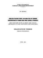
Figure 1 TBARS in grape leaves under heat stress and subsequent
recovery. It is showed that heat treatment (43°C for 6 h) significantly
increased the TBARS concentrations in grape leaves and after
subsequent recovery, there was no difference in TBARS concentrations
between heat-treated and control leaves. Each value represents the
mean ± standard error of the mean (S.E.M.) of three replicates. The
asterisks indicate the significance of differences between treatments
and their corresponding controls (* P < 0.05).
Among the 174 differentially expressed proteins, 127
were characterized as hypothetical or unknown proteins
under the grape genomics information category in uniprot ( To gain functional information about these proteins, BLASTP (i.
nlm.nih.gov/BLAST/) was used to search for homologous proteins against the NCBI non redundant (Nr) protein database. BLAST searching was able to align 117 of
the unknown proteins (Additional file 1). Among these
Liu et al. BMC Plant Biology 2014, 14:110
/>
Page 4 of 17
Figure 2 Donor side (Wk), reaction center (RCQA), acceptor side (φPo, ψEo, φEo) parameters of PSII and δRo (the efficiency with an
electron can move from plastoquinone (PQ) through PSI to the PSI end electron acceptor) in grape leaves under heat stress and
subsequent recovery. Each value represents the mean ± S.E. of five replicates. The asterisks indicate the significance of differences from their
corresponding control (* P < 0.05, ** P < 0.01). The detailed meanings of Wk, RCQA, φPo, ψEo, φEo and δRo were shown in Additional file 7.
aligned proteins, 90.6% had an E-value of less than 1.0E50 and showed very strong homology while the remaining 9.4% had an E-value of between 1.0E-10 and
1.0E-50. The identities distribution defined 27.4% of
these aligned proteins as having a matched identity
greater than 90%, 71.8% between 60% and 90% and only
one protein (59.93%) lower than 60%. These results indicating that the unknown proteins might have similar
function with the aligned proteins respectively. These
Figure 3 Venn diagram of differentially expressed proteins
that were up- or downregulated by heat stress or recovery.
The “ + “ and “- “indicate up- and downregulated proteins, respectively.
differentially expressed proteins were classified into 26
functional categories according to MapMan ontology as
shown in Figure 4 and Additional file 2. The main categories included photosynthesis, proteins and stress. In
addition, enrichment analysis against agriGO (http://
bioinfo.cau.edu.cn/agriGO/) showed that differentially
expressed proteins were mainly enrich in response to abiotic stimulus (GO: 0009628), generation of precursor metabolites and energy (GO: 0006091) and photosynthesis
(GO: 0015979) of biological process. Moreover, subcellular
localization of the 174 characterized proteins showed that
60 proteins (34%) were located in chloroplast, five proteins
(3%) were assigned to the mitochondria, 14 proteins (8%)
belonged to secretory pathway, and 21 proteins (12%) were
classified as belonging to other locations. Unfortunately,
74 of the differentially-expressed proteins had unknown
locations (Figure 5). These results indicated that quite a lot
of chloroplast proteins are related to thermotolerance of
grapevine.
Comparative analysis of common responsive proteins
between heat stress and subsequent recovery
There were 17 proteins that were upregulated by heat
stress, but were then downregulated after recovery
Liu et al. BMC Plant Biology 2014, 14:110
/>
Page 5 of 17
Figure 4 Functional characterization of heat stress and recovery–responsive proteins under heat stress and/or subsequent recovery.
Figure 5 Subcellular localization of the 174 differentially
expressed proteins under heat stress and/or subsequent
recovery. C: Chloroplast, i.e. the sequence contains cTP, a chloroplast
transit peptide; M: Mitochondrion, i.e. the sequence contains mTP, a
mitochondrial targeting peptide; S: Secretory pathway, i.e. the sequence
contains SP, a signal peptide; _: Any other location; *: “don't know”.
(Additional file 3). Three of these proteins were categorized as being related to photosynthesis, including PSI reaction center subunit N (PsaN), ATP synthase subunit
beta (fragment), and Rubisco large chain. Interestingly,
PsaN was upregulated 28 fold by heat stress but then
downregulated more than 5 fold after recovery, compared with their corresponding controls. In addition, two
of the proteins were related to metabolism: one is
acetoacetyl-CoA thiolase, which condenses two molecules of acetyl-CoA to give acetoacetyl-CoA, and this is
the first enzymatic step in the biosynthesis of isoprenoids
via mevalonate, the other is coproporphyrinogen-III oxidase (CPOX), a key enzyme in the biosynthetic pathway
of chlorophyll. Universal stress protein (USP), a transcription factor in abiotic stress, and thylakoidal ascorbate peroxidase (APX), involved in H2O2 detoxification,
were also induced by heat stress and decreased after
Liu et al. BMC Plant Biology 2014, 14:110
/>
subsequent recovery. Moreover, proteins related to protein metabolism included one chloroplastic large subunit
ribosomal protein (L12-1) and one translation initiation
factor (eIF3f). Peptidyl-prolyl cis-trans isomerase and two
transporters, the nascent polypeptide associated complex
alpha and the mitochondrial import inner membrane
translocase subunit Tim9 were also affected. One14-33-like protein, associated with a DNA binding complex
that binds to the G-box was also identified.
Only eight proteins were upregulated by both heat
stress and subsequent recovery (Additional file 3). One
PSII subunit R (PsbR), one PSI subunit H (PsaH) and a
Rubisco small submit were induced after heat stress and
recovery. Additionally, two ribosomal proteins (S21e, S9)
were also identified. Moreover, heat shock protein (HSP)
26 in chloroplast was induced 3.4 and 2.0 fold respectively by heat stress and recovery. Nucleoside diphosphate kinase 1 (NDPK1), involved in purine metabolism,
was also induced more than 10 fold under heat stress,
and returned to almost the control level after recovery.
Eight proteins were downregulated by heat stress but
upregulated after subsequent recovery (Additional file 3).
Among the eight proteins, two of them are related to
photosynthesis, PSI subunit l (PsaA), PSII protein D1
(PsbA). Biotin carboxylase subunit, a component of the
acetyl coenzyme A complex was downregulated 0.46 fold
by heat stress but upregulated 1.6 fold after subsequent
recovery. In addition, two stress-related proteins of the
HSP90 family (HSP90-5, HSP90-7) were also identified.
The three remaining proteins in this group were not
assigned.
Additional file 3 shows nine proteins that were downregulated both by heat stress and subsequent recovery.
Light-harvesting chlorophyll-protein complex II subunit
B1 (LHCB1.4) in photosynthesis and a magnesiumchelatase (MgCh) subunit ChlI-2 involved in chlorophyll
biosynthesis were identified in this group. Cyanate hydratase which catalyzes the bicarbonate-dependent breakdown
of cyanate to ammonia and bicarbonate in cyanogenic
glycosides was also repressed both by heat stress and
recovery. In addition, small subunit ribosomal protein SA
and protein phosphatase 2C in protein metabolism was
also repressed after heat stress and recovery.
Analysis of proteins only responsive to heat stress
A total of 71 proteins showed a specific response to heat
stress, with 23 upregulated proteins, and 48 downregulated proteins (Additional file 4). Five of the 23 upregulated proteins are related to photosynthesis, including
PsaF, three ATP synthase subunits (γ, δ, b) involved in
the photosystem electron-transfer reaction, and a fructose bisphosphate aldolase (FBA) involved in the Calvin
cycle. Of note, the ATP synthase CF (0) b subunit was
upregulated 8.4 fold by heat stress. Ribosomal protein S1
Page 6 of 17
involved in protein synthesis was also upregulated by
heat stress. HSP22, located in the endoplasmic reticulum,
and HSP21, located in the chloroplast, were induced 3.0
and 5.5 fold, respectively, under heat stress. Cytoplasmic
[Cu-Zn] superoxide dismutase (SOD), involved in redox,
was also induced 5.0 fold under heat stress. In addition,
14-3-3-like protein was upregulated 1.8 fold by heat
stress. Among the 48 downregulated proteins (Additional
file 4), eight of them were involved in photosynthesis, including LHCB1.3, PsbP, and PsaL. Many other proteins
were involved in a variety of metabolic mechanisms,
including glucose-1-phosphate adenylyltransferase, two
malate dehydrogenase enzymes (MDH), nitrite reductase
1 in N-metabolism and uracil phosphoribosyltransferase
involved in nucleotide metabolism. There are also some
carbohydrate metabolism-related proteins, such as UDPglucose pyrophosphorylase, which catalyze the reversible
reaction between glycose-1-phosphate and UDP-glycose,
dihydrolipoyl dehydrogenase in the tricarboxylic acid
cycle (TCA) and 6-phosphogluconate dehydrogenase in
the oxidative pentose phosphate pathway (OPP). Three
proteins were identified as being stress-related; including
osmotin-like protein and HSP70. Two identified proteins,
Beta-1-3 glucanase and alcohol dehydrogenase, were annotated to miscellaneous enzyme families. In addition,
ten proteins were involved in protein metabolism, including mitochondrial-processing peptidase subunit α and β,
in protein targeting; methionine sulfoxide reductase A, in
posttranslational modification; protease Do-like 8, and
proteasome subunit α type-5 in protein degradation and
a 20 kDa chaperonin, involved in protein folding. There
are also five proteins are not assigned.
Analysis of proteins only responsive to recovery from
heat stress
There were 25 proteins which were only upregulated
after recovery from heat stress (Additional file 5). Four
of these proteins are photosynthesis-related, including
LHCB2.1, PsbS, PetB. Two upregulated stress proteins
corresponded to the HSP70 family (HSP70-5, HSP70-11).
HSP70-5 is located in the cytoplasm, while HSP70-11 is
located in the endoplasmic reticulum and plays a role in
facilitating the assembly of multimeric protein complexes
inside the endoplasmic reticulum. Ribosomal proteins,
including L22, EF-Ts, were also upregulated only upon
recovery to heat stress.
Thirty-six proteins were downregulated only after recovery to heat stress (Additional file 5). Eight downregulated proteins were involved in photosynthesis, including
PsbE, PsaD, PetC, PetD, FNR in light reaction and phosphoribulokinase, FBA, fructose-1,6- bisphosphatase in
Calvin cycle. Two isoforms of FBA, glyceraldehyde-3phosphate dehydrogenase and phosphoglycerate kinase
involved in glycolysis were also repressed after recovery
Liu et al. BMC Plant Biology 2014, 14:110
/>
from heat stress. Down-expressed proteins involved in
amino acid metabolism included aspartate aminotransferase, serine-pyruvate aminotransferase, ketol-acid reductoisomerase, and aminomethyltransferase. Catalase (CAT)
and APX involved in H2O2 detoxification were also downregulated after recovery from heat stress. Several proteins
from this group were unfortunately unidentified.
Discussion
One of the many locations for heat stress injury in cells
is the membrane. TBARS is the product of lipid peroxidation in plants. The chlorophyll a fluorescence transient analysis (O-J-I-P test) is a powerful tool to probe the
PSII reactions, which may help determine the state of
the electron transport chain [54]. In this study, we investigated the TBARS content and chlorophyll fluorescence
parameters in grape leaves under heat stress and subsequent recovery (Figures 1 and 2). These results showed
that young grapevines of the ‘Cabernet Sauvignon’ varietal were damaged under heat stress at 43°C for 6 h, but
they subsequently recovered at 25°C for 18 h. Differential proteomic analysis of grapevines under these two
conditions were also performed, and the findings are further discussed below.
Electron transport chain and related proteins involved in
the photosynthesis
Photosynthesis is known to be one of the most heat sensitive processes due to its complex mechanisms and requirement for enzymes. It is directly related to plant
productivity and energy utilization. In this study we
identified 34 dysregulated proteins involved in photosynthesis, upon heat stress and subsequent recovery.
These accounted for one fifth of all differentially expressed
proteins in this study (Table 1 and Figure 6). Moreover, enrichment analysis showed that photosynthesis was enriched
under heat stress and/or recovery (Additional file 6 and
Additional file 7).
PSII is thermally labile and is considered to be the
most sensitive component of the electron transport
chain [55,56]. The peripheral antennas of PSII are composed of major trimeric and minor monomeric LHCII
proteins. In this study, the expression of LHCII1.3
and LHCII1.4 was inhibited under heat stress and increased after recovery, which indicated that LHCII1.3
and LHCII1.4, might be thermally labile. LHCB2.1 showed
the same expression as control under heat stress while increased about 2.7 fold after recovery, suggesting that
LHCB2.1 may be thermostable and solely involved in the
recovery from heat stress. The OEC activity is in close
association with the 33 kDa (PsbO) and 23 kDa (PsbP).
PsbO is a key structural component of many different
types of OECs and functions to stabilize the manganese
cluster and modulate the Ca2+ and Cl− requirements for
Page 7 of 17
oxygen evolution [57]. Additionally, the 10-kDa PsbR
protein has also been found play a role in stable association of the PsbP with the PSII core for water oxidation
[57,58]. In the present study, PsbO-2 levels were not altered upon heat stress or subsequent recovery, the PsbP
precursor was repressed under heat stress but returned
to control level after subsequent recovery, while PsbR
was elevated approximately eight fold with respect to
its control under heat stress, and remained upregulated
two fold upon subsequent recovery. In addition, the
chlorophyll fluorescence parameter Wk showed that
the OEC of PSII was damaged under heat stress, but
returned back to the normal physiological level in the
recovery phase (Figure 2). Therefore, these combined
results suggest that PsbR may play an important role in
maintaining the stability of the OEC of PSII compared
to PsbO and PsbP in grape leaves.
In the present study, RCQA values decreased under
heat stress and increased to the control level after subsequent recovery (Figure 2), indicating that the PSII reaction center was inhibited by heat stress and then
recovered when the stress was removed. The change of
D1 protein corroborated this result (Table 1). The multisubunits (PetA, PetB, PetC and PetD) complex of Cytb6/f
is a crucial component for the acceptor side of electron
transport chain of PSII [59]. In the present study, three
subunits PetB, PetD and PetC were differentially
expressed. The expression level of PetB, PetC and PetD
did not change significantly under heat stress, however,
after recovery, the expression of PetC and PetD was
largely inhibited while PetB was induced. In addition, φEo
and ψEo were reduced in grape leaves under heat stress,
then returned to control levels with the subsequent recovery (Figure 2). This suggests that the function of the
acceptor portion of the electron transport chain of PSII
including Cytb6/f complex recovered from heat stress.
These combined results suggest that PetB may promote
the Cytb6/f complex to recover from heat stress.
The study showed that many proteins in the PSI complex changed upon heat stress (Table 1). PSI consists of
a core complex and a peripheral antenna. In plants,
these two functional units result from the assembly of at
least 19 protein subunits. The PSI core complex contains
15 subunits, including PsaA to PsaL and PsaN to PsaP
which play important roles in PSI function. For example,
PsaF is located in the thylakoid lumen, and contains a
lysine-rich helix-loop-helix motif that has been demonstrated to interact with plastocyanin in plants and with
plastocyanin (PC) or Cytochrome c6 in algae [60]. PsaN
is necessary for the docking PC to the PSI complex, and
is the only subunit located entirely on the lumenal side
of PSI. In the present study, it was shown from the
chlorophyll a fluorescence parameter δRo that PSI was
damaged under heat stress and recovered to the control
Liu et al. BMC Plant Biology 2014, 14:110
/>
Page 8 of 17
Table 1 Proteins involved in photosynthesis under heat stress and/or subsequent recovery
Protein
accession
Fold change
HS
RC
Bin
Species
Description
A5ASG6
0.924
2.708
1.1.1.1
Arabidopsis thaliana
Photosystem II light harvesting complex protein 2.1, LHCB2.1
A5BPB2
A5B5I4
0.438
0.524
1.1.1.1
Arabidopsis thaliana
Putative light-harvesting chlorophyll-protein complex II subunit B1, LHCB1.4
0.456
1.084
1.1.1.2
Arabidopsis thaliana
Chlorophyll a/b-binding protein 1, chloroplastic, LHCB1.3
D7UA58
0.59
1.176
1.1.1.2
Gossypium hirsutum
PsbP precursor
Q67H94
1.045
0.608
1.1.1.2
Muscari comosum
Cytochrome b559 subunit alpha (Fragment), PsbE (cytb559α)
E0CR63
1.041
1.603
1.1.1.2
Ricinus communis
Photosystem II 22 kDa protein, PsbS, chloroplast precursor
B6VJV1
0.601
1.928
1.1.1.2
Vitis vinifera
Photosystem II protein D1, PsbA (D1)
A5AWT3
7.737
2.387
1.1.1.2
Nicotiana tabacum
Photosystem II 10 kDa polypeptide, PsbR, chloroplastic
F6GY64
NA*
1.645
1.1.1.2
Populus trichocarpa
One helix protein 2
A5AW35
0.656
1.226
1.1.2.2
Ricinus communis
Photosystem I reaction center subunit XI, PsaL, chloroplastic
A5B2H3
7.317
1.234
1.1.2.2
Ricinus communis
Photosystem I reaction center subunit III, chloroplast precursor, PsaF
A5AEB4
0.878
0.582
1.1.2.2
Ricinus communis
Photosystem I reaction center subunit II, PsaD, chloroplast precursor
Q0ZJ20
0.545
5.057
1.1.2.2
Vitis vinifera
photosystem I P700 apoprotein A1, PsaA
F6I0D9
28.065
0.185
1.1.2.2
Medicago truncatula
Photosystem I reaction center subunit N, PsaN
A5BHE6
5.11
2.172
1.1.2.2
Ricinus communis
Photosystem I reaction center subunit VI, PsaH
A5BX41
0.846
0.236
1.1.3
Vitis vinifera
Cytochrome b6/f complex iron-sulfur subunit, PetC
Q0ZIY8
1.479
0.417
1.1.3
Vitis vinifera
Cytochrome b6/f complex subunit IV, PetD
Q0ZIY9
0.8
1.881
1.1.3
Vitis vinifera
Cytochrome b6, PetB
Q67H40
0.392
0.818
1.1.4
Muscari comosum
ATP synthase subunit beta, chloroplastic
Q0ZJ34
8.386
0.957
1.1.4
Vitis vinifera
ATP synthase CF (0) b subunit
F6H7M1
1.502
1
1.1.4
Vitis vinifera
ATP synthase gamma chain, chloroplastic-like isoform 1
F6HVW3
1.995
1.03
1.1.4
Nicotiana tabacum
ATP synthase delta chain, chloroplastic
Q95FU2
1.83
0.401
1.1.4
Coccoloba uvifera
ATP synthase beta subunit
E0CQ75
1.234
0.554
1.1.5.3
Ricinus communis
Ferredoxin–NADP reductase, FNR
D7TQZ8
0.666
0.726
1.2.2
Glycine max
Peroxisomal (S)-2-hydroxy-acid oxidase GLO1-like
A5BTM9
2.969
0.505
1.3.1
Vitis vinifera
Ribulose-1,5-bisphosphate carboxylase/oxygenase large subunit, RbcL
Q2I314
1.627
1.886
1.3.2
Vitis pseudoreticulata
ribulose-1,5-bisphophate carboxylase/oxygenase small subunit
A5BHS5
0.61
1.167
1.3.4
Glycine max
NADP-dependent glyceraldehyde-3-phosphate dehydrogenase-like
F6HFL6
1.833
0.733
1.3.6
Vitis vinifera
Fructose-bisphosphate aldolase, FBA
F6GWQ0
0.799
0.626
1.3.6
Vitis vinifera
Fructose-bisphosphate aldolase
A5AYR7
1.476
0.664
1.3.7
Glycine max
Fructose-1,6-bisphosphatase, chloroplastic-like
A5BE19
0.84
0.447
1.3.12
Vitis vinifera
Phosphoribulose kinase, putative
D7THJ7
0.482
0.739
1.3.13
Ricinus communis
Ribulose bisphosphate carboxylase/oxygenase activase 1, chloroplast precursor
F6HBT1
0.594
1.004
1.3.13
Glycine max
Ribulose bisphosphate carboxylase/oxygenase activase, chloroplastic-like
*The proteins were not quantified under heat stress or subsequent recovery.
HS refers to the fold change in heat stressed proteins, with respect to controls, while RC refers to the fold change in proteins after recovery, with respect to controls.
level when returned to normal temperatures (Figure 2).
Consistent with this observation, the levels of PsaA and
PsaL declined under heat stress. However, the expression
level of PsaA remained 5 fold higher compared to the
control after subsequent recovery, suggesting that PsaA
may have a positive effect in the recovery phase of PSI.
In addition, the expression of PsaF, PsaH and PsaN increased by a 7.3, 5.1 and 28.1 fold respectively under
heat stress, which indicated that PsaF, PsaH and PsaN
might play a role of protection from heat stress in the
PSI complex of grape leaves. It is especially interesting
that while all proteins of the PSI complex inhibited
under heat stress were hydrophobic, all proteins induced
under heat stress were hydrophilic.
ATP synthase produces ATP from ADP in the presence of a proton gradient across the membrane. F-type
Liu et al. BMC Plant Biology 2014, 14:110
/>
Page 9 of 17
Figure 6 MapMan visualization of photosynthesis in grapevine leaves under heat stress (A) and subsequent recovery (B).
ATPase has two components, CF (1) - the catalytic
core - and CF (0) - the membrane proton channel. CF
(1) has five subunits: α, β, γ, δ and ε while CF (0) has
four main subunits: a, b, b′ and c. The α chain is the
largest subunit of the ATP synthase. The γ chain is believed to be important in regulating ATPase activity
and the flow of protons through the CF (0) complex. In
the study, all the identified ATP synthase subunits (γ, δ and
b of CF (0)) were upregulated under heat stress, and all
of them recovered to their control levels after subsequent recovery. Especially, the expression of subunit b
is increased by 8.4 fold under heat stress. These result
suggested that these three subunits may have a protective
role against heat stress for ATP synthase, and continue to
provide energy for maintaining the normal physiological
processes of grapevines.
Proteins involved in abiotic stress and redox regulation
Nineteen identified dysregulated proteins were functionally characterized as being involved in stress response
(Table 2). Most of them were assigned to one of the four
major classes of molecular chaperones, HSP90, HSP70,
HSP60 and sHSPs, however, no proteins belonged to
HSP100 family. Plants respond to different abiotic stress
by inducing the synthesis of proteins from the heat
shock protein (HSP)/chaperone family which have been
shown to play a crucial role in protecting plants against
stress by re-establishing normal protein conformations
and thus cellular homeostasis [61]. In this study, nine
HSPs were differentially expressed under heat stress or
after subsequent recovery. Proteins from the HSP90
family do not only manage protein folding [62,63], but
also play a major role in signal-transduction networks,
cell-cycle control, protein degradation and protein trafficking [64-66]. A previous study in P. euphratica showed
that a putative HSP90 was upregulated early upon heat
stress and later returned to control values [14]. In our
study, three members of HSP90 family were identified
and differentially expressed. Two of them were inhibited,
while the expression of HSP90-1 was not affected by heat
stress. However, all of them were upregulated during subsequent recovery. Proteins from the HSP70 family are essential for preventing aggregation and assisting re-folding
of non-native proteins under both normal and stressing
environmental conditions [62,67]. They are involved in
protein import and translocation processes, and in facilitating the proteolytic degradation of unstable proteins by
targeting these proteins to lysosomes or proteasomes
[62]. Previous reports have documented that HSP70 were
accumulated under heat stress [9,68] . In our research,
three members of the HSP70 family were identified. One
of the HSP70 family proteins was repressed under heat
stress and recovered to the control level during the subsequent recovery (Table 2) while the other two had no
difference compared to their control under heat stress
but were downregulated during the recovery phase
(Table 2). This suggests that the many isoforms of HSP70
play different roles under heat stress. In plants, the sHSPs
are abundant and diverse, and can be classified into five
families according to their cellular localization; including
cytosol (class I and II), chloroplast (class III), endoplasmic reticulum (class IV), and mitochondrion (class V)
[9,69-71]. In addition, sHSPs have been reported to be involved in protecting macromolecules like enzymes, lipids,
nucleic acid, and mRNAs from dehydration [72]. In our
study, one protein (HSP22) was predicted to be an endoplasmic reticulum-targeted sHSP, whereas the other
sHSP (HSP21) was predicted to be chloroplast-targeted.
A previous study in Arabidopsis showed the expression
of HSP21 and HSP22 significantly increased under heat
stress [73]. In our study, the similar results were observed, and moreover, the expression of HSP21 and
HSP22 return to control levels after subsequent recovery.
This also agrees with our previous findings, in which the
mRNA level of HSP21 and HSP22 exhibited similar increases [29]. In addition, increased thermotolerance has
Liu et al. BMC Plant Biology 2014, 14:110
/>
Page 10 of 17
Table 2 Proteins involved in abiotic stress and redox under heat stress and/or subsequent recovery
Protein
accession
Fold change
HS
RC
A5BS35
0.412
1.031
Bin
Species
Description
20.1
Nicotiana tabacum
NtPRp27
A5C2C9
0.877
0.324
20.1
Ricinus communis
Protein MLO, putative
A5AHJ5
0.114
1.149
20.2
Vitis vinifera
Osmotin-like protein
F6HYG1
0.409
0.933
20.2.1
Ricinus communis
Heat shock 70 kDa protein
F6HJZ4
3.046
0.978
20.2.1
Corylus heterophylla
Heat shock protein 22, endoplasmic reticulum, HSP22
A5B868
5.531
1.45
20.2.1
Solanum lycopersicum
Heat shock protein 21, chloroplast, HSP21
F6HU55
0.878
1.989
20.2.1
Cucumis sativus
Heat shock protein 70
F6HYK6
1.005
2.591
20.2.1
Vitis vinifera
Similar to PsHSP71.2
A5ADL7
1.312
2.959
20.2.1
Arabidopsis thaliana
Heat shock protein 90.1, cytoplasmic, HSP90-1
A5BX00
0.382
1.782
20.2.1
Arabidopsis thaliana
HSP90-like protein 7, HSP90-7
F6HGF1
0.598
2.854
20.2.1
Ipomoea nil
Heat shock protein 90
E0CVB4
3.416
1.983
20.2.1
Nicotiana tabacum
Heat shock protein 26
F6HKZ7
5.463
0.748
20.2.99
Ricinus communis
ATOZI1
D5LN28
1.8
0.472
20.2.99
Vitis pseudoreticulata
Universal stress protein (USP) family protein
E0CQM3
9.846
2.943
21.1
Populus trichocarpa
Thioredoxin M
D7SKR5
1.32
0.648
21.2.1
Vitis vinifera
Ascorbate peroxidase, APX
F6H0K6
1.508
0.434
21.2.1
Glycine max
L-ascorbate peroxidase T, chloroplastic-like isoform 2
F6HTX9
4.904
0.99
21.6
Vitis vinifera
Cytoplasmic [Cu-Zn] SOD
D7UD99
1.071
0.604
21.6
Vitis vinifera
Catalase, CAT
been previously achieved by overexpressing the plastidial
Hsp21 in tomato [74]. Therefore, these sHSPs may have
the important functions in alleviating heat stress in
grapevines.
The antioxidant enzymes are known to play important
roles in scavenging or reducing excessive reactive oxygen
species (ROS) which are produced under stress conditions, in order to maintain cell redox homeostasis [9]. In
this study, we identified a group of antioxidant enzymes
including [Cu-Zn] SOD, CAT, APX and thioredoxin.
[Cu-Zn] SOD which plays a central role in protecting
against oxidative stress is generally found in the cytosol
and chloroplasts (Table 2). The cytoplasmic [Cu-Zn]
SOD showed considerable upregulation (approximately
5 fold) under heat stress, followed by a return to the
control level after subsequent recovery. This is in agreement with published results in the heat-tolerant Agrostis
scabra, while these redox proteins were not detected in
the heat-sensitive Agrostis stolonifera [75]. In addition,
the expression of APX increased under heat stress in
our study. Thioredoxins are small proteins catalyzing
thiol-disulfide interchange, which is involved in the
regulation of the redox environment in cells [76,77]. The
most prominent candidates of proteins are thioredoxin h
in Populus euphratica Oliv. and rice leaves, upon heat
stress [9,14]. Thioredoxin M4 was predicted to be located in chloroplast in our study, and was upregulated
almost 10 fold under heat stress and maintained approximately 3 fold after subsequent recovery (Table 2).
These results suggest that cytosolic [Cu-Zn] SOD, APX
and chloroplastic thioredoxin have important roles in
maintaining redox homeostasis in grapevine cells under
heat stress (Figure 7).
Proteins involved in metabolism
The expression of most proteins predicated to be involved in metabolism was slightly downregulated in
grape leaves under heat stress (Table 3), indicating that
the metabolism of ‘Cabernet Sauvignon’ grapevine was
mildly affected under heat stress. In the present study,
three proteins identified were involved in nucleotide
metabolism. Most significantly, NDPK1, which plays a
major role in the synthesis of nucleoside triphosphates
other than ATP was upregulated more than 10 fold
under heat stress, and declined to 1.7 fold following recovery, compared to controls. Fukamatsu et al. showed
that Arabidopsis NDPK1 is a component of ROS signaling pathways by interacting with three CATs [78]. Furthermore, in Neurospora crassa, NDPK1 is suggested to
control CATs in response to heat, oxidative stress and
light, and results have indicated that NDPK1 protein
was translocated from the plasma membrane to the
cytoplasm in response to light, and may interact with
CAT [79]. Together with our findings, we suggest that
Liu et al. BMC Plant Biology 2014, 14:110
/>
Page 11 of 17
Figure 7 Overview of cellular response in grapevine leaves under heat stress (A) and subsequent recovery (B) visualized by MapMan.
Table 3 Proteins involved in metabolism under heat stress and/or subsequent recovery
Protein
accession
Fold change
Bin
Species
Description
HS
RC
D7TDB6
0.547
F6HDM4
0.516
1.053
2.1.2.1
Vitis vinifera
ADP-glucose pyrophosphorylase catalytic subunit
1.178
2.1.2.1
Vitis vinifera
Glucose-1-phosphate adenylyltransferase
Q9S944
F6HJU7
0.169
0.882
2.2.1.3.3
Vitis vinifera
Vacuolar invertase 1, GIN1
0.667
1.854
3.1.2.2
Ricinus communis
Stachyose synthase precursor
E0CU00
0.805
0.542
3.5
Ricinus communis
Aldo/keto reductase
F6HHH7
NA*
1.714
3.5
Glycine max
Putative aryl-alcohol dehydrogenase C977.14c-like
D7TMQ2
0.666
0.783
6.1
Vitis vinifera
Citrate synthase, glyoxysomal
F6HJJ4
0.624
1.095
6.3
Ricinus communis
Malate dehydrogenase
A5BEJ8
0.524
1.328
6.3
Vitis vinifera
Malate dehydrogenase, putative
F6H9P9
0.455
1.618
11.1.1
Camellia oleifera
Biotin carboxylase, CAC2
G3G8J7
0.425
0.696
12.1.2
Vitis vinifera
Nitrite reductase 1
D7SW04
0.744
0.519
13.1.1.2.1
Petunia x hybrida
Prephenate aminotransferase
A5ACX0
0.389
1.125
13.1.1.3.1
Arabidopsis thaliana
Alanine-2-oxoglutarate aminotransferase 2
F6HA09
0.696
0.616
13.1.1.3.11
Ricinus communis
Serine-pyruvate aminotransferase
F6GST3
0.614
0.949
13.1.2.3.22
Ricinus communis
Argininosuccinate synthase
A5AGN5
0.896
0.157
13.1.4.1
Catharanthus roseus
Ketol-acid reductoisomerase
A5AFH5
0.397
0.825
13.1.5.3.1
Vitis vinifera
Cysteine synthase
F6HHQ7
1.652
0.553
13.2.3.5
Hevea brasiliensis
Acetyl-CoA C-acetyltransferase
F6H7I9
1.038
0.559
13.2.5.2
Vitis vinifera
Aminomethyltransferase, mitochondrial-like
A5BQ64
1.09
0.474
16.1.3.3
Hevea brasiliensis
2-methyl-6-phytylbenzoquinone methyltranferase
A5BJL8
0.5
0.376
16.4.3.1
Vitis vinifera
Cyanate hydratase
D7SLA9
0.835
0.627
17.7.1.2
Vitis vinifera
Lipoxygenase
A5BEM6
1.25
0.643
19.3
Ricinus communis
Glutamate-1-semialdehyde-2,1-aminomutase,GSA-AT
A5BF85
1.526
0.635
19.8
Ricinus communis
Coproporphyrinogen III oxidase, CPOX
F6HM73
0.353
0.358
19.10
Ricinus communis
Magnesium-chelatase subunit chlI, chloroplast precursor
F6HL38
0.2
1.02
23.3.1.3
Glycine max
Uracil phosphoribosyltransferase-like
A5B878
10.227
1.695
23.4.10
Vitis vinifera
Nucleoside diphosphate kinase 1, NDPK1
F6HBJ7
0.813
0.602
23.4.99
Ricinus communis
Inorganic pyrophosphatase
*The proteins were not quantified under heat stress or subsequent recovery.
Liu et al. BMC Plant Biology 2014, 14:110
/>
Page 12 of 17
NDPK1 may play an important role in grape leaves in
response to heat stress.
Proteins involved in glycolysis and TCA in mitochondrial
respiration
The regulation of the enzymes involved in respiratory
carbon metabolism under heat stress has been a subject
of debate. As shown in Table 4, there were six enzymes
identified that are involved in glycolysis, which did not
significantly change in expression level under heat stress
while were downregulated after subsequent recovery. In
addition, we found that five enzymes (dihydrolipoyl dehydrogenase, aconitase, malate dehydrogenase, succinatesemialdehyde dehydrogenase and carbonic anhydrase),
which are involved in the TCA cycle, were dysregulated in
the study. With the exception of aconitase, the expression
of these enzymes was inhibited under heat stress and recovered to the control level or showed a slight increase
after subsequent recovery. The above results suggest that
the glycolysis pathway was not influenced, while the TCA
cycle was inhibited by heat stress. We also hypothesize
that during recovery, the TCA cycle recovereds to control
levels to consume the excess pyruvic acid produced
by glycolysis. Therefore, the glycolysis pathway may be
more heat tolerant than the TCA cycle in respiration in
grapevines.
Conclusion
This study provides a global look at the dysregulated
proteins in grapevine leaves exposed to heat stress and
after subsequent recovery using the iTRAQ technique. A
total of 174 differentially expressed proteins were identified in response to heat stress and/or subsequent recovery. On the basis of these findings, we propose that
some proteins related to the electron transport chain
of photosynthesis, antioxidant enzymes, HSPs and the
glycolysis pathway may play key roles in protecting
grapevines from heat stress and enhancing their recovery capacity.
Methods
Plant materials and treatments
One-year old ‘Cabernet sauvignon’ (V. vinifera L.) grapevine cuttings were planted in pots, then grown in a
greenhouse at 70-80% relative humidity under a 18-25°C,
with the maximum photosynthetically active radiation
(PAR) at approximately 1,000 μmol photons m−2 s−1.
When the sixth leaves (from bottom to top) of grapevines
became mature, all grapevines were divided into two
groups and acclimated for two days in a controlled environment room (70% average relative humidity, 25/18
(12 h/12 h) day/night cycle and PAR at 800 μmol m−2 s−1).
On day three, the grapevines were subjected to the following treatments: (1) the plants of the control group were
maintained at the optimal day/night temperature (25°C/
18°C) in the above growth chamber; (2) the plants of the
treatment group were exposed to 43°C from 9:30 to
15:30 (the conditions were the same as the control, except for temperature). The stressed grapevines were then
allowed to recover at 25°C rapidly (from 43°C to 25°C in
Table 4 Proteins involved in respiration under heat stress and subsequent recovery
Protein
accession
Fold change
F6I0H8
0.548
1.144
4.1
Gossypium hirsutum
UDP-D-glucose pyrophosphorylase
F6HFF7
0.931
1.93
4.2
Ricinus communis
Phosphoglucomutase
HS
Bin
Species
Description
RC
A5B118
1.356
0.481
4.7
Vitis vinifera
Fructose-bisphosphate aldolase, FBA
A5BX43
1.222
0.461
4.7
Vitis vinifera
Fructose-bisphosphate aldolase, FBA, cytoplasmic isozyme 1-like
F6GSG7
1.105
0.653
4.9
Ricinus communis
Glyceraldehyde 3-phosphate dehydrogenase
A5CAF6
1.017
0.5
4.10
Vitis vinifera
Phosphoglycerate kinase, cytosolic-like
A5BGC9
0.507
1.296
7.1.3
Vitis vinifera
6-phosphogluconate dehydrogenase
A5BDU8
0.537
0.938
8.1.1.3
Vitis vinifera
Dihydrolipoamide dehydrogenase, putative
D7TEL2
0.672
1.64
8.1.3
Ricinus communis
Aconitase
F6HZK0
0.499
1
8.2.9
Vitis vinifera
Malate dehydrogenase
F6H9T6
0.466
0.972
8.2.99
Solanum lycopersicum
Succinic semialdehyde dehydrogenase
A5BQL5
0.628
0.795
8.3
Vitis vinifera
Chloroplast carbonic anhydrase
A5C9C0
0.833
1.722
9.1.2
Ricinus communis
NADH-ubiquinone oxidoreductase flavoprotein
A5ASP2
1.286
2.886
9.1.2
Ricinus communis
NADH-ubiquinone oxidoreductase 24 kD subunit
D7TQ15
NA*
2.667
9.1.2
Solanum tuberosum
NADH:ubiquinone oxidoreductase-like
D7SUP9
NA*
0.653
9.5
Camellia sinensis
Ubiquinol-cytochrome C reductase complex
*The proteins were not quantified under heat stress or subsequent recovery.
Liu et al. BMC Plant Biology 2014, 14:110
/>
about 10 min), then, all conditions were the same as the
control until 9:30 h on Day 4. The fourth to sixth leaves
(from bottom to up) of each plant were detached from
each plant at 15:30 Day 3 (the end of the heat stress
treatment) and 9:30 Day 4 (the day of recovery) (Additional
file 8). Each biological replicate included three plants, and
three replicates were used for both treatment and controls.
Leaves were frozen in liquid nitrogen immediately and
stored at −80°C for further analysis.
Analysis of chlorophyll fluorescence parameters
The chlorophyll a fluorescence transient (O-J-I-P test)
was measured by a Handy Plant Efficiency Analyzer after
the leaves adapted for 15 min in the dark. The chlorophyll a fluorescence transient was induced by a saturating photon flux density at 3000 μmol photons m−2 s−1,
provided by an array of six light-emitting diodes (peak
650 nm). The fluorescence signals were recorded within
a time span from 10 μs to 1 s, with a data acquisition
rate of 10 μs for the first 2 ms and every 1ms thereafter.
The following data from the original measurements were
used: Fk: fluorescence intensity at 300 μs [required for
calculation of the initial slope (M) of the relative variable
fluorescence (V) kinetics and Wk]; Fj: the fluorescence
intensity at 2 ms (the J-step); Fi: the fluorescence intensity
at 30 ms (the I-step); Fm: maximal fluorescence intensity
(the P-step). The derived parameters are as follows: Fo:
fluorescence intensity at 50 μs; the parameter Wk on donor
side of photosystem II (PSII), represents the damage to
OEC, Wk = (Fk-Fo)/(Fj-Fo); the parameter RCQA on reaction center of PSII, represents the density of QA-reducing
reaction centers, RCQA = φPo × (Vj/Mo) × (ABS/CSm); the
parameter Fv/Fm on acceptor side of PSII, represents maximum quantum yield of primary photochemistry at t = 0;
the parameter φEo on acceptor side of PSII, represents
quantum yield for electron transport (at t = 0), φEo = ETo/
ABS = (Fm-Fj)/Fm. The calculation and derivation of a
range of new parameters from O-J-I-P transients is shown
in Additional file 9. Five independent replicates were used
in both treatments and controls respectively, and each replicate consisted of a plant. The chlorophyll a fluorescence
transient was measured on the same plants under heat
stress and subsequent recovery.
Measurement of thiobarbituric acidreactivesubstances
(TBARS)
The content of TBARS was determined according to
the methods of Heath and Packer [80] with minor
modifications. About 1 g of frozen leaves were homogenized in 0.5% thiobarbituric acid and 20% trichloroacetic acid. After heating for 30 min at 95°C, samples
were cooled quickly in an ice-water bath. Air bubbles
were then removed from each tube by shaking, and
samples were centrifuged at 14,000 rpm for 20 minutes
Page 13 of 17
at 20°C. The absorbance of the supernatant was read at
532 nm, corrected for nonspecific turbidity by subtracting the absorbance at 600 nm. The amount of TBARS
was calculated by using an extinction coefficient of
155 mM−1 cm−1.
Protein extraction
Total protein was extracted using the cold-acetone
method. The three biological replicates of the frozen
grape leaves were pooled for iTRAQ analysis [81,82],
and 10% m/m polyvinyl polypyrrolidone (PVPP) were
transferred to a mortar with liquid nitrogen and ground
until a fine powder was obtained. Approximately 500 mg
of the ground up leaf powder was combined with 4 ml
of 10% m/v trichloroacetic acid (TCA) in acetone to
each sample, and the samples were incubated at −20°C
for 2 h. The samples were then centrifuged at 20,000 g
for 30 min at 4°C. The supernatant was discarded without disturbing the pellets. In order to reduce acidity,
the pellets were washed with acetone and incubated
at −20°C for 30 min, and centrifuged at 20,000 g for
30 min at 4°C. The washing step with acetone was repeated several times until the pellets were white. The
dried pellets were lysed with 1 ml protein extraction
reagent [8 M urea, 30 mM HEPES, 1 mM PMSF, 2 mM
EDTA and 10 mM DTT]. The pellets were then dissolved
by ultrasound (pulse on 2 s, pulse off 3 s, power 180 w)
for five minutes. After dissolution, the solution was centrifuged at 20,000 g for 30 min at 4°C to remove nonsoluble impurities. Proteins were reduced with 10 mM
DTT at 56°C for 1 h and alkylated immediately by
55 mM iodoacetamide (IAM) in the dark at room
temperature for 1 h. The treated proteins were precipitated in acetone at −20°C for 3 h. After centrifugation at
20,000 g for 20 min at 4°C, the pellets were resuspended
and ultrasonicated in pre-chilled 50% TEAB buffer with
0.1% SDS and dissolved by ultrasound. The proteins were
regained after centrifugation at 2000 g and protein concentration was determined by the Bradford assay using
BSA as a standard.
Digestion and iTRAQ labeling
Total of 100 μg protein in TEAB buffer was incubated
with 3.3 μg of trypsin (1 μg/μl) (Promega, Madison, WI,
USA) at 37°C for 24 h in a sealed tube. The tryptic peptides were lyophilized and dissolved in the 50% TEAB
buffer and the trypsin digested samples were analyzed
using MALDI-TOF/TOF to ensure complete digestion.
The protocol of iTRAQ labelling was followed the company manual. The tryptic peptides were incubated with
8-plex iTRAQ labeling kit (AB Sciex, Foster City, CA,
USA) (116 for HS-CK; 121 for HS-TR; 114 for RC-CK;
118 for RC-TR) for 2 h at room temperature, which was
dissolved in 70 μl isopropanol.
Liu et al. BMC Plant Biology 2014, 14:110
/>
Peptide fractionation by strong cation exchange (SCX)
The labeled samples were fractionated using an HPLC
system (Shimadzu, Kyoto, Japan) connected to an SCX
column (Luna 5u column, 4.6 mm × 250 mm, 5 μm,
100 Å; Phenomenex, Torrence, CA). The retained peptides were eluted using Buffer A (10 mM KH2PO4 in an
aqueous solution of 25% acetonitrile and acidified to a
pH of 3.0 with H3PO4) and Buffer B, where Buffer B was
composed of Buffer A with 2 M KCl. The fractions were
collected in 1.5 ml microfuge tubes with flow rate at
1 ml/min. The following chromatographic gradient
was applied: 0 ~ 25 min 100% Buffer A; 25 ~ 26 min 5%
Buffer B; 26 ~ 46 min 5-30% Buffer B; 46 ~ 51 min 30-50%
Buffer B, 51-56 min 50% Buffer B; 56–61 min increasing
to 100% Buffer B. All solutions used were centrifuged
again at 20,000 g for 30 min at 4°C. Fraction collection
started 26 min after the injection with a sample collected
every 1 min to obtain a total of 38 fractions. For fractions
containing a high concentration of salt, an additional step
was used to remove the salt with Strata-X 33u polymeric
reversed phase column (Phenomenex). Eluted fractions
were dried in a vacuum concentrator, and each fraction
was dissolved in 0.1% formic acid solution prior to
reversed-phase nano-LC-tandem mass spectrometry
(LC-MS/MS).
Reverse-Pphase nano liquid xhromatography tandem MS
The SCX peptide fractions were pooled together to obtain 10 final fractions, to reduce the number of samples
and collection time. A 10 μl sample from each fraction
was injected twice to the Proxeon Easy Nano-LC system.
Peptides were separated on C18 analytical reverse phase
column (100 mm × 75 mm, 300 Å, 5 μm) at a flow rate
of 400 nl/min and a linear LC gradient profile was used
to elute peptides from the column. The fractions were
then analyzed using a hybrid Quadrupole/Time-offlight MS (Triple-TOF 5600, AB SCIEX, USA) with
nano electrospray ion source. The MS/MS scans from
50–2000 m/z were recorded. Nitrogen was used as the
collision gas. The ionization tip voltage and interface
temperature were set at 1250 V and 150°C, respectively.
Database search and protein quantification
All the mass spectral data were collected using Micro
TOF (AB5600, Applied Biosystems) control software, and
processed and analyzed using Data Analysis 4.0. The database of uniprot_grape (12/1/2011, 55416 sequences) was
downloaded ( and integrated into
the Mascot search engine version 2.3.01 by its database
maintenance unit. All parameters were set as follows: specifying trypsin as the digestion enzyme, cysteine carbamido methylation as fixed modification, iTRAQ 8-Plex on
N-terminal residue, iTRAQ 8-Plex on tyrosine, iTRAQ
8-Plex on lysine, glutamine as pyroglutamic acid and
Page 14 of 17
oxidation on methionine as the variable modification. The
tolerance settings for peptide identification in Mascot
searches were set at 0.05 Da for MS and 0.05 Da for MS/
MS. The maximum missed cleavages were set as 1. Finally,
the Mascot search results were exported into a DAT file,
quantified using Mascot 2.3.01 with the following criterias:
protein ratio type = median, minimum unique peptides = 1,
peptide threshold type = at least homolog. Peptides were
not quantified for the following reasons: peptide score was
too low, or the deviation was too large. The final ratios of
protein were then normalized by taking the median of all
the proteins quantified. All quantified proteins are listed in
Additional file 10.
Functional classification, enrichment analysis and
subcellular localization
Differentially expressed proteins functionally classified according to MapMan ontology [83]. Enrichment analysis
was conducted using the Singular Enrichment Analysis
(SEA) tool in the agriGO toolkit [84]. Uniprot IDs were
submitted to the SEA tool as the query list and suggested
backgrounds were as the select reference. Under advanced
options the statistical test method chosen was Fisher, the
multi-test method was Yekutieli (FDR under dependency),
the significance level was 0.05, and the gene ontology type
chosen was Plant GO slim. Subcellular localizations of
proteins were determined using TargetP [85].
Additional files
Additional file 1: The homologs of unknown proteins. BLASTP
( was used to search for homologs
of the unknown proteins.
Additional file 2: The functional categories of the 174 differentially
expressed proteins according to MapMan ontology.
Additional file 3: Differentially expressed proteins under heat stress
and subsequent recovery.
Additional file 4: Differentially expressed proteins only response to
heat stress.
Additional file 5: Differentially expressed proteins only response to
recovery from heat stress.
Additional file 6: The temperature conditions of grapevine in the
present study.
Additional file 7: Enrichment analysis against agriGO of grapevine
proteins under heat stress and/or subsequent recovery.
Additional file 8: File containing the GO-terms annotated by agriGO
for the proteins differentially expressed under heat stress and/or
subsequent recovery.
Additional file 9: Summary of parameters, formulae and their
description using data extracted from chlorophyll a fluorescence
transient (O-J-I-P test).
Additional file 10: Detailed information of the identified proteins
under heat stress and/or subsequent recovery.
Abbreviations
APX: Ascorbate peroxidase; CAT: Catalase; CK: Control; FBA: Fructose
bisphosphate aldolase; HS: Heat stress; HSP: Heat shock protein;
iTRAQ: Isobaric tags for relative and absolute quantitation;
Liu et al. BMC Plant Biology 2014, 14:110
/>
LHC: Light-harvesting chlorophyll-protein complex; NDPK: Nucleoside
diphosphate kinase; OEC: Oxygen evolution complex; PS: Photosystem;
RC: Recovery; SOD: Superoxide dismutase; TBARS: Thiobarbituric
acidreactivesubstances; TCA: Tricarboxylic acid cycle; TR: Treatment.
Competing interests
The authors declare that they have no competing interests.
Authors’ contributions
GTL and LJW designed the study, performed the proteomic experiments
and wrote the manuscript. LM assisted with experiment design, proteomic
experiments, data analysis and manuscript writing. WD, JHL, HGX and BFY
assisted to conduct the measurement of TBARS and chlorophyll fluorescence
parameters. XQY performed the blast analysis and wrote this part. BCW and
SHL revised the draft of the manuscript. All authors read, revised and
approved the final manuscript.
Acknowledgements
This work was supported by the National Natural Science Foundation of
China (No. 30771758 and 31130047).
Author details
1
Key laboratory of Plant Resources and Beijing Key Laboratory of Grape
Science and Enology, Institute of Botany, Chinese Academy of Sciences,
Beijing 100093, P. R., China. 2University of China Academy of Sciences, Beijing
100049, P. R., China. 3Key Laboratory of Photobiology, Institute of Botany,
Chinese Academy of Sciences, Beijing 100093, P. R., China. 4Beijing
Computing Center, Beijing 100094, P. R. China. 5Key laboratory of Plant
Germplasm Enhancement and Specialty Agriculture, Wuhan Botany Garden,
Chinese Academy of Sciences, Wuhan 430074, P. R., China.
Received: 18 February 2014 Accepted: 17 April 2014
Published: 28 April 2014
References
1. Wahid A, Gelani S, Ashraf M, Foolad M: Heat tolerance in plants: An
overview. Environ Exp Bot 2007, 61(3):199–223.
2. Lobell DB, Burke MB, Tebaldi C, Mastrandrea MD, Falcon WP, Naylor RL:
Prioritizing climate change adaptation needs for food security in 2030.
Science 2008, 319(5863):607–610.
3. Howarth C: Genetic improvements of tolerance to high temperature. In
Abiotic Stresses: Plant Resistance through Breeding and Molecular Approaches.
New York: Howarth Press Inc; 2005.
4. Bokszczanin KL, Solanaceae Pollen Thermotolerance Initial Training
Network C, Fragkostefanakis S: Perspectives on deciphering mechanisms
underlying plant heat stress response and thermotolerance. Front Plant
Sci 2013, 4:315.
5. Govindjee: Sixty-three years since Kautsky: chlorophyll a fluorescence.
Aust J Plant Physiol 1995, 22(2):131–160.
6. Stirbet A: On the relation between the Kautsky effect (chlorophyll a
fluorescence induction) and photosystem II: basics and applications
of the OJIP fluorescence transient. J Photochem Photobiol 2011,
104(1):236–257.
7. Smertenko A, Draber P, Viklicky V, Opatrny Z: Heat stress affects the
organization of microtubules and cell. Plant Cell Environ 1997,
20:1534–1542.
8. Schöffl F, Prandl R, Reindl A: Molecular responses to heat stress. In
Molecular Responses to Cold, Drought, Heat and Salt Stress in Higher Plants.
Austin, Texas: RG Landes Co; 1999:83–93.
9. Lee DG, Ahsan N, Lee SH, Kang KY, Bahk JD, Lee IJ, Lee BH: A proteomic
approach in analyzing heat-responsive proteins in rice leaves. Proteomics
2007, 7(18):3369–3383.
10. Lin SK, Chang MC, Tsai YG, Lur HS: Proteomic analysis of the expression of
proteins related to rice quality during caryopsis development and
the effect of high temperature on expression. Proteomics 2005,
5(8):2140–2156.
11. Skylas DJ, Cordwell SJ, Hains PG, Larsen MR, Basseal DJ, Walsh BJ,
Blumenthal C, Rathmell W, Copeland L, Wrigley CW: Heat shock of
wheat during grain filling: proteins associated with heat-tolerance.
J Cereal Sci 2002, 35(2):175–188.
Page 15 of 17
12. Majoul T, Bancel E, Triboi E, Ben Hamida J, Branlard G: Proteomic analysis
of the effect of heat stress on hexaploid wheat grain: characterization
of heat-responsive proteins from total endosperm. Proteomics 2003,
3(2):175–183.
13. Sule A, Vanrobaeys F, Hajos G, Van Beeumen J, Devreese B: Proteomic
analysis of small heat shock protein isoforms in barley shoots.
Phytochemistry 2004, 65(12):1853–1863.
14. Ferreira S, Hjerno K, Larsen M, Wingsle G, Larsen P, Fey S, Roepstorff P,
Pais MS: Proteome profiling of Populus euphratica Oliv. Upon heat
stress. Ann Bot 2006, 98(2):361–377.
15. Valcu C-M, Lalanne C, Plomion C, Schlink K: Heat induced changes in
protein expression profiles of Norway spruce (Picea abies) ecotypes
from different elevations. Proteomics 2008, 8(20):4287–4302.
16. Ng ZX, Chua KH, Kuppusamy UR: Proteomic analysis of heat treated bitter
gourd (Momordica charantia L. var. Hong Kong Green) using 2D-DIGE.
Food Chem 2014, 148:155–161.
17. Zieske LR: A perspective on the use of iTRAQ reagent technology for
protein complex and profiling studies. J Exp Bot 2006, 57(7):1501–1508.
18. Wu WW, Baek GWSJ, Shen R-F: Comparative study of three proteomic
quantitative metheods, DIGE, clCAT, and iTRAQ, using 2D Gel- or
LC- MALDI TOF/TOF. J Proteome Res 2005, 5:651–658.
19. Aggarwal K, Choe LH, Lee KH: Shotgun proteomics using the iTRAQ
isobaric tags. Brief Funct Genomic Proteomic 2006, 5(2):112–120.
20. Ross PL, Huang YN, Marchese JN, Williamson B, Parker K, Hattan S,
Khainovski N, Pillai S, Dey S, Daniels S, Purkayastha S, Juhasz P, Martin S,
Bartlet-Jones M, He F, Jacobson A, Pappin DJ: Multiplexed protein
quantitation in Saccharomyces cerevisiae using amine-reactive isobaric
tagging reagents. Mol Cell Proteomics 2004, 3(12):1154–1169.
21. Vivier MA, Pretorius IS: Genetically tailored grapevines for the wine
industry. Trends Biotechnol 2002, 20(11):472–478.
22. Sepulveda G, Kliewer WM: Stomatal response of three grapevine
cultivars (Vitis vinifera L.) to high temperature. Am J Enol Vitic 1986,
37:44–52.
23. Howell GS: Sustainable grape productivity and the growth-yield relationship:
A review. Am J Enol Vitic 2001, 52(3):165–174.
24. Caprio JM, Quamme HA: Weather conditions associated with grape
production in the Okanagan Valley of British Columbia and potential
impact of climate change. Can J Plant Sci 2002, 82(4):755–763.
25. Wang LJ, Li SH: Salicylic acid-induced heat or cold tolerance in relation to
Ca (2+) homeostasis and antioxidant systems in young grape plants.
Plant Sci 2006, 170(4):685–694.
26. Mori K, Goto-Yamamoto N, Kitayama M, Hashizume K: Loss of anthocyanins in
red-wine grape under high temperature. J Exp Bot 2007, 58(8):1935–1945.
27. Wang LJ, Loescher W, Duan W, Li W-D, Yang S-H, Li S-H: Heat acclimation
induced acquired heat tolerance and cross adaptation in different grape
cultivars: relationships to photosynthetic energy partitioning. Funct Plant
Biol 2009, 36(6):516–526.
28. Luo HB, Ma L, Xi HF, Duan W, Li SH, Loescher W, Wang JF, Wang LJ:
Photosynthetic responses to heat treatments at different temperatures
and following recovery in grapevine (Vitis amurensis L) leaves. PLoS One
2011, 6(8):e23033.
29. Liu GT, Wang JF, Grant C, Dai ZW, Duan W, Xu HG, Wu BH, Fan PG, Wang LJ,
Li SH: Transcriptomic analysis of grape (Vitis vinifera L.) leaves during and
after recovery from heat stress. BMC Plant Biol 2012, 12:174.
30. Agrawal GK, Rakwal R: Rice proteomics: A cornerstone for cereal food
crop proteomes. Mass Spectrom Rev 2006, 25(1):1–53.
31. Martinez-Esteso MJ, Selles-Marchart S, Lijavetzky D, Pedreno MA, BruMartinez R: A DIGE-based quantitative proteomic analysis of grape berry
flesh development and ripening reveals key events in sugar and organic
acid metabolism. J Exp Bot 2011, 62(8):2521–2569.
32. Basha SM, Mazhar H, Vasanthaiah HKN: Proteomics approach to identify
unique xylem sap proteins in pierce's disease-tolerant Vitis species. Appl
Biochem Biotechnol 2010, 160(3):932–944.
33. Martinez-Esteso MJ, Casado-Vela J, Selles-Marchart S, Elortza F, Pedreno MA,
Bru-Martinez R: iTRAQ-based profiling of grape berry exocarp proteins
during ripening using a parallel mass spectrometric method. Mol Biosyst
2011, 7(3):749–765.
34. Sharathchandra RG, Stander C, Jacobson D, Ndimba B, Vivier MA: Proteomic
analysis of grape berry cell cultures reveals that developmentally
regulated ripening related processes can be studied using cultured cells.
PLoS One 2011, 6(2):e14708.
Liu et al. BMC Plant Biology 2014, 14:110
/>
35. Zamboni A, Di Carli M, Guzzo F, Stocchero M, Zenoni S, Ferrarini A,
Tononi P, Toffali K, Desiderio A, Lilley KS, Pe ME, Benvenuto E,
Delledonne M, Pezzotti M: Identification of putative stage-specific
grapevine berry biomarkers and omics data integration into networks. Plant
Physiol 2010, 154(3):1439–1459.
36. Zhao FX, Chen LH, Perl A, Chen SW, Ma HQ: Proteomic changes in grape
embryogenic callus in response to Agrobacterium tumefaciens-mediated
transformation. Plant Sci 2011, 181(4):485–495.
37. Margaria P, Palmano: Response of the Vitis vinifera L. cv. ‘Nebbiolo’
proteome to flavescence doree phytoplasma infection. Proteomics 2011,
11(2):212–224.
38. Giribaldi M, Purrotti M, Pacifico D, Santini D, Mannini F, Caciagli P, Rolle L,
Cavallarin L, Giuffrida MG, Marzachi C: A multidisciplinary study on the
effects of phloem-limited viruses on the agronomical performance
and berry quality of Vitis vinifera cv. Nebbiolo. J Proteomics 2011,
75(1):306–315.
39. Marsh E, Alvarez S, Hicks LM, Barbazuk WB, Qiu W, Kovacs L, Schachtman D:
Changes in protein abundance during powdery mildew infection of leaf
tissues of Cabernet Sauvignon grapevine (Vitis vinifera L). Proteomics
2010, 10(10):2057–2064.
40. Cobos R, Barreiro C, Maria Mateos R, Coque J-JR: Cytoplasmic- and
extracellular-proteome analysis of Diplodia seriata: a phytopathogenic
fungus involved in grapevine decline. Proteome Sci 2010, 8:46.
41. Grimplet J, Wheatley MD, Jouira HB, Deluc LG, Cramer GR, Cushman JC:
Proteomic and selected metabolite analysis of grape berry tissues under
well-watered. Proteomics 2009, 9(9):2503–2528.
42. Vincent D, Ergul A, Bohlman MC, Tattersall EAR, Tillett RL, Wheatley MD,
Woolsey R, Quilici DR, Joets J, Schlauch K, Schooley DA, Cushman JC,
Cramer GR: Proteomic analysis reveals differences between Vitis vinifera
L. cv. Chardonnay and cv. Cabernet Sauvignon and their responses to
water deficit and salinity. J Exp Bot 2007, 58(7):1873–1892.
43. Jellouli N, Ben Jouira H, Skouri H, Ghorbel A, Gourgouri A, Mliki A:
Proteomic analysis of Tunisian grapevine cultivar Razegui under salt
stress. J Plant Physiol 2008, 165(5):471–481.
44. Galmes J, Medrano H, Flexas J: Photosynthetic limitations in response to
water stress and recovery in Mediterranean plants with different growth
forms. New Phytol 2007, 175(1):81–93.
45. Sengupta D, Kannan M, Reddy AR: A root proteomics-based insight
reveals dynamic regulation of root proteins under progressive
drought stress and recovery in Vigna radiata (L.) Wilczek. Planta
2011, 233(6):1111–1127.
46. Mirzaei M, Pascovici D, Atwell BJ, Haynes PA: Differential regulation of
aquaporins, small GTPases and V-ATPases proteins in rice leaves
subjected to drought stress and recovery. Proteomics 2012, 12(6):864–877.
47. Salavati A, Khatoon A, Nanjo Y, Komatsu S: Analysis of proteomic changes
in roots of soybean seedlings during recovery after flooding. J Proteomics
2012, 75(3):878–893.
48. Vinocur B, Altman A: Recent advances in engineering plant tolerance to
abiotic stress: achievements and limitations. Curr Opin Biotechnol 2005,
16(2):123–132.
49. Strasser BJ: Donor side capacity of photosystem II probed by chlorophyll
a fluorescence transients. Photosynth Res 1997, 52:147–155.
50. Nguyen THN, Brechenmacher L, Aldrich JT, Clauss TR, Gritsenko MA,
Hixson KK, Libault M, Tanaka K, Yang F, Yao Q, Pasˇ a-Tolic L, Xu D,
Nguyen HT, Stacey G: Quantitative phosphoproteomic analysis of
soybean root hairs inoculated with bradyrhizobium japonicum. Mol
Cell Proteomics 2012, 11:1140–1155.
51. Tolin S, Arrigoni G, Trentin AR, Veljovic-Jovanovic S, Pivato M, Zechman B,
Masi A: Biochemical and quantitative proteomics investigations in
Arabidopsis ggt1 mutant leaves reveal a role for the gammaglutamyl cycle in plant's adaptation to environment. Proteomics
2013, 13(12–13):2031–2045.
52. Tolin S, Arrigoni G, Moscatiello R, Masi A, Navazio L, Sablok G, Squartini A:
Quantitative analysis of the naringenin-inducible proteome in Rhizobium
leguminosarum by isobaric tagging and mass spectrometry. Proteomics
2013, 13(12–13):1961–1972.
53. Wang L, Liang W, Xing J, Tan F, Chen Y, Huang L, Cheng CL, Chen W:
Dynamics of chloroplast proteome in salt-stressed mangrove Kandelia
candel (L) Druce. J Proteome Res 2013, 12(11):5124–5136.
54. Srivastava A, Guisse B, Greppin H, Strasser RJ: Regulation of antenna
structure and electron transport in photosystem II of Pisum sativum
Page 16 of 17
55.
56.
57.
58.
59.
60.
61.
62.
63.
64.
65.
66.
67.
68.
69.
70.
71.
72.
73.
74.
75.
76.
77.
78.
79.
80.
81.
under elevated temperature probed by the fast polyphasic chlorophyll a
fluorescence transient: OKJIP. Biochim Biophys Acta 1997, 1320:95–106.
Camejo D, Rodriguez P, Morales A, Dell'Amico JM, Torrecillas A, Alarcon JJ:
High temperature effects on photosynthetic activity of two tomato
cultivars with different heat susceptibility. J Plant Physiol 2005,
162(3):281–289.
Efeoglu B, Terzioglu S: Photosynthetic responses of two wheat varieties to
high temperature. EurAsia J BioSci 2009, 3:97–106.
Roose JL, Wegener KM, Pakrasi HB: The extrinsic proteins of photosystem
II. Photosynth Res 2007, 92(3):369–387.
Suorsa M, Sirpio S, Allahverdiyeva Y, Paakkarinen V, Mamedov F,
Styring S, Aro EM: PsbR, a missing link in the assembly of the
oxygen-evolving complex of plant photosystem II. J Biol Chem 2006,
281(1):145–150.
Ashraf M, Harris PJC: Photosynthesis under stressful environments: An
overview. Photosynthetica 2013, 51(2):163–190.
Amunts A, Drory O, Nelson N: The structure of a plant photosystem I
supercomplex at 3.4 angstrom resolution. Nature 2007, 447(7140):58–63.
Vierling E: The roles of heat shock proteins in plants. Annu Rev Plant Biol
1991, 42(1):579–620.
Frydman J: Folding of newly translated proteins in vivo: The role of
molecular chaperones. Annu Rev Biochem 2001, 70:603–647.
Buchner J: Hsp90 & Co. - a holding for folding. Trends Biochem Sci 1999,
24(4):136–141.
Young JC, Moarefi I, Hartl FU: Hsp90: a specialized but essential proteinfolding tool. J Cell Biol 2001, 154(2):267–274.
Richter K, Buchner J: Hsp90: Chaperoning signal transduction. J Cell Physiol
2001, 188(3):281–290.
Pratt WB, Krishna P, Olsen LJ: Hsp90-binding immunophilins in plants: the
protein movers. Trends Plant Sci 2001, 6(2):54–58.
Boston RS, Viitanen PV, Vierling E: Molecular chaperones and protein
folding in plants. Plant Mol Biol 1996, 32(1–2):191–222.
Kosova K, Vitamvas P, Prasil IT, Renaut J: Plant proteome changes under
abiotic stress - Contribution of proteomics studies to understanding
plant stress response. J Proteomics 2011, 74(8):1301–1322.
Vierling E: The role of heat-shock proteins in plants. Annu Rev Plant Physiol
Plant Mol Biol 1991, 42:579–620.
Waters ER, Lee GJ, Vierling E: Evolution, structure and function of the
small heat shock proteins in plants. J Exp Bot 1996, 47(296):325–338.
Wang W, Vinocur B, Shoseyov O, Altman A: Role of plant heat-shock
proteins and molecular chaperones in the abiotic stress response.
Trends Plant Sci 2004, 9(5):244–252.
Yamaguchi-Shinozaki K, Kasuga M, Liu Q, Nakashima K, Sakuma Y, Abe H,
Shinwari ZK, Seki M, Shinozaki K: Biological mechanisms of drought stress
response. JIRCAS Working Report 2002, 23:1–8.
Palmblad M, Mills DJ, Bindschedler LV: Heat-shock response in Arabidopsis
thaliana explored by multiplexed quantitative proteomics using
differential metabolic labeling. J Proteome Res 2008, 7(2):780–785.
Neta-Sharir I, Isaacson T, Lurie S, Weiss D: Dual role for tomato heat shock
protein 21: protecting photosystem II from oxidative stress and
promoting color changes during fruit maturation. Plant Cell 2005,
17(6):1829–1838.
Xu C, Huang B: Differential proteomic response to heat stress in thermal
Agrostis scabra and heat-sensitive Agrostis stolonifera. Physiol Plantarum
2010, 139(2):192–204.
Serrato AJ, Perez-Ruiz JM, Cejudo FJ: Cloning of thioredoxin h reductase
and characterization of the thioredoxin reductase-thioredoxin h system
from wheat. Biochem J 2002, 367:491–497.
Gelhaye E, Rouhier N, Navrot N, Jacquot JP: The plant thioredoxin system.
Cell Mol Life Sci 2005, 62(1):24–35.
Fukamatsu Y, Yabe N, Hasunuma K: Arabidopsis NDK1 is a component of
ROS signaling by interacting with three catalases. Plant Cell Physiol 2003,
44(10):982–989.
Yoshida Y, Hasunuma K: Light-dependent subcellular localization of
nucleoside diphosphate kinase-1 in Neurospora crassa. FEMS Microbiol
Lett 2006, 261(1):64–68.
Heath RL, Packer L: Photoperoxidation in isolated chloroplasts I. kinetics
and stoichiometry of fatty acid peroxidation. Arch Biochem Biophys 1968,
125:189–198.
Qiao J, Huang S, Te R, Wang J, Chen L, Zhang W: Integrated proteomic
and transcriptomic analysis reveals novel genes and regulatory
Liu et al. BMC Plant Biology 2014, 14:110
/>
82.
83.
84.
85.
Page 17 of 17
mechanisms involved in salt stress responses in Synechocystis sp. PCC
6803. Appl Microbiol Biotechnol 2013, 97(18):8253–8264.
Martinez-Esteso MJ, Vilella-Anton MT, Pedreno MA, Valero ML, Bru-Martinez R:
iTRAQ-based protein profiling provides insights into the central metabolism
changes driving grape berry development and ripening. BMC Plant Biol 2013,
13(1):167.
Usadel B, Poree F, Nagel A, Lohse M, Czedik-Eysenberg A, Stitt M: A guide
to using MapMan to visualize and compare Omics data in plants: a case
study in the crop species, Maize. Plant Cell Environ 2009, 32(9):1211–1229.
Du Z, Zhou X, Ling Y, Zhang Z, Su Z: agriGO: a GO analysis toolkit for the
agricultural community. Nucleic Acids Res 2010, 38(Web Server issue):W64–W70.
Emanuelsson O, Nielsen H, Brunak S, Heijne G: Predicting subcellular
localization of proteins based on their N-terminal amino acid sequence.
J Mol Biol 2000, 300:1005–1016.
doi:10.1186/1471-2229-14-110
Cite this article as: Liu et al.: Differential proteomic analysis of grapevine
leaves by iTRAQ reveals responses to heat stress and subsequent recovery.
BMC Plant Biology 2014 14:110.
Submit your next manuscript to BioMed Central
and take full advantage of:
• Convenient online submission
• Thorough peer review
• No space constraints or color figure charges
• Immediate publication on acceptance
• Inclusion in PubMed, CAS, Scopus and Google Scholar
• Research which is freely available for redistribution
Submit your manuscript at
www.biomedcentral.com/submit