Molecular characterization and phenotypic study of new pleurotus djamor isolate KKM 1
Bạn đang xem bản rút gọn của tài liệu. Xem và tải ngay bản đầy đủ của tài liệu tại đây (496.89 KB, 9 trang )
Int.J.Curr.Microbiol.App.Sci (2018) 7(8): 3574-3582
International Journal of Current Microbiology and Applied Sciences
ISSN: 2319-7706 Volume 7 Number 08 (2018)
Journal homepage:
Original Research Article
/>
Molecular Characterization and Phenotypic Study of
New Pleurotus djamor Isolate KKM 1
T. Praveen, R. Reihana, V.K. Parthiban and V. Ramamoorthy*
Department of Plant Pathology, Agricultural College and Research Institute, (Tamil Nadu
Agricultural University) Killikulam, Vallanadu,
Thoothukudi (Dist), Tamil Nadu, India
*Corresponding author
ABSTRACT
Keywords
Pleurotus,
Molecular
characterization,
ITS, Phenotype
analysis
Article Info
Accepted:
20 July 2018
Available Online:
10 August 2018
Recently we found new Pleurotus sp. isolate KKM 1 that grew on wood logs. The
experiments were conducted to study its morphological and phenotypic characters of
mycelium and basidiocarps of Pleurotus isolate KKM 1 compared with other cultivated
mushroom varieties such as P. eous var. APK1, and P. djamor var. MDU1 and P. florida.
Pleurotus isolate KKM 1 was confirmed as Pleurotus djamor by ITS sequencing of rDNA
region. Thus, the new Pleurotus sp. isolate KKM1 was named as P. djamor isolate
KKM1.The mycelial growth of P. djamor isolate KKM 1appeared loose, nonrhizomorphic type and thin mycelium whereas other cultivated mushroom varieties such as
P. djamor var. MDU1 and P. florida produced compact and rhizomorphic type of
mycelium. Basidiocarps of P. djamor isolate KKM 1 was distinct from other Pleurotus
spp. P. djamor isolate KKM 1produced fruiting bodies with rudimentary stipe or no stipe
at all, whereas other tested Pleurotus spp. have visible stipe. Pileus diameter was the
maximum in P. djamor isolate KKM1.P. djamor isolate KKM1 produced leathery type of
pileus having less plectenchymatous tissue and its thickness is the minimum among the
tested Pleurotus spp. P. eous var. APK1 produced pink coloured fruiting bodies whereas
other species produced white colored basidiocarp. P. djamor isolate KKM 1 produced
pileus with wavy margins/edges whereas in other Pleurotus spp. the margin/edges are
smooth. Thus the present study shows the new P. djamor isolate KKM would be a
potential isolate for mushroom cultivation and mushroom germplasm collection.
Introduction
Mushrooms are saprophytic fungi producing
conspicuous sporocarps (fruiting bodies) and
are collectively called as macrofungi.
Mushroom has a large sporocarp, enough to
be seen with the naked eye and can be picked
up by hand (Chang, 2012). Oyster mushrooms
are a diverse group of saprotrophic fungi
belonging to the genus Pleurotus (Kong,
2004). Oyster mushroom can grow at
moderate temperatures, ranging from 20 to
30°C, and produces sporocarp at a humidity
of 80–100%, on various agricultural waste
3574
Int.J.Curr.Microbiol.App.Sci (2018) 7(8): 3574-3582
materials used as substrate. The climatic
conditions and seasonal diversity is the most
important factors for the cultivation of the
oyster mushroom (Amin et al., 2007).
Pleurotus cultivation has recently increased
due to desired taste as well as numerous
nutritional
and
medicinal
benefits.
Development of new mushroom strains that
can be adapted to the hot climatic conditions,
higher yield potential and prolonged shelf life
are the present day needs of commercial
cultivation.
Giving more emphasis on development of
mushroom variety having short crop duration
and high biological efficiency, the present
study was carried out to characterize new
Pleurotus sp. isolate KKM1.
Materials and Methods
Isolation and culturing of
Pleurotus sp. isolate KKM1
the
new
Well grown disease free sporocarps were
collected and kept on a sterile tissue paper for
2-3 hours to evaporate the moisture present in
the mushroom. The mushroom was surface
sterilized with 70% ethyl alcohol using
absorbent cotton and it was split opened
longitudinally into two halves. Using a new
sterilized blade, a small piece of tissue was
cut from the centre of the split mushroom at
the junction point of the pileus and stipe. The
sterilized PDA medium was amended with
100 ppm of streptomycin sulphate to avoid
bacterial
contamination.
When
the
temperature of the medium was cooled to
bearable temperature (60 - 70°C), 20 ml was
poured into sterile Petri plates (90mm) and
allowed to solidify. Three tissue pieces, taken
from the junction point of pileus and stipe,
were then inoculated on the PDA medium at
equidistance in triangular position and
incubated at 28°C. The plates were observed
daily for the growth of the fungus. All these
works were carried out under aseptic
conditions. The pure culture of the Pleurotus
sp. isolate KKM1 was maintained on PDA
slants for further use in this study. The stock
cultures were maintained in PDA slants for
long term storage under refrigerated
conditions at 4°C.
Commercially produced mushrooms cultivars
viz., P. florida, P. djamor var. MDU1 and P.
eous var. APK1 were used in this study as
standard isolates for comparison. For isolation
of mycelium from these three isolates, the
same procedure as described above was
followed. The culture of all strains was
maintained in PDA slant for further studies.
Molecular characterization of Pleurotus sp.
Isolation of total genomic DNA from
Pleurotus sp.
Total genomic DNA was isolated from
Pleurotussp.as described by(Lee et al.,
1988)with slight modifications. About 100
mg of mycelial mat was used for the isolation
of the DNA. The mycelium was dried well
using the blotter paper and immersed in 95100 per cent ethanol for 10-15 mins and then
ethanol was blot-dried (Avin et al., 2013).
Then the mycelium was ground with equal
amount of acid-washed sand (for breaking the
cell wall) using sterile pestle and mortar until
mycelia tissue become paste. To ground
mycelium, 1 ml of 2X CTAB buffer
(hexadecyl trimethyl ammonium bromide)
was added and mixed well.750 μl of the
mixture was taken in 1.5 ml Eppendorf tubes
and incubated at 65°C for 25-30 mins. To
that, 750 μl of chloroform was added and
vortexed for 5 sec, incubated for 10 mins and
centrifuged at 14,000 rpm for 15 minutes. The
supernatant was collected and equal amount
of isopropanol was added and incubated at 20°C for 30 – 45 minutes or overnight for
DNA precipitation. The samples were
centrifuged at 14,000 rpm for10 minutes to
3575
Int.J.Curr.Microbiol.App.Sci (2018) 7(8): 3574-3582
pellet the nucleic acids and washed with 70%
ice cold ethanol. Finally the pelleted DNA
was dried at 37°C. Finally, the isolated DNA
was re-suspended in 50 μl of sterile water and
quality and quantity of the isolated DNA was
confirmed by resolving the DNA in the 1%
agarose gel.
ITS sequencing of Pleurotus spp.
A PCR reaction was carried out using
Emerald Amp® GT PCR master mix using
genomic DNA of Pleurotus spp. as a
template. ITS1-5.8S-ITS2 region of the rDNA
was amplified using the primers ITS1 and
ITS4. The following PCR conditions were
followed. Initial denaturation at 94°C for 5
min; 35 cycles of denaturation at 94°C for 30
sec, annealing temperature at 50°C for 30 sec
and extension at 72°C for 60 sec, and a final
extension at 72°C for 10 min. The PCR
products were verified by electrophoresis in
1% agarose gel.
The Primers used for amplification of ITS
region were
ITS1 - 5′ TCCGTAGGTGAACCTGCGG 3′
(forward primer)
ITS4 - 5′ TCCTCCGCTTATTGATATGC3′
(reverse primer)
Sequencing of ITS and identification of
species of Pleurotus
from the output is considered as the closely
related species to the test fungus used in the
study.
Mycelial growth pattern of Pleurotus spp.
To study the cultural and phenotypic
characters of mycelial growth of Pleurotus sp.
isolate KKM 1 along with the standard
cultures, five millimetre culture discs were cut
with sterilized cork borer from advancing
margins of the colonies of fungus and
inoculated on PDA plates supplemented with
streptomycin sulphate (100ppm). The plates
were incubated at 28°C. Three replications
were maintained for each temperature. Radial
growth of the mycelium was recorded when
anyone of the Petri plates of the treatments
was completely covered by mycelium.
Morphological
characterization
basidiocarp of the Pleurotus spp.
of
The following phenotypic characters were
gathered on Pleurotus sp. isolate KKM 1 with
existing cultivars viz., P.florida, P. djamor
var. MDU-1 and P. eous var. APK 1.
Information on morphological characters viz.,
diameter of the pileus, pileus phenotype
especially its margin/edge shape, stipe length,
colour of the basidiocarp, thickness of the
pileus, number of gills,and number of stipe
present per bunch was recorded.
Results and Discussion
The PCR products were purified using
FavorPrep GEL/ PCR purification kit.The
purified ITS product was sequenced at
Eurofins genomics India Pvt. Ltd. Bangalore.
Then DNA sequences, in which clear
chromatogram obtained, were made in Fasta
format. This was used as input sequence
(Query sequence) in nucleotide blast analysis
program at NCBI database. The output was
retrieved from the bioinformatics analysis tool
and then, the organism showing major score
Molecular characterization of Pleurotus sp.
isolate KKM1
Identification
and
confirmation
of
Pleurotus sp. isolate KKM1 by molecular
technique
Based on the morphological character of
basidiocarp, most of the mushroom fungi are
identified at genus level. Likewise, the new
3576
Int.J.Curr.Microbiol.App.Sci (2018) 7(8): 3574-3582
mushroom used in the study was identified at
genus level as Pleurotus sp. and named as
Pleurotus sp. isolate KKM1. Identification of
unknown species of Pleurotus can be done by
molecular technique such as ITS1-5.8S-ITS4
sequencing analysis which is one of the
commonly used molecular methods for the
identification of fungus at species and
subspecies level (Bruns et al., 1991; Hibbett,
1992).
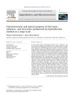
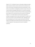
Genomic DNA from the Pleurotus isolate
KKM1,wasisolated by CTAB method of
DNA isolation. High molecular weight band
was visualized on the agarose gel (Fig. 1A).
Genomic DNA was used for amplification of
ITS 1-5.8S-ITS 2 rDNA sequence region
using primer pair ITS 1 and ITS 4.The PCR
fragments of 700 bp size fragment was
visualized as single band in agarose gel
stained with ethidium bromide (Fig. 1B).
ITS 1-5.8S-ITS2 PCR products were cleaned
up with PCR cleaned up kit to remove the
residual primers, polymerase and salts in the
PCR product, the cleaned up products were
sequenced at Eurofin genomic private Ltd,
India. The sequence was used for DNA
database search using BLAST program. The
BLAST search analysis of ITS 1-5.8S-ITS2
region of Pleurotus isolate KKM 1 matched
with that of Pleurotus djamor at 99 %
identified in the database.
Identification using the sequences of ITS
region is typically the most useful method and
also this method is applicable for molecular
systematics at the species levels(De Beeck et
al., 2014). For identification of specific
genera and species, the rDNA repeat unit,
consisting of the subunits 18S, 5.8S and 28S
rDNA interrupted by the internal transcribed
spacer (ITS) and the intergenic spacer (IGS)
is employed due to their specific sequences as
a target region. The advantage of ITS
sequencing is the identification of any
unknown fungal isolate using the database
containing the corresponding sequence of
previously identified fungal species or closely
related species (Schmidt et al., 2012). From
the new Pleurotus sp. isolate KKM1, ITS
sequence was PCR amplified and DNA
fragments of 700 bp was eluted and
sequenced. The results of ITS analysis
indicated that the sequence was similar to that
of ITS1-5.8S-ITS2 region of P. djamor. Thus,
this new Pleurotus isolate KKM1 was named
as Pleurotus djamor isolate KKM1.
Mycelial growth phenotype of Pleurotus
spp.
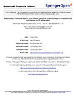
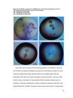
On PDA medium, mycelial growth of P.
djamor isolate KKM1 appeared loose, nonrhizomorphic type and thin mycelium
whereas, P. djamor var. MDU1 and P. florida
produced compact and rhizomorphic type of
mycelium because they produce more
mycelial branches from one place. P.eous var.
APK1 showed polar growth defects and that
formed constricted growth with cottony fluffy
mycelium (Fig. 2). Similarly, the mycelium of
P. djamor isolate KKM1 appeared loose, nonrhizomorphic type on the spawn substrate
whereas all other Pleurotus spp. tested
including P. eous produced compact and
rhizomorphic type of mycelium.
Among
the
Pleurotus
spp.
tested,
P.djamorvar MDU1 grew well and attained
the maximum growth of89.00mm on PDA
medium followed by P. florida and P. djamor
isolate KKM 1with the mycelial growth of 78
and 76 mm respectively. P. eous var. APK1
grew slowly on PDA medium (Table 1).
Mishra et al., (2015) reported similar type of
mycelial growth pattern in several Pleurotus
spp. The pattern of mycelial growth in P.
citrinopileatus was thick and fluffy, whereas,
that in P. fossulatus, P. flabellatus and P.
sapidus were slightly fluffy. P. djamor
showed the cottony growth. In the present
3577
Int.J.Curr.Microbiol.App.Sci (2018) 7(8): 3574-3582
also, all the tested Pleurotus spp. except P.
djamor isolate KKM1 produced thick cottony
mycelial growth.
3; Table 2).
Phenotypic characterization of basidiocarp
of Pleurotus spp.
Thickness of the pileus depends on the
amount of plectenchymatous tissue present in
the pileus. The thickness of the pileus was
measured near the junction point of pileus and
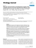
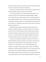
stipe. P. florida produced fruiting bodies with
maximum pileus (12.62mm). This was
followed by P. djamor var. MDU1 and P.eous
var.APK1 having the thickness of 8.62mm
and 7.87mm respectively. P. djamor isolate
KKM1 produced leathery type of pileus
having less plectenchymatous tissue and the
thickness of 5.62mm (Table 2).
The characterization of various Pleurotus
isolate had been attempted by many scientists
from time to time. In the present study,
phenotypic characters of basidiocarps of four
Pleurotus spp. were studied. Each Pleurotus
sp. has typical distinguishing characters for
easy identification. They are described below
Stipe length
Thickness of fruiting body
Among the four Pleurotus spp. tested,P.
djamor var. MDU1 produced fruiting bodies
with long stipe (54.50 mm), which was on par
with P. florida (52.25 mm). P. djamor isolate
KKM 1produced fruiting bodies with
rudimentary stipe or no stipe at all. P. eous
var.APK1 produced fruiting bodies having
small sized stipe (26.50mm) (Fig. 3; Table 2).
Margin of fruiting body
Diameter of the pileus
P. eous var. APK1 produced pink coloured
fruiting bodies. P. florida produced creamy
white coloured fruiting bodies. Whereas P.
djamor isolate KKM1 and P. djamor var.
MDU1 produced white coloured fruiting
bodies (Fig. 3; Table 2).
Pileus diameter was maximum inP. Djamor
isolate KKM1(119.25 mm) followed by
P.eous var. APK-1 (97.75mm) and P djamor
var. MDU1 (91.00mm). P. florida had small
sized pileus with the diameter 88.25 mm (Fig.
P. djamor isolate KKM1 has the pileus with
wavy margin whereas P. florida, P. djamor
var. MDU1. And P.eous var. APK1 have
pileus with smooth margins (Fig. 3; Table 2).
Colour of fruiting body
Table.1 Effect of temperature on the mycelial growth of Pleurotus spp. on PDA medium
Pleurotusspp.
P. eous var.APK-1
P. djamor var. MDU-1
P. florida
P. djamor isolate KKM 1
Mycelial growth (mm)*
4th day
23.00b
62.50a
60.50a
59.25a
7th day
34.75c
89.00a
78.25b
76.25b
* Mean of four replications
The treatment means are compared using Duncan Multiple Range Test (DMRT).
In a column, mean values followed by a common letter (s) are not significantly different (P = 0.05).
3578
Int.J.Curr.Microbiol.App.Sci (2018) 7(8): 3574-3582
Table.2 Phenotypic characterization of basidiocarp of P.djamorisolate KKM 1 and other Pleurotusspp
Pleurotus spp.
P.eous
var.APK-1
Diameter of the pileus(mm)*
Length of stripe(mm)*
appearance of Thickness Number Colour of
of
fruiting
Primordial Mature Harvesting Primordial Mature Harvesting pileus margin of pileus
2
(mm)*
gills/cm
body
stage
stage
stage
stage
stage
stage
b
C
b
a
c
c
c
b
11.25
57.75
97.75
12.35
21.75
26.50
Smooth
7.87
21.00
Pinkish
P.florida
5.25c
86.50a
88.25c
10.25ab
49.75a
52.25a
Smooth
12.62a
19.50c
P.djamor var.
MDU-1
16.50a
55.25b
91.00b
14.50a
43.00b
54.50a
Smooth
8.62b
11.50d
Creamy
white
White
P. djamor
isolate
KKM 1
15.25a
80.75b
119.25a
5.55b
11.50d
15.12b
Wavy and
broken
5.62d
23.50a
White
*Mean of four observations
Treatment means are compared using Duncan multiple range test(DMRT).
In a column, mean values followed by a common letter(s) are not significantly different(P=0.05)
3579
Int.J.Curr.Microbiol.App.Sci (2018) 7(8): 3574-3582
Figure.1 Molecular characterization new Pleurotus sp. isolate KKM1
A). Isolation of genomic DNA from Pleurotus sp. isolate KKM1. Lane 1: Lambda ladde
Lane 2: gDNA of Pleurotus isolate KKM
B) Amplification of ITS1-5.8S-ITS2 region from the genomic DNA of Pleurotus sp. isolate KKM1. Lane 1: 100 bp
ladder and 1 and Lane 2:ITS of Pleurotus isolate KKM 1
Figure.2 Mycelial growth pattern of Pleurotus spp. on PDA medium
3580
Int.J.Curr.Microbiol.App.Sci (2018) 7(8): 3574-3582
Figure.3 Phenotypic characterization of basidiocarps of Pleurotus spp
Number of gills
Generally P. djamor isolate KKM1 had thick
cluster of gills at the under surface of pileus
by recording 21 gills per cm2 area. This was
followed by P. eous var. APK1 which had
21gills /cm2. P. djamor var. MDU-1 had the
least number of gills (11.50 gills/cm2) (Table
2).
Mostly, the pileus of several Pleurotus spp. is
white in color. The pileus of P. djamor isolate
KKM1, P. djamor var. MDU1, and P.
floridais white in color. The other commercial
cultivar, P. eous var. APK1 has large pink
colored pileus and small stipe. However,
some studies reported that P. djamor species
are pink in color Mishra et al., (2015).
Diameter of the pileus usually ranges between
70 to 130 mm. In the present study also the
maximum pileus diameter was recorded in P.
djamor isolate KKM1 with 119 mm and
minimum in P. florida with the diameter of
88.25 mm. Among the various Pleurotus spp.,
tested, P. flabellatus showed the maximum
pileus length of 139 mm followed by P.
ostreatus (132mm) and minimum pileus
diameter was observed in P. sajorcaju with
54 mm (Mishra et al., 2015). Shukla and
Jaitly (2011) characterized the seven different
Pleurotus species based on the five different
morphological traits such as stipe length (cm),
cap diameter (cm), margin of fruit body,
peripheral architecture of the pileus, colour of
fruit body, total yield (kg), carbohydrate
content (%) and protein content (%). Out of
seven Pleurotus spp., five species were
named as follows P. citrinopileatus, P.
djamor, P. florida, H. ulmarius and P. sajorcaju. They also observed great diversity on
morphological characters among all the five
species of Pleurotus.
Usually, many Pleurotus spp. produce smooth
pileus with long stipe. But P. djamor isolate
KKM1 has large white colour pileus having
3581
Int.J.Curr.Microbiol.App.Sci (2018) 7(8): 3574-3582
wavy margin and has small or rudimentary
stipe. Thus, it is concluded that this new P.
djamor isolate KKM1 has typical phenotypic
features and characteristic mycelial pattern of
loose non-rhizomorphic mycelial characters
that distinguishes it from other Pleurotus spp.
and this P. djamor isolate KKM1 could be
used as new culture in mushroom germplasm
collection and mushroom cultivation.
References
Amin, S.M.R., Sarker, N.C., Khair, A. and
Alam, N.2007. Detection of novel
supplements of paddy straw substrate
on oyster mushroom cultivation.
Bangladesh Journal of Mushroom, 1:
33-37.
Avin, F. A., Bhassu, S. and Vikineswary, S.
2013. A simple and low-cost technique
of DNA extraction from edible
mushrooms examined by molecular
phylogenetics. Research on Crops,
14(3): 897-901.
Bruns, T. D., White, T. J. and Taylor, J. W.
1991. Fungal molecular systematics.
Annual Review of Ecology and
systematics, 22(1): 525-564.
Chang, S.T. 2012. Foreword. Mushroom
Science XVIII. Jinxia Zhang; Hexiang
Wang and Mingjie Chen (eds).
Proceedings the Internl. Society for
Mushroom Science.
De Beeck, M. O., Lievens, B., Busschaert, P.,
Declerck, S., Vangronsveld, J. and
Colpaert, J. V. 2014.Comparison and
validation of some ITS primer pairs
useful for fungal metabarcoding studies.
PLoS One, 9(6): e97629.
Hibbett, D. 1992. Ribosomal RNA and fungal
systematics. Transaction of Mycological
society of japan, 33: 533-556.
Lee, S.B., Milgroom, M.G. and Taylor, J.W.
1998. A rapid high yield mini-prep
method for isolation of total genomic
DNA from fungi. Fungal Genetics
Newsletter, 35: 23-24.
Kong,
W.S.
2004.Descriptions
of
commercially important Pleurotus
species. In: Mushroom world (Ed.).
Oyster mushroom cultivation. Part
II.Oyster mushrooms. Seoul: Heineart
Incorporation, pp.54-61. (Mushroom
growers’ handbook, 1).
Mishra, R., Shahid, M., Pandey, S., Pandey,
M.
and
Singh,
M.
2015.
Characterization of Pleurotus sp. of
mushroom based on phenotypic,
biochemical and yield parameter.
African Journal of Microbiology
Research, 9(13): 934-937.
Schmidt, O., Gaiser, O. and Dujesiefken, D.
2012. Molecular identification of decay
fungi in the wood of urban trees. Eur. J.
For. Res., 131: 885-891.
Shukla, S. and Jaitly, A. 2011. Morphological
and biochemical characterization of
different oyster mushroom (Pleurotus
spp.). Journal of Phytology, 3(8):18-20.
How to cite this article:
Praveen, T., R. Reihana, V.K. Parthiban and Ramamoorthy, V. 2018. Molecular
Characterization and Phenotypic Study of New Pleurotus djamor Isolate KKM 1.
Int.J.Curr.Microbiol.App.Sci. 7(08): 3574-3582. doi: />
3582