Microwave synthesis, crystal structure, antioxidant, and antimicrobial study of new 6-heptyl-5,6-dihydrobenzo[4,5]imidazo[1,2-c] quinazoline compound
Bạn đang xem bản rút gọn của tài liệu. Xem và tải ngay bản đầy đủ của tài liệu tại đây (3.91 MB, 15 trang )
(2018) 12:145
Hasan et al. Chemistry Central Journal
/>
RESEARCH ARTICLE
Chemistry Central Journal
Open Access
Microwave synthesis, crystal structure,
antioxidant, and antimicrobial study of new
6‑heptyl‑5,6‑dihydrobenzo[4,5]imidazo[1,2‑c]
quinazoline compound
Hiba Ali Hasan1,2,3* , Emilia Abdulmalek1,2*, Mohd Basyaruddin Abdul Rahman1,2, Khozirah Binti Shaari2,4,
Bohari Mohd. Yamin5 and Kim Wei Chan6
Abstract
Background: Although the development of antibiotic and antioxidant manufacturing, the problem of bacterial
resistance and food and/or cosmetics oxidation still needs more efforts to design new derivatives which can help
to minimize these troubles. Benzimidazo[1,2-c]quinazolines are nitrogen-rich heterocyclic compounds that possess
many pharmaceutical properties such as antimicrobial, anticonvulsant, immunoenhancer, and anticancer.
Results: A comparative study between two methods, (microwave-assisted and conventional heating approaches),
was performed to synthesise a new quinazoline derivative from 2-(2-aminophenyl)-1H-benzimidazole and octanal
to produce 6-heptyl-5,6-dihydrobenzo[4,5]imidazo[1,2-c]quinazoline (OCT). The compound was characterised using
FTIR, 1H and 13C NMR, DIMS, as well as X-ray crystallography. The most significant peak in the 13C NMR spectrum is
C-7 at 65.5 ppm which confirms the cyclisation process. Crystal structure analysis revealed that the molecule grows
in the monoclinic crystal system P21/n space group and stabilised by an intermolecular hydrogen bond between the
N1–H1A…N3 atoms. The crystal packing analysis showed that the molecule adopts zig-zag one dimensional chains.
Fluorescence study of OCT revealed that it produces blue light when expose to UV-light and its’ quantum yield equal
to 26%. Antioxidant activity, which included DPPH· and ABTS·+ assays was also performed and statistical analysis was
achieved via a paired T-test using Minitab 16 software with P < 0.05. Also, the antimicrobial assay against two Grampositive, two Gram-negative, and one fungus was screened for these derivatives.
Conclusions: Using microwave to synthesise OCT have drastically reduced reaction time, and increased yield. OCT
show good antioxidant activity in one of the tests and moderate antimicrobial activity.
Keywords: Single crystal, Antioxidant, ABTS, DPPH, Dihydrobenzo[4,5]imidazo[1,2-c]quinazoline
Background
Nitrogen-comprising heterocyclic compounds have
attracted the interest and attention of many researchers
within the medicinal chemistry field over recent years.
One of which is the benzimidazo[1,2-c]quinazoline
*Correspondence: ;
;
1
Integrated Chemical BioPhysics Research, Universiti Putra Malaysia, 43400
UPM Serdang, Selangor, Malaysia3 Department of Pharmacognosy and
Medicinal Plants, College of Pharmacy, Mustansiriyah University, Baghdad, Iraq
Full list of author information is available at the end of the article
nucleus, which is formed from the fusion of benzimidazole to quinazoline bioactive systems (Fig. 1). Literatures
revealed that benzimidazoquinazolines possess many
distinctive therapeutic properties such as antitumor, anticonvulsant, antioxidant, antimicrobial, antiviral, and as
potent imunosuppressors [1–5].
Free radicals and various reactive oxygen or nitrogen species are produced either exogenously from pollution, radiation and food, or endogenously inside the
human body from metabolic pathways, leading to oxidative stress. Oxidative stress is the primary cause of many
© The Author(s) 2018. This article is distributed under the terms of the Creative Commons Attribution 4.0 International License
(http://creativecommons.org/licenses/by/4.0/), which permits unrestricted use, distribution, and reproduction in any medium,
provided you give appropriate credit to the original author(s) and the source, provide a link to the Creative Commons license,
and indicate if changes were made. The Creative Commons Public Domain Dedication waiver (http://creativecommons.org/
publicdomain/zero/1.0/) applies to the data made available in this article, unless otherwise stated.
Hasan et al. Chemistry Central Journal
(2018) 12:145
disorders including atherosclerosis, cancer, diabetes, and
ageing [6]. Compounds which can scavenge free radicals
can, therefore, contribute towards the protection and
prevention of these illnesses [7]. Hence, the need for new
antioxidants is increasing to solve these problems.
Furthermore, bacterial infections have become a serious threat after many decades of treating the first patient
with antibiotic. That is because of the fast increasing in
bacterial resistance which become prevalent all over the
world. Bacterial resistance to antibiotic is a result of overuse and misuse of these drugs [8]. Therefore, there is continuous need for exploration new medication.
Attempting to solve the said problems, chemists and
pharmacists have tried for years to synthesis new nitrogen-comprising compounds which are known for their
biological activities. Nevertheless, the problem of using
organic solvent in chemical routes presents a significant
threat to the environment as it can cause pollution during processing handling, and storage. As a result, many
researchers have focused on developing alternative methods and procedures that not only facilitates organic synthesis but also reduces the amounts of solvents. One of
these methods uses microwave irradiation to perform
organic reactions [9].
Microwave technique to heat organic reactions have
been widely discussed and debated within the organic
and medicinal chemist community since the publication of the first scientific article in 1986 [10]. In recent
years, this fast-moving protocol has been used in many
laboratories to synthesise organic materials within a very
brief time, resulting in considerable yield, and enhancing
pure products. This technique includes direct interaction
between the microwave radiation and molecules in the
reaction system which dramatically reduces any undesired side-products and increases the yield of the target
product [10].
Since
6-heptyl-5,6-dihydrobenzo[4,5]imidazo[1,2-c]
quinazoline (OCT) is combining skeleton of bioactive
quinazoline and benzimidazole nucleolus, it is expected
Fig. 1 Benzimidazoquinazoline scaffold
Page 2 of 15
to have some pharmaceutical activities. Also, the literature survey resulted to only one study that have focused
on antioxidant activities of benzimidazoquinazoline
compounds [11]. Therefore, we report herein the crystal
structure, spectroscopic characterisation, antioxidant,
and antimicrobial activities of new 6-heptyl-5,6-dihydrobenzo[4,5]imidazo[1,2-c]quinazoline resulting from
two different synthetic methods.
Experimental section
Materials and experimental conditions
The analytical grade chemicals used for this project
were commercially available from several suppliers and
applied without any additional purification. The glacial
acetic acid was supplied from J. T. Baker/USA. The analytical grade methanol and Mueller–Hinton agar were
procured from Merck/Germany. The DMSO-d6 for
nuclear magnetic resonance was obtained from Merck/
Switzerland. The 2-(2-aminophenyl)-1H-benzimidazole, octanal, potassium persulfate, 2,2′-azino-bis(3ethylbenzothiazoline-6-sulfonic acid)diammonium salt
(ABTS),
(±)-6-hydroxy-2,5,7,8-tetramethylchromane2-carboxylic acid (Trolox), and 2,2-diphenyl-1-picrylhydrazyl (DPPH) were all supplied from Sigma-Aldrich.
Three-angstrom molecular sieves were supplied by Acros
Organics/USA and used to dry the solvents.
A 10-mL vial capacity single-mode CEM microwave
(USA) along with Synergy software were used to achieve
the condensation reaction. An IR Tracer-100 (Shimadzu/Japan) was activated to determine the functional
groups applying FTIR analysis and GCMS QP5050A
(Shimadzu/Japan) recorded the mass spectrum (DIMS). JEOL JNM ECA 400 was executed at ambient
room temperature to analyse the 1H-NMR (400 MHz)
and 13C-NMR (100 MHz) spectra. A Barnstead Electrothermal/UK instrument was used to measure the melting point, and a Thermo Scientific ELISA reader/UK
was used to measure the absorbance of the radical-OCT
mixture. A UV–Visible spectrophotometer (UV-1700,
Shimadzu/Japan) was operated at ambient room temperature to measure A
BTS·+ absorbance. An Autopol
VI, Automatic Polarimeter manufactured by Rudolph
Research Analytical/Hackettstown, NJ, USA was used
to measure the optical rotation, and a CHNS instrument
(LECO TruSpec Micro CHNS/US) was used to analyse
the carbon, hydrogen, and nitrogen percentage contents
in the compound. UV-1650 PC (UV–Visible spectrophotometer, SHIMADZU/Japan) was run to measure
the UV–Vis absorbance spectra of the studied compounds. Perkin Elmer LS 55 Fluorescence Spectrometer/UK was used to measure emission spectra. Lastly,
thin layer chromatography was carried out using silica
gel aluminium plates 60 F254 (Merck/Germany).
Hasan et al. Chemistry Central Journal
(2018) 12:145
Synthesis and characterisation
Microwave synthesis
The microwave-assisted synthesis was conducted
according to Negi et al. [12] with some modifications.
In a 10-mL volume microwave vial, octanaldehyde
(1.2 mmol, 186 µL) was dissolved in methanol (1 mL)
and added dropwise to 2-(2-aminophenyl)-1H-benzimidazole (1 mmol, 0.21 g) which was dissolved in 5 mL
methanol, followed by addition of two drops of glacial
acetic acid. The solution was irradiated in a singlemode benchtop microwave for 5 min at 102 °C, and the
reaction was monitored using Synergy software. The
TLC was performed to check the progress of the reaction and completion. After 5 min, the vial was cooled to
room temperature, dried in a vacuum oven, and washed
with hexane to provide the final pure product. The
crystals were obtained by slow evaporation of toluene
to produce off-white crystalline solid with a premium
yield of 91% (0.29 g).
Conventional heating synthesis
The conventional reflux method was performed according to Kapoor et al. [13] with slight modifications. In a
50-mL round bottom flask, octanaldehyde (1.2 mmol,
186 µL) dissolved in methanol (1 mL) was added dropwise to 2-(2-aminophenyl)-1H-benzimidazole (1 mmol,
0.21) which was dissolved in 15 mL hot methanol, followed by addition of two drops of glacial acetic acid.
The prepared mixture was refluxed at 95 °C for around
80 min over an oil bath. The reaction progress was
monitored every 15 min to check the reaction progression. Next, it was cooled to room temperature after
completion as evident by TLC. The target crystals were
obtained after vacuum drying, and vigorously washing
the crude product with hexane to produce the precipitate which was recrystallised from toluene to furnish
off-white, shiny crystals of 77% yield (0.24 g).
Characterization of 6‑heptyl‑5,6‑dihydrobenzo[4,5]
imidazo[1,2‑c]quinazoline (OCT)
White crystals. M.p.: 116–118 °C; R
f: 0.50 in hexane: ethyl acetate (2:1) solvent system. [α]20
D = + 347.3
(c = 0.01, DMSO). FTIR UATR ( cm−1) ʋmax: 3202 (N–
H stretching), 2928 (–C–H sp3 and =C–H sp2 stretching), 1614 (C=N stretching), 1520 (C=C aromatic),
1461 (N–H bending), 1261 (C–N stretching), 736 (C–H
bending out of plane for aromatic). 1H NMR (400 MHz,
DMSO-d6) δ ppm 0.78 (t, J = 7.3 Hz, 3H, CH3), 1.06–
1.22 (m, 8H, H-17, 18, 19, 20), 1.23–1.32 (m, 2H,
H-16), 1.61–1.72 (m, 1H, HA), 1.80 (dt, J = 13.8, 7.3 Hz,
1H, HB), 6.03–6.09 (m, 1H, H-7), 6.78 (ddd, J = 1.0,
Page 3 of 15
7.9 Hz, 1H, H-3), 6.88 (d, J = 7.8 Hz, 1H, H-5), 7.15
(s, 1H, N1-H), 7.17–7.27 (m, 3H, H-4, 10, 11), 7.55–
7.60 (m, 1H, H-12), 7.60–7.65 (m, 1H, H-9), 7.87 (dd,
J = 1.4, 7.9 Hz, 1H, H-2). 13C NMR (100 MHz, DMSO):
δc, ppm, 13.8 (CH3), 21.9 (C-19, 20), 23.7 (C-18), 28.5
(C-17), 31.0 (C-16), 35.6 (CHA,B), 65.5 (C-7), 110.0
(C-12), 112.0 (C-1), 114.9 (C-5), 117.7 (C-3), 118.5
(C-9), 121.8 (C-11), 121.9 (C-10), 124.5 (C-2), 131.5
(C-4), 132.6 (C-8), 143.2 (C-13), 143.7 (C-6), 146.5 (C14). MS: DIMS m/z: 319 (M+, 7%), 246 ([C16H12N3]+,
8), 233 ([C15H12N3]+, 27), 220 ([C14H10N3]+, 100), 194
([C13H10N2]+, 5), 110 ([C6H10N2]+, 6), 92 ([C6H6N]+, 6).
Anal. Calcd. for C21H25N3: C, 78.96; H, 7.89; N, 13.15%.
Found: C, 78.54; H, 7.92; N, 13.19%. UV–Vis in DMSO
λmax, nm (ɛ, L/mol/cm): 360 (ɛ, 0.191 × 10⁴), 304 (ɛ,
0.319 × 10⁴), 293 (ɛ, 0.228 × 10⁴), 267 (ɛ, 0.236 × 10⁴),
(Figs. 2, 3, 4, 5, 6).
Structure determination by X‑ray crystallography analysis
Single crystal X-ray determinations were conducted at
Center for Research and Instrumentation (CRIM), Universiti Kebangsaan Malaysia (UKM). A suitable crystal with appropriate size was mounted on a gonio head.
Reflection data was collected at 25 °C using (graphitemonochromated Mo Kα radiation, λ = 0.71073 Å) with
a photon detector distance of 4 cm and a swing angle
of − 30° maximum. The data collected were reduced
using the program SAINT [14] and an empirical absorption correction was carried out using SADABS [15]. The
structure was solved by direct methods and refined by
using the full- matrix least-squares method using the
SHELXTL [16] software package. All non-H atoms were
anisotropically refined. The hydrogen atoms were located
by difference syntheses and refined isotropically. The
molecular graphics were created using SHELXTL and
MERCURY softwares. PLATON program was used for
molecular structure calculation [17]. Atomic scattering
factors and anomalous dispersion corrections were taken
from the international table for X-ray crystallography.
Optical activity
Optical rotation of the studied compound was measured
for a 0.01 g/100 mL sample concentration dissolved in
DMSO at 20 °C, with a 589-nm wavelength. The sample was injected into a 1 dm long polarimeter cell after
removing all air bubbles and blanking the instrument.
Specific rotation calculated by applying Eq. (1) for the
average of five times reading:
[α]T =
α
l∗c
(1)
Hasan et al. Chemistry Central Journal
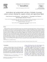
Fig. 2 1H-NMR spectrum of OCT
Fig. 3 13C-NMR of OCT
(2018) 12:145
Page 4 of 15
Hasan et al. Chemistry Central Journal
(2018) 12:145
Page 5 of 15
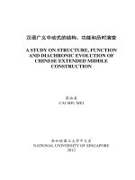
Fig. 4 FTIR spectrum of OCT
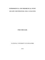
Fig. 5 DIMS spectrum of OCT with the main fragments
where, α = measured optical rotation. T = temperature at measurement process. λ = light wavelength in
nm, 589 nm using a D line of sodium. l = polarimetry
cell length in decimetre. c = sample concentration in
g/mL.
Elemental analysis
Carbon, hydrogen, and nitrogen percentage analyses
were performed to determine the actual ratios of these
elements in the OCT sample, comparing them with the
calculated ratios.
Hasan et al. Chemistry Central Journal
(2018) 12:145
Page 6 of 15
Fig. 6 UV–Vis spectrum of OCT
Fluorescent study
Electronic spectral analysis
UV–Vis absorbance of the studied compounds were
measured at room temperature at the concentration
of 1 × 10⁻⁴ M. The samples were dissolved in DMSO at
25 °C and measured at 250–500 nm wavelength. Each
spectrum was measured after blanking the instrument
with DMSO solvent, and loading the sample to 3 cm3
quartz cuvette that has path length of 1 cm. Molar
absorptivity calculated by applying Eq. (2):
ε = A/lc
(2)
where, ɛ = The molar absorptivity, L/mol/cm. A = the
amount of light absorbed by the sample for a given wavelength, without units. l = the distance that the light travels through the solution, 1 cm. c = the concentration of
the absorbing species per unit volume, mole/L.
Fluorescence emission study
Fluorescence study was measured at room temperature
for 1 × 10⁻⁴ M for both samples in DMSO and quinine
sulfate in 0.1 M solution of H2SO4 as standard. The quantum yield of all synthesized compounds was obtained
from the following method: First, UV–vis absorption
spectra for the compounds and quinine sulfate were
measured at RT. Then, the emission fluorescence spectra
were measured at the low energy excitation wave length
which was 360 nm for OCT compound and at 350 for
both AMINE and quinine sulfate. Finally, quantum yield
was calculated by applying Eq. (3)
ΦYsam = ΦYref
Isam Aref n2sam
Iref Asam n2ref
(3)
where, Subscripts indices “sam” and “ref” refer to sample
and reference, respectively. ΦYref = 0.54 when excited at
350 nm. I = Integrated area of emission peak at the excitation wavelength. A = UV–vis abortion correction factor
which is = 1 − 10−A . n = refractive index for both water
and DMSO.
Antioxidant activities
DPPH· scavenging activity of OCT
The DPPH· scavenging activity of OCT and AMINE was
conducted according to Chan et al. [18]. In a 96 well
microplate, 50 µL of the diluted OCT sample in DMSO
was reacted with 195 μL of 0.2 mM D
PPH· (methanolic
solution) and kept in a dark ambient room where the
mixture was kept for 1 h at 25 °C. Next, using the microplate ELISA reader and at 540 nm, the absorbance was
read. The analysis was conducted in triplicate, and the
antioxidant activity of both compounds was expressed in
mg Trolox equivalent/g sample.
Hasan et al. Chemistry Central Journal
(2018) 12:145
Page 7 of 15
ABTS·+ scavenging activity of OCT
·+
The ABTS scavenging activity of both samples was
conducted according to the previous study performed
by Chan et al. [19] with some additional modifications.
Briefly, ABTS·+ was generated by adding 10 mL of 7 mM
ABTS to 10 mL of 2.45 mM potassium persulfate and
kept in a dark place at room temperature for 24 h. Then,
the ABTS·+ solution was diluted to the absorbance of
1.40 ± 0.05 at 734 nm with the UV–vis spectrophotometer. Subsequently, 180 μL of ABTS·+ solution was added
to 20 μL of the OCT sample in a ninety-six well microplate. After 1 h of incubation at room temperature, the
absorbance was recorded at 734 nm using a microplate
ELISA reader. The analysis was conducted in triplicate,
and the scavenging activity of the OCT compound was
expressed in mg Trolox equivalent/g sample.
Statistical analysis
Antioxidant values were expressed as mean ± SD of three
replicates for both samples. Statistical analysis was performed by paired T-test using Minitab 16 software with
P < 0.05.
Antimicrobial assay
Microbial strain
All the microorganisms used in this study were human
clinical strains, provided by the Microbial Culture Collection Unit (UNiCC), Institute of Bioscience, University
Putra Malaysia. The microbes strain includes two Grampositive: Staphylococcus aureus ATCC 43300, Bacillus
sublitis UPMC 1175; two Gram-negative: Pseudomonas
aeruginosa ATCC 15542, Salmonella choleraesuis ATCC
10708; and one fungus: Aspergillus brasilliensis ATCC
16404.
Antimicrobial activity
The antimicrobial activities of the studied compounds
were evaluated using an agar-well diffusion assay [20]
with some modifications. Into each of the sterile Petri
dishes (Ø 90 mm), 20 mL of molten agar at 45 °C was
poured. After the plates were aseptically dried, the agar
surface of each plate was streaked using a sterilised cotton swab with the specified microbial strain. Then, with
a 5 mm Cork borer diameter, the wells were punctured
into the agar. The synthesised compounds were then
dissolved in DMSO to produce 100 mg/mL final concentration. Next, 20 μL of the studied samples were loaded
into each well, and the plates were incubated invertedly
between 30 and 37 °C for 18 and 24 h. or until proper
growth had occurred. Once the incubation was completed, the circular inhibition zones were measured using
callipers, including the well diameter. The DMSO was
used as a negative control while the tetracycline or nystatin was used as a positive control. The experiments were
performed in triplicate.
Results and discussion
Synthesis
Classical heating, together with microwave heating techniques were undertaken to synthesise the titled crystal (OCT) via the condensation of octanaldehyde with
AMINE to compare the reaction time, % yield, purity
of the product, and the efficiency of both methods. The
results revealed that a microwave-assisted reaction
not only produces pure crystals in higher yield but also
within a brief reaction time, as summarised in Table 1.
Furthermore, the reaction time drastically decreased by
93% when the microwave was applied, and the product
percentage yield moderately increased by 14% to produce
a very pure product without requiring further purification. From an environmental perspective, this technique
is more benign concerning the environment as compared
to normal reflux, since the total amount of used methanol was only one-third of the amount used in the conventional heating method.
As illustrated in Fig. 7, the reaction begins by the activation of the carbonyl group of an aldehyde via a protonation step. This is followed by the nucleophilic amine
attacking the protonated carbonyl carbon to form the
intermediate 3 which was then protonated under acidic
reaction conditions to produce carbinolamine intermediate 4. Notably, this step is considered as a rate-determining step. Meanwhile, carbinolamine is in equilibrium
with iminium cation 5 formed by losing a water molecule. Presumably, the imine carbon is quite electrophilic
and proceeds to react with the basic secondary amine of
the benzimidazole ring to form a new ring following the
loss of a proton. Interestingly, the cyclised compound was
obtained instead of the expected Schiff base 6 under the
same reaction conditions which means that the position
Table 1 Reaction time and % yield of OCT under conventional reflux and microwave irradiation, respectively
OCT
Reaction time, min.
% yield
Conventional
MW
% decreased
Conventional
MW
% increase
75
5
93
77
91
14
Hasan et al. Chemistry Central Journal
(2018) 12:145
Page 8 of 15
Fig. 7 Plausible mechanism for 6-heptyl-5,6-dihydrobenzo[4,5]imidazo[1,2-c]quinazoline formation
of an ortho-amino group of the parent amine is the main
reason behind the cyclisation process and benzimidazoquinazoline creation.
Seemingly, Schiff base could initially be forming but
reacts to create benzimidazoquinazoline, which is applicable for all aldehydes. In the future, the R group in
amine can be changed to decrease its’ reactivity to obtain
isolate Schiff base compounds.
Characterisation
The structure of the OCT crystal was confirmed via
FTIR, 1H and 13C NMR, and DIMS and it immediately
became apparent by observing the 1H, and 13C NMR
spectra (Figs. 2 and 3) that there was no Schiff base
formed, but, a new diazine ring had been formed. Furthermore, there is a new aliphatic multiplet at 6.03–
6.09 ppm which belongs to H-7 of the newly formed ring,
and the N1–H proton appears as a singlet at 7.15 ppm.
This, therefore, proved that the cyclisation process rather
than Schiff base formation occurred. Moreover, there is
no singlet peak around 8.5 to 9 ppm which would belong
to the imine proton (–N=C–H). The 1H NMR also displayed four different peaks in the aliphatic area belonging
to protons CH3, H-17, 18, 19 and 20. The other characteristic peaks are diastereotopic protons HA and HB
which rose up at different chemical shifts as a multiplet
at 1.61–1.72 and doublet of the triplet at 1.80 ppm for
HA and HB respectively. In the 13 C NMR spectrum, the
most important peak is C-7 at 65.5 at the aliphatic area
which confirms the cyclisation process and the formation
of OCT. Otherwise, there will be a peak around 165 to
170 ppm belonging to carbon (C=N) of the Schiff base.
Figure 3 illustrates the remaining peaks.
The FTIR spectrum of OCT exhibited two medium
absorption bands at the 3202 and 2928 cm−1 regions corresponding to N–H and –C–H sp2 stretching, respectively. Also, the band at 2859 and the medium sharp
band at 1614 cm−1 corresponds to –C–H s p3 and C=N
stretching absorptions, respectively. The C=C aromatic absorption peaks resulted in a medium peak at
1520 cm−1, and at 1461 cm −1 the N–H bending band
is observed. Also, the C–N stretching band appears at
1261 cm −1 and C–H aromatic out of plane bending at
736 cm −1. Figure 4 summarises all distinctive peaks for
the mentioned derivative.
Hasan et al. Chemistry Central Journal
(2018) 12:145
The molecular ion peak was determined for OCT and
is equivalent to its molecular weight (C21H25N3 = 319.
44). The peak at 220 m/z with 100% intensity is considered as the base peak belonging to the [C14H10N3]+
fragment. The remainder of the fragments with their
molecular weights is illustrated in Fig. 5.
As shown in the mass spectrum of the compound
6-heptyl-5,6-dihydrobenzo[4,5]imidazo[1,2-c]quinazoline in Fig. 5, the molecular ion peak at 319 m/z (7%),
which is precisely equal to the calculated molecular
weight and the other fragmentation peaks, are also displayed. This molecular ion also underwent α-cleavage to
eliminate 6-heptyl moiety to produce a fragment at m/z
220 with 100% abundance as a base peak. Further, under
the same type of cleavage, a radical ion at m/z 110 formed
by cutting off C15H15N moiety. However, under inductive
cleavage (i-cleavage), a radical ion at m/z 92 was formed
via cutting C
15H19N2 off, (Fig. 8). Same type of cleavage
also occurred to produce a fragment at m/z 194 with 4%
abundance. Also, both 246 and 233 fragments resulted
from the carbon–carbon bond breaking the straight
hydrocarbon chain.
Crystallography study of 6‑heptyl‑5,6‑dihydrobenzo[4,5]
imidazo[1,2‑c]quinazoline (OCT)
6-Hepty5,6-dihydrobenzo[4,5]imidazo[1,2-c]quinazoline crystalized in monoclinic system with space group
Fig. 8 Fragmentation pattern of OCT
Page 9 of 15
P21/n, a = 9.37 (4), b = 17.14 (5), c = 11.27 (4) Å, α = 90°,
β = 101.5 (2)°, ɤ = 90°, z = 4 and volume = 1773 (11) Å3.
The crystal system and refinement parameters are given
in Table 2. The isotopic displacement parameters and
structure parameters are given in Additional file 1.
The molecule is discrete, having only one molecule in
the asymmetric unit. The heptyl group is attached to the
diazine ring at C7 atom. The molecular structure with the
numbering scheme is illustrated in Fig. 9. Notably, the
relative configuration at the chiral centre C7 is R which
means it is an enantiopure compound.
The benzimidazole ring N2/N3/(C8–C14) is planar
with a maximum deviation of 0.012 (5) Å and 0.012 (7) Å
for C8 and C11, respectively from the least square plane.
The benzene ring (C1–C6) is planar with a maximum
deviation of 0.007 (5) Å for C1 from the least square
plane. The dihedral angle between the benzimidazole
plane and the benzene ring is 7.26 (17)°.
The diazine ring, N1/N2/C1/C6/C7/C14 adopts halfchair conformation with a maximum deviation of 0.209
(5) Å for atom C7 from the least square plane (Fig. 10).
The N3-C14 is 1.318 (7) Å indicating a double bond
character while the other bond lengths and angles
(Table 3) are in normal ranges and are comparable to
those in its analogues of 6-butyl-5,6-dihydrobenzo-[4, 5]
imidazo[1,2-c]quinazoline [21].
Hasan et al. Chemistry Central Journal
(2018) 12:145
Page 10 of 15
Table
2
Refinement of structure and crystal data
for
6-heptyl-5,6-dihydrobenzo[4,5]imidazo[1,2-c]
quinazoline
In the crystal structure, the molecules are linked by
N1–H1A…N3 intermolecular hydrogen bonds (symmetry code as in Table 4) to form zig-zag one dimensional
chains (Fig. 11).
Identification code
OCT
Empirical formula
C21H25N3
Formula weight
319.20
Fluorescent study
Temperature
293 (2) K
Wave length
0.71076 Å
Crystal system
Monoclinic
Space group
P21/n
The handling and experimental work with this compound
unexpectedly disclosed that this compound fluoresces
and emits a bright blue colour when exposed to ultraviolet light either from the sun or a UV-lamp. Therefore, it
is meaningful if not necessary, to study the fluorescent
properties of this compound as a part of the characterisation process which hopefully will expose new potential
applications.
Unit cell dimensions
a = 9.37 (4) Å α = 90°
b = 17.14 (5) Å β = 101.5 (2)°
c = 11.27 (4) Å ɣ = 90°
Volume
1773 (11) Å3
Z
4
Density (calculated)
1.196 Mg/m3
Absorption coefficient
0.071 mm−1
F(000)
688
Electronic spectral data
3
Crystal size
0.500 × 0.430 × 0.270 mm
Theta range for data collection
3.009 to 25.249°
Index ranges
− 11 ≤ h ≤ 11, − 20 ≤ k ≤ 20,
− 13 ≤ l ≤ 13
Reflections collected
16,139
Independent reflections
3186 [R(int) = 0.1192]
Completeness to θ = 25.243°
99.0%
Refinement method
Full-matrix least-squares on F2
Data/restraints/parameters
3186/1/223
Goodness-of-fit on F2
1.046
Final R indices [I > 2 sigma (I)]
R1 = 0.1062, wR2 = 0.2552
The UV–Vis spectrum of the OCT compound was measured in DMSO solvent at 25 °C and the result exhibited
various absorption bands at 267 (ɛ, 0.236 × 10⁴), 293 (ɛ,
0.228 × 10⁴), 304 (ɛ, 0.319 × 10⁴), and 360 (ɛ, 0.191 × 10⁴)
nm which are ascribed to π–π* and n–π* intramolecular
transitions between electronic energy levels. When the
OCT compound is exposed to ultraviolet radiation, the
Table 3 Selected bond lengths (Å) and angles (°)
Bonds
lengths (Å)
and angles (°)
R indices (all data)
R1 = 0.1858, wR2 = 0.3193
Extinction coefficient
0.015 (4)
N1–C7
1.439 (7)
Largest diff. peak and hole
0.330 and − 0.297 e Å−3
N2–C7
1.465 (8)
N2–C14
1.348 (8)
C1–C14
1.455 (8)
C6–C1
1.421 (8)
N1–C6
1.375 (7)
C6–N1–C7
122.2 (5)
N1–C7–N2
107.2 (5)
N1–C7–C15
114.2 (5)
N2–C7–C15
110.9 (5)
CCDC reference no.
1830213
Fig. 9 Molecular structure of OCT compound
Fig. 10 The conformation of diazine ring of OCT
Hasan et al. Chemistry Central Journal
Table
4
Hydrogen
compound
bonds
(2018) 12:145
parameters
(Å)
Page 11 of 15
of OCT
Donor—H…acceptor D–H (Å)
H…A (Å)
D…A (Å)
N1–H1A…N3i
2.08 (6)
3.050 (14) 173 (5)
0.97 (6)
D—H…A (°)
i = 1/2 + x, 1/2 − y, 1/2 + z
electrons are excited and transfer from the highest occupied molecular orbital (HOMO) to the lowest unoccupied molecular orbital (LUMO). The molar absorptivity
εmax values (molar extinction coefficient) of this derivative have medium intensities for π → π* transitions which
are higher than that of n → π* transition which refers to
the higher probability of π electron transitions rather
than non-bonding electrons transfer (Fig. 6).
Emission spectral data
Luminescence is the process that describes the electronic
transfer from the excited electronic state to the lower
unexcited state. When the emission occurs due to light
excitation (usually the UV part of the electromagnetic
spectrum), it is called photoluminescence (PL). Notably, fluorescence is one of the members of the luminescence family, and presently, luminescence spectroscopy
has wide-ranging applications [22]. Fluorescence spectra of OCT and its’ starting AMINE were measured for
a very diluted dimethyl sulfoxide (DMSO) solutions at
room temperature. These two solutions are colourless
under ambient light but display a very intense blue and
purplish-blue colour for OCT and AMINE, respectively
under long wave UV light. Also, the PL-spectrum for
those compounds gave a wide band in the visible region
at 425 and 414 nm for both OCT and AMINE, respectively (Figs. 12 and 13). Table 5 depicts all absorption
and emission maxima, stock shifts and quantum yield of
these derivatives. Accordingly, the quantum yield (QY) is
the ratio of a number of emitted photons to the number
of absorbed protons, and its’ evaluation is considered as
a key step to characterise fluorescent compounds. The
quantum yields for OCT and AMINE were found to be
26% and 13%, respectively compared to 54% of quinine
sulphate as the standard. This means that OCT emits
around double the amount of light as compared to its’
parent compound.
Antioxidant activities
The antioxidant activity of both OCT and the starting material AMINE was next evaluated spectrophotometrically by measuring the ability of both compounds
to reduce the reagent radicals, which will be confirmed
by decreasing the absorbance of the radical-sample mixture. This can be performed by employing two antioxidant assays, i.e., D
PPH· and A
BTS·+ scavenging activities.
The results are illustrated in Fig. 14. Next, the antiradical activity of the OCT and AMINE compounds were
evaluated by the reaction of respective crystals with
Fig. 11 Molecular packing of OCT compound viewed down a-axis. All hydrogen atoms except hydrogen bonded are omitted for clarity
Hasan et al. Chemistry Central Journal
(2018) 12:145
Page 12 of 15
Fig. 12 Fluorescence spectrum of OCT compound
two types of the mentioned stable radicals. The ABTS·+
scavenging activities were found to be 658.34 ± 41.01
and 48.61 ± 3.58 mg Trolox eq./g (sample) for the OCT
and AMINE samples, respectively, (P < 0.05). The D
PPH·
scavenging activity of the same samples were 22.27 ± 1.34
and 50.90 ± 1.44 mg Trolox eq./g (sample) for the OCT
Fig. 13 Fluorescence spectrum of AMINE compound
and AMINE, respectively, (P
<
0.05). Therefore, from
the results, the ABTS·+ scavenging activity of the OCT
compound was surprisingly found to be around 30-fold
higher than that for D
PPH·. Indeed, the absence of
·
DPPH scavenging activity compared to ABTS·+ has also
been highlighted in many studies [18, 19, 23, 24] and is
Hasan et al. Chemistry Central Journal
(2018) 12:145
Page 13 of 15
Table 5 Absorption and emission maxima and quantum
yields ( Φ ) for OCT and AMINE compounds
Compounds
λex (nm) λem (nm) Stock
shifts
(nm)
Quantum Quantum
yield ( Φ) yield (%)
OCT
360
425
65
0.259
26
AMINE
350
414
64
0.129
13
Quinine sulfate 350
458
108
0.54
54
attributed to the stearic accessibility of the DPPH· radical
which is considered as a major hindrance to the chemical reaction. Furthermore, it was found that many small
molecules which have better access to the radical site of
DPPH·, have enhanced scavenging activities as compared
to the bulky or rigid molecules which not only slowly
react but are also inert in this assay [25]. Notwithstanding, this is also explained in the high reactivity of the
starting material AMINE compared to the OCT derivative in this type of test.
Antimicrobial assay
The preliminary information to test the in vitro antimicrobial activity of the synthesised OCT compound and
its initiating material AMINE against five different pathogenic micro-organisms was achieved by applying the
agar-well diffusion method. The results confirmed that
the range of the inhibition zone mainly depends on the
strain of bacteria and fungi. Also, the AMINE compound
showed no inhibitory activity for all microorganisms
used in this study. In contrast, OCT displayed a moderate
inhibitory effect on the selected Gram-positive bacteria
with negligible activity against the selected Gram-negative bacteria and Aspergillus brasiliensis fungus in this
study, as compared to tetracycline and nystatin as a
standard antibiotic and anti-fungus, respectively, (refer
Table 6). Therefore, it is evident from these results that
OCT is a more potent compound compared to its parent
because of the bioactive characteristic of the benzimidazoquinazoline compounds [6, 7, 26–28]. Furthermore,
the inhibitory activity of OCT was only active against
the Gram-positive kind of bacteria, due to the highly
resistant Gram-negative bacteria compared to the Grampositive bacteria. Since the external membrane of the
Gram-negative type is rendered with a highly hydrophilic
surface, this, therefore, makes it more resistant to antibiotics as compared to Gram-positive bacteria. Also, the
negative charge on the Gram-positive wall surface may
decrease its resistance to antibacterial derivatives [20].
For the antifungal activity, there is no biological activity
for both the studied compounds.
Fig. 14 DPPH· and ABTS·+ scavenging activity of OCT and AMINE. Results are displayed as mean ± SD (n = 3)
Hasan et al. Chemistry Central Journal
(2018) 12:145
Page 14 of 15
Table 6 Antimicrobial activities of studied compounds
Seq.
Compounds
concentration in 100
(mg/mL)
Inhibition zone diameter in (mm)a
Target microbes
Gram positive
Staphylococcus
aureus ATCC 43300
Gram negative
Bacillus subtilis
UPMC 1175
Pseudomonas
aeruginosa ATCC
15542
Fungus
Salmonella
choleraesuis ATCC
10708
Aspergillus
brasiliensis ATCC
16404
1
OCT
9
7
–
–
–
2
AMINE
–
–
–
–
–
3
+ve controlb
DMSO (−ve control)
28.3
23.6
18.3
25
26
–
–
–
–
–
Values are given as mean of triplicate experiment
–, no inhibition was observed
a
Diameter of inhibition zones including diameter of 5 mm well
b
Tetracycline or nystatin in case of antibacterial and antifungal respectively
Conclusion
The 6-heptyl-5,6-dihydrobenzo[4,5]imidazo[1,2-c] quinazoline was successfully synthesised at an excellent yield
of 91% using the microwave approach. The FTIR, NMR,
and DIMS along with single crystal analysis of titled
benzimidazoquinazoline (OCT) confirmed the building structure of this new crystal. The fluorescence study
of this compound further disclosed that it fluoresces
with double the amount of light compared to the starting AMINE compound. Hence, it could be a potential
candidate for further cell imaging applications or single
cell level studies for physiological applications. From
the antioxidant results, the A
BTS·+ test revealed higher
scavenging activity as compared to the D
PPH· test for the
same compound. Furthermore, the antimicrobial study of
these derivatives demonstrated that OCT is a more active
compound as compared to its parent against each of the
Staphylococcus aureus and Bacillus subtilis types of bacteria. Therefore, it could be a good candidate to suppress
antibiotic resistant bacteria.
Author details
1
Integrated Chemical BioPhysics Research, Universiti Putra Malaysia, 43400
UPM Serdang, Selangor, Malaysia. 2 Department of Chemistry, Faculty of Science, Universiti Putra Malaysia, 43400 UPM Serdang, Selangor, Malaysia.
3
Department of Pharmacognosy and Medicinal Plants, College of Pharmacy,
Mustansiriyah University, Baghdad, Iraq. 4 Laboratory of Natural Products,
Institute of Bioscience, Universiti Putra Malaysia, 43400 Serdang, Selangor,
Malaysia. 5 Faculty of Science and Technology, Universiti Sains Islam Malaysia,
71800 Nilai, Negeri Sembilan, Malaysia. 6 Laboratory of Molecular Biomedicine,
Institute of Bioscience, Universiti Putra Malaysia, 43400 Serdang, Selangor,
Malaysia.
Acknowledgements
All Authors gratefully acknowledge the financial funding of this work from
Ministry of Higher Education, Malaysia under Grant Nanomite 5526306. First
Author would like to thank Mustansiriyah University (us
tansiriyah.edu.iq) Baghdad-Iraq for its supporth in the present work. She is
thankful to Ministry of Higher Education and Scientific research, Iraq for Ph.D.
scholarship.
Competing interests
The authors declare that they have no competing interests.
Availability of data and materials
All the data and material support this manuscript are available either in the
article or attached as additional file.
Funding
Ministry of Higher Education/Malaysia under Grant Nanomite 5526306.
Additional file
Additional file 1. Additional tables.
Authors’ contributions
HAH designed the study and performed most experimental works as well as
wrote the manuscript draft. EA helped in designing an overall perspective of
the study. She provided advice and support as well as she read and corrected
the draft. MBAR covered all financial support for this project. BMY did the
crystallography part and helped in its’ writing. KWC helped in designing and
calculation the antioxidant activity part. KBS helped and supported the overall
study. All authors read and approved the final manuscript.
Publisher’s Note
Springer Nature remains neutral with regard to jurisdictional claims in published maps and institutional affiliations.
Received: 30 April 2018 Accepted: 3 December 2018
References
1. Galarce GD, Foncea RE, Edwards AM, Pessoa-Mahana H, Pessoa-Mahana
CD, Ebensperger RA (2008) Biological evaluation of novel 6-arylbenzimidazo [1,2-c]quinazoline derivatives as inhibitors of LPS-induced TNFalpha secretion. Biol Res 41:1–7
Hasan et al. Chemistry Central Journal
(2018) 12:145
2. Bahekar RH, Rao ARR (2000) Bronchodilation and structure-activity relationship studies on new 6-substituted benzimidazo [1,2-c] quinazolines.
Arzneim Forsch Drug Res. 50(8):712–716
3. Kuarm BS, Reddy YT, Madhav JV, Crooks PA, Rajitha B (2011) 3-[Benzimidazoand 3-[benzothiadiazoleimidazo-(1,2-c)quinazolin-5-yl]-2Hchromene-2-ones as potent antimicrobial agents. Bioorg Med Chem Lett
21:524–527
4. Rohini R, Shanker K, Reddy PM, Ravinder V (2010) Synthesis and antimicrobial activities of a new class of 6-arylbenzimidazo [1, 2-c] quinazolines.
J Braz Chem Soc 21(1):49–57
5. Joshi PP, Shirodkar SG (2014) A new approach for the synthesis of 6-aryl5,6-dihydrobenzimidazo[1,2-c]quinazoline derivatives and its biological
study. World J Pharm Pharm Sci. 3(9):950–958
6. Sankaran M, Kumarasamy C, Chokkalingam U, Mohan PS (2010) Synthesis,
antioxidant and toxicological study of novel pyrimido quinoline derivatives from 4-hydroxy-3-acyl quinolin-2-one. Bioorg Med Chem Lett.
20(23):7147–7151
7. Roopan SM, Khan FRN (2009) Synthesis, antioxidant, hemolytic and
cytotoxicity activity of AB ring core of mappicine. Arkivoc. 13:161–169
8. Ventola CL (2015) The antibiotic resistance crisis part 1: causes and
threats. P&T 40(4):277–283
9. Wardencki W, Curylo JNJ (2005) Green chemistry—current and future
issues. Polish J Environ Stud. 14(4):389–395
10. Kappe CO, Dallinger D (2009) Controlled microwave heating in modern
organic synthesis: highlights from the 2004–2008 literature. Mol Divers.
13:71–193
11. Mahire VN, Patel VE, Mahulikar PP (2016) Facile DES-mediated synthesis
and antioxidant potency of benzimidazoquinazolinone motifs. Spectrochim Acta Part A. 44:1–15
12. Negi A, Alex JM, Amrutkar SM, Baviskar AT, Joshi G, Singh S, Banerjee
UC, Kumar R (2015) Imine/amide–imidazole conjugates derived from
5-amino-4-cyano-N1-substituted benzyl imidazole: microwave-assisted
synthesis and anticancer activity via selective topoisomerase-II-a inhibition. Bioorg Med Chem 23:5654–5661
13. Kapoor P, Fahmi N, Singh RV (2011) Microwave assisted synthesis, spectroscopic, electrochemical and DNA cleavage studies of lanthanide(III)
complexes with coumarin based imines. Spectrochim Acta Part A.
83:74–81
14. Sheldrick GM (1996) SAINT V4, Software reference manual siemens analytical Xray systems. Brukel AXS Inc., Madison
Page 15 of 15
15. Sheldrick GM (1996) SADABS, program for empirical absorption correction of area detector data. University of Gottingen, Germany
16. Sheldrick GM (1997) SHELXTL V5.1, software reference manual. Brukel AXS
Inc., Madison
17. Spek AL (2009) Structure validation in chemical crystallography. Acta
crystallogr sect D. Int Union Crystallogr 65:148–155
18. Chan K, Khong NMH, Iqbal S, Mansor SM, Ismail M (2013) Defatted kenaf
seed meal (DKSM): prospective edible flour from agricultural waste with
high antioxidant activity. LWT Food Sci Technol. 53(1):308–313
19. Chan KW, Khong NMH, Iqbal S, Ismail M (2013) Isolation and antioxidative
properties of phenolics-saponins rich fraction from defatted rice bran. J
Cereal Sci 57:480–485
20. Hasan HA, Raauf AMR, Razik BMAR, Hassan BA (2012) Chemical compositionand-antimicrobial activity of the crude extracts isolated from zingiber
officinale by different solvents. Pharm Anal Acta. 3(9):1–5
21. Naveen S, Anandanwar SM, Prasad JS, Gayathri V, Bhattacharjee R (2006)
Synthesis and crystal structure of 6-Butyl-5,6-dihydrobenzo- [4,5] imidazo
[1,2-c] quinazoline. Anal Sci 22:185–186
22. Porres L, Holland A, Palsson L-O, Monkman AP, Kemp C, Beeby A (2006)
Absolute measurements of photoluminescence quantum yields of solutions using an integrating sphere. J Fluoresc. 16(2):267–272
23. Foo SC, Yusoff FM, Ismail M, Basri M, Khong NMH, Chan KW, Yau SK (2015)
Efficient solvent extraction of antioxidant-rich extract from a tropical
diatom, Chaetoceros calcitrans (Paulsen) Takano 1968. Asian Pac J Trop
Biomed. 5(10):834–840
24. Foo SC, Yusoff FM, Ismail M, Basri M, Khong NMH, Chan KW et al (2015)
Production of fucoxanthin-rich fraction (FxRF) from a diatom, Chaetoceros
calcitrans (Paulsen) Takano 1968. Algal Res 12:26–32
25. Boligon AA, Machado MM, Athayde ML (2014) Technical evaluation of
antioxidant activity. Med Chem (Los Angeles). 4(7):517–522
26. Rohini R, Shanker K, Reddy PM, Ho Y-P, Ravinder V (2009) Mono and bis-6arylbenzimidazo[1,2-c]quinazolines: a new class of antimicrobial agents.
Eur J Med Chem 44:3330–3339
27. Insuasty BA, Torres H, Quiroga J, Abonia R, Rodriguez R, Nogeras M,
Sanchez A, Saitz C, Alvarez SL, Zacchino SA (2006) Synthesis, characterization and in vitro antifungal evaluation of novel benzimidazo[1,2-c]
quinazolines. J Chil Chem Soc 51(2):927–932
28. Shri CN, Aruna A, Vetrichelvan T (2015) Synthesis of 6-Aryl
Benzimidazo[1,2-c]quinazoline derivatives and their antimicrobial evaluation. Int J Sci Res. 4(8):713–714
Ready to submit your research ? Choose BMC and benefit from:
• fast, convenient online submission
• thorough peer review by experienced researchers in your field
• rapid publication on acceptance
• support for research data, including large and complex data types
• gold Open Access which fosters wider collaboration and increased citations
• maximum visibility for your research: over 100M website views per year
At BMC, research is always in progress.
Learn more biomedcentral.com/submissions