A study on the antioxidant and antimicrobial activities in the chloroformic and methanolic extracts of 6 important medicinal plants collected from North of Iran
Bạn đang xem bản rút gọn của tài liệu. Xem và tải ngay bản đầy đủ của tài liệu tại đây (1.11 MB, 11 trang )
BMC Chemistry
(2020) 14:33
Hadadi et al. BMC Chemistry
/>
Open Access
RESEARCH ARTICLE
A study on the antioxidant and antimicrobial
activities in the chloroformic and methanolic
extracts of 6 important medicinal plants
collected from North of Iran
Zahra Hadadi1, Ghorban Ali Nematzadeh2* and Somayeh Ghahari2
Abstract
Background: As possible sources of natural bioactive molecules, the plant essential oils and extracts have been used
globally in new antimicrobial compounds, food preservatives, and alternatives to treat infectious disease.
Methods: In this research, the antimicrobial activities of chloroformic and methanolic extracts of Sophora flavescens,
Rhaponticum repens, Alhagi maurorum, Melia azedarach, Peganum harmala, and Juncus conglomeratus were evaluated
against 8 bacteria (S. aureus, B. subtilis, R. toxicus, P. aeruginosa, E. coli, P. syringae, X. campestris, P. viridiflava) and 3 fungi
(Pyricularia oryzae, Fusarium oxysporum and Botrytis cinerea), through disc diffusion method. Furthermore, the essential
oils of plants with the highest antibacterial activity were analyzed utilizing GC/MS. Moreover, the tested plants were
exposed to screening for possible antioxidant effect utilizing DPPH test, guaiacol peroxidas, and catalase enzymes.
Besides, the amount of total phenol and flavonoid of these plants was measured.
Results: Among the tested plants, methanolic and chloroformic extracts of P. harmala fruits showed the highest antibacterial activity against the tested bacteria. Besides, the investigation of free radical scavenging effects of the tested
plants indicated the highest DPPH, protein, guaiacol peroxidase, and catalase in P. harmala, M. azedarach, J. conglomeratus fruits, and J. conglomeratus fruits, respectively. In addition, the phytochemical analysis demonstrated the greatest
amounts of total phenolic and flavonoid compositions in J. conglomeratus and P. harmala, respectively.
Conclusion: The results indicated that these plants could act as a promising antimicrobial agent, due to their short
killing time.
Keywords: Antibacterial activities, Antifungal effects, Antioxidant activities, Plant extracts
Introduction
The plant essential oils and extracts, considered as possible sources of natural bioactive molecules, have been
utilized globally in new antimicrobial compounds, food
preservatives, and alternatives to treat infectious disease
[1]. There are many researches about the antibacterial
*Correspondence: ;
2
Sari University of Agricultural Sciences and Natural Resources, Genetics
and Agricultural Biotechnology Institute of Tabarestan (GABIT), Sari, Iran
Full list of author information is available at the end of the article
and antifungal activities of plant extracts and essential
oils [2–6]. For example, Srinivasan et al. [7] measured
the antimicrobial activity of 50 medicinal plants including Eucalyptus globulus. The results showed that
Eucalyptus globulus had antimicrobial activity versus
Chromobacterium, Escherichia coli, Klebsiella pneumonia, Enterobacter faecalis, Pseudomonas aeruginosa,
Proteus mirabilis, Salmonella partyphy, S. typhi, Bacillus subtilis, and Staphylococcus aureus bacteria and did
not show any antifungal activity on the tested fungus.
Nagata et al. [8] investigated the antimicrobial activity
© The Author(s) 2020. This article is licensed under a Creative Commons Attribution 4.0 International License, which permits use, sharing,
adaptation, distribution and reproduction in any medium or format, as long as you give appropriate credit to the original author(s) and
the source, provide a link to the Creative Commons licence, and indicate if changes were made. The images or other third party material
in this article are included in the article’s Creative Commons licence, unless indicated otherwise in a credit line to the material. If material
is not included in the article’s Creative Commons licence and your intended use is not permitted by statutory regulation or exceeds the
permitted use, you will need to obtain permission directly from the copyright holder. To view a copy of this licence, visit http://creativeco
mmons.org/licenses/by/4.0/. The Creative Commons Public Domain Dedication waiver (http://creativecommons.org/publicdomain/
zero/1.0/) applies to the data made available in this article, unless otherwise stated in a credit line to the data.
Hadadi et al. BMC Chemistry
(2020) 14:33
Page 2 of 11
of macrocarpals, phloroglucinol derivatives contained in
Eucalyptus leaves, versus a diversity of bacteria containing oral bacteria. Among the tested bacteria, P. gingivalis
presented the maximum sensitivity to macrocarpals. Furthermore, its trypsin-like proteinase activity and binding
to saliva-coated hydroxyapatite beads were inhibited by
macrocarpals. Hayet et al. [9] evaluated the antibacterial activities of ethyl acetate, chloroform, butanol and
methanol extracts of peganum harmala leaves against
some pathogens containing 11 g-positive and 6 g-negative bacteria, among which methanol and chloroform
extracts exhibited a higher antibacterial activity versus gram-positive than gram-negative bacteria. Han
and Guo [10] investigated the antibacterial activity of
Angelica sinensis extract (AE), Sophora flavescens extract
(SE), and herb pair A. sinensis and S. flavescens extract
(HPE), according to the result of which HPE had strong
antibacterial activity on Escherichia coli, Staphylococcus aureus, Shigella castellani, and Chalmers. Besides,
SE was moderately active to E. coli. Moreover, Sen and
Batra [11] examined the antimicrobial activity of ethanol,
methanol, petroleum ether and water extracts of Melia
azedarach L. leaves versus 8 human pathogens including Staphylococcus aureus, Bacillus cereus, Pseudomonas
aeruginosa, Escherichia coli, Aspergillus flavus, Aspergillus niger, Fusarium oxisporum, and Rhizopus stolonifera.
All the extracts indicated considerable activity versus all
pathogens; however, the alcoholic extract exhibited the
maximum inhibitory concentration versus all the microorganisms. Ahmad et al. [12] studied the antibacterial
effect of Alhagi maurorum leaves extract and showed
that the crude extract, chloroform, and ethyl acetate fractions had prominent effects, giving over 80% inhibition
versus Bacillus anthrax. The crude extract displayed 80%
inhibition versus Shigella dysenteriae. Similarly, the ethyl
acetate and crude extract acted well versus Salmonella
typhe by 78.35% and 76.50% inhibition respectively.
Furthermore, antioxidants helped to prevent cancer or
heart diseases, as they could act as scavengers of free radicals and neutralized the damaging reactive free radicals
in body cells before they could cause protein and lipid
oxidation and decrease potential mutation [13]. Generally, plants include considerable extents of phytochemical
antioxidants such as flavonoids, phenolics, carotenoids,
and tannins, which can be utilized to scavenge the extra
free radicals existing in the body [14]. Many researches
have reported the antioxidant effect of essential oils and
plant extracts. For example, Hayet et al. [9] examined the
antioxidant activity of ethyl acetate, chloroform, butanol
and methanol extracts of Peganum harmala leaves,
demonstrating that methanol extract had the highest
antioxidant activity. Nesrin and Tolan [15] proved the
antioxidant effect of Hyssopus officinalis; however, it was
lower than butylated hydroxytoluene and ascorbic acid.
Ahmad et al. [12] indicated that extracts/fractions from
Alhagi maurorum leaves displayed powerful radical scavenging activity, probably because of the existence of phenolic compounds in the plant.
The main aim of the present work was to study the
chemical composition, antioxidant effects, and antimicrobial activities, while doing the phytochemical analysis
of some important medicinal plants.
Materials and methods
Plant materials
The plants studied in this research are displayed in
Table 1. All plants were collected from the research field
of Sari Agricultural and Natural Resources University
(SANRU), located at 53º 04′ E and 36º 39′ N (Iran), and
identified from flora resources. A botanist authenticated
the samples (different parts of the mentioned plants)
and the voucher specimen deposited in the laboratory
(Table 1).
Plant extracts preparation
The collection of plant materials complied with institutional guidelines, and whole plant materials were wild
type requiring no licenses for the application. The fresh
selected parts of each plant were washed by the distilled
water, shade-dried and then powdered in a mechanical
mill. Afterward, 10 g of powdered materials was soaked
into 170 mL methanol and chloroform, separately. The
Table 1 Characteristics, DPPH radical scavenging activity, Total phenol and flavonoid content of the investigated plants
Scientific name
Family
Parts of sample
Voucher specimen no.
IC50 (µg mL−1)
Total phenol content
Total flavonoid content
S. flavescens
Fabaceae
Aerial
966,510,282
6.12 ± 0.77
39.07 ± 0.01
69.39 ± 0.01
R. repens
Asteraceae
Aerial
966,510,574
6.94 ± 1.12
24.72 ± 0.03
68.86 ± 0.03
146.71 ± 0.02
A. maurorum
Fabaceae
Aerial
966,510,515
7.87 ± 1.09
45.43 ± 0.02
M. azedarach
Meliaceae
Fruit
966,510,063
11.02 ± 1.36
21.96 ± 0.00
48.68 ± 0.00
P. harmala
Nitrariaceae
Fruit
966,510,482
0.46 ± 0.12
39.30 ± 0.20
155.29 ± 0.20
J. conglomeratus
Juncaceae
Fruit
966,510,126
7.19 ± 0.89
45.66 ± 0.10
46.54 ± 0.10
Hadadi et al. BMC Chemistry
(2020) 14:33
plugged flasks of samples solution were placed at room
temperature for 48 h by persistent shaking. The crude
solutions were filtered through glass funnel and then
dried via a rotary vacuum evaporator at 40 °C temperature. Finally, the extracts were filter sterilized by a
0.22 µm Ministart (Sartorius) and stored at 4 °C before
utilization [16].
Essential oils separation
The powdered samples (75 g) were exposed to hydrodistillation for 4 h, using a Clevenger-type apparatus. The
essential oils were dehydrated by sodium sulfate anhydrous and stored at 4 °C before GC/MS analysis [17–19].
Gas chromatography coupled to mass spectrometry (GC/
MS) analysis
GC/MS analysis was performed on an Agilent Technologies 7890A (GC) coupled with Agilent Technologies
5975C, equipped with a fused silica capillary HP-5MS
column (30 m × 0.25 mm iD, film thickness 0.25 µm). The
oven temperature was increased from 50 to 220 °C at a
speed of 15 °C min−1, retained at 220 °C for 7 min; and
then incremented to 260 °C at a speed of 15 °C min−1.
Transfer line temperature was 250 °C. Helium was used
as the carrier gas, at a flow speed of 1 mL min−1. The
inlet temperature was 280 °C.
Antioxidant assays
Dry samples (0.5 g) were homogenized in the extraction
buffer (1 mL) containing; EDTA (1 mM), PVP (1%) and
sodium phosphate buffer (50 mM, pH = 7) by mortar and
pestle. Afterwards, the homogenates were centrifuged
(Eppendorf centrifuge 5430R) at 10,000 g for 15 min.
Finally, the supernatant fractions were utilized for the
measurement of protein content and enzyme activities
[20].
Measurement of catalase (CAT)
Catalase was examined via evaluating the primary rate
of disappearance of H
2O2, according to the Chance and
Meahly [21] method. The reaction mixture, including
phosphate buffer (2.5 mL, 50 mM, pH = 7), H2O2 (0.1 mL,
1%) and enzyme extracts (50 µL), was diluted in order to
keep the measurements within the linear range of the
analysis. The absorbance of the reaction mixtures was
recorded at 240 nm via spectrophotometer (Biochrom
WPA Biowave II UV/Visible), in which the reduction in
the absorbance at 240 nm was because of the reduction
of H2O2. The activity was stated as µmole activity mg−1
protein.
Page 3 of 11
Measurement of guaiacol peroxidase
Guaiacol peroxidase (GPX) activity was studied according to the Upadhyaya et al. [22] method. The reaction
combination included phosphate buffer (2.5 mL, 50 mM,
pH = 7), H2O2 (1 mL, 1%), guaiacol (1 mL, 1%), and
enzyme extracts (20 µL). The absorbance of the reaction
mixtures was recorded at 470 nm via spectrophotometer
(Biochrom WPA Biowave II UV/Visible), and the increment in absorbance at 470 nm was followed for 1 min.
The activity was stated as mmole activity m
g−1 protein.
Measurement of protein
Protein concentrations were specified based on the Bradford [23] method, by Bovine Serum Albumin (BSA), as
standard protein.
2, 2‑ Di‑Phenyl‑1‑Picryl Hydrazyl (DPPH) scavenging
The antiradical activity of the methanol extract of samples was evaluated using a spectrophotometer, via
Liyana-Pathirana and Shahidi [24] method. A solution
of 0.135 mM DPPH in methanol was made, and then,
1.0 mL of this solution was blended with 1.0 mL of the
methanol extract of the samples in methanol including
40–270 µg of the methanol extract. The reaction mixtures were vortexed completely and placed for 30 min in
the dark at room temperature. The mixtures absorbance
was recorded spectrophotometrically at 517 nm. Ascorbic acid was utilized as a reference. The capability to
scavenge DPPH radical was computed using the following equation:
DPPH scavenging assay (% )
= [(Abscontrol − Abssample )/Abscontrol ]
× 100.
where, Abscontrol is the absorbance of DPPH radical + methanol; and Abssample is the absorbance of DPPH
radical + samples methanol extract. The radical scavenger activity was stated as the extent of antioxidants
required to reduce the primary DPPH absorbance by 50%
(IC50). The IC50 amount for any sample was calculated
graphically through plotting the percentage of disappearance of DPPH as a function of the sample concentration.
Phytochemical analysis
Total Phenolic Content (TPC) of the test samples was
assayed using Yu et al. [25] Folin–Ciocalteu method, utilizing gallic acid as the standard. Briefly, double distilled
water (900 µL) was added to the methanolic solution of
test samples (100 µL, 100 µg mL−1). Then, Folin–Ciocalteu reagent (500 µL) was added, followed by the addition of sodium carbonate (1.5 mL, 20%). The volume of
Hadadi et al. BMC Chemistry
(2020) 14:33
the mixture was reached to 10 mL by the distilled water.
The mixture was afterward incubated at room temperature for 2 h. After that, the absorbance was assayed via
spectrophotometer (Biochrom WPA Biowave II UV/
Visible) at 725 nm. The same method was used for the
standard solutions of gallic acid. Based on the evaluated
absorbance, the concentration of phenolic content was
determined from the calibration line. Finally, the total
phenolic content of methanol extracts was stated as mg
Gallic Acid Equivalents (GAE) g −1 dry matter.
In order to determine the flavonoid content, the colorimetric aluminum chloride method was utilized [26]. Each
sample in methanol (0.5 mL, 1:10 g mL−1) was blended
with methanol (1.5 mL), potassium acetate (0.1 mL, 1 M),
aluminum chloride (0.1 mL, 10%), and the distilled water
(2.8 mL). Then, the extracts were placed at room temperature for 30 min. Afterwards, the absorbance of the reactions was recorded using spectrophotometer (Biochrom
WPA Biowave II UV/Visible) at 415 nm. The calibration
curve was plotted through making quercetin solutions
(12.5 to 100 µg mL−1) in methanol. Finally, the total flavonoid content was stated as mg of quercetin equivalents
g−1 of dry sample.
Antibacterial screening
Microorganisms Staphylococcus aureus PTCC 1431,
Bacillus subtilis PTCC 1023, Pseudomonas aeruginosa
PTCC 1074, Escherichia coli PTCC 1330, Pseudomonas
syringae subsp. Syringae ICMP 5089, Pseudomonas viridiflava ICMP 2848, Rathayibacter toxicus ICMP 9525,
and Xanthomonas campestris pv. Campestris ICMP 13
were obtained from the Sari Agricultural and Natural
Resources University (SANRU) microbiology laboratory.
The antibacterial effect of the methanol and chloroform extracts of the samples was assessed with the disk
diffusion method utilizing Mueller–Hinton agar [17, 33],
and investigation of inhibition zones of the extracts. The
filter paper discs of 6 mm diameter (Padtan, Iran) were
sterilized then impregnated with 25 µL of methanol and
chloroform extracts, separately. The sterile impregnated
discs were put on the agar surface by the flamed forceps
and softly compressed down to ensure perfect contact of
the discs with the agar surface. The incubation condition
was 37 °C for quality control strains and 27 °C for plant
bacteria for 24 h. All trials were performed in triplicate
and the results were stated as mean ± SD.
The antibacterial activity was evaluated by determining the Minimum Inhibitory Concentration (MIC),
employing broth dilution method [18]. Each strain was
tested with an extract serially diluted in Luria broth, to
obtain concentrations ranging from 100 to 0.8 µg mL−1.
The samples were thereafter stirred, inoculated with
50 µg mL−1 of physiologic solution containing 5 × 10⁸
Page 4 of 11
microbial cells, and incubated at 37 °C for quality control
strains and 27 °C for plant bacteria for 24 h. A number of
wells were reserved on each plate for sterility control (no
inoculum), inoculum viability (no extract added), and the
positive control (Gentamicin). The MIC was stated as the
lowest concentration of extract that visibly inhibited the
growth of bacterial spots. The assays were performed in
triplicate.
To determine the Minimum bactericidal Concentration
(MBC), 10 µL of aliquot broth were taken from each well,
and plated in Mueller–Hinton agar for 24 h at 37 °C for
quality control strains, and 27 °C for plant bacteria. The
MBC represents the concentration required to kill 99.9%
or more of the initial inoculum [18]. The assays were performed in triplicate.
Antifungal effect
The following microorganisms were utilized: Fusarium
oxysporum, Pyricularia oryzae, and Botrytis cinerea.
The antifungal property of the methanol and chloroform extracts was examined with the agar-well diffusion
method [16]. Potato Dextrose Agar (PDA) was seeded by
tested fungus. Sterile paper discs of 6 mm diameter (Padtan, Iran) were impregnated by 25 µL of the methanol
and chloroform extracts of samples, separately. The sterile impregnated discs were put on the level of the seeded
agar plate. The incubation conditions utilized were 28 °C
and 70% RH for 12–14 days for Pyricularia oryzae and
7–9 days for Botrytis cinerea, and Fusarium oxysporum.
The antifungal activity was visualized as a zone of inhibition of fungal growth around the paper disc and the
results were stated as mean ± SD after three repetitions.
Pathogen grown on PDA without plant extract was utilized as control.
Statistical analysis
Methanol and chloroform extracts tested in triplicate for
chemical analysis and bioassays. The obtained data were
exposed to Analysis of Variance (ANOVA), following a
completely randomized design to determine the Least
Significant Difference (LSD) at P < 0.05 by SPSS statistical software package (SPSS v. 11.5, IBM Corporation,
Armonk, NY, USA). All results were stated as mean ± SD.
Independent-sample t-test was used for selected comparisons between samples. Alpha value was set a priori
at P < 0.05.
Results and discussion
Essential oils compounds
As S. flavescens and P. harmala plants showed the best
antimicrobial activities, they were selected for GC/MS
(2020) 14:33
analysis to identify the effective compounds. The results
are shown below, separately.
S. flavescens
Thirty-three constituents were recognized in the essential oil of S. flavescens aerial parts, representing 93.70%
of the total essential oil. The essential oil combinations
are listed in the order of their elution on the HP-5MS
column as follows: Decane (0.44%), p-Cymene (0.31%),
γ-Terpinene (0.39%), α-Terpinolene (0.26%), Terpinen4-ol (0.35%), 4-isopropyl-2-cyclohexenone (0.46%),
1,6- cyclodecadiene (4.59%), Benzaldehyde, 4-(1-methylethyl)- (1.12%), Thymol (1.70%), Carvacrol (0.26%),
β-Damascenone (0.91%), Caryophyllene (1.09%), Nerylacetone (0.44%), 2,6,10,14-Tetramethylheptadecane
(0.49%), Alloaromadendrene (6.59%), α-curcumene
(0.55%), β-Ionone (0.55%), 3,5-Di-tert-butylphenol
(0.48%), Germacrene D (0.35%), Dodecanoic acid (3.37%)
(+)-spathulenol (15.39%), Caryophyllene oxide (1.43%),
Ledene (0.67%), Tetradecanoic acid (1.13%), 6,10,14-trimethylpentadecan-2-one (5.15%), Diisobutyl phthalate (0.65%), methyl 14-methylpentadecanoate (1.99%),
n-Hexadecanoic acid (8.86%), Butyl 2-ethyl hexyl phthalate (1.20%), Squalene (8.87%), Ethyl linoleolate (4.99%),
Neophytadiene (17.61%), and Linoleic acid (1.06%).
GC/MS analysis showed that the main components of
the essential oil were Neophytadiene (17.61%), Spathulenol (15.39%), and Squalene (8.87%).
P. harmala
Eighteen components were identified in the essential
oil of P. harmala fruits representing 91.76% of the total
essential oil. The essential oil compounds are listed in
the order of their elution on the HP-5MS column as follows: Decane (1.05%), m-Cymene (0.78%), γ-Terpinene
(0.74%), 4-carvomenthenol (1.52%), 4-isopropyl-2-cyclohexenone (0.81%), Cuminaldehyde (2.58%), Thymol
(2.46%), β-caryophyllene (1.44%), 6,10-dimethyl-5,9-undecadiene-2-one (0.88%), Alloaromadendrene (5.00%)
(-)-Spathulenol (37.83%) (+)-Aromadendrene (1.07%),
β-oplopenone (0.39%), Methyl palmitate (1.14%), n-Hexadecanoic acid (13.21%), Methyl linoleate (1.04%), Linoleic acid (11.08%), and Elaidic acid (8.72%).
GC/MS analysis showed that the main components of
the essential oil were Spathulenol (37.83%), n-Hexadecanoic acid (13.21%), and Linoleic acid (11.08%).
Page 5 of 11
from the activated oxygen species, produced as the result
of external environmental stresses, such as dryness, chilling and air pollution. Certain enzymatic antioxidant
defense systems contain Super Oxide Dismutase (SOD),
Catalase (CAT), and Guaiacol Peroxidase (GPX) [27]. In
this research, the activity of 2 enzymes (CAT and GPX)
was evaluated. Moreover, protein content was measured
by bovine serum albumin as a standard. The results are
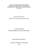
exhibited in Fig. 1. As shown, the maximum and the minimum activities of catalase were found in J. conglomeratus
and S. flavescens plants, respectively. Besides, guaiacol
peroxidase activity assay indicated that J. conglomeratus
plant had the highest activity. Furthermore, the minimum guaiacol peroxidase activity was related to R. repens
plant. Moreover, the maximum and the minimum protein contents were observed in M. azedarach fruit and J.
conglomeratus plant, respectively.
DPPH radical scavenging effect
The effect of antioxidants on DPPH. was assumed to
be because of their hydrogen donating capability [28].
Table 1 shows the DPPH radical scavenging effect of the
tested plants. As presented, the highest free radical scavenging capacity of the plants was determined in P. harmala extract with an I C50 value of 0.46 ± 0.12 µg mL−1.
Total phenol and flavonoid content of the extracts
Plants have unlimited capability to produce aromatic secondary metabolites, which most of them are phenols or
their oxygen-substituted derivatives. Key subclasses in
this set of compounds contain phenols, phenolic acids,
quinones, flavones, flavonoids, flavonols, tannins, and
coumarins. These collections of compounds indicate
catalase
Activity
Hadadi et al. BMC Chemistry
6
Guaiacol peroxidase
5
protein
4
3
2
1
0
Protein content and enzymes activity
Plants have evolved antioxidant pathways that are usually sufficient to protect them from oxidative injury during periods of natural growth and moderate stress. Both
enzymatic and non-enzymatic systems protected tissue
Plant
Fig. 1 Enzymes activity and protein content
Hadadi et al. BMC Chemistry
(2020) 14:33
antimicrobial activity and apply as plant defense mechanisms versus pathogenic microorganisms. Phenolic
toxicity to microorganisms is because of the number of
hydroxyl groups and site(s) existing in the phenolic compounds. Phenolic compounds cause cell membrane disruption, increase of ion permeability and leakage of vital
intracellular constituents or impairment of bacterial
enzyme systems in pathogenic microorganisms [34, 35].
It has been recognized that the antioxidant effect of
the flavonoids and their effectiveness on human health
and nutrition are considerable. Chelating or scavenging
procedures are the action mechanism of flavonoids [29].
The evaluation of total flavonoid content was based on
the determining the absorbance amount of tested plant
solutions reacting with aluminum chloride reagent, and
comparing with the standard solution of quercetin equivalents. The standard curve of quercetin was performed
utilizing quercetin concentration ranging from 12.5 to
100 µg mL−1. The following equation stated the absorbance of the standard solution of quercetin as a function of
concentration:
Y = 0.0056x + 0.1764, R2 = 0.9878
where, x is the absorbance and Y is the quercetin
equivalent (mg g−1). The flavonoid content of samples is
shown in Table 1. As shown, the highest phenol content
was determined in A. maurorum, P. harmala and S. flavescens extracts with a value of 45.43, 39.3 and 39.07 mg of
quercetin equivalents g−1 of dry matter, respectively.
Phenolic compounds gained from plants are a class
of secondary metabolites, acting as an antioxidant or
free radical terminators. Therefore, it is necessary to
evaluate the total content of phenols in the tested plants
[30]. The designation of the total phenolic amount was
based on the absorbance amount of sample solutions
(100 µg mL−1) reacting with Folin-Ciocalteu reagent,
and comparing with the standard solution of gallic acid
equivalents. The standard curve of gallic acid was performed utilizing gallic acid concentration ranging from
12.5 to 100 µg mL−1. The following equation stated the
absorbance of the gallic acid standard solution as a function of concentration:
Y = 0.0954x + 0.196, R2 = 0.9973
where, x is the absorbance and Y is the gallic acid equivalent (mg g−1). The phenol content of the samples is presented in Table 1. As shown, the highest phenol content
was determined in P. harmala and A. maurorum extracts
with a value of 155.29 ± 0.20 and 146.71 ± 0.02 mg Gallic
Acid Equivalents (GAE) g−1 dry matters, respectively.
Page 6 of 11
Antibacterial screening
The antibacterial activity of methanolic and chloroformic extracts including A. maurorum, S. flavescens, R.
repens, M. azedarach, P. harmala and J. conglomeratus
in different concentrations (0.01, 0.03, 0.06, 0.12, 0.25
and 0.5 ppm) were tested versus 3 g-positive (B. subtilis,
S. aureus, R. toxicus) and 5 g-negative (P. aeruginosa, E.
coli, X. campestris, P. viridiflava, P. syringae) bacteria. The
results at 0.5 ppm are shown in Figs. 2, 3. In addition, as
in other concentrations, similar results were observed, for
simplifying the discussion we considered only 0.5 ppm
concentration. As shown in Fig. 2, methanolic extracts
of S. flavescens, P. harmala fruit and J. conglomeratus
and chloroformic extracts of P. harmala fruit, S. flavescens, and P. harmala showed the maximum antibacterial activity on P. aeruginosa, respectively. Furthermore,
methanolic extract of J. conglomeratus fruits and chloroformic extracts of M. azedarach and J. conglomeratus
fruit had no antibacterial effect on P. aeruginosa (Fig. 2a).
The methanolic extract of P. harmala and chloroformic
extracts of P. harmala fruit, R. repens, and M. azedarach had the maximum antibacterial activity against B.
subtilis, respectively. Besides, chloroformic extract of
A. maurorum extract had no antibacterial activity on B.
subtilis (Fig. 2b). The methanolic extracts of P. harmala
fruit, P. harmala, and J. conglomeratus and chloroformic
extracts of M. azedarach and P. harmala fruit indicated
the maximum antibacterial activity on E. coli, respectively (Fig. 2c). Moreover, the methanolic extracts of P.
harmala fruit, the aerial part and chloroformic extracts
of S. flavescens and P. harmala fruit had the maximum
antibacterial activity on S. aureus, respectively (Fig. 2d).
Moreover, the antibacterial activity of tested plants on
plant bacteria strains is shown in Fig. 3. As indicated,
methanolic extracts of P. harmala fruit and S. flavescens
and chloroformic extracts of R. repens and M. azedarach
showed the maximum antibacterial activity against R.
toxicus, respectively (Fig. 3a). Furthermore, methanolic
extracts of R. repens and P. harmala fruit and chloroformic extracts of P. harmala fruit, J. conglomeratus fruit
and, A. maurorum presented the maximum antibacterial
activity against X. campestris, respectively (Fig. 3b). The
methanolic extract of P. harmala fruit and chloroformic
extracts of P. harmala and J. conglomeratus displayed the
maximum antibacterial activity on P. viridiflava (Fig. 3c).
Besides, the methanolic extracts of S. flavescens, P. harmala fruit and R. repens and chloroformic extracts of
R. repens represented the maximum antibacterial activity on P. syringae, respectively. However, the methanolic
extract of J. conglomeratus fruit showed no antibacterial
activity (Fig. 3d).
Hadadi et al. BMC Chemistry
(2020) 14:33
Page 7 of 11
P. aeruginosa
B. subtilis
40
*
*
30
*
20
*
*
10
0
1
2
3
4
5
6
7
45
*
40
35
30
*
25
*
20
*
10
0
8
1
2
3
*
*
20
*
15
*
10
5
0
1
2
3
4
5
6
7
8
6
7
8
Plant extract
60
*
50
*
40
*
30
20
*
*
*
*
10
0
Methanol
Chloroform
Methanol
Chloroform
Methanol
Chloroform
Methanol
Chloroform
Methanol
Chloroform
Methanol
Chloroform
Methanol
Chloroform
Methanol
Chloroform
Zone of growth inhibition (mm)
*
25
Methanol
Chloroform
Methanol
Chloroform
Methanol
Chloroform
Methanol
Chloroform
Methanol
Chloroform
Methanol
Chloroform
Methanol
Chloroform
Methanol
Chloroform
Zone of growth inhibition (mm)
*
30
5
S. aureus
*
35
4
Plant extract
E. coli
40
*
5
Plant extract
45
*
15
Methanol
Chloroform
Methanol
Chloroform
Methanol
Chloroform
Methanol
Chloroform
Methanol
Chloroform
Methanol
Chloroform
Methanol
Chloroform
Methanol
Chloroform
*
50
Zone of growth inhibition (mm)
50
Methanol
Chloroform
Methanol
Chloroform
Methanol
Chloroform
Methanol
Chloroform
Methanol
Chloroform
Methanol
Chloroform
Methanol
Chloroform
Methanol
Chloroform
Zone of growth inhibition (mm)
60
1
2
3
4
5
6
7
8
Plant extract
Fig. 2 The antibacterial activity of methanolic and chloroformic extracts including 1: S. flavescens; 2: P. harmala fruit; 3: P. harmala; 4: R. repens; 5:
M. azedarach; 6. J. conglomeratus fruit; 7: A. maurorum; 8: J. conglomeratus on standard bacteria strains. Data were exposed to Analysis of Variance
(ANOVA), following a completely randomized design to determine the Least Significant Difference (LSD) at P < 0.05 by SPSS statistical software
package (SPSS v. 11.5, IBM Corporation, Armonk, NY, USA). All consequences were stated as mean ± SD. Also, * using independent t-test between
the two groups
In order to compare the antibacterial activities of
methanolic and chloroform extracts, independent-sample t-test was used, indicated with asterisk in Figs. 2,
3. For example, in Fig. 2a, methanolic and chloroform
extracts of plants 1, 2, 3, 5, 7 and 8 showed significant
differences on Pseudomonas bacteria. In Fig. 2b, methanolic and chloroform extracts of plants 2, 3, 4, 5, 6 and 7
displayed significant differences on B. subtilis. In Fig. 2c,
methanolic and chloroform extracts of plants 1, 2, 3, 4,
5, 7 and 8 exhibited significant differences on E. coli. In
Hadadi et al. BMC Chemistry
(2020) 14:33
Page 8 of 11
*
*
*
20
*
15
10
5
0
45
40
2
3
4
5
6
7
*
30
20
15
*
*
*
5
1
8
2
3
4
5
6
7
8
Plant extract
P. viridiflava
P. syringae
40
*
30
25
15
*
*
*
20
*
*
*
10
5
1
2
3
4
5
6
7
8
Plant extract
35
30
25
20
15
10
5
0
*
*
*
*
*
*
Methanol
Chloroform
Methanol
Chloroform
Methanol
Chloroform
Methanol
Chloroform
Methanol
Chloroform
Methanol
Chloroform
Methanol
Chloroform
Methanol
Chloroform
35
Zone of growth inhibition (mm)
40
*
Methanol
Chloroform
Methanol
Chloroform
Methanol
Chloroform
Methanol
Chloroform
Methanol
Chloroform
Methanol
Chloroform
Methanol
Chloroform
Methanol
Chloroform
Zone of growth inhibition (mm)
*
10
Plant extract
0
*
*
25
0
1
*
35
Methanol
Chloroform
Methanol
Chloroform
Methanol
Chloroform
Methanol
Chloroform
Methanol
Chloroform
Methanol
Chloroform
Methanol
Chloroform
Methanol
Chloroform
30
25
*
*
Zone of growth inhibition (mm)
35
X. campestris
Methanol
Chloroform
Methanol
Chloroform
Methanol
Chloroform
Methanol
Chloroform
Methanol
Chloroform
Methanol
Chloroform
Methanol
Chloroform
Methanol
Chloroform
Zone of growth inhibition (mm)
R. toxicus
1
2
3
4
5
6
7
8
Plant extract
Fig. 3 The antibacterial activity of methanolic and chloroformic extracts including 1: S. flavescens; 2: P. harmala fruit; 3: P. harmala; 4: R. repens; 5: M.
azedarach; 6. J. conglomeratus fruit; 7: A. maurorum; 8: J. conglomeratus on plant bacteria strains. Data were exposed to Analysis of Variance (ANOVA),
following a completely randomized design to determine the Least Significant Difference (LSD) at P < 0.05 by SPSS statistical software package (SPSS
v. 11.5, IBM Corporation, Armonk, NY, USA). All consequences were stated as mean ± SD. Also, * using independent t-test between the two groups
Fig. 2d, methanolic and chloroform extracts of plants
1, 2, 3, 4, 5, 7 and 8 exhibited significant differences on
S. aureus. While in Fig. 3a, methanolic and chloroform
extracts of plants 1, 2, 4, 5, 7 and 8 presented significant
differences on R. toxicu, in Fig. 3b, methanolic and chloroform extracts of plants 1, 2, 3, 4, 5, 6, 7 and 8 presented
Hadadi et al. BMC Chemistry
(2020) 14:33
Page 9 of 11
Table 2 The Minimal Inhibitory Concentration (MIC, µg mL−1) and the Minimum Microbicidal Concentration (MBC,
µg mL−1) of the methanolic extract of the tested medicinal plants against bacteria
Strain
MIC (MBC)
Gentamicin
S. flavescens
R. repens
A. maurorum
M. azedarach
P. harmala fruit
J. conglomeratus
fruit
B. subtilis
–a
50 (100)
–
–
50 (100)
100 (−)
6.24
S. aureus
–
50 (100)
100 (−)
–
1.56 (3.12)
50 (100)
3.12
R. toxicus
25 (50)
–
–
–
12.5 (25)
–
NSb
E. coli
–
50 (100)
–
–
1.56 (3.12)
50 (100)
1.56
P. aeruginosa
12.5 (25)
–
100 (−)
50 (100)
25 (50)
–
12.48
P. syringae
–
100 (−)
–
–
100 (−)
–
NS
P. viridiflava
–
25 (50)
–
–
25 (50)
–
NS
X. campestris
50 (100)
25 (50)
–
–
25 (50)
–
NS
a
No inhibition with the highest concentration in the test conditions
b
Not specified
Table 3 The Minimal Inhibitory Concentration (MIC, µg mL−1) and the Minimum Microbicidal Concentration (MBC,
µg mL−1) of the chloroformic extract of the tested medicinal plants against bacteria
Strain
MIC (MBC)
Gentamicin
S. flavescens
R. repens
A. maurorum
M. azedarach
P. harmala fruit
J. conglomeratus
fruit
B. subtilis
–a
50 (100)
–
50 (100)
25 (50)
–
6.24
S. aureus
1.56 (3.12)
50 (100)
50 (100)
50 (100)
25 (50)
50 (100)
3.12
R. toxicus
50 (100)
12.5 (25)
25 (50)
12.5 (25)
25 (50)
25 (50)
NSb
E. coli
100 (100)
50 (100)
50 (100)
12.5 (25)
25 (50)
50 (100)
1.56
P. aeruginosa
12.5 (25)
–
–
–
1.56 (3.12)
–
12.48
P. syringae
–
100 (−)
–
100
–
–
NS
P. viridiflava
–
–
50 (100)
25
12.5 (25)
50 (100)
NS
X. campestris
100 (100)
–
25 (50)
–
12.5 (25)
12.5 (25)
NS
a
No inhibition with the highest concentration in the test conditions
b
Not specified
significant differences on X. campestris. Besides, in
Fig. 3c, methanolic and chloroform extracts of plants 1, 2,
3, 4, 5, 6, 7 and 8 showed significant differences on P. viridiflava, whereas in Fig. 3d, methanolic and chloroform
extracts of plants 1, 2, 3, 4, 5, 6, 7 and 8 showed significant differences on P. syringae.
Furthermore, Tables 2, 3 illustrate the MIC and MBC
values of the methanolic and chloroformic extracts of
the tested medicinal plants against bacteria, respectively.
The methanolic extract of P. harmala fruits showed the
maximum activity against S. aureus and E. coli with
MIC = 1.56 µg mL−1. In addition, chloroformic extracts
of S. flavescens and P. harmala fruit indicated maximum activity against S. aureus and P. aeruginosa with
MIC = 1.56 µg mL−1, respectively.
Antifungal activity
The antifungal properties of the methanolic and chloroformic extracts were tested using the agar well diffusion
method. The results of the experiments showed that none
of the tested plants had antifungal activity.
The use of herbal extracts as antioxidant and antimicrobial agents has two separate advantages: the natural origin and the related low risk. This means that they
cause fewer side effects for people and the environment
[31]. Based on the results, methanolic and chloroformic extracts of P. harmala fruit showed the maximum
antibacterial activity against most of the tested bacteria
pathogens, attributable to higher content of phenolic and
flavonoid compounds. In addition, our findings were in
agreement with those of Hayet et al. [9] and Guergour
Hadadi et al. BMC Chemistry
(2020) 14:33
et al. [32]. Methanolic and chloroformic extracts of S.
flavescens indicated the maximum antibacterial activity against P. aeruginosa and S. aureus, respectively. Our
findings were in according with Han and Guo [10] and
Yang et al. [31]. Chloroformic extract of M. azedarach
represented the maximum antibacterial activity on E.
coli, in accordance with Sen and Batra [11]. methanolic
and chloroformic extracts of A. maurorum indicated
antibacterial activity against all tested bacteria pathogens,
in agreement with the study of Ahmad et al. [12].
Conclusion
In this work, the antimicrobial and antioxidant activities
of extracts of some plants used in Iranian folklore medicine were reported. Based on the results, methanolic
and chloroformic extracts of P. harmala fruit showed
the maximum antibacterial activity against most of the
tested bacteria pathogens, attributable to higher content of phenolic and flavonoid compounds. According to
the obtained results, a high resolution GC/MS method
reported for the evaluation of the constituents of P.
harmala and S. flavescens plants, while in both plants,
Spathulenol was the main component of the essential oil.
Furthermore, in this study, the antibacterial and antifungal activities of medicinal plants extracts on plant bacteria and fungi strains were evaluated for the first time.
Furthermore, antioxidant assays including measurement of catalase, guaiacol peroxidase and protein were
reported for the first time in this study.
In conclusion, the results confirmed the traditional use
of the herb against antimicrobial diseases. These plants
could act as a potential antimicrobial agent; however, further studies are required for them to be safely used in the
control of disease and pests.
Acknowledgements
The financial support of this work from Genetics and Agricultural Biotechnology Institute of Tabarestan (GABIT) is gratefully acknowledged.
Authors’ contributions
GN and SG designed the experiment and revised the manuscript with
co-author. ZH conducted the experimental work. GN, SGh and ZH analyzed
the data and wrote the manuscript. All authors read and approved the final
manuscript.
Funding
The research was funded by Genetics and Agricultural Biotechnology Institute
of Tabarestan (GABIT), Sari Agricultural Sciences and Natural Resources
University, Iran.
Availability of data and materials
All data and materials are all provided.
Competing interest
The authors have no conflicts of interest.
Author details
1
Department of Plant Breeding, Sari Agricultural Sciences and Natural
Resources University, Sari, Iran. 2 Sari University of Agricultural Sciences
Page 10 of 11
and Natural Resources, Genetics and Agricultural Biotechnology Institute
of Tabarestan (GABIT), Sari, Iran.
Received: 9 October 2019 Accepted: 9 April 2020
References
1. Chouhan S, Sharma K, Guleria S (2017) Antimicrobial activity of some
essential oils—present status and future perspectives. Medicines 4(3):58
2. Kamonwannasit S, Nantapong N, Kumkrai P, Luecha P, Kupittayanant
S, Chudapongse N (2013) Antibacterial activity of Aquilaria crassna leaf
extract against Staphylococcus epidermidis by disruption of cell wall. Ann
Clin Microbiol Antimicrobials 12(1):20
3. Dash BK, Sen MK, Alam K, Hossain K, Islam R, Banu NA, Rahman S, Jamal
AM (2013) Antibacterial activity of Nymphaea nouchali (Burm. f ) flower.
Ann Clin Microbiol Antimicrobials 12(1):27
4. Adamu M, Naidoo V, Eloff JN (2014) The antibacterial activity, antioxidant
activity and selectivity index of leaf extracts of thirteen South African tree
species used in ethnoveterinary medicine to treat helminth infections.
BMC Vet Res 10(1):52
5. de Mélo Silva IS, da Silva ALL, dos Santos RFEP, de Souza RR, Barbosa
AM, Santos KS, Amorim MR, da Trindade LS, Krause LC, Campesatto EA:
Evaluation of antibacterial and toxicity activity in vitro of extracts from
Tournefortia bicolor SW (Boraginaceae). In: BMC proceedings: 2014.
BioMed Central: p. 31
6. de Paula Bicudo B, Rodrigues AB, Mendonça MM, Borges RR, de Almeida
AA, de Oliveira KMP: Evaluation of antibacterial and antifungal activity
of ethanolic extract of Cochlospermum regium (Cochlospermaceae) leaf,
a medicinal plant from the Cerrado of Brazil. In: BMC proceedings: 2014.
BioMed Central: p. 72
7. Srinivasan D, Nathan S, Suresh T, Perumalsamy PL (2001) Antimicrobial
activity of certain Indian medicinal plants used in folkloric medicine. J
Ethnopharmacol 74(3):217–220
8. Nagata H, Inagaki Y, Yamamoto Y, Maeda K, Kataoka K, Osawa K, Shizukuishi S (2006) Inhibitory effects of macrocarpals on the biological activity
of Porphyromonas gingivalis and other periodontopathic bacteria. Oral
Microbiol Immunol 21(3):159–163
9. Hayet E, Maha M, Mata M, Mighri Z, Laurent G, Mahjoub A (2010) Biological activities of Peganum harmala leaves. Afr J Biotech 9(48):8199–8205
10. Han C, Guo J (2012) Antibacterial and anti-inflammatory activity of
traditional Chinese herb pairs, Angelica sinensis and Sophora flavescens.
Inflammation 35(3):913–919
11. Sen A, Batra A (2012) Evaluation of antimicrobial activity of different
solvent extracts of medicinal plant: Melia azedarach L. Int J Curr Pharm
Res 4(2):67–73
12. Ahmad N, Shinwari ZK, Hussain J, Perveen R (2015) Phytochemicals,
antibacterial and antioxidative investigations of Alhagi maurorum medik.
Pak J Bot 47(1):121–124
13. Borra SK, Gurumurthy P, Mahendra J (2013) Antioxidant and free radical
scavenging activity of curcumin determined by using different in vitro
and ex vivo models. J Med Plants Res 7(36):2680–2690
14. Kumar M, Chandel M, Kumar S, Kaur S (2012) Studies on the antioxidant/
genoprotective activity of extracts of Koelreuteria paniculata laxm. Am J
Biomed Sci 1:177–189
15. Nesrin H, Tolan V (2010) Chemical composition, antimicrobial and antioxidant activities of hyssop (Hyssopus officinalis L.) essential oil. Notulae
Botanicae Horti Agrobotanici Cluj-Napoca 38(3):99–103
16. Ghahari S, Alinezhad H, Nematzadeh GA, Ghahari S (2015) Phytochemical
screening and antimicrobial activities of the constituents isolated from
Koelreuteria paniculata leaves. Nat Prod Res 29(19):1865–1869
17. Ghahari S, Alinezhad H, Nematzadeh GA, Tajbakhsh M, Baharfar R (2017)
Chemical composition, antioxidant and biological activities of the essential oil and extract of the seeds of glycine max (soybean) from North Iran.
Curr Microbiol 74(4):522–531
18. Ghahari S, Alinezhad H, Nematzadeh GA, Tajbakhsh M, Baharfar R (2017)
Biochemical composition, Antioxidant and biological activities of the
essential oil and fruit extract of Xanthium strumarium Linn. From Northern
Iran. J Agric Sci Technol 19:1603–1616
Hadadi et al. BMC Chemistry
(2020) 14:33
19. Ghahari S, Alinezhad H, Nematzadeh GA, Tajbakhsh M, Baharfar R (2018)
Phytochemical, Antioxidant and Biological Activities of the essential oil
of Astragalus alopecurus Pall. Fruits from Northern Iran. J Essent Oil Bear
Plants 21(1):103–115
20. Gapińska M, Skłodowska M, Gabara B (2008) Effect of short-and longterm salinity on the activities of antioxidative enzymes and lipid peroxidation in tomato roots. Acta Physiol Plant 30(1):11–18
21. Chance B, Maehly A (1955) [136] Assay of catalases and peroxidases.
Methods Enzymol 2:764–775
22. Upadhyaya A, Sankhla D, Davis TD, Sankhla N, Smith B (1985) Effect of
paclobutrazol on the activities of some enzymes of activated oxygen
metabolism and lipid peroxidation in senescing soybean leaves. J Plant
Physiol 121(5):453–461
23. Bradford MM (1976) A rapid and sensitive method for the quantitation
of microgram quantities of protein utilizing the principle of protein-dye
binding. Anal Biochem 72(1–2):248–254
24. Liyana-Pathirana CM, Shahidi F (2005) Antioxidant activity of commercial
soft and hard wheat (Triticum aestivum L.) as affected by gastric pH conditions. J Agri Food Chem 53(7):2433–2440
25. Yu L, Haley S, Perret J, Harris M, Wilson J, Qian M (2002) Free radical scavenging properties of wheat extracts. J Agri Food Chem 50(6):1619–1624
26. Chang C-C, Yang M-H, Wen H-M, Chern J-C: Estimation of total flavonoid
content in propolis by two complementary colorimetric methods. J Food
Drug Anal. 2002, 10(3)
27. Kang HM, Saltveit ME (2001) Activity of enzymatic antioxidant defense
systems in chilled and heat shocked cucumber seedling radicles. Physiol
Plant 113(4):548–556
28. Brighente I, Dias M, Verdi L, Pizzolatti M (2007) Antioxidant activity and total phenolic content of some Brazilian species. Pharm Biol
45(2):156–161
Page 11 of 11
29. Pourmorad F, Hosseinimehr S, Shahabimajd N. Antioxidant activity, phenol and flavonoid contents of some selected Iranian medicinal plants. Afr
J Biotechnol. 2006, 5(11)
30. Kumar M, Chandel M, Kumar S, Kaur S (2011) Protective effects of
Koelreuteria paniculata Laxm. on oxidative stress and hydrogen peroxideinduced DNA damage. Phytopharmacology 1(5):177–189
31. Yang J-F, Yang C-H, Wu C-C, Chuang L-Y (2015) Antioxidant and antimicrobial activities of the extracts from Sophora flavescens. J Pharmacog
Phytochem 3(6):26–31
32. Guergour H, Allouni RR, Bouzidi A (2018) A: phytochemical screening,
antioxidant activity of the various extracts from Peganum harmala (Zygophyllaceae). Der Pharma Chemica 10(3):146–150
33. Jahantighi A, Kiani G, Moghaddam TT, Ghahari S. In vitro antibacterial
activity of selected medicinal plants traditionally used in Iran against
plant and human pathogenic bacteria. 2016:4221. .
34. Gurjar MS, Ali S, Akhtar M, Singh KS (2012) Efficacy of plant extracts in
plant disease management. Agri Sci 3(3):425
35. Sandri I, Zacaria J, Fracaro F, Delamare A, Echeverrigaray S (2007) Antimicrobial activity of the essential oils of Brazilian species of the genus
Cunila against foodborne pathogens and spoiling bacteria. Food Chem
103(3):823–828
Publisher’s Note
Springer Nature remains neutral with regard to jurisdictional claims in published maps and institutional affiliations.
Ready to submit your research ? Choose BMC and benefit from:
• fast, convenient online submission
• thorough peer review by experienced researchers in your field
• rapid publication on acceptance
• support for research data, including large and complex data types
• gold Open Access which fosters wider collaboration and increased citations
• maximum visibility for your research: over 100M website views per year
At BMC, research is always in progress.
Learn more biomedcentral.com/submissions