Identification of structural alerts for liver and kidney toxicity using repeated dose toxicity data
Bạn đang xem bản rút gọn của tài liệu. Xem và tải ngay bản đầy đủ của tài liệu tại đây (1.08 MB, 11 trang )
Pizzo et al. Chemistry Central Journal (2015) 9:62
DOI 10.1186/s13065-015-0139-7
RESEARCH ARTICLE
Open Access
Identification of structural alerts for liver
and kidney toxicity using repeated dose toxicity
data
Fabiola Pizzo1*, Domenico Gadaleta1,2, Anna Lombardo1, Orazio Nicolotti2 and Emilio Benfenati1
Abstract
Background: The potential for a compound to cause hepatotoxicity and nephrotoxicity is a matter of extreme interest for human health risk assessment. To assess liver and kidney toxicity, repeated-dose toxicity (RDT) studies are conducted mainly on rodents. However, these tests are expensive, time-consuming and require large numbers of animals.
For early toxicity screening, in silico models can be applied, reducing the costs, time and animals used. Among in
silico approaches, structure–activity relationship (SAR) methods, based on the identification of chemical substructures
(structural alerts, SAs) related to a particular activity (toxicity), are widely employed.
Results: We identified and evaluated some SAs related to liver and kidney toxicity, using RDT data on rats taken from
the hazard evaluation support system (HESS) database. We considered only SAs that gave the best percentages of
true positives (TP).
Conclusions: It was not possible to assign an unambiguous mode of action for all the SAs, but a mechanistic explanation is provided for some of them. Such achievements may help in the early identification of liver and renal toxicity
of substances.
Keywords: Liver, Kidney, Structural alerts, Toxicity, In silico, Mechanism of action
Background
Early identification of the potential for substances to
cause hepatotoxicity and nephrotoxicity is of the utmost
importance for human health risk assessment [1]. The
liver is often involved in chemically-induced injuries and
several factors actively contribute to the liver’s susceptibility. Since most xenobiotics enter the body orally, are
absorbed through the gastrointestinal tract and then are
transported to the liver, this organ is the most exposed
to their attack [2, 3]. The second reason is that the biotransformation of chemicals in the body takes place in
the liver itself [4]. Most of time, biotransformation leads
to the formation of a molecule that is no longer- or, at
least, less-biologically active, more polar and water-soluble hence more easily excreted from the body; however
*Correspondence:
1
Laboratory of Environmental Chemistry and Toxicology, IRCCS-Istituto di
Ricerche Farmacologiche “Mario Negri”, Via La Masa 19, 20159 Milan, Italy
Full list of author information is available at the end of the article
in some cases the metabolic activity of the liver produces
toxic reactive chemicals [5].
Microsomal cytochrome P450 monooxygenases
(CYP450) are important in the metabolism of several
xenobiotics [6]. The liver is the organ with the richest
source of P450s and other enzymes, but P450s are also
expressed in various extra-hepatic tissues [7]. P450s are
expressed in kidney mainly in the renal proximal tubule,
which is also the primary target for xenobiotic-induced
renal toxicity [8, 9]. Indeed, the biotransformation of
chemicals into reactive metabolites is a key event for
nephrotoxicity. The nephrotoxic metabolites may be produced locally by the action of P450s in the kidney or they
can be produced in the liver or in other organs and transported into the kidney through the systemic circulation
[10]. The high renal blood flow and the heavy concentrations of excretory products, deriving from the re-absorption of water from the tubular fluid, are further important
factors in the kidney’s susceptibility to xenobiotics [11].
© 2015 Pizzo et al. This article is distributed under the terms of the Creative Commons Attribution 4.0 International License
( which permits unrestricted use, distribution, and reproduction in any medium,
provided you give appropriate credit to the original author(s) and the source, provide a link to the Creative Commons license,
and indicate if changes were made. The Creative Commons Public Domain Dedication waiver ( />publicdomain/zero/1.0/) applies to the data made available in this article, unless otherwise stated.
Pizzo et al. Chemistry Central Journal (2015) 9:62
Since early evaluation of the potential risk to humans
is not possible in humans, in vivo repeated-dose toxicity (RDT) studies are run in rodents [12–14]. One of the
main aims of RDT is to define the no observed adverse
effect level (NOAEL) and the lowest observed adverse
effect level (LOAEL); these parameters indicate respectively the dosage at which there is no significant response
and lowest dosage at which adverse effects arise, compared to a control group [15].
Some current legislations require the reduction of
in vivo studies when possible. These include the European Community (EC) Regulation No 1907/2006 (Registration, Evaluation, Authorisation and restriction of
Chemicals, REACH) [16]. In other cases, experiments on
animals are already banned, such as by Cosmetic Directive 76/768/EEC [17].
From the regulatory point of view, no alternatives to
animal testing are currently acceptable for the assessment of RDT. However, several attempts to assess
in vitro target-organ toxicities have been reported
[13]. As a further alternative to animal testing, in silico
approaches, such as structure–activity relationship
(SAR) can help in prioritizing laboratory tests, preclinical and clinical studies [18, 19]. The identification of
structural alerts (SAs) which are chemical substructures
whose presence may be related to the ability of a substance to cause adverse effects to organs, has met with
some success. Such approach, alongside in vitro models,
is effective for screening purposes [1]. Beside the statistical aspects related to in silico models, in the last decade
the concept of mode of action (MoA) has been introduced referring to a series of key biological events from
the initial interaction of chemicals with biological systems to the adverse outcome, and now it plays a key role
in predictive toxicology [20]. These mechanistic details
can be employed as a basis for generating SAR or as a
support of them.
In the last years, some research groups have successfully developed SAs or chemical classes for identifying hazardous substances for liver and kidney [15,
21]. Machine learning methods such as multiple linear
regression (MLR) [22–24], linear discriminant analysis
(LDA) [23], partial least square (PLS) [22] and k-nearest
neighbors (k-NN) [25, 26] have been applied for the prediction of RDT. Unlike SAs based strategies, that enables
toxicity predictions on the basis of a qualitative representation of chemical structures (SAR), such methods
employ numerical representations of chemicals for the
derivation of predictive models (quantitative structure–
activity relationship, QSAR,). For the ease of example,
molecular descriptors [27] and fingerprints [28, 29] are
widely used methods that enable quantitative representation of chemical structures.
Page 2 of 11
Some software (mostly commercial) and literature
models have been developed for predicting liver and
renal injury [30]. However, consistent and reliable data
for obtaining accurate models are still scarce and thus
developing predictive systems for systemic toxicity still
remains an open challenge [31].
This work proposes some SAs related to liver and renal
toxicity, using RDT data on rats, which may be useful for
the early evaluation of toxicity of substances. These rules
will be implemented into the ToxRead software [32], a
new freely available tool that assists users in read-across
approach.
Results and discussion
To consider SAs with good ability to predict the toxicity under investigation, we selected only SAs with a likelihood ratio (LR) of two or more and with at least 70 %
true positive (TP). However, when where was only a very
small number of total occurrences (three) we decided to
retain only those rules that gave 100 % TP.
We report the SAs identified for liver and kidney toxicity. We could not always assign an unambiguous mode
of action (MoA) for all the fragments. However, in some
cases we provided a plausible mechanistic explanation,
which was confirmed and supported by examples available in literature. It is important to keep in mind that
the data available to derive these rules are limited, thus
sometimes there are very few occurrences.
The SAs are encoded as SMiles Arbitrary Target Specification (SMARTS) that is a language used for specifying
substructures using rules that are extensions of simplified
molecular input line entry specification (SMILES) notation including, for instance, wildcards characters and for
describing the chemical structure in a more general way
[33].
Structural alerts for liver toxicity
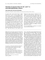
Table 1 reports the complete list of SAs for liver toxicity with their statistical performance. Out of the nine SAs
found, four had 100 % TP. In the other cases the TP % was
lower; however the number of occurrences was higher.
The SA having ID = 3 is 1,2,4,5-tetrachlorobenzene,
it was found four times in our dataset and it always
matched experimentally-hepatotoxic compounds, so
there was 100 % TP. The chlorobenzenes are important
environmental contaminants employed for several private and industrial applications [34]. They are hepatotoxic in rodents and mice after repeated exposure [35]. In
particular, 1,2,4,5-tetrachlorobenzene is a hepatic carcinogen that promotes glutathione S-transferase (GSTP11)-positive pre-neoplastic foci in rat liver [34].
The toxicological pathway shared by many halobenzenes is suggested by Sakuratani et al. [15] and Greim
Pizzo et al. Chemistry Central Journal (2015) 9:62
Page 3 of 11
Table 1 SAs recognized as harmful for liver
ID
SMARTS
1
Structure
LR
occurrences
No TP
TP%
Cc1c(c(ccc1)C(C))
inf
7
7
100
2
C(C(COC)CC)
inf
7
7
100
3
c1c(c(c(c(c1Cl)Cl))Cl)Cl
inf
4
4
100
4
C(CO)Cl
inf
3
3
100
5
c1cc2ccccc2cc1
5.61
8
7
87.5
6
c1c(c(cc(c1)CC))[OH]
8.02
11
9
81.8
7
c1cc(ccc1Cc2ccc(cc2))
4.28
19
16
84.2
8
c1ccc(cc1)c2ccccc2
2.4
8
6
75
9
CBr
2.4
8
6
75
For each structure the percentage of likelihood ratio (LR) as calculated by SARpy, the total number of occurrences and percentage of true positives (TP %) are
reported. Marvin Sketch was used for drawing the structures
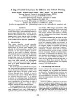
[35]. Briefly, halobenzenes are metabolically activated
by cytochrome P450, which transforms them into epoxides, highly reactive electrophilic species. The spontaneous conversion of the epoxide to phenol and then the
secondary oxidation of phenols by CYP450 enzymes
lead to the formation of hydroquinones, which can be
subsequently oxidized to quinones. Quinones too are
electrophilic and can bind tissue proteins or lead to the
Pizzo et al. Chemistry Central Journal (2015) 9:62
generation of reactive oxygen species harmful for hepatic
cells [15, 25] (Fig. 1).
The SA having ID = 5 reports the naphthalene ring, a
polycyclic aromatic hydrocarbon, known as an environmental contaminant, and classified as a potential human
carcinogen [36]. It is widely used commercially in the
synthesis of dyes, resins, plastics, pharmaceuticals, dispersants and tanning agents in the rubber and leather
industries [36, 37]. In humans and laboratory animals,
the eyes and lungs are the organs mostly involved after
exposure to naphthalene [38]. However, naphthalene is
also implicated in hepatocyte injury and liver dysfunction [37]. Indeed, early studies demonstrated that it
caused lipid peroxidation in liver as well as increasing
liver weight and aniline hydroxyalase activity [39–41].
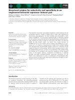
In in vitro and in vivo models, metabolism of naphthalene is a key event in its toxicity [36]. Its main metabolic
pathways in mammals are described in Fig. 2. Once
absorbed, naphthalene can be metabolized by various
CYP 450 [42]. Briefly, CYP450 converts naphthalene into
naphthalene epoxide, which can undergo several reactions: conjugation to glutathione (GSH), transformation
into naphthol or into dihydrodiol. Naphthol and dihydrodiol are both enzymatically converted to naphthalenediol, which is further oxidized to naphthoquinone
through redox cycling; this final reaction generates reactive oxygen species (ROS). ROS induce oxidative stress,
leading to cell death. In addition, quinones can form
adducts with proteins or DNA, leading to cell damage
[36, 42].
Page 4 of 11
The SA having ID = 6 is the para-alkyl phenol. It was
found 11 times in the dataset. In nine cases it was found
in molecules labelled as hepatotoxic. Phenols, commonly
present in the environment, are substances largely used
in chemical and pharmaceutical industry [43]. The key
event that leads to phenol toxicity is its interaction with
cell biomolecules combined with the donation of free
electrons from oxidized substrates [43]. The main effect
of these reactions, catalyzed by oxidative enzymes in the
liver, is the formation of phenoxy radicals, semiquinones
and quinine methide that, finally, bind and damage DNA
or enzymes. As a consequence of these reactions, ROS
such as superoxide radicals and hydrogen peroxide, are
also created [43]. Phenolic compounds with ortho- or
para-alkyl groups (alkylphenols) can also form quinone
methides that interact with biomolecules in the cell [44].
The SA having ID = 8 is the biphenyl. It occurred eight
times in the datasets and in six cases it was correctly
associated with hepatotoxic compounds. Several in vivo
studies on rodents reported liver toxicity, including histopathological changes and increases in liver weight and
serum liver enzymes after exposure to this chemical
[45–47]. However, only few human data are available for
biphenyl and these are even limited to two occupational
epidemiology studies involving workers handling this
chemical [48, 49]. These studies provided some evidence
of liver toxicity, such as increases of serum enzyme levels.
The last SA selected is bromomethane reported with
ID = 9. It was found eight times in the dataset and in six
cases it was correctly associated with compounds labelled
Fig. 1 Metabolic hepatic pathway of halobenzenes mediated by CYP450. X stands for any halogenated atom
Pizzo et al. Chemistry Central Journal (2015) 9:62
Page 5 of 11
Fig. 2 Partial metabolic pathways of naphthalene in mammalians
as hepatotoxic. A previous study [50] reported that rats
exposed through inhalation to bromomethane showed
histopathological changes and hepatocellular degeneration, such as foci of hepatocellular coagulative necrosis.
However, no mechanism of action of this compound on
liver tissue is reported in the literature.
It was not possible to find a mechanistic explanation in
the literature for SAs having ID 1, 2, 4 and 7; however, the
percentage of TP was high for these substructures. SAs 1,
2 and 4 had 100 % TP and SA 7 84.2 % TP.
Structural alerts for renal and urinary tract toxicity
Table 2 gives the complete list of SAs identified for renal
and urinary tract toxicity with their statistical performance. The fragments give 100 % TP except for the last
SA (ID = 6), which has 71.4 % TP since there were two
errors.
The second SA (ID = 2) found for renal toxicity is sulfanilamide. It was found four times into our dataset with
100 % TP. The LR, calculated by SARpy software [51], is
infinite. Sulfanilamide belongs to the chemical class of
sulfonamides which are antibiotics widely used for the
treatment of bacterial and protozoa infections in veterinary and human medicine [52, 53]. The literature for this
chemical category indicated that their relatively insolubility in acid urine means these compounds can precipitate
in the tubular lumen forming insoluble crystals, leading
to hematuria, albuminuria, crystalluria, renal colic and
even acute renal insufficiency [54, 55]. Acid urine and
dehydration promote sulfonamide crystallization [55]
(Fig. 3).
Benzonitriles (SA, ID = 3) are solvents with many
industrial
applications.
Bromoxynil,
chloroxynil,
dichlobenil, and ioxynil are chemically similar pesticides
that share the same benzonitrile structure [56]. A recent
investigation [57] reported that the benzonitriles had
adverse effects in vitro on the human embryonic renal
cell line HEK293T, with significant cytotoxicity.
SA having ID = 5 is the chloroform structure. It was
found three times, in all cases in molecules related to
kidney toxicity. Chloroform is used as a solvent in many
industrial applications [10]. It causes renal toxicity in
Pizzo et al. Chemistry Central Journal (2015) 9:62
Page 6 of 11
Table 2 SAs recognized as harmful for kidney and urinary tract
ID
SMARTS
1
Structure
LR
occurrences
No. TP
TP%
c1ccc(cc1)S(=O)(=O)O
inf
4
4
100
2
NS(=O)(=O)c1ccc(cc1)N
inf
4
4
100
3
c1cc(cc(c1)C#N)
inf
3
3
100
4
CC(=CCCC(=C))
inf
3
3
100
5
C(Cl)(Cl)Cl
inf
3
3
100
6
c1ccc(cc1)c2ccccc2
3.3
7
5
71.4
For each structure the percentage of likelihood ratio (LR) as calculated by SARpy, the total number of occurrences and percentage of true positives (TP %) are
reported. Marvin Sketch was used for drawing the structures
several species through a P450-dependent metabolism
that leads to the formation of nephrotoxic chloroform
metabolites [58, 59]. It has been reported that chloroform
induces renal cancer, not via direct DNA reactivity, but
for events associated with cytolethality and regenerative
cell proliferation caused by exposure to chloroform [60,
61]. Regenerative cell proliferation is an important part
of the repair process and this mechanism has been positively linked to the carcinogenicity of some non-genotoxic chemicals in animal bioassays [10].
The last SA, having ID = 6, found for renal and urinary
tract toxicity was biphenyl. This fragment was identified
Pizzo et al. Chemistry Central Journal (2015) 9:62
Page 7 of 11
Fig. 3 Toxicity pathway for sulfamides. Ar stands for aryl group, AH stands for any atoms including hydrogen
seven times and in five cases the molecules were actually labelled as nephrotoxic. A large number of studies
on animals have reported the toxicological role of biphenyl in serious injury of the urinary tract [45, 62–65].
The effects on animals were hematuria, increased urinary pH, increased kidney weight, formation of calculi
accompanied by the induction of urinary tract tumours.
Potassium 4-hydroxy-biphenyl-O-sulfate is one of main
biphenyl metabolites involved in the formation of urinary
calculi, due to its low solubility. The presence of urine
crystals, promoted by higher pH and potassium concentrations, is the first step in urinary calculi formation [65].
However, the mechanism that leads to the formation of
the urine crystals induced by exposure to biphenyl still
needs to be fully elucidated [65].
To the best of our knowledge a mechanistic explanation for SAs having ID 1 and 4 was no available in the literature. The percentage of TP for both of them was 100.
Besides those we identified, other SAs were developed
for liver and kidney toxicity [15, 21, 66]. Some of them are
the same that we here reported. Similarly to our findings,
Sakuratani et al. [15] identified halobenzenes (Table 1, SA
ID = 3), para alkyl phenols (Table 1, SA ID = 6), halogenated aliphatic compounds (Table 1, SA ID = 9) and
aromatic hydrocarbons (Table 1, SAs ID = 1, 5, 6, 7 and
8) as alerts related to hepatotoxicity and sulphonamide
group (Table 2, SA ID = 2) to urinary tract toxicity. Phenols (Table 1, SA ID = 6) were identified as hepatotoxic
by a recent study [21] that used a dataset of pharmaceutical chemicals as starting point to identify SAs for liver
toxicity.
The overlap of these results should not be interpreted
as a redundancy of the findings, rather a confirmation
of the data obtained. Indeed, the key point is that starting from different sets of data and even applying different methods, all these studies come to same results. This
increases the reliability of the SAs for the prediction of
toxicity.
Compared to hepatotoxicity, nephrotoxicity is less
investigated from a computational point of view. The
major contribution of this work is related to kidney toxicity since most of our results on liver toxicity confirm
those previously obtained by other authors with the
exception of SAs having ID 2 and 4.
Experimental
Selection of data
RDT data for modeling are present in the Hazard Evaluation Support System (HESS) database [15], which
was downloaded from the OECD QSAR Toolbox [67].
This database provides NOAEL and LOAEL values and
gives information on the organ toxicity for 503 chemicals tested on rats by oral exposure over periods ranging
from 28 to 120 days. More details on these data can be
found in [15]. For the selection of the liver toxicity data
to be used for modeling, we considered the compounds
for which LOAEL related to effects on liver was reported
and we labelled them as “active” substances. Those compounds with reported LOAEL effects on organs other
than liver were considered negative controls and were
labelled as “inactive”. We applied the same procedure to
build a dataset for renal toxicity.
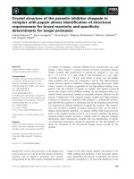
We finally obtained two datasets: one containing 218
liver toxicity data (121 of which were “active”) and the
other with 202 data related to kidney toxicity (89 labelled
as “active”). Some compounds appear in both datasets
since at the LOAEL they reported effects both on liver
and kidney. We labelled “active” the data that indicated
liver or renal effects after 28 or 90 days of exposure and
“inactive” those had no effect on the organ of interest
after 90 days of exposure, since if no effect is reported
after 28 days it may occur later (90 days) (Fig. 4). We considered only organic compounds; salts were neutralized
and we double-checked the correspondence between
CAS number and chemical structures using Pubchem
compound [68] and ChemID plus [69]. For the dataset on
Pizzo et al. Chemistry Central Journal (2015) 9:62
Page 8 of 11
Fig. 4 Procedure for selecting data in the HESS database
nephrotoxicity, we also included compounds reported to
have effects on the urinary tract.
Extraction and evaluation of structural alerts
In order to obtain SAs related to liver and kidney toxicity,
we used the software SARpy, developed by Politecnico di
Milano and described in Ferrari et al. [51]. Briefly, SARpy
is able to extract sets of rules by automatically generating
and selecting substructures on the sole basis of their prediction performance on a training set used as input [51]
and irrespective of any a priori knowledge. This is done
in three steps. The first step is the fragmentation of the
input chemicals (training set) in order to extract all the
substructures within a customizable size range. Then, the
software analyses the correlation between the occurrence
of each molecular substructure and the experimental
activity of the compounds that contain it in the training
set. This is a validation step aimed at assessing the predictive power of each fragment. Finally, a subset of fragments is selected and provided to the user in the form of
rules ‘‘IF fragment THEN activity’’ [70]. The input and the
output chemical structures of SARpy are all expressed as
SMILES [33]. The statistical parameter used for defining
the precision of a fragment to predict the activity under
investigation is the LR, calculated for each SA as:
Likelihood ratio =
TP negatives
×
FP
positives
TP are experimentally positive (toxic) compounds correctly predicted as positive, false positives (FP) are experimentally negative but wrongly predicted as positive.
For each SA we calculated the TP %, the percentage of
Pizzo et al. Chemistry Central Journal (2015) 9:62
correctly predicted compounds out of the total number
of occurrences.
TP % =
TP
occurences
SARpy can be customized so as to minimize the number of FP, or in a more balanced way, to improve the accuracy. We used SARpy with different settings (min, max,
optimal) in order to get a large number of SAs, then each
fragment was evaluated and those did not meet our criteria were eliminated and not considered further. Indeed,
we did not use SARpy like a black box, but we carefully
checked every SA generated by the software and in some
cases they were generalized so as to have rules to match
correctly with a larger number of compounds.
Conclusions
Liver and kidney toxicities are key points in the evaluation of safety for existing and new substances. Many
in vivo RDT studies have been done to assess the ability
of a chemical to induce hepatotoxicity and nephrotoxicity. However, in many regulatory contexts, the tendency
is to strongly reduce the number of in vivo tests. Thus
there is an urgent need for reliable alternatives to animal testing, in order to protect human health. In silico
methods may be useful to minimize the number of animals required and to reduce time and costs. We have
proposed some SAs that are chemical substructures that
may be related to hepatotoxicity and nephrotoxicity. For
some of them a mechanistic explanation is also provided
as further evidence. The aim is not to fully replace in vivo
studies, but to provide a supporting tool that may be used
for early identification and prioritization of the potential
toxicity of substances.
Abbreviations
SA: structural alerts; RDT: repeated dose toxicity; LOAEL: lowest observed
adverse effect level; NOAEL: no observed adverse effect level; QSAR: quantitative structure-activity relationship; SAR: structure-activity relationship; LR:
likelihood ratio; TP: true positive; TP %: percentage of true positive; FP: false
positive; SMILES: simplified molecular input line entry specification; SMARTS:
smiles arbitrary target specification; MoA: mechanism of action; MLR: multiple
linear regression; LDA: linear discriminant analysis; PLS: partial least square;
k-NN: k nearest neighbors.
Authors’ contributions
This work was carried out in collaboration between all authors. FP and DG
compiled the datasets. FP performed the study and drafted the manuscript.
EB supervised the work. EB, AL, ON read and corrected the manuscript. All
authors checked and validated the draft. All authors read and approved the
final manuscript.
Author details
1
Laboratory of Environmental Chemistry and Toxicology, IRCCS-Istituto di
Ricerche Farmacologiche “Mario Negri”, Via La Masa 19, 20159 Milan, Italy.
2
Dipartimento di Farmacia‑Scienze del Farmaco, Università degli Studi di Bari
“Aldo Moro”, Bari, Italy.
Page 9 of 11
Acknowledgements
The authors are grateful for the contribution of the project HEALTHF5-2010-267042 ToxBank (Supporting Integrated Data Analysis and servicing
of Alternative Testing Methods in Toxicology) funded by European Commission and Cosmetics Europe under the Seventh Framework programme.
Competing interests
The authors declare that they have no competing interests.
Received: 28 July 2015 Accepted: 27 October 2015
References
1. Abernethy DR, Woodcock J, Lesko LJ (2011) Pharmacological mechanismbased drug safety assessment and prediction. Clin Pharmacol Ther
89:793–797
2. Roberts SM, James RC, Franklin MR (2010) Hepatotoxicity: toxic effects on
the liver. In: Williams PL, James RC, Roberts SM (eds) Principles of toxicology: environmental and industrial applications. Wiley, USA
3. Eipel C, Abshagen K, Vollmar B (2010) Regulation of hepatic blood
flow: hepatic arterial buffer response revisited. World J Gastroentero
16:6046–6057
4. Hodgson E, Levi PE (2004) Hepatotoxicity. In: Hodgson E (ed) A textbook
of modern toxicology. Wiley, USA, p 2004
5. Williams DP, Park BK (2003) Idiosyncratic toxicity: the role of toxicophores
and bioactivation. Drug Discov Today 18:1044–1050
6. Porter TD, Coon MJ (1991) Cytochrome P-450. Multiplicity of isoforms,
substrates, and catalytic and regulatory mechanisms. J Biol Chem
266:13469–13472
7. Ding X, Kaminsky LS (2003) Human extrahepatic cytochromes P450:
function in xenobiotic metabolism and tissue-selective chemical toxicity
in the respiratory and gastrointestinal tracts. Ann Rev Pharmacol Toxicol
43:149–173
8. Cummings BS, Zangar RC, Novak RF, Lash LH (1999) Cellular distribution
of cytochromes P-450 in the rat kidney. Drug Met Dispos 27:542–548
9. Van Vleet TR, Schnellmann RG (2003) Toxic nephropathy: environmental
chemicals. Semin Nephrol 23:500–508
10. Fang C, Behr M, Xie F, Lu S, Doret M, Luo H, Yang W, Aldous K, Ding X, Gu
J (2008) Mechanism of chloroform-induced renal toxicity: non-involvement of hepatic cytochrome P450-dependent metabolism. Toxicol Appl
Pharmacol 227:48–55
11. Hodgson E, Levi PE (2004) Nephrotoxicity. In: Hodgson E (ed) A textbook
of modern toxicology, 3rd edn. Wiley, USA
12. Suter L, Babiss LE, Wheeldon EB (2004) Toxicogenomics in predictive
review toxicology in drug development. Chem Biol 11:161–171
13. Prieto P, Baird AW, Blaauboer BJ, Castell Ripoll JV, Corvi R, Dekant W, Dietl P,
Gennari A, Gribaldo L, Griffin JL, Hartung T, Heindel JJ, Hoet P, Jennings P,
Marocchio L, Noraberg J, Pazos P, Westmoreland C, Wolf A, Wright J, Pfaller
W (2006) The assessment of repeated dose toxicity in vitro: a proposed
approach. ATLA 34:315–341
14. Lilienblum W, Dekant W, Gebel T, Hengstler JG, Kahl R, Kramer PJ, Schweinfurth H, Wollin KM (2008) Alternative methods to safety studies in
experimental animals: role in the risk assessment of chemicals under the
new European Chemicals Legislation (REACH). Arch Toxicol 82:211–236
15. Sakuratani Y, Zhang H, Nishikawa S, Yamazaki K, Yamada T, Yamada J,
Gerova K, Chankov G, Mekenyan O, Hayashi M (2013) Hazard evaluation
support system (HESS) for predicting repeated dose toxicity using toxicological categories. SAR QSAR Env Res 24:351–363
16. European Commission (2006) Regulation (EC) of No 1907/2006 of the
European parliament and of the council 18 December 2006 concerning
the Registration, Evaluation, Authorisation and Restriction of Chemicals
(REACH), establishing a European Chemicals Agency, amending Directive
1999/45/EC and repealing Council Regulation (EEC) No 793/93 and Commission Regulation (EC) No 1488/94 as well as Council Directive 76/769/
EEC and Commission Directives 91/155/EEC, 93/67/EEC, 93/105/EC and
2000/21/EC
Pizzo et al. Chemistry Central Journal (2015) 9:62
17. European Commission (2009) Regulation (EC) No 1223/2009 of the European parliament and of the council of 30 November 2009 on cosmetic
products
18. Raunio H (2011) In silico toxicology-non-testing methods. Front Pharmacol 2: 33/1
19. Nicolotti O, Benfenati E, Carotti A, Gadaleta D, Gissi A, Mangiatordi GF,
Novellino E (2014) REACH and in silico methods: an attractive opportunity for medicinal chemists. Drug Discov Today 19:1757–1768
20. Vinken M (2013) The adverse outcome pathway concept: a pragmatic
tool in toxicology. Toxicol 312:158–165
21. Hewitt M, Enoch SJ, Madden JC, Przybylak KR, Cronin MT (2013) Hepatotoxicity: a scheme for generating chemical categories for read-across, structural
alerts and insights into mechanism(s) of action. Crit Rev Toxicol 43:537–558
22. Mazzatorta P, Estevez MD, Coulet M, Schilter B (2008) Modeling oral rat
chronic toxicity. J Chem Inf Model 48:1949–1954
23. De Julián-Ortiz JV, García-Domenech R, Gálvez L et al (2005) Predictability
and prediction of lowest o.bserved adverse effect levels in a structurally
heterogeneous set of chemicals. SAR QSAR Environ Res 16:263–272
24. García-Domenech R, de Julián-Ortiz JV, Besalú E (2006) True prediction of
lowest observed adverse effect levels. Mol Diversity 10:159–168
25. Gadaleta D, Pizzo F, Lombardo A, Carotti A, Escher SE, Nicolotti O, Benfenati E (2014) A k-NN algorithm for predicting oral sub-chronic toxicity in
the rat. ALTEX 31:4–14
26. Chavan S, Friedman R, Nicholls IA (2015) Acute toxicity-supported chronic
toxicity prediction: a k-nearest neighbor coupled read-across strategy. Int
J Mol Sci 16:11659–11677
27. Todeschini R, Consonni V (2000) Handbook of molecular descriptors.
Wiley-VCH, Weinheim
28. Floris M, Manganaro A, Nicolotti O, Medda R, Mangiatordi GF, Benfenati E
(2014) A generalizable definition of chemical similarity for read-across. J
Cheminf 6:39
29. Willett P (2011) Similarity searching using 2D structural fingerprints. In:
Bajorath J (ed) Chemoinformatics and computational chemical biology,
vol 672. Humana Press, pp 133–158
30. Lapenna S, Fuart-Gatnik M, Worth A (2010) Review of QSAR models and
software tools for predicting acute and chronic systemic toxicity. JRC
Scientific and Technical Reports
31. Tsakovska I, Lessigiarska I, Netzeva T, Worth AP (2007) A mini review of
mammalian toxicity (Q)SAR models. QSAR Comb Sci 27:41–48
32. Gini G, Franchi AM, Manganaro A, Golbamaki A, Benfenati E (2014)
ToxRead: a tool to assist in read across and its use to assess mutagenicity
of chemicals. SAR QSAR Environ Res 25:999–1011
33. Daylight, Chemical Information System Inc [ />dayhtml/doc/theory/theory.smarts.html]
34. Gustafson DL, Long ML, Thomas RS, Benjamin SA, Yang RSH (2000) Comparative hepatocarcinogenicity of hexachlorobenzene, pentachlorobenzene, 1,2,4,5-tetrachlorobenzene, and 1,4-dichlorobenzene: application
of a medium-term liver focus bioassay and molecular and cellular indices.
Toxicol Sci 53:245–252
35. Greim H (2003) Mechanistic and toxicokinetic data reducing uncertainty
in risk assessment. Toxicol Lett 138:1–8
36. Cho TM, Rose RL, Hodgson E (2006) In vitro metabolism of naphthalene
by human liver microsomal cytochrome P450 enzymes. Drug Metab
Dispos 34:176–183
37. Ahmed AAE, Fatani AJ (2007) Protective effect of grape seeds proanthocyanidins against naphthalene-induced hepatotoxicity in rats. Saudi
Pharma J 15:38–47
38. Stohs SJ, Ohia S, Bagchi D (2002) Naphthalene toxicity and antioxidant
nutrients. Toxicol 180:97–105
39. Rao GS, Pandya KP (1981) Biochemical changes unduced by naphthalene
after oral administration in albino rats. Toxicol Lett 8:311–315
40. Yamauchi T, Komura S, Yagi K (1986) Serum lipid peroxide levels of albino
rats administered naphthalene. Biochem Intern 13:1–6
41. Vuchetich PJ, Bagchi D, Bagchi M, Hassoun EA, Tang L, Stohs SJ (1996)
Naphthalene-induced oxidative stress in rats and the protective effects of
vitamin E succinate. Free Rad Biol Med 21:577–590
42. Preuss R, Angerer J, Drexler H (2003) Naphthalene-an environmental and
occupational toxicant. Int Arch Occup Environ Health 76:556–576
Page 10 of 11
43. Michałowicz J, Duda W (2007) Phenols-sources and toxicity. Polish J
Environ Stud 16:347–362
44. Thompson DC, Perera K, London R (1995) Quinone methide formation
from para isomers of methylphenol (cresol), ethylphenol, and isopropylphenol: relationship to toxicity. Chem Res Toxicol 8:55–60
45. Umeda Y, Arito H, Kano H, Ohnishi M, Matsumoto M, Nagano K, Yamamoto S, Matsushima T (2002) Two-year study of carcinogenicity and
chronic toxicity of biphenyl in rats. J Occup Health 44:176–183
46. Umeda Y, Aiso S, Arito H, Nagano K, Matsushima T (2004) Induction of
peroxisome proliferation in the liver of biphenyl-fed female mice. J Occup
Health 46:486–488
47. Umeda Y, Aiso S, Yamazaki K, Ohnishi M, Arito H, Nagano K, Yamamoto S,
Matsushima T (2005) Carcinogenicity of biphenyl in mice by two years
feeding. J VetMed Sci 4:417–424
48. Seppalainen AM, Hakkinen I (1975) Electrophysiological findings in
diphenyl poisoning. J Neurol Neurosur Psych 38:248–252
49. Carella G, Bettolo PM (1994) Reversible hepatotoxic effects of diphenyl:
report of a case and a review of the literature. J Occup Med 36:575–576
50. Hurtt ME, Morgan KT, Working PK (1987) Histopathology of acute toxic
responses in selected tissues from rats exposed by inhalation to methyl
bromide. Fund Appl Toxicol 9:352–365
51. Ferrari T, Cattaneo D, Gini G, Golbamaki Bakhtyari N, Manganaro A, Benfenati E (2013) Automatic knowledge extraction from chemical structures:
the case of mutagenicity prediction. SAR QSAR Environ Res 24:365–383
52. Garcıa-Galan MJ, Dıaz-Cruz MS, Barcelo D (2008) Identification and
determination of metabolites and degradation products of sulfonamide
antibiotics. Trends Anal Chem 27:11
53. Chen J, Zhou X, Zhang Y, Gao H (2012) Potential toxicity of sulfanilamide
antibiotic: binding of sulfamethazine to human serum albumin. STOTEN
432:269–274
54. Perazella MA (1999) Crystal-induced Acute Renal Failure. Am J Med
106:459–465
55. Wawruch M, Bozekova L, Krcmery S, Kriska M (2002) Risks of antibiotic
treatment. Brat Med J 103:270–275
56. Holtze MS, Sørensen SR, Sørensen J, Aamand J (2008) Microbial degradation of the benzonitrile herbicides dichlobenil, bromoxynil and ioxynil in
soil and subsurface environments e Insights into degradation pathways,
persistent metabolites and involved degrader organisms. Environ Poll
154:155–168
57. Lovecka P, Thimova M, Grznarova P, Lipov J, Knejzlik Z, Stiborova H, Gde
T, Nindhia T, Demnerova K, RumlT (2015) Study of cytotoxic effects of
benzonitrile pesticides. BioMed Res Internat (Article ID 381264)
58. Smith JH, Hook JB (1984) Mechanism of chloroform nephrotoxicity III.
Renal and hepatic microsomal metabolism of chloroform in mice. Toxicol
Appl Pharmacol 73:511–524
59. Branchplower RV, Nunn DS, Highet RJ, Smith JH, Hook JB, Pohl LR (1984)
Nephrotoxicity of chloroform: metabolism to phosgene by the mouse
kidney. Toxicol Appl Pharmacol 72:159–168
60. Hard G (1998) Mechanisms of chemically induced renal carcinogenesis
laboratory rodent. Toxicol Pathol 26(101–1):12
61. Constan AA, Sprankle CS, Peters JM, Kedderis GL, Everitt JI, Wong BA, Gonzalez FL, Butterworth BE (1999) Metabolism of chloroform by cytochrome
P450 2E1 is required for induction of toxicity in the liver, kidney, and nose
of male mice. Toxicol Appl Pharmacol 160:120–126
62. Booth A, Ambrose AM, Deeds F, Cox AJ (1961) The reversible nephrotoxic
effects of biphenyl. Toxicol Appl Pharmacol 3:560–567
63. Kluwe WM (1981) Renal function tests as indicators of kidney injury in
subacute toxicity studies. Toxicol Appl Pharmacol 57:414–424
64. Ohnishi M, Yajima Y, Yamamoto S, Matsushima T, Ishii T (2000) Sex
dependence of the components and structure of urinary calculi induced
by biphenyl administration in rats. Chem Res Toxicol 13:727–735
65. Ohnishi M, Yajima H, Takeuchi T, Saito M, Yamazaki K, Kasai T, Nagano K,
Yamamoto S, Matsushima T, Ishii T (2001) Mechanism of urinary tract
crystal formation following biphenyl treatment. Toxicol Appl Pharmacol
174:122–129
66. Marchant CA, Fisk L, Note RR, Patel ML, Suarez D (2009) An expert system
approach to the assessment of hepatotoxic potential. Chem Biodiver
6:2107–2114
Pizzo et al. Chemistry Central Journal (2015) 9:62
67. EOCD QSAR toolbox. Accessed 23 July
2015
68. PubChem compound website. />Accessed 23 July 2015
69. ChemID plus. Accessed 23 July
2015
Page 11 of 11
70. Lombardo A, Pizzo F, Benfenati E, Manganaro A, Ferrari T, Gini G (2014)
A new in silico classification model for ready biodegradability, based on
molecular fragments. Chemosphere 108:10–16
Publish with ChemistryCentral and every
scientist can read your work free of charge
Open access provides opportunities to our
colleagues in other parts of the globe, by allowing
anyone to view the content free of charge.
W. Jeffery Hurst, The Hershey Company.
available free of charge to the entire scientific community
peer reviewed and published immediately upon acceptance
cited in PubMed and archived on PubMed Central
yours you keep the copyright
Submit your manuscript here:
/>