Disposable screen printed sensor for the electrochemical detection of methamphetamine in undiluted saliva
Bạn đang xem bản rút gọn của tài liệu. Xem và tải ngay bản đầy đủ của tài liệu tại đây (1.5 MB, 9 trang )
Bartlett et al. Chemistry Central Journal (2016) 10:3
DOI 10.1186/s13065-016-0147-2
RESEARCH ARTICLE
Open Access
Disposable screen printed
sensor for the electrochemical detection
of methamphetamine in undiluted saliva
Carrie‑Ann Bartlett, Sarah Taylor, Carlos Fernandez, Ceri Wanklyn, Daniel Burton, Emma Enston,
Aleksandra Raniczkowska, Murdo Black and Lindy Murphy*
Abstract
Background: Methamphetamine has an adverse effect on the ability to drive safely. Police need to quickly screen
potentially impaired drivers therefore a rapid disposable test for methamphetamine is highly desirable. This is the first
proof-of-concept report of a disposable electrochemical test for methamphetamine in undiluted saliva.
Results: A screen printed carbon electrode is used for the N,N′-(1,4-phenylene)-dibenzenesulfonamide mediated
detection of methamphetamine in saliva buffer and saliva. The oxidized mediator reacts with methamphetamine to
give an electrochemically active adduct which can undergo electrochemical reduction. Galvanostatic oxidation in
combination with a double square wave reduction technique resulted in detection of methamphetamine in undi‑
luted saliva with a response time of 55 s and lower detection limit of 400 ng/mL.
Conclusions: Using a double square wave voltammetry technique, rapid detection of methamphetamine in undi‑
luted saliva can be achieved, however there is significant donor variation in response and the detection limit is signifi‑
cantly higher than desired. Further optimization of the assay and sensor format is required to improve the detection
limit and reduce donor effects.
Keywords: Square wave voltammetry, SWV, Galvanostatic oxidation, Screen printed electrode, Mediator,
Methamphetamine, Saliva, Detection
Background
Two thirds of US trauma centre admissions are due
to motor vehicle accidents with almost 60 % of such
patients testing positive for drugs or alcohol [1]. Cannabis, cocaine and methamphetamine are the drugs most
frequently detected in drivers randomly stopped for
roadside drug screening [2–5]. In Norway prior to the
year 2000 there was almost no methamphetamine on the
Norwegian market. There was a steady increase in methamphetamine usage till 2010 where it appeared to have
stabilized. The data for this study was confirmed by testing venous blood of convicted motorists, customs seizures and wastewater analysis [6]. A US survey, using a
questionnaire which annually monitored adolescent drug
*Correspondence:
Oxtox Limited, Warren House, 5 Mowbray Street, Stockport SK1 3EJ, UK
use, showed a gradual decline in methamphetamine use
from 3.7 % in 1981 (peak year) to 1.2 % in 2008 [7]. A
recent study showed conflicting trends when comparing
the questionnaire survey approach and wastewater analysis. Over the period 2010–2013 the population survey
showed a slight decline in methamphetamine use while
wastewater analysis showed a doubling of methamphetamine usage [8].
Methamphetamine remains a significant public health
concern with known neurotoxic and neurocognitive
effects to the user [9]. It is frequently abused as a recreational drug due to its stimulant and euphoric effects.
The physiological and psychological side effects of
methamphetamine include confusion, paranoia, depression, nausea and blurred vision. Driving a vehicle while
under the influence of methamphetamine is thus clearly
undesirable.
© 2016 Bartlett et al. This article is distributed under the terms of the Creative Commons Attribution 4.0 International License
( which permits unrestricted use, distribution, and reproduction in any medium,
provided you give appropriate credit to the original author(s) and the source, provide a link to the Creative Commons license,
and indicate if changes were made. The Creative Commons Public Domain Dedication waiver ( />publicdomain/zero/1.0/) applies to the data made available in this article, unless otherwise stated.
Bartlett et al. Chemistry Central Journal (2016) 10:3
Page 2 of 9
Roadside screening for methamphetamine in oral
fluid has a number of requirements: it needs to be fast,
ideally 15–30 s, i.e. ideally the same speed as a breath
alcohol test; it must be very sensitive, ideally <10 ng/mL
(25 ng/mL was used as the cut-off concentration in the
European DRUID [Driving under the influence of Drugs,
Alcohol, and Medicines] project [5]); and it should be
non-invasive, difficult to tamper with and be portable.
The currently available drug screening products require
a minimum of 5–10 min for a test [10]. Test time and
cost are restricting the roadside drug screening market to
<10 % the volume of the alcohol screening market.
Oral fluid which contains saliva and other liquid substances present in the oral cavity are of great interest
for roadside drug screening. Although this fluid is easy
to collect there is considerable inter-sample variability
in the fluid matrix that generates issues when developing a testing methodology [11]. Dilution of the sample
can reduce the donor variability, however this dilutes
the drug of interest and therefore requires the device to
have greater sensitivity. The current devices on the market are primarily lateral flow immunodiagnostic tests,
where the presence or absence of a coloured bar can be
read either visually or in a meter in response to the drug
of interest; these were used in the DRUID project. The
response times are typically several minutes. The clinical sensitivity of these devices in saliva can be relatively
poor at 16–75 % although clinical specificity can be close
to 100 % [12].
There are only a few reports of the electrochemical
sensing of amphetamines, and there are no reports of the
determination of amphetamines in undiluted saliva using
disposable electrochemical sensors. Electrochemical sensing of methamphetamine by direct oxidation has been
reported at a pretreated pencil graphite electrode [LOD
50 nM (7.5 ng/mL) in aqueous solution, response time
>10 min] [13], at a self-assembled boron-doped diamond
electrode [LOD 50 nM (7.5 ng/mL) in aqueous solution,
response time not given] [14], and in alkaline solution
using a gold nanoparticle-multiwalled carbon nanotube
modified screen printed electrode [LOD 0.3 nM (0.05 ng/
mL), response time not given] [15]. The indirect electrochemical detection of amphetamine in saliva has been
reported using 1,2-naphthoquinone-4-sulfonate at an edge
plane pyrolytic graphite electrode [LOD 41 μM (6.2 μg/
mL) in aqueous solution, response time not given] [16].
This paper reports a mediated screen printed carbon electrode for the detection of methamphetamine in
undiluted saliva using substituted N,N′-(1,4-phenylene)dibenzenesulfonamide mediator. Screen printed electrodes are well established as cheap and disposable single
use sensors which can be manufactured with high reproducibility [17].
The sensor is optimized for speed of response and for
response in undiluted saliva.
Results and discussion
Initial mediator screen
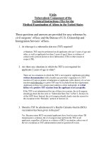
The mechanism of reaction between oxidized N,N′-(1,4phenylene)-dibenzenesulfonamide and primary and
secondary amines has been described by Adams and
Schowalter [18]. The mechanism is shown schematically in Fig. 1. The oxidized form of the mediator (II)
reacts with secondary amines such as methamphetamine
(MAMP) by 1,4-addition resulting in the reduced form
of the MAMP-mediator adduct (III). Electron exchange
between (III) and a further molecule of (II) results in
the oxidized form of the adduct (IV) which can undergo
reduction at the electrode at the appropriate reduction
potential i.e. it can give rise to a new reduction peak in
addition to the reduction peak for unreacted oxidized
mediator, (II).
With primary amines such as amphetamine (AMP),
1,2-addition can take place, resulting in elimination of the
two benenesulfonamide groups from the mediator and
formation of an AMP-mediator adduct. This adduct can
SO2Ph
SO2Ph
SO2Ph
SO2Ph
NH
N
NH
N
N
-2H+, -2e-
+ MAMP
Ph
N
+ II
NH
N
NH
N
SO2Ph
SO2Ph
SO2Ph
SO2Ph
I
II
III
Fig. 1 Reaction of N,N′-(1,4-phenylene)-dibenzenesulfonamide with methamphetamine (MAMP)
IV
Ph + I
Bartlett et al. Chemistry Central Journal (2016) 10:3
also undergo oxidation via II and subsequently undergo
electrochemical reduction.
An initial mediator screen was performed
with several substituted N,N′-(1,4-phenylene)dibenzenesulfonamide compounds, described in
Additional file 1. The mediators were screened for
electrochemical response using differential pulse
voltammetry (DPV) and reaction with MAMP. The
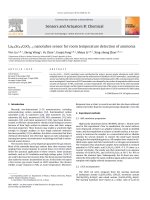
sensors used were of the format shown in Fig. 2a, consisting of a two electrode system of carbon working
electrode and Ag/AgCl combined counter/reference
electrode. The preferred mediator was OX1006 (N,N′(2-nitro-1,4-phenylene)dibenzenesulfonamide) on the
basis of giving a large, clearly defined peak response
to MAMP without adsorption of the parent mediator to the electrode. At pH 10.8, the mediator is fully
deprotonated [pKas 6.05 and 8.00 (25 °C)] and soluble
at 1 mg/mL, and this pH was used for the development
of the sensor.
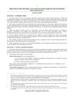
The cyclic voltammetry of OX1006 with MAMP is
shown in Fig. 3. In the absence of MAMP, there is a
single oxidation peak at +0.38 V and negligible reduction peak. In the presence of MAMP, two new peaks
are present at +0.15 V and −0.046 V, and a new reduction peak is present at −0.088 V. In addition, the parent
mediator peak height at +0.38 V is increased by 29 and
47 % in the presence of 25 and 50 μg/mL MAMP. The
increase in the parent peak height and the new peaks
are due to the oxidation/reduction of the mediatorMAMP adduct.
Optimization of electrochemical procedure with dried
reagent
It was desired that the mediator and buffer solution be
dried down in some way on the sensor. Deposition of
Page 3 of 9
Fig. 3 Cyclic voltammetry of OX1006 in the absence and presence of
MAMP. The MAMP concentration was 0, 25 or 50 μg/mL MAMP (solid
line, dotted line and dashed lines) in 0.1 M sodium carbonate buffer
(pH 10.4), 0.2 M NaCl. 15 μL of solution was pipetted onto the sensor.
The scan rate was 50 mV/s
mediator solution directly onto the sensor requires tight
control of the volume and position of the dispensed reagent. Therefore an alternative technique was used comprising a porous overlayer onto which mediator was dried
and which is then secured over the sensor. On application of sample, the mediator dissolves and diffuses to the
working electrode where it can be oxidized, react with
MAMP and produce a reduction response to MAMP.
Sensors with overlayer applied are shown in Fig. 2b.
Galvanostatic oxidation of OX1006 was investigated
in combination with the overlayer. The advantage of galvanostatic oxidation compared to potentiostatic oxidation is that the amount of oxidized mediator should be
relatively independent of the concentration of mediator
which has dissolved off the overlayer and reached the
electrode surface, provided there is sufficient mediator. A
Fig. 2 Screen printed electrodes a without and b with overlayer. The sensor comprises a circular carbon working electrode (2 mm diameter) and
outer Ag/AgCl counter/reference electrode
Bartlett et al. Chemistry Central Journal (2016) 10:3
potential disadvantage of galvanostatic oxidation is that
if there is insufficient mediator, other species present will
be oxidized, resulting in a large increase in working electrode potential.
There are very few reported examples of galvanostatic
oxidation to generate reactant, and these examples are
for electrochemical titrations using separate generatorcollector electrodes [19, 20]. For example, Tomcik et al.
[21] have reported the galvanostatic generation of hypobromite at an interdigitated microelectrode array, for
end-point titration of the drugs Antabus and Celaskon.
In our application, the working electrode is used to both
generate the reactant (oxidized mediator) and detect the
mediator-MAMP adduct.
The shift in working electrode potential during galvanostatic oxidation is shown in Fig. 4, for sensors with
mediator in the overlayer and using a saliva sample.
A 10 s wait time during which the sensor was at open
circuit potential was employed at the start of the test
sequence to ensure the mediator had dissolved off the
overlayer. With higher galvanostatic currents there is a
larger shift in potential starting at +0.4 V, with the shift
seen at an earlier time for higher current, indicating the
mediator has been depleted more quickly with higher
galvanostatic current setting. A galvanostatic current of
800 nA was selected.
The square wave voltammetry (SWV) response to
MAMP in saliva buffer or saliva using galvanostatic oxidation and the mediator overlayer is shown in Fig. 5. In
saliva buffer, the main reduction peak at +0.38 V was
reduced in the presence of MAMP (1800–1260 nA, 30 %
reduction), and two new peaks were observed at +0.14
and −0.06 V (717 and 1430 nA). The reduction peak
heights were very significantly reduced in saliva compared to saliva buffer, by approximately 85–95 % (peak
heights at +0.34, +0.15 and −0.04 V were 205, 38 and
88 nA in the presence of 5 μg/mL MAMP). The overall
response time with the SWV procedure was 122 s. Ideally the response time of the sensor would be in the range
15–30 s, although a response time of less than 120 s
would still be acceptable for a roadside test as it would
be considerably faster than the existing roadside tests.
Therefore the electrochemical procedure was optimized
for speed of response.
In order to increase the speed of the SWV technique,
the first part of the scan between +0.6 and +0.1 V was
conducted at a higher scan rate compared to the second
part of the scan between +0.1 V and −0.4 V. Both parts
of the scan were optimized for amplitude, step size and
frequency. The third peak height is independent of frequency (Additional file 2), therefore a faster scan rate can
be used for the first part of the scan without any adverse
effect on the 3rd peak height.
Page 4 of 9
Fig. 4 Varying the current during the galvanostatic oxidation step.
The overlayer was treated with 0.12 mg/mL of OX1006 in 0.4 M
sodium carbonate buffer (pH 10.8), containing 0.23 M NaCl and 0.1 %
TX-100. The procedure consisted of a 10 s wait time after application
of 7 μL of saliva, followed by galvanostatic oxidation at 300, 800, 1200,
1500 or 3000 nA for 30 s
Fig. 5 SWV response to MAMP in a saliva buffer or b saliva. The
MAMP concentration was 0 μg/mL (solid line) or 5 μg/mL (dashed
line). The overlayer was treated with 1.0 mg/mL of OX1006 in 0.4 M
sodium carbonate buffer (pH 10.8), containing 1.0 M NaCl and 0.1 %
TX-100. The SWV procedure consisted of a 10 s wait time after appli‑
cation of 7 μL of sample, then (1) galvanostatic oxidation at 800 nA
for 30 s, (2) SWV with start voltage +0.6 V, stop voltage −0.4 V, 4.25 Hz
frequency, 2.85 mV step potential and 50 mV amplitude
Bartlett et al. Chemistry Central Journal (2016) 10:3
The split SWV responses to MAMP in saliva buffer
and saliva are shown in Fig. 6, using frequencies of 20
and 4.25 Hz for the first and second parts of the scan.
The new peak in response to MAMP is clearly observed
at −0.06 V for saliva buffer and −0.04 V for saliva. The
overall response time is 55 s. The calibration plot for
response to MAMP in a saliva sample using the third
peak of the optimized split SWV technique is shown
in Fig. 7. Good linearity of response to MAMP was
obtained (R2 0.9877). The lower limit of detection was
400 ng/mL (0 ng/mL response +3 SD) which is considerably higher than that required for a commercial device
(<10 ng/mL).
The LOD compares favourably with that obtained using
indirect electrochemistry with 1,2-naphthoquinone4-sulfonate [16], and it is considerably higher than the
LODs obtained using direct electrochemical methods
[13–15], although all these methods use aqueous solution
and not undiluted saliva. Use of microelectrodes should
provide greater sensitivity of response, since increased
Page 5 of 9
Fig. 7 Calibration plot for response to MAMP in saliva obtained from
a single donor, using the 3rd peak height obtained with the split
SWV technique. Each sample was tested with 12 sensors. Error bars
are 1 SD. The overlayer treatment and electrochemical procedure are
described in Fig. 6
mass transport of MAMP to the electrode should result
in increased peak heights i.e. higher nA per ng/mL
MAMP. However this would require reproducible screen
printed microelectrodes and development of a suitable
manufacturing methodology was beyond the time and
budgetary restraints of this work.
The response to MAMP and amphetamine in saliva
using the split SWV technique showed a new peak
formed in response to MAMP at −0.04 V, and no new
peak observed in response to amphetamine (Additional
file 3). This demonstrates the selectivity of the mediator
to secondary amines over primary amines.
Variation in response with different donor saliva samples
Fig. 6 Split SWV response to MAMP in a saliva buffer or b saliva. The
MAMP concentrations were 0 (solid line) or 5 μg/mL (dashed line). The
overlayer was treated with 1.0 mg/mL of OX1006 in 0.4 M sodium
carbonate buffer (pH 10.8), containing 1.0 M NaCl and 0.1 % TX-100.
The SWV procedure consisted of a 10 s wait time after application of
7 μL of sample, then (1) galvanostatic oxidation at 800 nA for 30 s; (2)
SWV-1 with start voltage +0.6 V, stop voltage +0.1 V, 20 Hz frequency,
10 mV step potential and 50 mV amplitude; (3) SWV-2 with start
voltage +0.1 V, stop voltage −0.4 V, 4.25 Hz frequency, 10 mV step
potential and 100 mV amplitude
The response to saliva obtained from 10 donors is
shown in Fig. 8. There is considerable variation in 1st
and 3rd peak heights, and to a lesser extent the 2nd
peak height, between the donors. At 0 μg/mL MAMP,
the average peak heights range from 95 to 1878 nA
for the 1st peak, 1523–2882 nA for the 2nd peak and
0–6 nA for the 3rd peak. At 1 μg/mL MAMP, the average peak heights range from 129 to 1578 nA for the 1st
peak, 1813–2573 nA for the 2nd peak and 0–113 nA
for the 3rd peak. The individual donor samples can give
very different responses. For example, while the majority of the donor samples do not show a decrease in 1st
and 2nd peak heights in response to MAMP, donors 6
and 10 do show a decrease in 1st and 2nd peak heights
in response to MAMP (donor 6 gave 90 and 37 %
decrease and donor 10 gave 59 and 30 % in 1st and 2nd
peak heights, for response to 0 and 1 μg/mL MAMP).
However for the 3rd peak, donor 6 gave no response
to MAMP, whereas for donor 10 the 3rd peak height
increased from 2.4 to 18 nA for 0–1 μg/mL MAMP. It
can also be observed that only the samples from donors
2 and 8 show an increase in the 3rd peak height in
Bartlett et al. Chemistry Central Journal (2016) 10:3
Page 6 of 9
Fig. 8 Donor variation in response to MAMP in saliva from 10 donors. a 1st and 2nd peak heights and b 3rd peak height. The MAMP concentrations
were 0, 0.1, 0.25 and 1 μg/mL. Each sample was tested with 6 sensors. Error bars are 1 SD. The overlayer treatment and SWV procedure are described
in Fig. 6
response to 100 ng/mL MAMP (donor 2, 6.1–13.5 nA
and donor 8, 4.3–28.9 nA for response to 0 and 100 ng/
mL MAMP).
To further investigate the effect of donor variation in
saliva on response, saliva from two donors was centrifugally filtered using filters with cut-offs of 3, 10, 30 and
100 kDa. The results are shown in Fig. 9. There was a significant increase in the 2nd peak height and also in the
3rd MAMP peak height for 100 kDa filtered saliva compared to unfiltered saliva; with 1 μg/mL MAMP, the peak
heights increase from 218 to 629 nA (donor 1, 2nd peak),
and 15–142 nA (donor 1, 3rd peak), and from 329 to
539 nA (donor 2, 2nd peak) and 88–285 nA (donor 2, 3rd
peak). This indicates high molecular weight species such
as proteins and mucin have a significant negative impact
on the peak height. For donor 1, the 1st peak is not present except for the 3 kDa filtered sample, while for donor
2 the 1st peak was not present for the unfiltered samples,
but was present for the filtered samples.
The response to MAMP increased with decreasing
molecular weight cut-off of the filter e.g. for donor 1, the
3rd peak heights in response to 1 μg/mL MAMP were
15, 142 and 353 nA for unfiltered saliva, 100 and 3 kDa
filters. However there is still considerable donor variation in response with the filtrate from the 3 kDa filter
(the 3rd peak heights for donors 1 and 2 were 353 and
Bartlett et al. Chemistry Central Journal (2016) 10:3
Page 7 of 9
Fig. 9 Response to MAMP in saliva from two donors, in unfiltered saliva and saliva filtrate. a 1st and 2nd peak heights and b 3rd peak height. Saliva
filtrate was collected using centrifugal filters with 3, 10, 30 or 100 kDa cut-offs. The MAMP concentrations were 0 or 1 μg/mL. Each sample was
tested with 6 sensors. Error bars are 1 SD. The overlayer treatment and SWV procedure is described in Fig. 6, except the galvanostatic current was
700 nA
512 nA respectively). While this filter will have removed
larger proteins and mucins, some small proteins and
protein fragments will remain, which may compete for
adsorption sites on the electrode surface with the mediator MAMP adduct. In addition, the filtrate will contain
endogenous amines which may react with the mediator.
The effect of the saliva components mucin and
lysozyme on response are shown in Table 1. Addition
of mucin had little effect, whereas addition of lysozyme
resulted in significant reduction in peak heights, demonstrating the adverse effect of saliva proteins on response.
Experimental
(+)-Methamphetamine
hydrochloride
(M8750),
d-amphetamine sulphate (A5880), human recombinant
lysozyme (L1667) and mucin from bovine submaxillary glands (M3895) were obtained from Sigma-Aldrich
Co. Ltd (Poole, UK). The mediators were synthesized by
Peakdale Molecular (High Peak, UK). All other chemicals
were purchased from Sigma-Aldrich Co. Ltd. All chemicals were used as received without further purification.
All solutions were prepared using deionized water with
resistivity no less than 18.2 MΩ cm.
Bartlett et al. Chemistry Central Journal (2016) 10:3
Page 8 of 9
Table 1 Response to saliva buffer containing added protein
Average peak height/nA (±1 SD)
% Decrease in peak height
1st peak (at +0.40 V) 2nd peak (at +0.25 V) 3rd peak (at −0.06 V) 1st peak 2nd peak 3rd peak
SSB
2865 ± 1369
3257 ± 1939
436 ± 30
SSB + 0.021 mg/mL mucin
2665 ± 728
2581 ± 893
481 ± 59
SSB + 0.021 mg/mL mucin + 0.3 mg/mL
lysozyme
1952 ± 1009
1781 ± 1018
382 ± 70
−26.7
−31.0
−20.5
SSB + 0.021 mg/mL mucin + 3.0 mg/mL
lysozyme
985 ± 275
717 ± 233
54 ± 29
−49.5
−59.7
−85.8
−7.0
−20.8
10.4
(A) No addition and with the addition of (B) 0.021 mg/mL mucin; (C) 0.3 mg/mL lysozyme and 0.021 mg/mL mucin; and (D) 3 mg/mL lysozyme and 0.021 mg/mL
mucin. The overlayer was treated with 0.2 mg/mL of OX1006 in 0.4 M sodium carbonate buffer (pH 10.8), containing 0.23 M NaCl and 0.1 % TX-100. Each sample was
tested with 6 sensors. The SWV procedure is described in Fig. 6
Screen printed electrodes were fabricated in house with
appropriate stencil designs using a DEK Horizon printing
machine (DEK, Weymouth, UK). Successive layers of carbon-graphite ink (C2120403D1, modified in house by the
addition of 0.1 % TX-100), dielectric ink (D2070423P5)
and Ag/AgCl ink (60:40, C2030812P3) obtained from
Gwent Electronic Materials Ltd. (Pontypool, UK) were
printed onto a polyester substrate. The layers were
cured using a tunnel drier at 70 °C (Natgraph, Nottingham, UK). The reproducibility of response of a sample
of sensors from each print batch was determined using
square wave voltammetry (SWV) with 1 mM OX1006 in
0.4 M sodium carbonate buffer (pH 10.8), 0.23 M NaCl,
0.0018 % TX-100. The SWV settings were as follows:
start potential +0.6 V, stop potential −0.5 V, frequency
10 Hz, amplitude 0.05 V and step size 0.00285 V. Each
sensor batch comprised 15 sheets with 4 rows of 48 sensors per sheet. A sample of 12 sensors from the second
sheet of each batch were tested for SWV response to
OX1006, and the responses were characterized for peak
position and peak height. The %CVs were typically in
the range 0.5–1.7 and 3–5 % for peak position and peak
height respectively.
Voltammetric measurements were performed using
either a MultiAutolab M101 or a μ-Autolab III potentiostat (Eco Chemie). The screen printed sensors were used
as a two electrode system, with a combined counter/reference electrode (Ag/AgCl ink).
The overlayer material was composed of abaca and
cellulosic fibres (75 %) in a polypropylene thermoplastic matrix (25 %), dry weight 16.5 g/m2 (CD020010,
Ahlstrom) in reel format (1 cm wide) was obtained
from Ahlstrom (Duns, UK). The overlayer was coated
with OX1006 as follows: 1 mg/mL OX1006 was prepared in 0.4 M sodium carbonate buffer solution (pH
10.8) containing 1 M NaCl and 0.1 % Triton X-100.
The solution was dispensed onto the membrane at a
loading of 0.1–1 mg/mL and dried at 40 °C. The dried
overlayer was heat soldered to each sensor along the
edges.
Saliva buffer, which mimics real saliva except for the
absence of proteins, consisted of 27.5 mM sodium chloride, 6.3 mM ammonium chloride, 4.9 mM sodium phosphate (monobasic), 2.9 mM potassium chloride, 1.1 mM
sodium citrate (anhydrous), 0.02 mM magnesium
chloride (anhydrous), 0.27 mM sodium carbonate and
0.2 mM calcium chloride.
Each saliva sample was collected immediately before
use by spitting into a pot. Saliva samples containing
MAMP were prepared by dissolving MAMP directly into
the saliva sample at 1 mg/mL. Saliva samples containing
lower MAMP concentrations were obtained by dilution
of the 1 mg/mL sample with neat saliva.
Centrifugal filtration of saliva was performed using
Amicon Ultra 0.5 mL centrifugal filters with molecular
cut-off weights of 100, 30, 10, and 3 kDa. The samples
were centrifuged at 14,000g for 10 min. The filters were
weighed before and after centrifugation and deionised
water was added to each filtrate to adjust for volume lost.
Conclusions
The detection of 400 ng/mL MAMP in undiluted saliva
has been reported using mediated disposable screen
printed sensors with a response time of 55 s. While the
response time is significantly faster than existing lateral
flow immunodiagnostic tests, the limit of detection of the
sensors is considerably higher (400 ng/mL compared to
10 ng/mL) and is too high to be acceptable as a screening test. The precision of the sensor response is adversely
affected by saliva proteins and further development of the
sensor is required to overcome these effects and obtain
a commercially viable sensor. Saliva samples are notoriously variable in terms of composition and viscosity, even
within the same donor sample collected over a short
period of time, and it is probable that an on-strip dilution of the sample would decrease adverse effects arising
Bartlett et al. Chemistry Central Journal (2016) 10:3
from sample variability and viscosity, however this would
require controlled sample dilution. It would also require
greater sensitivity of response which may be achieved by
the use of microelectrodes and this is a route that should
be investigated further. In conclusion, development of a
disposable roadside test for the rapid determination of
methamphetamine in undiluted saliva is challenging, and
requires significant further effort.
Additional files
Additional file 1: Mediator screen.
Additional file 2: Effect of SWV-1 frequency.
Additional file 3: Response to MAMP and AMP.
Abbreviations
MAMP: (+)-methamphetamine; AMP: d-amphetamine; SWV: square wave
voltammetry; DPV: differential pulse voltammetry; OX1006: N,N′-(2-nitro-1,4phenylene)dibenzenesulfonamide.
Authors’ contributions
LM and MB co-directed the study. CF demonstrated the initial concept. EE
and AR characterised the electrode performance. DB and CW performed
the mediator screen. CAB optimized the electrochemical procedure and ST
investigated donor variation. LM and MB wrote the manuscript. All authors
read and approved the final manuscript.
Acknowledgements
The authors gratefully acknowledge Professor Richard Compton and Professor
Craig Banks for helpful discussions. Professors Compton and Banks are the
company founders and are shareholders in Oxtox.
Competing interests
The authors declare that they have no competing interests.
Received: 7 August 2015 Accepted: 7 January 2016
References
1. Walsh JM, Flegel R, Cangianelli LA, Atkins R, Soderstrom CA, Kerns TJ
(2004) Epidemiology of alcohol and other drug use among motor vehicle
crash victims admitted to a trauma center. Traffic Inj Prev 5:254–260
2. Lacey JH, Kelley-Baker T, Furr-Holden D, Voas RB, Romano E, Ramirez A,
Brainard K, Moore C, Torres P, Berning A (2007) National roadside survey
of alcohol and drug use by drivers: drug results. National Highway Traffic
Safety Administration, Washington DC. Report number DOT HS 811 249
3. Van der Linden T, Legrand SA, Silverans P, Verstraete AG (2012) DUID:
oral fluid and blood confirmation compared in Belgium. J Anal Toxicol
36:418–421
4. Chu M, Gerostamoulos D, Beyer J, Rodda L, Boorman M, Drummer OH
(2012) The incidence of drugs of impairment in oral fluid from random
roadside testing. Forensic Sci Int 215:28–31
5. Houwing S, Hagenzieker M, Mathijssen R, Bernhoft IM, Hels T, Janstrup K,
Van der Linden T, Legrand S-A, Verstraete A (2011) Prevalence of alcohol
and other psychoactive substances in drivers in general traffic. Part I:
general results. DRUID Deliverable 2.2.3. www.druid-project.eu
Page 9 of 9
6. Bramness JG, Reid MJ, Solvik KF, Vindenes V (2015) Recent trends in the
availability and use of amphetamine and methamphetamine in Norway.
Forensic Sci Int 246:92–97
7. Johnson LD, O’Malley PM, Bachman JG, Schulenberg JE (2008) Monitor‑
ing the future. National results on adolescent drug use. Overview of key
findings, 2008. (NIH Publication No 09-7401). National Institute on Drug
Abuse, Bethseda
8. Tscharke BJ, Chen C, Gerber JP, White JM (2015) Trends in stimulant use in
Australia: a comparison of wastewater analysis and population surveys.
Sci Total Environ 536:331–337
9. Courtney KE, Ray LA (2014) Methamphetamine: an update on epidemiol‑
ogy, pharmacology, clinical phenomenology, and treatment literature.
Drug Alcohol Depend 143:11–21
10. Alere Toxicology. />11. Chiappin S, Antonelli G, Gatti R, De Palo EF (2007) Saliva specimen: a new
laboratory tool for diagnostic and basic investigation. Clin Chim Acta
383:30–40
12. Vanstechelman S, Isalberti C, Van der Linden T, Pil K, Legrand SA,
Verstraete AG (2012) Analytical evaluation of four on-site oral fluid drug
testing devices. J Anal Toxicol 36:136–140
13. Oghli AH, Alipour E, Asadzadeh M (2015) Development of a novel
voltammetric sensor for the determination of methamphetamine in
biological samples on the pretreated pencil graphite electrode. RSC Adv
5:9674–9682
14. Svorc L, Vojs M, Michniak P, Marton M, Rievaj M, Bustin D (2014) Electro‑
chemical behaviour of methamphetamine and its voltammetric determi‑
nation in biological samples using self-assembled boron-doped diamond
electrode. J Electroanal Chem 717–718:34–40
15. Rafiee B, Fakhari AR, Ghaffarzadeh M (2015) Impedimetric and stripping
voltammetric determination of methamphetamine at gold nanoparti‑
cles-multiwalled carbon nanotubes modified screen printed electrode.
Sens Actuators, B 218:271–279
16. Goodwin A, Banks CE, Compton RG (2006) Tagging of model ampheta‑
mines with sodium 1,2-naphthoquinone-4-sulfonate: application to the
indirect electrochemical detection of amphetamines in oral (saliva) fluid.
Electroanal 18(18):1833–1837
17. Thiyagarajan N, Chang J-L, Senthilkumar K, Zen J-M (2014) Disposable
electrochemical sensors: a mini review. Electrochem Commun 38:86–90
18. Adams R, Schowalter KA (1952) Quinone imides. X. Addition of amines to
p-quinonedibenzenesulfonimide. JACS 74:2597–2602
19. Rajantie H, Strutwolf J, Williams DE (2001) Theory and practice of electro‑
chemical titrations with dual microband electrodes. J Electroanal Chem
500(1–2):108–120
20. Rajantie H, Williams DE (2001) Potentiometric titrations with dual micro‑
band electrodes. Analyst 126:1882–1887
21. Tomcik P, Krajcikova M, Bustin D (2001) Determination of pharmaceutical
dosage forms via diffusion layer titration at an interdigitated microelec‑
trode array. Talanta 55:1065–1070