α-Glucosidase inhibitors from the bark of Mangifera mekongensis
Bạn đang xem bản rút gọn của tài liệu. Xem và tải ngay bản đầy đủ của tài liệu tại đây (1021.88 KB, 6 trang )
Nguyen et al. Chemistry Central Journal (2016) 10:45
DOI 10.1186/s13065-016-0193-9
Open Access
RESEARCH ARTICLE
α‑Glucosidase inhibitors from the bark
of Mangifera mekongensis
Hai Xuan Nguyen1,2, Tri Cong Le1, Truong Nhat Van Do1, Tho Huu Le1, Nhan Trung Nguyen1,2
and Mai Thanh Thi Nguyen1,2*
Abstract
Background: Mangifera mekongensis (Anacardiaceae) is cultivated for its edible fruit and has been used in traditional
Vietnamese medicine for its anti-aging properties and for treating diabetes, vermifuge, and dysentery. As part of a
search for biologically active compounds with reduction of the rate of glucose absorption, a screening has been initiated to evaluate natural product extracts for the inhibition of enzyme α-glucosidase. A n-hexane extract of the bark of
M. mekongensis showed strong α-glucosidase inhibitory activity with IC50 value of 1.71 µg/mL. Thus, the constituents
of this plant were examined.
Results: Two new steroids named mekongsterol A (1) and mekongsterol B (2), were isolated from the n-hexane
extract of the bark of M. mekongensis (Anacardiaceae), together with seven known compounds (3–9). Their chemical
structures were elucidated on the basis of spectroscopic data. All compounds possessed significant α-glucosidase
inhibitory activity in a concentration-dependent manner, except for 3 and 4. Compounds 1, 2, 5–9 showed more
potent inhibitory activity, with IC50 values ranging from 1.2 to 112.0 µM, than that of a positive control acarbose (IC50,
214.5 µM).
Conclusions: These results suggested that the traditional use of the bark of M. mekongensis for the treatment of
diabetes diseases in Vietnam may be attributable to the α-glucosidase inhibitory activity of its steroid and cycloartane
constituents.
Keywords: Mangifera mekongensis, Anacardiaceae, α-Glucosidase inhibition, Sterols
Background
Mangifera mekongensis (Anacardiaceae), commonly
known as mango, is widely distributed in tropical and subtropical regions of Asia. In Vietnam, M. mekongensis is
called as “Xoai Thanh Ca”, and this plant is cultivated for
its edible fruit and has been used in traditional Vietnamese medicine for treating anti-aging, diabetes, vermifuge,
dysentery [1, 2]. A research for biologically active compounds with reduction of the rate of glucose absorption,
a screening has been initiated to evaluate natural product
extracts for the inhibition of enzyme α-glucosidase. It is
effective in controlling postprandial hyperglycaemia and
prevents complications associated with type-II diabetes,
*Correspondence:
1
Faculty of Chemistry, University of Science, Vietnam National University
Hochiminh City, 227 Nguyen Van Cu, District 5, Hochiminh City, Vietnam
Full list of author information is available at the end of the article
such as microvascular (i.e., retinal, renal, and possibly neuropathic), macrovascular (i.e., coronary and peripheral
vascular), and neuropathic (i.e., autonomic and peripheral)
complications [3, 4]. Previously, we reported that the methanolic extracts of Embelia ribes, Oroxylum indicum, and
Artocarpus altilis exhibited significant inhibitory activity
on α-glucosidase [5–8]. In a part of our continued research
on the screening of medicinal plants of different origins,
we also found that the n-hexane extract of the bark of M.
mekongensis showed strong α-glucosidase inhibitory activity with IC50 value of 1.71 µg/mL. Thus, we carried out the
bioactivity-guided fractionation of n-hexane extract of this
plant and isolated two new steroids, mekongsterols A (1)
and B (2), together with seven known compounds (3–9)
(Fig. 1). In this paper, we describe the isolation and structural elucidation of these compounds by spectroscopic
methods as well as their α-glucosidase inhibitory activity.
© 2016 The Author(s). This article is distributed under the terms of the Creative Commons Attribution 4.0 International License
( which permits unrestricted use, distribution, and reproduction in any medium,
provided you give appropriate credit to the original author(s) and the source, provide a link to the Creative Commons license,
and indicate if changes were made. The Creative Commons Public Domain Dedication waiver ( />publicdomain/zero/1.0/) applies to the data made available in this article, unless otherwise stated.
Nguyen et al. Chemistry Central Journal (2016) 10:45
Page 2 of 6
O
O
O
1
O
HO
O
2
OH
OH
HO
HO
3
O
O
O
4
O
COOH
O
6
HO
O
COOH
COOH
7
O
5
O
8
HO
COOH
9
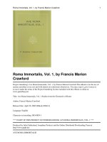
Fig. 1 Structures of the isolated compounds from the bark of M. mekongensis
Result and discussion
Chemistry
The dried powdered bark of M. mekongensis was
extracted with n-hexane in Soxhlet extractor to yield
n-hexane fraction. Further separation and purification
of this fraction led to the isolation of two new steroids,
mekongsterols A (1) and B (2), together with seven
known compounds (3–9). The known compounds were
identified by the analysis of their spectroscopy data and
comparing with the literature data to be as β-sitosterol
(3) [9], stigmastane-3,6-dione (4) [10], β-sitosteryl-3O-β-D-glucopyranosyl-6′-O-palmitate (5) [11], mangiferonic acid (6) [12], mangiferolic acid (7) [12], ambonic
acid (8) [13], and ambolic acid (9) [12] (Fig. 1).
Mekongsterol A (1) was obtained as a white crystal and showed the quasimolecular ion at m/z 733.6223
[M + K]+, corresponding to the molecular formula
C48H86O2K in HR-ESI–MS. The IR spectrum of 1 showed
absorption of ester carbonyl (1720 cm−1), double bond
(1610 cm−1), and methyl, methylene, and methine (2950
and 2870 cm−1) groups. The 1H NMR spectrum of 1
(Table 1) displayed signals due to two methyl singlets
(δH 0.68, 1.02, each s), three methyl doublets (δH 0.81, d,
J = 6.8 Hz; δH 0.84, d, J = 6.8 Hz; δH 0.92, d, J = 6.5 Hz),
a methyl triplet (δH 0.82, t, J = 7.5 Hz), an oxymethine
(δH 4.62, m), and trisubstituted olefinic bond (δH 5.38,
d, J = 4.4 Hz), together with many aliphatic methylene
and aliphatic methine groups (δH 0.95–2.30). The 13C
NMR (Table 1) and DEPT spectra of 1 exhibited signals for six methyls (δC 12.0, 12.1, 18.9, 19.2, 19.5, 19.9),
an oxymethine (δC 73.8), and two olefinic carbons (δC
122.7 and 139.9). These data closely resembled those of
β-sitosterol (3) [9], a common steroid found in plants,
but they were characterized by the presence of additional
signals due to a saturated fatty ester chain having 19C,
which showed ester carbonyl (δC 173.5), many methylenes (δH 1.20–2.27; δC 22.8–34.9), and one methyl triplet
(δH 0.88, t, J = 6.9 Hz). The location of saturated fatty
ester chain was determined to be at C-3 on the basis of
the low-field shift of H-3 (δH 4.62) compared to that of 3
(δH 3.51), which was confirmed by the HMBC correlation
from H-3 to C-1′ (Fig. 2). The orientation of saturated
fatty ester group at C-3 was determined β-equatorial
from the NOESY correlations H-3/H-2α and H-3/H-4α,
and large J value (7.7 Hz) between H-3 and H-4β (Fig. 3).
The relative stereochemistry of 1 was assigned on the
basis of NOESY correlations and coupling constant
data. The NOESY correlations H-3/H-4α, H-3/H-2α,
H-14/H-17, H-2β/H3-19, H-4β/H-19, H-19/H-8, H-8/
H3-18, and H3-18/H-20, together with the large coupling
constant (J = 11.9) between H-8 and H-14 suggested
that rings C and D to be trans-fused. From this spectroscopic evidence, the structure of 1 was concluded as
3β-nonadecanoylsitosterol (mekongsterol A).
Mekongsterol B (2) was obtained as a white amorphous solid and showed the quasimolecular ion at m/z
607.4719 [M + Na]+, corresponding to the molecular
formula C38H64O4Na in HR-ESI–MS. Absorption bands
at 3500, 1710, 1730, 1600, 2960 and 2860 cm−1 in the IR
spectrum of 2 indicated the presence of hydroxyl, acid
carbonyl, ester carbonyl, double bond, methyl, methylene, and methine groups. The 1H NMR spectrum of
2 (Table 1) displayed signals due to two methyl singlets
(δH 0.68, 1.02, each s), three methyl doublets (δH 0.81, d,
J = 6.8 Hz; δH 0.84, d, J = 6.8 Hz; δH 0.92, d, J = 6.5 Hz),
a methyl triplet (δH 0.84, t, J = 7.5 Hz), an oxymethine
Nguyen et al. Chemistry Central Journal (2016) 10:45
Page 3 of 6
Table 1 1H and 13C NMR (500 and 125 MHz) of 1 and 2
in CDCl3 (δ in ppm, multiplicities, J in Hz)
42.5
13
14
1.07 ddd (11.9,
6.0, 5.8)
56.9
14
1.07 m
56.9
15
1.61 m
1.08 m
24.4
15
1.61 m
1.08 m
24.4
16
1.85 m
1.28 m
28.4
16
1.85 m
1.28 m
28.4
NMR (Table 1) and DEPT spectra of 2 exhibited 38 carbons including six methyls (δC 12.0, 12.1, 18.9, 19.2, 19.5,
19.9), an oxymethine (δC 73.8), two olefinic carbons (δC
122.7 and 139.9), an ester carbonyl carbon (δC 173.4),
and an acid carbonyl carbon (δC 178.5). These 1H and
13
C data were similar to those of β-sitosterol (3) [9], the
steroid isolated from the same extract, except for the
presence of additional signals due to monoester derivative of nonadioic acid. This was confirmed by the COSY
and HSQC spectra, and from them, the partial structure C(2′)H2–C(3′)H2–C(4′)H2–C(5′)H2–C(6′)H2–C(7′)
H2–C(8′)H2 were deduced. Furthermore, the HMBC
correlations from two methylene groups H2-2′ and H2-3′
to the ester carbonyl carbon C-1′, while two methylene
groups H2-7′ and H2-8′ gave significant correlations to
the acid carbonyl carbon C-9′ suggesting the monoester
azelaic acid. The location of this moiety was determined
to be at C-3 based on HMBC correlations from H-3 to
C-1′ (Fig. 2). The configuration of monoester nonadioic
acid moiety at C-3 to be β-equatorial orientation from
the NOESY correlations H-3/H-2α and H-3/H-4α, and
large J value (7.6 Hz) between H-3 and H-4β (Fig. 3). The
relative stereochemistry of 2 was confirmed to be the
same as 1 based on the results of difference NOE experiments. Thus, the structure of 2 was concluded as 3β-(8carboxyoctanoyl)sitosterol (mekongsterol B).
17
1.11 m
56.2
17
1.11 m
56.2
Biological assay
18
0.68 s
12.0
18
0.68 s
12.0
19
1.02 s
19.5
19
1.02 s
19.5
20
1.35 m
36.3
20
1.35 m
36.3
21
0.92 d (6.5)
18.9
21
0.92 d (6.5)
18.9
22
0.98 m
34.1
22
0.98 m
34.1
23
1.15 m
26.2
23
1.15 m
26.2
24
0.95 m
46.0
24
0.95 m
46.0
25
1.33 m
29.2
25
1.33 m
29.2
26
0.84 d (6.8)
19.9
26
0.84 d (6.8)
19.9
27
0.81 d (6.8)
19.2
27
0.81 d (6.8)
19.2
28
1.25 m
23.2
28
1.25 m
23.2
29
0.82 t (7.5)
12.1
29
0.84 t (7.5)
173.5
1′
Position 1
Position 2
δH
δC
1
1.15 m
1.86 m
37.2
1
1.14 m
1.86 m
37.2
2
1.84 m
1.57 m
27.9
2
1.84 m
1.57 m
27.9
3
4.62 m
73.8
3
4.62 m
73.8
4
2.30 d (7.7)
38.3
4
2.30 d (7.6)
139.9
5
5
δH
δC
38.3
139.9
6
5.38 d (4.4)
122.7
6
5.38 d (4.5)
7
1.98 m
1.48 m
32.0
7
1.98 m
1.48 m
32.0
8
1.44 m
32.0
8
1.43 m
32.0
9
0.95 m
50.2
9
0.95 m
50.2
36.7
10
10
122.7
36.7
11
1.00 m
1.47 m
21.1
11
1.47 m
1.00 m
21.1
12
1.20 m
2.02 m
39.9
12
1.20 m
2.02 m
39.9
13
1′
42.5
12.1
173.4
2′
2.27 t (7.6)
34.9
2′
2.27 t (7.6)
34.7
3′
1.62 m
25.2
3′
1.61 m
25.1
4′-17′
1.20–1.40 m
29.3–30.0 4′-6′
1.20–1.40 m
29.0
22.8
7′
1.62 m
24.9
14.3
8′
2.34 t (7.7)
18′
19′
0.88 t (6.9)
9′
33.8
178.5
(δH 4.62, m), and trisubstituted olefinic bond (δH 5.38,
d, J = 4.5 Hz), together with many aliphatic methylene
and aliphatic methine groups (δH 0.95–2.30). The 13C
Among three fractions extracted from the bark of M.
Mekongensis, n-hexane fraction showed α-glucosidase
inhibitory activity with IC50 value of 17.1 µg/mL. This
fraction was subjected to silica gel column chromatography to yield twelve fractions. All these fractions possessed inhibitory activity, with IC50 values ranging from
1.9 to 69.3 μg/mL (Table 2).
The isolated compounds were tested for their
α-glucosidase inhibitory activity (Table 3). The assay was
carried out at various concentrations ranging from 1
to 250 µM. Compounds 1, 2, 5–9 possessed significant
α-glucosidase inhibitory activity in a concentrationdependent manner, and showed more potent inhibitory
activity, with IC50 values ranging from 1.2 to 112.0 μM,
than that of a positive control acarbose (IC50, 214.5 μM),
which is currently used clinically in combination with
either diet or anti-diabetic agents to control blood glucose level of patients [14]. Among isolated compounds,
the sterol compounds (1–5) with saturated fatty ester
chain or sugar group at C-3 (1, 2, and 5) showed potent
α-glucosidase inhibitory activity, while the compounds
with hydroxyl or ketone grop at C-3 (3 and 4) were inactive. On the other hand, all isolated cycloartane triterpenes (6–9) showed strong α-glucosidase inhibitory
activity, however, their structure–activity relationships
Nguyen et al. Chemistry Central Journal (2016) 10:45
21
20
19'
17'
1'
13
O
7
5
3
HO
1
7'
27
O
3'
9'
1'
26
25
13
19
1
15
O
20
17
11
27
O
3'
26
25
13
29
22
18
17
11
19
21
29
22
18
1
Page 4 of 6
15
O
7
5
3
2
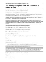
Fig. 2 Connectivity (bold lines) deduced by the 1H-1H Correlation Spectroscopy (COSY) spectrum and significant HMBC correlations (arrows)
observed for 1 and 2
29
18
H
C18H37COO
19
H
11
H
13
H
H
H
4
22
17
18
26
20
25
H
H
21
27
8
3
H
29
7
5
14
H
H
HOOCC7H14COO
19
11
8
3
H
H
4
13
22
20
17
26
25
21
27
H
1
H
H
7
5
14
H
H
2
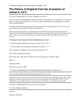
Fig. 3 Key NOESY correlations observed for compounds 1 and 2
Table 2 α-Glucosidase inhibitory activity of fractions
Fractions
IC50 (µg/mL)
Fractions
IC50 (µg/mL)
17.1
Fr. 6
69.3
EtOAc
>100
Fr. 7
5.1
MeOH
>100
Fr. 8
2.8
n-Hexane
Fr. 1
21.1
Fr. 9
4.7
Fr. 2
46.8
Fr. 10
1.9
Fr. 3
39.2
Fr. 11
11.6
Fr. 4
3.9
Fr. 12
28.9
Fr. 5
4.2
Acarbosea
138.4
a
Positive control
have not been discussed yet due to the limited number of
compounds. These results indicated that the strong active
compounds such as mekongsterol B (2; IC50, 2.5 μM)
and magiferonic acid (8; IC50, 1.2 μM) can potentially be
developed as a novel natural nutraceutical to decrease the
blood glucose level because of their strong α-glucosidase
inhibitory activity.
Methods
General experimental procedures
The IR spectra were measured with a Shimadzu IR-408
spectrophotometer in CHCl3 solution. The NMR spectra were taken on a Bruker Advance III 500 spectrometer
with tetramethylsilane (TMS) as an internal standard,
and chemical shifts are expressed in δ values. The HRESI–MS was performed on a Bruker MicroTOF-QII
spectrometer. The absorbance (OD) was measured with a
Shimadzu UV-1800 UV–Vis spectrophotometer.
Chemicals
α-Glucosidase (EC 3.2.1.20) from Saccharomyces cerevisiae (750 UN) and p-nitrophenyl-α-d-glucopyranoside
were obtained from Sigma Chemical Co. (St. Louis,
MO, USA). Acarbose and dimethylsulfoxide were purchased from Merck (Darmstadt, Germany). Silica gel
60, 40–63 µm (230–400 mesh ASTM), for column chromatography was purchased from Scharlau (Barcelona,
Spain). Analytical and preparative TLC were carried out
on precoated Kiesegel 60F254 or RP-18F254 plates (0.25 or
0.5 mm thickness) (Merck, Germany). Other chemicals
were of the highest grade available.
Plant material
The bark of M. mekongensis was collected at Ben Tre
province, Vietnam, in March 2013, and was identified by
Ms. Hoang Viet, Faculty of Biology, University of Science,
Vietnam National University-Hochiminh City (VNUHCMC). A voucher specimen (MDE0047) was deposited
at the Division of Medicinal Chemistry, Faculty of Chemistry, University of Science, VNU-HCMC.
Extraction and isolation
The dried powdered bark of M. mekongensis (6.0 kg) was
refluxed with n-hexane (5.0 L) in Sohxlet extractor to
yield a n-hexane fraction (14.7 g), continuously extracted
with EtOAc (5.0 L) to obtain EtOAc fraction (65.0 g), and
Nguyen et al. Chemistry Central Journal (2016) 10:45
Page 5 of 6
Table 3 α-Glucosidase inhibitory activity of the isolated compounds
Compounds
Inhibition (I %)
IC50 (µM)
250 (µM)
100 (µM)
50 (µM)
25 (µM)
10 (µM)
1
*
91.8 ± 1.1
67.7 ± 1.4
38.6 ± 1.2
24.4 ± 1.8
3
–
4
–
5
*
90.9 ± 1.4
75.9 ± 2.6
49.7 ± 3.1
32.1 ± 2.3
21.1
6
*
95.2 ± 2.3
85.6 ± 1.0
70.8 ± 1.2
39.0 ± 1.8
13.2
7
*
88.5 ± 1.0
75.7 ± 1.2
68.0 ± 1.1
32.9 ± 1.6
95.9 ± 1.0
32.2 ± 1.0
15.9 ± 1.1
59.8 ± 1.2
21.2 ± 2.2
9.8 ± 1.1
9
Acarbosea
Compounds
–
–
–
–
–
–
>250
–
>250
–
–
3.2 ± 1.7
–
Inhibition (I %)
25 (µM)
27.7
–
16.7
112.0
214.5
IC50 (µM)
10 (µM)
5.0 (µM)
2.5 (µM)
1.0 (µM)
2
*
93.1 ± 1.2
82.6 ± 1.4
50.8 ± 1.1
15.2 ± 1.2
2.5
8
*
87.5 ± 1.0
78.4 ± 1.6
62.5 ± 1.1
46.5 ± 1.1
1.2
* Not tested due to inessential result (IC50 values can be identified without these results)
– Not identified
a
Positive control
then extracted with MeOH (5.0 L) to give MeOH fraction
(108.0 g). The n-hexane fraction (12.5 g) was subjected to
silica gel column (6.5 × 120 cm) chromatography, eluted
with acetone–n-hexane (0–80 %) to yield 12 fractions: fr.
1 (0.1 g), fr. 2 (1.8 g), fr. 3 (1.1 g), fr. 4 (2.6 g), fr. 5 (1.4 g),
fr. 6 (0.8 g), fr. 7 (0.3 g), fr. 8 (0.8 g), fr. 9 (0.7 g), fr. 10
(0.6 g), fr. 11 (0.9 g), fr. 12 (1.4 g). All extractions and fractions were tested for their α-glucosidase inhibitory activity (Table 2).
Fraction 2 (1.8 g) was applied to silica gel column chromatography with acetone-n-hexane gradient system
to give six subfractions (fr. 2.1, 1.2 g; fr. 2.2, 134 mg; fr.
2.3, 75 mg; fr. 2.4, 47 mg; fr. 2.5, 89 mg; fr. 2.6, 270 mg).
Subfraction 2.1 was chromatographed further using an
CHCl3-n-hexane (0–80 %) to yield six subfractions fr.
2.1.1–6; fr. 2.1.1 (451 mg) was separated further using an
EtOAc-n-hexane (0–30 %) to afford 1 (25.0 mg).
Fraction 4 (2.6 g) was chromatographed on silica gel
column chromatography, eluted with EtOAc-n-hexane
gradient system to give six subfractions (fr. 4.1, 717 mg;
fr. 4.2, 202 mg; fr. 4.3, 993 mg; fr. 4.4, 150 mg; fr. 4.5,
78 mg; fr. 4.6, 460 mg). Subfraction 4.4 was recrystallized
with MeOH-CHCl3 to give 4 (12.0 mg).
Fraction 5 (1.4 g) was rechromatographed to silica gel
column chromatography with CHCl3-n-hexane gradient system to yield seven subfractions (fr. 5.1, 81 mg; fr.
5.2, 94 mg; fr. 5.3, 57 mg; fr. 5.4, 260 mg; fr. 5.5, 190 mg;
fr. 5.6, 88 mg; fr. 5.7, 630 mg). Subfraction 5.3 was chromatographed with EtOAc-n-hexane (0–50 %), and then
purified by normal-phase preparative TLC with CHCl3
(100 %) to give 3 (2.5 mg).
Fraction 6 (0.8 g) was applied to silica gel column
chromatography, eluted with CHCl3-n-hexane gradient
system to yield five subfractions (fr. 6.1, 124 mg; fr. 6.2,
192 mg; fr. 6.3, 272 mg; fr. 6.4, 42 mg g; fr. 6.5, 130 mg).
Subfraction 6.1 was also chromatographed on silica
gel with EtOAc-n-hexane (0–80 %), and then followed
by normal-phase preparative TLC with ethyl acetaten-hexane (25:75) to give 2 (8.0 mg). Subfraction 6.2 was
rechromatographed further using EtOAc-n-hexane
(0–80 %) and then purified by normal-phase preparative
TLC with CHCl3-n-hexane (10:90) to give 6 (6.0 mg) and
8 (10.0 mg).
Fraction 9 (0.7 g) was chromatographed on silica gel column chromatography, eluted with CHCl3-n-hexane gradient system to give four subfractions (fr. 9.1, 150 mg; fr.
9.2, 125 mg; fr. 9.3, 360 mg; fr. 9.4, 47 mg). Subfraction 9.3
was subjected to silica gel with EtOAc-n-hexane (0–80 %)
to yield two subfractions fr. 9.3.1–2; fr. 9.3.1 (190 mg)
was separated further using a CHCl3-n-hexane (0–80 %),
and then purified by normal-phase preparative TLC with
EtOAc-n-hexane (10:90) to give 7 (6.0 mg) and 9 (10.0 mg).
Fraction 11 (0.9 g) was chromatographed on silica gel
column chromatography, eluted with CHCl3-MeOH
gradient system to give five subfractions (fr. 11.1, 42 mg;
fr. 11.2, 139 mg; fr. 11.3, 93 mg; fr. 11.4, 30 mg; fr. 11.5,
570 mg). Subfraction 11.2 was subjected to silica gel with
EtOAc-n-hexane (0–50 %) to yield two subfractions fr.
11.1.1–2; fr. 11.2.1 (60 mg) was separated further using
an CHCl3-MeOH (0–30 %), and then purified by normal-phase preparative TLC with CHCl3-MeOH (96:4) to
afford 5 (8.0 mg).
Nguyen et al. Chemistry Central Journal (2016) 10:45
Mekongsterol A (1): white crystal; IR νmax (CHCl3)
2950, 2870, 1720, 1610 cm−1; HR-ESI–MS positive m/z
733.6223 [M + K]+ (calcd for C48H86O2K+, 733.6259,
error of – 3.6 mmu); 1H NMR (CDCl3, 500 MHz) and 13C
NMR (CDCl3, 125 MHz), see Table 1 (For further information, see Additional file 1).
Mekongsterol B (2): white crystal; IR νmax (CHCl3) 3500,
2960, 2860, 1730, 1710, 1600 cm−1; HR-ESI–MS positive m/z 607.4719 [M + Na]+ (calcd for C38H64O4Na+,
607.4697, error of 2.2 mmu); 1H NMR (CDCl3, 500 MHz)
and 13C NMR (CDCl3, 125 MHz), see Table 1 (For further
information, see Additional file 1).
Page 6 of 6
and design of the study, read and brought some corrections to the paper. All
the authors read and approved the final manuscript.
Author details
1
Faculty of Chemistry, University of Science, Vietnam National University
Hochiminh City, 227 Nguyen Van Cu, District 5, Hochiminh City, Vietnam.
2
Cancer Research Laboratory, Vietnam National University Hochiminh City,
227 Nguyen Van Cu, District 5, Hochiminh City, Vietnam.
Acknowledgements
This research is funded by Vietnam National University Hochiminh City (VNUHCM) under Grant number A2015-18-02.
Competing interests
The authors declare that they have no competing interests.
Received: 19 April 2016 Accepted: 14 July 2016
α‑Glucosidase inhibitory assay
The inhibitory activity of α-glucosidase was determined according to the modified method of Kim et al.
[15]. 3 mM p-nitrophenyl-α-d-glucopyranoside (25 μL)
and 0.2 U/mL α-glucosidase (25 μL) in 0.01 M phosphate buffer (pH = 7.0) were added to the sample solution (625 μL) to start the reaction. Each reaction was
carried out at 37 °C for 30 min and stopped by adding
0.1 M Na2CO3 (375 μL). Enzymatic activity was quantified by measuring absorbance at 401 nm. One unit of
α-glucosidase activity was defined as amount of enzyme
liberating p-nitrophenol (1.0 μM) per min. The IC50
value was defined as the concentration of α-glucosidase
inhibitor that inhibited 50 % of α-glucosidase activity.
Acarbose, a known α-glucosidase inhibitor, was used as
positive control.
Conclusions
In this paper, we have reported two new compounds,
mekongsterol A (1) and mekongsterol B (2), together
with seven known compounds isolated from the
bark of M. mekongensis. Seven compounds possessed
α-glucosidase inhibitory activity. This is the first report
on α-glucosidase inhibitory activity of the bark of this
plant. These results suggested that the traditional use
of the bark of M. mekongensis for the treatment of diabetes diseases in Vietnam may be attributable to the
α-glucosidase inhibitory activity of its steroid and
cycloartane constituents.
Additional file
Additional file 1. 1H, 13C, DEPT, COSY, HSQC, HMBC, and NOESY NMR,
and MS spectra of new compounds (1 and 2) have been provided as an
online file
Authors’ contributions
HXN and TCL isolated and elucidated the compounds, TNVD and THL carried
out the bioassay, NTN wrote the manuscript, MTTN carried out conception
References
1. Pham HH An illustrated flora of Vietnam. Youth Publishing House:
Hochiminh City. 2000
2. Dang BK, Pham MT, Ngo VQ, Dang TBO (2013) Total phenolic content and
anti-oxidant capacity of some spices and herbs grown in Vietnam. J Post
Harvest Technol 1:22–28
3. Bischoff H (1994) Pharmacology of α-glucosidase inhibition. Eur J Clin
Invest 24:3–10
4. Baron AD (1998) Postprandial hyperglycaemia and α-glucosidase inhibitors. Diabetes Res Clin Pract 40:S51–55
5. Dang PH, Nguyen HX, Nguyen NT, Le HN, Nguyen MT (2014)
α-Glucosidase inhibitors from the stems of Embelia ribes. Phytother Res
28:1632–1636
6. Dang PH, Nguyen NT, Nguyen HX, Nguyen LB, Le TH, Do TN, Can MV,
Nguyen MT (2015) α-Glucosidase inhibitors from the leaves of Embelia
ribes. Fitoterapia 100:201–207
7. Nguyen MTT, Nguyen NT, Nguyen HX, Huynh TNN, Min BS (2012) Screening of alpha-glucosidase inhibitory activity of Vietnamese medicinal
plants: Isolation of active principles from Oroxylum indicum. Nat Prod Sci
18:47–51
8. Mai NTT, Hai NX, Phu DH, Trong PNH, Nhan NT (2012) Three new geranyl
aurones from the leaves of Artocarpus altilis. Phytochem Lett 5:647–650
9. Chaturvedula VSP, Prakash I (2012) Isolation of stigmasterol and
β-sitosterol from the dichloromethane extract of Rubus suavissimus. Int
Curr Pharm J 1:239–242
10. Wei K, Li W, Koike K, Pei Y, Chen Y, Nikaido T (2004) Complete 1H and 13C
NMR assignments of two phytosterols from roots of Piper nigrum. Magn
Reson Chem 42:355–359
11. Yoon NY, Min BS, Lee HK, Park JC, Choi JS (2005) A potent anti-complementary acylated sterol glucoside from Orostachys japonicus. Arch
Pharmacal Res 28:892–896
12. Escobedo-Martínez C, Concepción Lozada M, Hernández-Ortega S,
Villarreal ML, Gnecco D, Enríquez RG, Reynolds W (2012) 1H and 13C NMR
characterization of new cycloartane triterpenes from Mangifera indica.
Magn Reson Chem 50:52–57
13. Anjaneyulu V, Satyanarayana P, Viswanadham KN, Jyothi VG, Rao KN,
Radhika P (1999) Triterpenoids from Mangifera indica. Phytochemistry
50:1229–1236
14. Van de Laar FA, Lucassen PL, Akkermans RP, Van de Lisdonk EH, Rutten GE,
Van Weel C (2005) α-Glucosidase inhibitors for type 2 diabetes mellitus.
Cochrane Database Syst Rev 18:CD003639
15. Kim KY, Nam KA, Kurihara H, Kim SM (2008) Potent α-glucosidase inhibitors purified from the red alga Grateloupia elliptica. Phytochemistry
69:2820–2825