Synthesis of acyl oleanolic acid-uracil conjugates and their anti-tumor activity
Bạn đang xem bản rút gọn của tài liệu. Xem và tải ngay bản đầy đủ của tài liệu tại đây (2.52 MB, 11 trang )
Mo et al. Chemistry Central Journal (2016) 10:69
DOI 10.1186/s13065-016-0217-5
Open Access
RESEARCH ARTICLE
Synthesis of acyl oleanolic acid‑uracil
conjugates and their anti‑tumor activity
Wei‑bin Mo1,3†, Chun‑hua Su1,2†, Jia‑yan Huang1,2, Jun Liu4*, Zhen‑feng Chen1,2* and Ke‑guang Cheng1,2*
Abstract
Background: Oleanolic acid, which can be isolated from many foods and medicinal plants, has been reported to
possess diverse biological activities. It has been found that the acylation of the hydroxyl groups of the A-ring in the
triterpene skeleton of oleanolic acid could be favorable for biological activities. The pyrimidinyl group has been con‑
structed in many new compounds in various anti-tumor studies.
Results: Five acyl oleanolic acid-uracil conjugates were synthesized. Most of the IC50 values of these conjugates were
lower than 10.0 μM, and some of them were even under 0.1 μM. Cytotoxicity selectivity detection revealed that con‑
jugate 4c exhibited low cytotoxicity towards the normal human liver cell line HL-7702. Further studies revealed that
4c clearly possessed apoptosis inducing effects, could arrest the Hep-G2 cell line in the G1 phase, induce late-stage
apoptosis, and activate effector caspase-3/9 to trigger apoptosis.
Conclusions: Conjugates of five different acyl OA derivatives with uracil were synthesized and identified as possess‑
ing high selectivity toward tumor cell lines. These conjugates could induce apoptosis in Hep-G2 cells by triggering
caspase-3/9 activity.
Keywords: Acyl oleanolic acid, Uracil, Anti-tumor activity, Cytotoxicity, Apoptosis
Background
Pentacyclic triterpenes, which are ubiquitous in the plant
kingdom, have important ecological and agronomic functions, and contribute greatly to pest and disease resistance and to food quality in crop plants [1]. They are also
applied in a variety of commercial uses in the food, cosmetic and pharmaceutical fields. For example, pentacyclic triterpene imberbic acid, isolated from Combretum
imberbe (Engl. and Diels), has been found to have particularly potent activity against Mycobacterium fortuitum and Staphylococcus aureus [2]. Other pentacyclic
triterpenes have been reported to possess antioxidant,
*Correspondence: ; ;
†
Wei-bin Mo and Chun-hua Su contributed equally to this work
1
State Key Laboratory for the Chemistry and Molecular Engineering
of Medicinal Resources, Guangxi Normal University, Guilin 541004,
People’s Republic of China
2
School of Chemistry and Pharmacy, Guangxi Normal University,
Guilin 541004, People’s Republic of China
4
Jiangsu Key Laboratory of Drug Screening, China Pharmaceutical
University, 24 Tongjia Xiang, Nanjing 210009, People’s Republic of China
Full list of author information is available at the end of the article
antiproliferative, and pro-apoptotic capacities on MCF-7
human breast cancer cells [3]. They were also reported as
a new class of glycogen phosphorylase inhibitors [4] and
further proved to be multi-target therapeutic agents for
the prevention and treatment of metabolic and vascular
diseases [5]. Oleanolic acid (3β-hydroxyolean-12-en-28oic acid, OA, 1, Fig. 1), which belongs to the family of
oleanane pentacyclic triterpenes, has been isolated from
more than 1620 plant species, including many food and
medicinal plants [6]. It is among the major effective components of some well-known traditional chinese medicines (TCM) such as Rehmannia Six Formula (Liu Wei Di
Huang Wan), which is one of the most commonly used
Chinese herb formulas in the world. It has been used as a
nonprescription antihepatitis drug for almost 35 years in
China [7]. Oleanolic acid and its derivatives have recently
attracted much attention due to their diverse biological
activities [8]. For instance, oleanolic acid and its derivatives were reported to be inhibitors of protein tyrosine
phosphatase 1B with cellular activities [9] and osteoclast
formation [10, 11]. These compounds were also focused
© The Author(s) 2016. This article is distributed under the terms of the Creative Commons Attribution 4.0 International License
( which permits unrestricted use, distribution, and reproduction in any medium,
provided you give appropriate credit to the original author(s) and the source, provide a link to the Creative Commons license,
and indicate if changes were made. The Creative Commons Public Domain Dedication waiver ( />publicdomain/zero/1.0/) applies to the data made available in this article, unless otherwise stated.
Mo et al. Chemistry Central Journal (2016) 10:69
Page 2 of 11
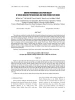
Fig. 1 Chemical structures of oleanane triterpene skeleton, oleanolic acid, maslinic acid and 2
on cytotoxicity evaluation [12]. Furthermore, some synthetic oleanane triterpenoids (CDDO, CDDO-Me and
CDDO-Im) have demonstrated potent antiangiogenic
and antitumor activities in rodent cancer models [13, 14].
Other biological activities of oleanolic acid and its derivatives, including antiproliferative activity in solid tumor
cells [15], inhibition of α-glucosidase [16], and others [6,
8], were also revealed.
The importance of C-3 in the oleanolic acid skeleton
was elucidated (Fig. 1). The SAR analysis of oleanolic
acid derivatives modified at C-3 and C-28 indicated that
hydrogen-bond acceptor substitution at the C-3 position
of oleanolic acid may be advantageous for the improvement of cytotoxicity against PC-3, A549 and MCF-7 cell
lines [12]. Gnoatto found that the derivative with an acetylation at C-3 of the oleanolic acid backbone had much
better activity against the L. amazonensis strain [17].
3-Oxo oleanolic acid (3-oxo-olea-12-en-28-oic acid), a
derivative of oleanolic acid modified at C-3, was found
to significantly inhibit the growth of cancer cells derived
from different tissues, particularly on melanoma in vivo
[18]. Some other acyl compounds, generated from the
modification of the hydroxyl groups of the A-ring in the
triterpene skeleton of oleanolic acid and maslinic acid
(MA, Fig. 1) with 10 different acyl groups, displayed
cytotoxic effects against b16f10 murine melanoma cells
and showed apoptotic effects with high levels of early and
total apoptosis (up to 90%). These acyl compounds also
exhibited better inhibition effects to anti-HIV-1-protease,
with IC50 values between 0.31 and 15.6 μM, which are
4–186 times lower than their non-acylated precursors
[19]. Compound 2 (Fig. 1), un benzyl (2α,3β) 2,3-diacetoxy-olean-12-en-28-amide, exhibited much better cytotoxicity against human tumor cell lines compared with its
deacylation product, while it showed a rather low cytotoxicity for human fibroblasts (WW030272) [20].
On the other hand, pyrimidine has been widely used
as an anti-tumor pharmacophore in medicinal chemical research [21]. For instance, some new pyridines and
pyrazolo [1,5-α] pyrimidines exhibited potent anti-tumor
cytotoxic activity in vitro against different human cell
lines [22]. The evaluation of several ring-A fused hybrids
of oleanolic acid against seven human cancer cell lines
showed that the fused pyrimidine moiety seemed important to enhance the antiproliferative activity of oleanolic
acid [23]. Thus, the pyrimidinyl group has been constructed in many new compounds in various anti-tumor
studies [24].
Results and discussion
Synthesis
Inspired by the cited evidence, in this study, we conjugated five different acyl OA derivatives (3a–3e) [15, 19,
20, 25, 26] with uracil. The synthetic routes are outlined
in Schemes 1 and 2. The treatment of 1 (1 equiv) with
anhydride (1.5 equiv) and DMAP (0.1 equiv) in anhydrous
CH2Cl2/pyridine (1/7 = v/v) at room temperature afforded
3-O-acyl derivatives 3a–3c [15, 19, 20, 25] (64–89%). The
Mo et al. Chemistry Central Journal (2016) 10:69
Page 3 of 11
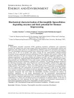
Scheme 1 Synthesis of acyl oleanolic acid derivatives. Reagents and conditions: (i) anhydride, DMAP, anhydrous CH2Cl2/pyridine, rt (64–89%); (ii)
acyl chloride, Et3N, anhydrous THF, rt (75–88%)
Scheme 2 Synthesis of oleanolic acid-uracil conjugates, where n is the number of methylene groups in the acyl group
treatment of 1 (1 equiv) with acyl chloride (3 equiv) and
Et3N (3.5 equiv) in anhydrous THF at room temperature
gave acyl derivatives 3d–3e [19, 26] (75–88%). The acyl
oleanolic acid compounds (3a–3e, 1 equiv) were then first
treated with oxalyl chloride (18 equiv) to give the corresponding acyl chloride, which was then treated with uracil
(3 equiv) in the presence of Et3N to generate the corresponding acyl oleanolic acid-uracil conjugates (4a–4e,
Scheme 2) in 11–60% yields. The structures of compounds
4a–4e were confirmed by NMR and mass spectra.
Cytotoxicity
As anti-tumor effects are the most classical activities of
oleanolic acid and its derivatives [1, 27–29], these conjugates have been evaluated by MTT assay [30, 31] against
5 adherent tumor cell lines (Hep-G2, A549, BGC-823,
MCF-7 and PC-3) with 1 as the positive control. 5-Fluorouracil (5-FU), a medication used in the clinical treatment of cancer, is also a pyrimidine analog and was used
as a positive control in this study. The results are presented in Table 1.
The results showed that these compounds exhibited
excellent antiproliferative activities against the tested
cells, with the IC50 values mainly under 10.0 μM, except
for compounds 4a and 4b which showed no inhibition
against the PC-3 cell line. In the Hep-G2, A549, BGC823 and MCF-7 cell line assays, all the synthesized compounds displayed much better inhibition than that of 1
and 5-FU. With a propionyloxy group at C-3, compound
4b possessed the best inhibition activity against the
Hep-G2 cell line, almost 5.5-fold and 20-fold stronger
than 1 and 5-FU, respectively. With a dodecanoyloxy
group at C-3, compound 4d showed the best inhibition
activity against the A549 cell line, almost 60-fold and
84-fold stronger than 1 and 5-FU, respectively. Meanwhile, compound 4a, with an acetoxy group at C-3,
exhibited the best inhibition activity against the MCF-7
cell line, more than 126-fold and 215-fold more effective
than 1 and 5-FU respectively. Compounds 4a (acetoxy),
4b (propionyloxy) and 4e (palmitoxy) exhibited excellent
antiproliferative activities against the BGC-823 cell line
(IC50 < 0.1 μM). Although compounds 4a (acetoxy) and
4b (propionyloxy) possessed good antiproliferative activities against the Hep-G2, A549, BGC-823 and MCF-7
cell lines, they showed no inhibition against the PC-3
cell line. In the PC-3 assay, the butyryloxy compound 4c
exhibited the best antiproliferative activity, being 260fold and 44-fold stronger than 1 and 5-FU, respectively.
The results above reveal that in general, the acyl groups
at the C-3 position of these uracil conjugates have primarily made a great contribution to the antiproliferative
activities against the tested cell lines.
For further analysis, conjugate 4c was selected to determine its cytotoxicity selectivity and mechanism of growth
inhibition on an adherent Hep-G2 cell line. The controls
of the figures were reused from our previous work [32].
Cytotoxicity selectivity
As shown in Fig. 2, though the inhibition rate of 4c
against human liver cell line HL-7702 (L-O2) at the
Mo et al. Chemistry Central Journal (2016) 10:69
Page 4 of 11
Table 1 Evaluation of 4a–4e against different tumor cell lines
Compounds
IC50 (μM)a
Hep-G2
4a
7.83 ± 0.69
A549
4.01 ± 0.37
BGC-823
MCF-7
PC-3
<0.1
<0.1
NIb
4b
2.81 ± 0.22
15.5 ± 1.34
<0.1
6.53 ± 0.35
NI
4c
5.24 ± 0.05
3.01 ± 0.28
0.22 ± 0.02
6.99 ± 0.57
0.28 ± 0.05
4d
8.49 ± 0.68
0.27 ± 0.03
0.11 ± 0.01
3.51 ± 0.22
10.61 ± 1.13
4e
4.19 ± 0.35
8.76 ± 0.07
< 0.1
5.26 ± 0.41
2.51 ± 0.05
1
15.90 ± 1.13
16.29 ± 1.36
23.74 ± 1.53
12.60 ± 1.09
72.74 ± 6.88
5-FU
55.74 ± 5.09
22.62 ± 2.19
8.82 ± 0.78
21.47 ± 1.99
12.23 ± 1.18
a
IC50 values are presented as the mean ± SD (standard deviation) from three separated experiments
b
No inhibition detected
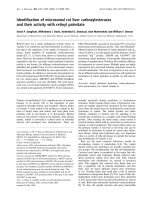
Fig. 2 Cytotoxicity of 4c against Hep-G2 tumor cells and
HL-7702 human liver cells. Hep-G2 and HL-7702 cells were cultured
in medium in the presence of the indicated concentrations of 4c for
72 h
concentration of 50 μM was equivalent to that of human
hepatoma cell line Hep-G2, its inhibition rate against the
HL-7702 cell line was only approximately 15% at the concentration of 10 μM, while the inhibition rate against the
Hep-G2 cell line was up to 90% at the same concentration. Thus, it was exhibited that compound 4c showed
strong cytotoxicity selectivity to human hepatocellular
carcinoma cells in vitro at the therapeutically effective
concentration.
Fluorescence staining
After sequentially staining with acridine orange (AO)/
ethidium bromide (EB) (Fig. 3) and Hoechst 33258
(Fig. 4), the living cells were treated with compound 4c
(2.5 and 5.0 μM, 24 h). As depicted in Fig. 3, the living
cells excluded EB and staining by AO caused a green
color (Fig. 3a), whereas the Hep-G2 cells treated with 4c
had obviously changed (Fig. 3b, c). Under fluorescence
microscopy, early apoptosis cells were observed to emit
orange or dark orange fluorescence, with nuclear morphological changes, which suggested that 4c could
induce apoptosis in Hep-G2 cells. This is consistent with
the results of Hoechst 33258 staining shown in Fig. 4.
The nuclei of the Hep-G2 cells retained the regular
round contours in the control group (Fig. 4a), and cells
with smaller nuclei and condensed chromatin were rarely
observed. It was found that the contours of some of the
Hep-G2 cells became irregular even when they were
exposed to 4c at lower concentration of 2.5 μM, accompanied with the nuclei being condensed (as the bright
blue fluorescence indicates) and the apoptotic bodies
appeared (Fig. 4b). When treated with 4c at a higher concentration of 5.0 μM, the nuclei of many more cells were
highly condensed and the apoptotic bodies were pervasive in the visual field (Fig. 4c). These clear changes to the
cell morphology suggested the significant cell apoptosis
induction of 4c on Hep-G2 cells.
Cell cycle analysis
To confirm whether the decrease of cell viability was
caused by cell cycle arrest, the Hep-G2 cells were treated
with compound 4c for 48 h at different concentrations
and then the cell cycle distribution was determined by a
flow cytometry assay after propidium iodide (PI) staining
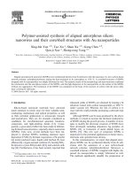
(Fig. 5). The results indicate that conjugate 4c enhanced
the cell cycle arrest at the G1 phase at different concentrations, resulting in a concomitant population increase
in the G1 phase (60.65–86.49, 87.66 and 73.54%), and
declines in the cell population in the G2/M (13.83–2.09,
1.40 and 3.05%) and S-phases (25.52–11.42, 10.95 and
23.41%).
AnnexinV/propidium iodide assay
To determine whether the observed cell death induced by
conjugate 4c was due to apoptosis or necrosis, the interactions of Hep-G2 cells with 4c were further investigated
Mo et al. Chemistry Central Journal (2016) 10:69
Page 5 of 11
Fig. 3 Cell morphological observation for cell apoptosis induction on the Hep-G2 cells treated by 4c. a Control cells; b, c cells treated by 4c for
24 h; cells were stained by AO/EB, and selected visual fields illustrating the corresponding live cells, early apoptotic cells (white arrow) are shown
(magnification ×200)
Fig. 4 Cell morphological observation for cell apoptosis induction on the Hep-G2 cells treated by conjugate 4c. a Control cells; b, c cells treated by
4c for 24 h; cells were stained by Hoechst 33258, and selected visual fields illustrating the condensed chromatin (white arrow) as occurrence of cell
apoptosis are shown (magnification ×200)
using an Annexin V-FITC/PI assay (Fig. 6). The apoptosis
ratios (including the early and late apoptosis ratios) of 4c
measured at different concentration points were found
to be 16.31% (2.5 μM) and 26.43% (5.0 μM), respectively,
while that of the control was 4.06%. This revealed that 4c
could mainly induce later period apoptosis in Hep-G2
cells.
analysis were tested (Fig. 7). The results indicated that 4c
induced a marked concentration-dependent decrease of
Rhodamine 123 fluorescence (decreasing from 86.2% to
85.8, 81.3 and 65.7% with the increase concentration of
4c). This indicated that compound 4c can induce mitochondrial membrane potential disruption in Hep-G2
cells.
Mitochondrial membrane potential detection
Caspase‑3/9 activity assay
As the mitochondrial membrane potential (Δψ) has been
considered a new antitumor target [33, 34], the changes
in Δψ in Hep-G2 cells (treated with conjugate 4c) stained
with Rhodamine 123 indicated by flow-cytometric
FITC-DEVD-FMK (for caspase-3) and FITC-LEHDFMK (for caspase-9) probe assays were carried out to
determine the death signaling in the caspase family after
treatment with 4c (5.0 μM, 24 h) in Hep-G2 cells. The
Mo et al. Chemistry Central Journal (2016) 10:69
Page 6 of 11
Fig. 5 Cell cycle progress detection by flow cytometry assay after PI staining of Hep-G2 cells following treatment with compound 4c: a control
cells; b, c, d cells treated by 4c for 48 h; e corresponding histograms. *P < 0.05 and **P < 0.01 compared with the control
Fig. 6 Apoptosis ratio detection by Annexin V/PI assay on the Hep-G2 cells treated by conjugate 4c: a control cells; b, c cells treated by 4c for 24 h
proportion of activated-caspase-3 cells after 4c treatment
was enhanced to 22.7% (Fig. 8a), while that of activatedcaspase-9 cells was enhanced to 22.9% (Fig. 8b). These
results indicate that 4c could induce cell apoptosis by
triggering caspase-3/9 activity in Hep-G2 cells.
Conclusions
Five acyl oleanolic acid-uracil conjugates were synthesized and their anti-tumor activities were
evaluated. These conjugates exhibited excellent antiproliferative activities against the tested cells (Hep-G2, A549,
Mo et al. Chemistry Central Journal (2016) 10:69
Page 7 of 11
Fig. 7 Collapse of mitochondrial membrane potential in the Hep-G2 cells treated by conjugate 4c. a Control cells; b, c, d cells treated by 4c for
24 h; cells were stained with Rhodamine 123 for 30 min. The results are expressed as relative fluorescent intensity
BGC-823, MCF-7 and PC-3) except compounds 4a and
4b, which showed no inhibition against the PC-3 cell line.
Most of the IC50 values were under 10.0 μM, with some
of them even under 0.1 μM.
Conjugate 4c was selected for further analysis including for its cytotoxicity selectivity and its mechanism of
growth inhibition on the Hep-G2 cell line. The inhibition
rate of 4c against the HL-7702 cell line was only approximately 15% (90% against Hep-G2) at the concentration of
10 μM, indicating that it had strong cytotoxicity selectivity to human hepatocellular carcinoma cells in vitro. The
treatment of Hep-G2 cells with this compound, could
induce changes in the permeability of the mitochondrial
membrane, and thus cause a decline in the mitochondrial membrane potential. With the disruption of the
mitochondrial membrane potential, changes in cellular
morphology appeared as a result of significant apoptosis induction. Then, cell proliferation in the G1 phase
was arrested and apoptotic signaling activated caspase-9.
Caspase-9, as a protease, can activate the apoptotic
effector caspase-3, eventually causing nuclear apoptosis. Further studies of the specific mechanisms of these
compounds in human malignant tumors are currently
underway.
Mo et al. Chemistry Central Journal (2016) 10:69
Page 8 of 11
Fig. 8 Activation of caspase-3/9 by conjugate 4c in Hep-G2 cells after treatment with 5.0 μM for 24 h: a activation of caspase-3; b activation of
caspase-9
Methods
General
All commercially available solvents and reagents used were
of analytical grade and were used without further purification. All commercial reagents were purchased from Aladdin (Shanghai) Industrial Corporation. Melting points
were measured on a RY-1 melting point apparatus. 1H
and 13C-NMR spectra were recorded on Bruker AV-500
(500/125 MHz for 1H/13C) spectrometers. Chemical shifts
are reported as values relative to an internal tetramethylsilane standard. The low-resolution mass spectra were
obtained on a Bruker Esquire HCT spectrometer, and
HRMS were recorded on a Thermo Scientific Accela—
Exactive High Resolution Accurate Mass spectrometer.
Chemistry
General procedures for synthesis of oleanolic acid‑uracil
conjugates 4a–4e
To a solution of different acyl oleanolic acid compound
(3a–3e, 0.2 mmol) in anhydrous CH2Cl2 (3 mL) at 0 °C,
oxalyl chloride (0.34 mL, 3.6 mmol) was added. After
stirring at room temperature for 12 h, the mixture was
evaporated, and co-evaporated with CH2Cl2 (3 × 1 mL).
The residue was dissolved in dry THF (3 mL), and then
Et3N (1 mL, 0.7 mmol) and uracil (0.067 g, 0.6 mmol)
were added at 0 °C. After stirring at r.t. for 24 h, the
solvent was evaporated. The residue was then taken up
in H2O (35 mL) and extracted with CH2Cl2 (3 × 20 mL).
The combined organic layers were washed with H2O and
brine, dried with MgSO4, filtered and concentrated to
give a crude product. The crude product was purified by
flash column chromatography to afford the corresponding product (4a–4e) respectively (Additional file 1).
Compound 4a
Compound 4a was prepared from 3a [15, 19, 20] (1.000 g,
2.00 mmol) and uracil (0.673 g, 6.00 mmol) according to
the general procedure. The residue was purified by flash
column chromatography (Petroleum ether:EtOAc = 4:
1). Yield: 0.349 g, 29%, white solid, mp 209–211 °C. 1H
NMR (500 MHz, CDCl3) δ 0.69, 0.85, 0.86, 0.99 and 1.17
(5 s, each 3H, 5 × CH3), 0.92 (s, 6H, 2 × CH3), 0.63–2.15
(m, 22H), 2.05 (s, 3H, CH3COO), 2.99 (dd, 1H, J = 3.2,
13.2 Hz, H-18), 4.48 (dd, 1H, J = 6.6, 9.3 Hz, H-3), 5.28
(t, 1H, J = 3.2 Hz, H-12), 5.74 (d, 1H, J = 8.3 Hz, H-5Ura),
7.51 (d, 1H, J = 8.3 Hz, H-6Ura), 8.20 (brs, 1H, NH). 13C
NMR (125 MHz, CDCl3) δ 15.6, 16.8, 18.2, 18.3, 21.4,
23.0, 23.7, 24.0, 26.1, 27.7, 28.2, 30.0, 30.7, 32.7, 33.2,
33.9, 37.0, 37.9, 38.4, 39.8, 42.0, 43.2, 46.5, 47.5, 53.3,
55.3, 81.0, 103.0, 123.6, 141.7, 143.3, 148.8, 162.7, 171.2,
182.0. HRMS (ESI) m/z: [M−H]+ calcd for C36H51N2O5,
591.3798; found 591.3808.
Mo et al. Chemistry Central Journal (2016) 10:69
Compound 4b
Compound 4b was prepared from 3b [19, 25] (0.300 g,
0.59 mmol) and uracil (0.196 g, 1.75 mmol) according to the general procedure. The residue was purified by flash column chromatography (Petroleum
ether:EtOAc = 5: 1). Yield: 0.213 g, 60%, white solid, mp
119–121 °C. 1H NMR (500 MHz, CDCl3) δ 0.69, 0.84,
0.85, 0.98 and 1.17 (5 s, each 3H, 5 × CH3), 0.92 (s, 6H,
2 × CH3), 1.14 (t, J = 7.6 Hz, CH3), 0.63–2.13 (m, 22H),
2.32 (q, 2H, J = 7.3 Hz, CH2COO), 2.99 (dd, 1H, J = 2.5,
12.8 Hz, H-18), 4.48 (dd, 1H, J = 6.5, 9.3 Hz, H-3), 5.28
(s, 1H, H-12), 5.74 (d, 1H, J = 8.3 Hz, H-5Ura), 7.50 (d,
1H, J = 8.3 Hz, H-6Ura), 8.97 (brs, 1H, NH). 13C NMR
(125 MHz, CDCl3) δ 9.4, 15.6, 16.9, 18.1, 18.2, 22.9,
23.6, 24.0, 26.1, 27.7, 28.1, 28.2, 30.0, 30.6, 32.7, 33.1,
33.9, 37.0, 37.9, 38.4, 39.8, 42.0, 43.2, 46.5, 47.5, 53.2,
55.3, 80.6, 103.0, 123.6, 141.7, 143.3, 149.0, 163.4, 174.4,
182.0. HRMS (FTMS + pESI) m/z: [M−H]+ calcd for
C37H53N2O5, 605.3955; found 605.3979.
Compound 4c
Compound 4c was prepared from 3c [19, 25] (0.200 g,
0.38 mmol) and uracil (0.127 g, 1.14 mmol) according to
the general procedure. The residue was purified by flash
column chromatography (Petroleum ether:EtOAc = 3: 1).
Yield: 0.131 g, 56%, white solid, mp 285–287 °C. 1H NMR
(500 MHz, CDCl3) δ 0.69, 0.85, 0.86, 0.95, 0.99 and 1.18
(6 s, each 3H, 6 × CH3), 0.92 (s, 6H, 2 × CH3), 0.63–2.15
(m, 24H), 2.28 (t, 2H, J = 7.1 Hz, CH2COO), 2.99 (dd, 1H,
J = 3.3, 13.2 Hz, H-18), 4.49 (dd, J = 5.7, 10.1 Hz, 1H,
H-3), 5.28 (s, 1H, H-12), 5.74 (d, 1H, J = 8.3 Hz, H-5Ura),
7.50 (d, 1H, J = 8.3 Hz, H-6Ura), 8.30 (brs, 1H, NH). 13C
NMR (100 MHz, CDCl3) 13.7, 15.4, 16.8, 17.2, 18.2, 18.6,
23.4, 23.6, 25.7, 25.8, 27.8, 28.0, 30.7, 32.0, 32.7, 33.0, 33.8,
36.8, 36.9, 37.7, 38.1, 40.0, 41.3, 41.9, 45.8, 47.4, 47.5,
55.3, 80.5, 104.5, 112.3, 123.0, 139.6, 142.9, 149.0, 161.8,
173.5, 175.2. APCI-MS m/z: 619.4 [M−H]+. HRMS (ESI)
m/z: [M−H]+ calcd for C38H55N2O5, 619.4111; found
619.4113.
Compound 4d
Compound 4d was prepared from 3d [19] (0.280 g,
0.44 mmol) and uracil (0.148 g, 1.32 mmol) according to
the general procedure. The residue was purified by flash
column chromatography (Petroleum ether:EtOAc = 5:
1). Yield: 0.072 g, 22%, white solid. 1H NMR (500 MHz,
CDCl3) δ 0.70, 1.00 and 1.19 (3 s, each 3H, 3 × CH3), 0.87
and 0.93 (2 s, each 6H, 4× CH3), 0.63–2.10 (m, 43H), 2.30
(s, 2H, CH2COO), 3.01 (d, 1H, J = 11.8 Hz, H-18), 4.50
(s, 1H, H-3), 5.30 (s, 1H, H-12), 5.74 (d, 1H, J = 7.5 Hz,
H-5Ura), 7.52 (d, 1H, J = 7.7 Hz, H-6Ura), 8.72 (brs, 1H,
NH). 13C NMR (125 MHz, CDCl3) δ 14.3, 15.6, 16.9, 18.1,
18.2, 22.8, 23.0, 23.6, 24.0, 25.3, 26.1, 27.7, 28.2, 29.3,
Page 9 of 11
29.4, 29.5, 29.6, 29.7, 30.0, 30.7, 32.1, 32.6, 33.2, 33.9,
35.0, 37.0, 37.9, 38.4, 39.8, 42.0, 43.2, 46.5, 47.5, 53.2,
55.3, 80.5. 103.0, 123.6, 141.7, 143.3, 148.9, 163.1, 173.9,
182.0. HRMS (ESI) m/z: [M−H]+ calcd for C46H71N2O5,
731.5363; found 731.5387.
Compound 4e
Compound 4e was prepared from 3e [26] (0.347 g,
0.50 mmol) and uracil (0.168 g, 1.50 mmol) according to
the general procedure. The residue was purified by flash
column chromatography (Petroleum ether:EtOAc = 5:
1). Yield: 0.044 g, 11%, white solid, mp 89–91 °C. 1H
NMR (500 MHz, CDCl3) δ 0.69, 0.85, 0.86, 0.99 and 1.18
(5 s, each 3H, 5× CH3), 0.92 (s, 6H, 2× CH3), 0.63–2.10
(m, 51H), 2.29 (t, 2H, J = 7.5 Hz, CH2COO), 2.99 (dd, 1H,
J = 2.7, 12.9 Hz, H-18), 4.48 (dd, 1H, J = 5.9, 9.8 Hz, H-3),
5.28 (s, 1H, H-12), 5.74 (d, 1H, J = 8.3 Hz, H-5Ura), 7.50
(d, 1H, J = 8.3 Hz, H-6Ura), 8.17 (brs, 1H, NH). APCI-MS
m/z: 787.7 [M−H]+. HRMS (ESI) m/z: [M−H]+ calcd for
C50H79N2O5, 787.5989; found 787.6003.
Cell lines and culture
The human hepatocellular cell line Hep-G2, human
lung cancer cell line A549, human gastric tumor cell line
BGC-823, human breast tumor cell line MCF-7, human
prostate cancer cell line PC-3 and human hepatocyte cell
line HL-7702, these adherent cells were purchased from
the Cell Bank of Type Culture Collection of the Chinese
Academy of Sciences (Shanghai) and cultured in DMEM
medium supplemented with 10% FCS (Fetal Calf Serum).
The cells were incubated in an atmosphere of 5% CO2
and 95% air at 37 °C.
MTT assay
The MTT assay was carried out according to a description in a published study [30, 31]. Cells were seeded in
96-well plates and incubated in a CO2 incubator at 37 °C.
The tested compounds were dissolved in fresh culture
medium with 2% DMSO to afford various concentrations
(100, 50, 10, 5, 1 or 0.1 μmol/L). When the cells adhered,
compounds at different concentrations were added to
every well. The control wells contained medium supplemented with 2% DMSO. After incubation for another
72 h, 20 μL MTT (5%) was added to each well, and the
cells were incubated for an additional 4 h at 37 °C. At last,
the medium was removed carefully and dimethyl sulfoxide (100 μL) was added to each well. Then the plate was
kept on a shaker for 10 min to mix these solutions properly. The absorbance of each well was scanned with an
electrophotometer at 570 nm. Each concentration treatment was performed in triplicate wells. The IC50 values
were estimated by fitting the inhibition data to a dosedependent curve using a logistic derivative equation.
Mo et al. Chemistry Central Journal (2016) 10:69
AO/EB staining
This assay was carried out according to a description in
a published study [35]. Cells were seeded at a concentration of 5 × 10⁴ cell/mL in a volume of 2 mL on a sterile cover slip in six-well tissue culture plates. Following
incubation, the RPMI 1640 medium was removed and
replaced with fresh medium plus 10% FCS and supplemented with compound 4c at the indicated concentration. After the treatment period (24 h), the cover slip
with monolayer cells was inverted on a glass slide with
20 μL of AO/EB stain (100 mg/mL). The fluorescence was
read on a Nikon ECLIPSETE2000-S fluorescence microscope (Japan).
Hoechst 33258 staining
This assay was carried out according to a description in a
published study [35]. Cells grown on a sterile cover slip in
six-well tissue culture plates were treated with compound
for a certain range of time. The culture medium containing compounds was removed, and the cells were fixed
in 4% paraformaldehyde for 10 min. After being washed
twice with PBS, the cells were stained with 0.5 mL of
Hoechst 33258 (0.5 μg/mL, Beyotime) for 5 min and
then again washed twice with PBS. The stained nuclei
were observed under a Nikon ECLIPSETE2000-S fluorescence microscope using 350 nm excitation and 460 nm
emission.
Mitochondrial membrane potential measurement
This assay was carried out as described in a published
study [34]. The depolarization of the mitochondrial
membrane potential for cell apoptosis results in the loss
of Rhodamine123 from the mitochondria and a decrease
in the intracellular fluorescence intensity. Prepared HepG2 cells were harvested and washed twice in cold PBS
and then resuspended in Rhodamine 123 (2 μM) for
30 min in the dark. The fluorescence was measured by
flow cytometry with an excitation wavelength of 485 nm
and emission wavelength of 530 nm.
Flow cytometric analysis of cell cycle and apoptosis
This assay was carried out according to a description in a
published study [35]. The induced apoptosis was assayed
by the Annexin V-FITC Apoptosis Detection kit (Beyotime, China), according to the manufacturer’s instructions. Briefly, the prepared Hep-G2 cells (1 × 10⁶ cells/
mL) were washed twice with ice-cold PBS and then
resuspended gently in 500 μL of binding buffer. Thereafter, the cells were stained in 5 μL of Annexin V-FITC and
shaken well. Finally, the cells were mixed with 5 μL of PI,
incubated for 20 min in the dark and subsequently analyzed using an FACS AriaII (Becton–Dickinson).
Page 10 of 11
Determination of caspase‑3 and caspase‑9 activities
by flow cytometric analysis
According to a description in a published study [35], the
measurement of the caspase-3 and caspase-9 activities
was performed by a CaspGLOW™ Fluorescein Active
Caspase-3 and Caspase-9 Staining Kit. The prepared
Hep-G2 cells were harvested at a density of 1 × 10⁶ cells/
mL in RPMI 1640 medium supplemented with 10% FCS.
A total of 300 μL each from the induced and control cultures were incubated with 1 μL of FITC-DEVD-FMK
(caspase-3) or FITC-LEHD-FMK (caspase-9) for 1 h in a
37 °C incubator with 5% CO2. Flow cytometric analysis
was performed using a FACS AriaII flow cytometer (Becton–Dickinson) equipped with a 488 nm argon laser.
Statistical analysis
The experiments were repeated three times, and the
results were presented as mean ± standard deviation
(SD). Student’s t test was used to process the statistical
significance and the differences between groups with
P < 0.05 were considered significant.
Additional file
Additional file 1. The copies of NMR spectra of compounds 4a–4e
Abbreviations
A549: human alveolar adenocarcinoma cell line; Annexin V-FITC: apoptosis
detection kit; AO/EB: acridine orange/ethidium bromide; BGC-823: human
grastic cancer cell line; CH2Cl2: dichloromethane; CDDO: 2-cyano-3,12-diox‑
ooleana-1,9-dien-28-oic acid; CDDO-Im: 2-cyano-3,12-dioxooleana-1,9-dien28-imidazolide; CDDO-Me: 2-cyano-3,12-dioxooleana-1,9-dien-28-oic acid
methyl ester; DMAP: 4-dimethylaminopyridine; DMEM: dulbecco’s modified
eagle’s medium; DMSO: dimethyl sulfoxide; Et3N: triethylamine; EtOAc: ethyl
acetate; 5-FU: 5-fluorouracil; FCS: fetal calf serum; FITC-DEVD-FMK: caspase-3
detection kit; FITC-LEHD-FMK: caspase-9 detection kit; G1 phase: gap1,
pre-synthetic period; G2/M: gap2, post-synthetic period/mitosis; H2O: water;
Hep-G2: human hepatoma cell line; HL-7702 (L-O2): hepatic immortal cell
line; HRMS: high resolution mass spectrometry; IC50: half maximal inhibitory
concentration; MA: maslinic acid; MCF-7: human breast adenocarcinoma cell
line; MgSO4: magnesium sulfate; MTT: 3-(4, 5-dimethyl-2-thiazolyl)-2,5-diphe‑
nyl-2-H-tetrazolium bromide; NMR: nuclear magnetic resonance; OA: oleanolic
acid; PBS: phosphate buffered saline; PC-3: human prostatic carcinoma cell
line; PI: propidium iodide; RPMI: roswell park memorial institute; SAR: struc‑
ture–activity relationships; SD: standard deviation; S-phase: synthetic period;
TCM: traditional Chinese medicines; THF: tetrahydrofuran; Δψ: the mitochon‑
drial membrane potential; μM: micromolar.
Authors’ contributions
WBM developed the pharmacology part and co-wrote the manuscript, CHS
developed the synthesis and co-wrote the manuscript, JYH participated in
the synthesis, JL, ZFC and KGC conceived and designed the experiments. All
authors read and approved the final manuscript.
Author details
State Key Laboratory for the Chemistry and Molecular Engineering of Medici‑
nal Resources, Guangxi Normal University, Guilin 541004, People’s Republic
of China. 2 School of Chemistry and Pharmacy, Guangxi Normal University,
Guilin 541004, People’s Republic of China. 3 Biochemistry and Pharmacology
of Sport School, Guangxi Normal University, Guilin 541004, People’s Republic
1
Mo et al. Chemistry Central Journal (2016) 10:69
of China. 4 Jiangsu Key Laboratory of Drug Screening, China Pharmaceutical
University, 24 Tongjia Xiang, Nanjing 210009, People’s Republic of China.
Acknowledgements
The controls of the figures in this study were reused from our previous work
with permission from the Royal Society of Chemistry (Med. Chem. Commun.,
2016, 7, 972. doi: 10.1039/c6md00061d). This study was financially supported
by Grants from the National Natural Science Foundation of PRC (21562006),
the Guangxi Natural Science Foundation of China (2015GXNSFAA139186),
the Key Laboratory for the Chemistry and Molecular Engineering of Medicinal
Resources (Guangxi Normal University), the Ministry of Education of China
(CMEMR2012-B03/B04, CMEMR2013-A01/C02), Guangxi’s Medicine Talented
Persons Small Highland Foundation (1506), IRT1225 and the Bagui Scholar
Program of Guangxi Province of China.
Competing interests
The authors declare that they have no competing interests.
Received: 4 February 2016 Accepted: 10 November 2016
References
1. Patlolla JMR, Rao CV (2012) Triterpenoids for cancer prevention and
treatment: current status and future prospects. Curr Pharm Biotechnol
13:147–155
2. Katerere DR, Gray AI, Nash RJ, Waigh RD (2003) Antimicrobial activity of
pentacyclic triterpenes isolated from African Combretaceae. Phytochem‑
istry 63:81–88
3. Allouche Y, Warleta F, Campos M, Sánchez-Quesada C, Uceda M, Beltrán
G, Gaforio JJ (2011) Antioxidant, antiproliferative, and pro-apoptotic
capacities of pentacyclic triterpenes found in the skin of olives on MCF-7
human breast cancer cells and their effects on DNA damage. J Agric
Food Chem 59:121–130
4. Wen X, Sun H, Liu J, Cheng K, Zhang P, Zhang L, Hao J, Zhang L, Ni P,
Zographos SE, Leonidas DD, Alexacou KM, Gimisis T, Hayes JM, Oikon‑
omakos NG (2008) Naturally occurring pentacyclic triterpenes as inhibi‑
tors of glycogen phosphorylase: synthesis, structure-activity relationships,
and X-ray crystallographic studies. J Med Chem 51:3540–3554
5. Sheng H, Sun H (2011) Synthesis, biology and clinical significance of pen‑
tacyclic triterpenes: a multi-target approach to prevention and treatment
of metabolic and vascular diseases. Nat Prod Rep 28:543–593
6. Pollier J, Goossens A (2012) Oleanolic acid. Phytochemistry 77:10–15
7. Liu J (1995) Pharmacology of oleanolic acid and ursolic acid. J Ethnophar‑
macol 49:57–68
8. Dzubak P, Hajduch M, Vydra D, Hustova A, Kvasnica M, Biedermann
D, Markova L, Urban M, Sarek J (2006) Pharmacological activities of
natural triterpenoids and their therapeutic implications. Nat Prod Rep
23:394–411
9. Zhang YN, Zhang W, Hong D, Shi L, Shen Q, Li JY, Li J, Hu LH (2008)
Oleanolic acid and its derivatives: new inhibitor of protein tyrosine phos‑
phatase 1B with cellular activities. Bioorg Med Chem. 16:8697–8705
10. Zhang Y, Li JX, Zhao JW, Wang SZ, Pan Y, Tanaka K, Kadota S (2005) Syn‑
thesis and activity of oleanolic acid derivatives, a novel class of inhibitors
of osteoclast formation. Bioorg Med Chem Lett 15:1629–1632
11. Li JF, Chen SJ, Zhao Y, Li JX (2009) Glycoside modification of oleanolic acid
derivatives as a novel class of anti-osteoclast formation agents. Carbo‑
hydr Res 344:599–605
12. Hao J, Liu J, Wen X, Sun H (2013) Synthesis and cytotoxicity evaluation of
oleanolic acid derivatives. Bioorg Med Chem Lett 23:2074–2077
13. Wang YY, Zhe H, Zhao R (2014) Preclinical evidences toward the use of
triterpenoid CDDO-Me for solid cancer prevention and treatment. Mol
Cancer. 13:30
14. Shanmugam MK, Dai X, Kumar AP, Tan BK, Sethi G, Bishayee A (2014)
Oleanolic acid and its synthetic derivatives for the prevention and ther‑
apy of cancer: preclinical and clinical evidence. Cancer Lett 346:206–216
Page 11 of 11
15. Leal AS, Wang R, Salvador JAR, Jing YK (2013) Synthesis of novel hetero‑
cyclic oleanolic acid derivatives with improved antiproliferative activity in
solid tumor cells. Org Biomol Chem 11:1726–1738
16. Ali MS, Jahangir M, ul Hussan SS, Choudhary MI (2002) Inhibition of
α-glucosidase by oleanolic acid and its synthetic derivatives. Phytochem‑
istry 60:295–299
17. Gnoatto SC, Vechia LD, Lencina CL, Dassonville-Klimpt A, Da Nascimento
S, Mossalayi D, Guillon J, Gosmann G, Sonnet P (2008) Synthesis and
preliminary evaluation of new ursolic and oleanolic acids derivatives as
antileishmanial agents. J Enzyme Inhib Med Chem 23(5):604–610
18. Huang D, Ding Y, Li Y, Zhang W, Fang W, Chen X (2006) Anti-tumor activity
of a 3-oxo derivative of oleanolic acid. Cancer Lett 233:289–296
19. Parra A, Martin-Fonseca S, Rivas F, Reyes-Zurita FJ, Medina-O’Donnell
M, Martinez A, Garcia-Granados A, Lupianez JA, Albericio F (2014)
Semi-synthesis of acylated triterpenes from olive-oil industry wastes for
the development of anticancer and anti-HIV agents. Eur J Med Chem
74:278–301
20. Siewert B, Pianowski E, Obernauer A, Csuk R (2014) Towards cytotoxic and
selective derivatives of maslinic acid. Bioorg Med Chem. 22:594–615
21. Zhu WF, Zhai X, Li S, Cao YY, Gong P, Liu YJ (2012) Synthesis and cytotoxic
activity of novel 2,6-disubstituted-4-morpholinothieno[3,2-d]pyrimidines
as potent anti-tumor agents. Chin Chem Lett 23:703–706
22. Ahmed OM, Mohamed MA, Ahmed RR, Ahmed SA (2009) Synthesis and
anti-tumor activities of some new pyridines and pyrazolo [1,5-α]pyrimi‑
dines. Eur J Med Chem 44:3519–3523
23. Mallavadhani UV, Vanga NR, Jeengar MK, Naidu VGM (2014) Synthesis
of novel ring-A fused hybrids of oleanolic acid with capabilities to arrest
cell cycle and induce apoptosis in breast cancer cells. Eur J Med Chem
74:398–404
24. Abbas SE, Gawad NMA, George RF, Akar YA (2013) Synthesis, antitu‑
mor and antibacterial activities of some novel tetrahydrobenzo[4, 5]
thieno[2,3-d]pyrimidine derivatives. Eur J Med Chem 65:195–204
25. Silva M, David JP, Silva LCRC, Santos RAF, David JM, Lima LS, Reis PS,
Fontana R (2012) Bioactive oleanane, lupane and ursane triterpene acid
derivatives. Molecules 17:12197–12205
26. Mallavadhani UV, Mahapatra A, Raja SS, Manjula C (2003) Antifeedant
activity of some pentacyclic triterpene acids and their fatty acid ester
analogues. J Agric Food Chem 51:1952–1955
27. Salvador JAR, Leal AS, Alho DPS, Goncalves BMF, Valdeira AS, Mendes VIS,
Jing Y (2014) Highlights of pentacyclic triterpenoids in the cancer set‑
tings. Stud Nat Prod Chem 41:33–73
28. Liu Q, Liu H, Zhang L, Guo T, Wang P, Geng M, Li Y (2013) Synthesis and
antitumor activities of naturally occurring oleanolic acid triterpenoid
saponins and their derivatives. Eur J Med Chem 64:1–15
29. Moreira VM, Salvador JAR, Simões S, Destro F, Gavioli R (2013) Novel
oleanolic vinyl boronates: synthesis and antitumor activity. Eur J Med
Chem 63:46–56
30. Mosmann T (1983) Rapid colorimetric assay for cellular growth and
survival: application to proliferation and cytotoxicity assays. J Immunol
Methods 65:55–63
31. Zhang P, Xu L, Qian K, Liu J, Zhang L, Lee KH, Sun H (2011) Efficient
synthesis and biological evaluation of epiceanothic acid and related
compounds. Bioorg Med Chem Lett 21:338–341
32. Cheng KG, Su CH, Huang JY, Wang HS, Liu J, Zheng YT, Chen ZF (2016)
Synthesis and cytotoxic evaluation of several oleanolic acid–uracil/thy‑
mine conjugates. Med Chem Commun. 7:972–981
33. Green DR (1998) Apoptotic pathways: the roads to ruin. Cell 94:695–698
34. Wang M, Ruan Y, Chen Q, Li S, Wang Q, Cai J (2011) Curcumin induced
HepG2 cell apoptosis-associated mitochondrial membrane potential and
intracellular free Ca2+ concentration. Eur J Pharmacol 650:41–47
35. Chen ZF, Qin QP, Qin JL, Liu YC, Huang KB, Li YL, Meng T, Zhang GH, Peng
Y, Luo XJ, Liang H (2015) Stabilization of G-Quadruplex DNA, inhibition
of telomerase activity, and tumor cell apoptosis by organoplatinum(II)
complexes with oxoisoaporphine. J Med Chem 58:2159–2179