Structural optimization and evaluation of novel 2-pyrrolidone-fused (2-oxoindolin-3-ylidene)methylpyrrole derivatives as potential VEGFR-2/PDGFRβ inhibitors
Bạn đang xem bản rút gọn của tài liệu. Xem và tải ngay bản đầy đủ của tài liệu tại đây (2.04 MB, 17 trang )
Yang et al. Chemistry Central Journal (2017) 11:72
DOI 10.1186/s13065-017-0301-5
Open Access
RESEARCH ARTICLE
Structural optimization
and evaluation of novel 2‑pyrrolidone‑fused
(2‑oxoindolin‑3‑ylidene)methylpyrrole
derivatives as potential VEGFR‑2/PDGFRβ
inhibitors
Ting‑Hsuan Yang1, Chun‑I Lee2, Wen‑Hsin Huang2 and An‑Rong Lee1,2*
Abstract
Background: Tumor angiogenesis, essential for tumor growth and metastasis, is tightly regulated by VEGF/VEGFR
and PDGF/PDGFR pathways, and therefore blocking those pathways is a promising therapeutic target. Compared to
sunitinib, the C(5)-Br derivative of 2-pyrrolidone-fused (2-oxoindolin-3-ylidene)methylpyrrole has significantly greater
in vitro activities against VEGFR-2, PDGFRβ, and tube formation.
Results and discussion: The objective of this study was to perform further structural optimization, which revealed
certain new products with even more potent anti-tumor activities, both cellularly and enzymatically. Of these, 15
revealed ten- and eightfold stronger potencies against VEGFR-2 and PDGFRβ than sunitinib, respectively, and showed
selectivity against HCT116 with a favorable selective index (SI > 4.27). The molecular docking results displayed that
the ligand–protein binding affinity to VEGFR-2 could be enhanced by introducing a hydrogen-bond-donating (HBD)
substituent at C(5) of (2-oxoindolin-3-ylidene)methylpyrrole such as 14 (C(5)-OH) and 15 (C(5)-SH).
Conclusions: Among newly synthetic compounds, 7 and 13–15 exhibited significant inhibitory activities against
VEGFR-2 and PDGFRβ. Of these, the experimental results suggest that 15 might be a promising anti-proliferative
agent.
Keywords: Multi-target kinase inhibitor, VEGFR-2 inhibitor, PDGFRβ inhibitor angiogenesis, (2-oxoindolin-3-ylidene)
methylpyrrole, Hydrogen-bond-donating
Introduction
Angiogenesis is a highly ordered process in which new
capillaries are formed from pre-existing vessels in physiological conditions such as reproductive angiogenesis,
pregnancy, and wound healing. Angiogenesis is up-regulated in many diseases, including rheumatoid arthritis
and especially tumor angiogenesis, which is critical for
tumor growth and metastasis [1, 2]. New blood vessels
*Correspondence:
1
Graduate Institute of Medical Sciences, National Defense Medical
Center, No. 161, Section 6, Mingchuan East Road, Taipei 11490, Taiwan
Full list of author information is available at the end of the article
are required for tumor tissues, when beyond 2 mm3, to
provide oxygen, nutrients, and paths for metastasis, and
to remove metabolic wastes [3]. In the absence of vascular support, tumor tissues would become necrotic
or apoptotic [4, 5]. Thus, anti-angiogenesis could be an
effective therapeutic treatment for cancer.
Pro-angiogenic growth factors secreted by tumor cells,
such as angiopoietin-2, epidermal growth factors (EGFs),
fibroblast growth factors (FGFs), vascular endothelial
growth factors (VEGFs), and platelet-derived growth
factors (PDGFs) can stimulate angiogenesis around
tumor tissue [6]. Among them, VEGFs, PDGFs, and their
receptor tyrosine kinases (RTKs) are the keys of tumor
© The Author(s) 2017. This article is distributed under the terms of the Creative Commons Attribution 4.0 International License
( which permits unrestricted use, distribution, and reproduction in any medium,
provided you give appropriate credit to the original author(s) and the source, provide a link to the Creative Commons license,
and indicate if changes were made. The Creative Commons Public Domain Dedication waiver ( />publicdomain/zero/1.0/) applies to the data made available in this article, unless otherwise stated.
Yang et al. Chemistry Central Journal (2017) 11:72
angiogenesis signal transduction [7]. Specific binding of
VEGFs and PDGFs to their RTKs triggers downstream
signal pathways that induce proliferation, migration,
and cell survival of endothelial cells, fibroblast, and vascular smooth muscle cells [8–11]. Therefore, targeting
both VEGF and PDGF signal pathways is a promising
approach for anti-angiogenesis drug development [9, 10,
12, 13]. Many small-molecule anti-angiogenesis agents
targeting VEGFRs and PDGFRs have been developed and
approved for clinical use. Of these, sunitinib, an orally
bioavailable indolinone-based RTK inhibitor, inhibits
angiogenesis by targeting VEGFR-2 and PDGFRβ, and
therefore triggers cancer cell apoptosis. The USFDA has
approved the use of sunitinib for treating advanced renal
cell carcinoma (RCC), gastrointestinal stromal tumors
(GISTs) and pancreatic neuroendocrine tumors (pNETs)
[7, 14].
Jun et al. showed that the VEGFR-2 and PDGFRβ
inhibitory activity of sunitinib was not as potent as those
of some novel bicyclic N-substituted pyrrolo-fused six-,
seven-, and eight-membered-heterocycle derivatives,
which are conformation-modified sunitinib analogs. The
optimized fused-ring sizes of the products were found to
be six and seven. The most potent analog was famitinib,
a C(5)-F 2-piperidinone-fused (2-oxoindolin-3-ylidene)
methylpyrrole [15]. Famitinib is a tyrosine kinase inhibitor agent targeting at c-Kit, VEGFR-2, PDGFR, VEGFR3, Flt1, and Flt3. In Phase IIb study, compared to placebo,
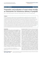
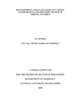
Fig. 1 Drug design of target compounds
Page 2 of 17
famitinib showed significantly improved progression free
survival (PFS) in patients with advanced colorectal cancer while its toxicity was manageable [16–18].
Given the effectiveness of famitinib, our previous study
successfully synthesized a series of novel five-memberedheterocycle derivatives of 2-pyrrolidone fused (2-oxoindolin-3-ylidene)methylpyrrole I (Fig. 1) [19]. In contrast
to famitinib, our synthetic compounds possess a more
rigid conformation than sunitinib and demonstrated
superior inhibitory activity of VEGFR-2 and PDGFRβ
to sunitinib. Among them, C(5)-Br 2-pyrrolidone-fused
(2-oxoindolin-3-ylidene)methylpyrrole showed that
potency against VEGFR-2 was fivefold higher in comparison to sunitinib [19].
Structure–activity relationships (SARs) of (2-oxoindolin-3-ylidene)methylpyrrole have been comprehensively
investigated in previous works [20–26]. The oxindole
scaffold, capable to provide two hydrogen bonds, is critical for the binding of (2-oxoindolin-3-ylidene)methylpyrroles to the ATP-binding site of the kinases, such as
VEGFRs [26–28]. The C(5) position of (2-oxoindolin3-ylidene)methylpyrroles is also considered one of most
effective positions for interaction with the ATP-binding site [20–25]. The significant VEGFRs and PDGFRs
inhibitory activity of C(5)-halogen substituted 2-pyrrolidone-fused (2-oxoindolin-3-ylidene)methylpyrroles
demonstrated in our previous report was at least partly
due to increased interaction between the synthetic
Yang et al. Chemistry Central Journal (2017) 11:72
compounds and the active sites of the receptors [19].
However, it remains unclear whether 2-pyrrolidonefused (2-oxoindolin-3-ylidene)methylpyrroles with C(5)
substituents other than C(5)-halogens, such as groups
producing electronic effects by induction or conjugation,
are still better VEGFRs and PDGFRs inhibitors. For an
improved understanding of the SARs of 2-pyrrolidonefused (2-oxoindolin-3-ylidene)methylpyrrole with C(5)
substituent replacement and in the hope of obtaining
novel compounds with more potent anti-proliferative
activity and lower toxicity, this study synthesized a series
of 2-pyrrolidone-fused (2-oxoindolin-3-ylidene)methylpyrrole with various C(5)-substituents to alter physical
and chemical properties for the purpose of ameliorating
anti-tumor activity. These experiments revealed several
new compounds with favorable selective indexes and
potent activities. The most promising of these, 14 and 15,
were chosen for further preclinical development.
Results and discussion
Chemistry
Scheme 1 shows the approach used to synthesize the
target products. Preparation of the key intermediate
5-(2-(diethylamino)ethyl)-3-methyl-4-oxo-1,4,5,6tetrahydropyrrolo[3,4-b]pyrrole-2-carbaldehyde
(3)
was essentially performed as described in the literature
[19]. Condensation of 3 with various 5-substitued oxindoles in the presence of piperidine at room temperature
readily afforded target compounds 4–15 in the yield of
46–66%. Most of the requisite 5-substitued oxindoles
Page 3 of 17
were prepared by modifying methods described in the
literature [26, 29–35] or were obtained commercially.
The exceptions were N,N-diethyl-2-oxoindoline-5-sulfonamide (16) and N,N-bis(2-chloroethyl)-2-oxoindoline-5-sulfonamide (17), which were produced by direct
amidation of 2-oxoindoline-5-sulfonyl chloride in dichloromethane at room temperature using triethylamine as
a base [32] (Scheme 2). The resulting oxindoles 16 and
17 were used to synthesize the desired products 6 and 7,
respectively, as described in Scheme 1.
All the target compounds were isolated as free bases
which were precipitated out during the synthesis. Compounds were purified by simply washing with EtOH.
However, most cases required further purification by column chromatography (silica gel, 90:10:1 EtOAc–MeOH–
TEA) with TEA to facilitate elution and to remove trace
impurities with the exclusion of compound 7. Purification of 7 by column chromatography using various solvent systems only led to rapid decomposition and then
a string of unidentifiable spots from the eluent appeared
in TLC. Our experiment results showed that analytically
pure 7 could be obtainable smoothly by recrystallization
from tetrahydrofuran (THF). All the structures of synthetic intermediates and products were determined by
spectroscopy and specific data of high-resolution mass
analysis (Additional file 1).
Anti‑proliferation activity
The in vitro anti-proliferation activity of synthetic compounds 4–15 and sunitinib (positive control) were
Scheme 1 Synthesis of key intermediate 3 [19] and 5-substituted 2-pyrrolidone-fused (2-oxoindolin-3-ylidene)methylpyrrole derivatives
Yang et al. Chemistry Central Journal (2017) 11:72
Page 4 of 17
Scheme 2 Synthesis of oxindoles 16 and 17
evaluated in three different human cancer cell lines
(human colon cancer cells HCT116, human non-small
cell lung cancer cells NCI-H460, and human renal cell
carcinoma 786-O) and a normal human fibroblast cell
line Detroit 551. Table 1 summarizes the experimental
results.
Compared to sunitinib, compounds 4, 8–12 showed
less activity against HCT116 cells (IC50 > 10 μM), indicating that electron-withdrawing groups (EWG) substituted
at C(5) appeared detrimental to the anti-tumor activity of the 2-pyrrolidone-fused (2-oxoindolin-3-ylidene)
methylpyrrole products [e.g., 11 (C(5)-CF3) and 12
(C(5)-NO2)]. However, introducing hydrogen bond
donating (HBD) groups at C(5) in the 2-oxindole ring,
e.g., 14 (C(5)-OH) and 15 (C(5)-SH), markedly inhibited
HCT116 cells. From lowest to highest, the anti-proliferative activities against HCT116 cells based on the IC50 values were enhanced as follows: 15 (2.34 ± 0.20 μM) > 14
(2.83 ± 0.40 μM) > 13 (3.06 ± 0.67 μM) ≈ 7
(3.65 ± 0.19 μM) > 6 (4.20 ± 0.57 μM) ≈ sunitinib
(4.60 ± 0.23 μM) > 5 (8.98 ± 0.92 μM). The presence and
probably the appropriately positioning of HBD groups
were apparently the main determinants of anti-proliferation potency. These experimental results indicated
that C(5) substituted 2-pyrrolidone-fused (2-oxoindolin3-ylidene)methylpyrroles against HCT116 cells had the
descending order as follows: C(5)-HBD > C(5)-sulfonamide > C(5)-EWG. Regarding anti-proliferative effects
on NCI-H460 cells, the IC50 values of 4–10, 14, and 15
were higher than 10 μM. Compounds 11–13 revealed
approximately equal activity to sunitinib; however, their
anti-proliferative activities did not significantly differ
(p ≥ 0.05). For 786-O cells, the IC50 values of 4–10, 12,
14, and 15 exceeded 10 μM. The order of anti-proliferative activities of 11, 13 and sunitinib against 786-O cells
was 13 ≈ sunitinib > 11. Comparisons with our previously reported data confirmed the superior activity of
13 (C(5)-OMe) against 786-O cells to the corresponding C(5)-halogen 2-pyrrolidone-fused (2-oxoindolin3-ylidene)methylpyrroles [19].
Since the proliferation of HCT116 cells is stimulated by
HCT116-produced VEGF and VEGFR-1/2 via an autocrine mechanism, inhibiting VEGFR-1/2 of HCT116
cells with VEGFR-1/2 inhibitor AAL993 significantly
decreases proliferation of HCT116 cells [36]. Table 1
shows that our experiments revealed a strong correlation between anti-proliferation activities of 4–15 against
HCT116 cells and VEGFR-2 inhibition percentage at
80 nM.
Although NCI-H460 cells express both VEGF and
VEGFR-2, proliferation of NCIH-460 cells is not promoted by VEGF/VEGFR-2 pathway [37]. Sunitinib has
been approved for treating renal cell carcinoma (RCC);
however, it inhibits RCC growth through an anti-angiogenesis mechanism rather than by directly targeting RCC
cells [38]. Moreover, 786-O cells express VEGF and neuropilin-1 (NRP-1) rather than VEGFR-2. The VEGF promoted 786-O cell proliferation in an autocrine manner
via VEGF/NRP-1 pathway [39]. Therefore, the IC50 values
of most VEGFR-2 inhibiting compounds (5, 7, 14, 15,
and sunitinib) against either NCI-H460 or 786-O cells
were higher than those of HCT116 cells. Interestingly, 11
(C(5)-CF3) showed cytotoxicity to both NCI-H460 and
786-O but not to HCT116 cells; 12 (C(5)-NO2) was toxic
to NCI-H460; 13 (C(5)-OMe) was toxic to all three tested
cancer cell lines. These experimental results suggest that
the C(5) substituent replacement in this structural system
significantly affected the selectivity of cancer cell growth
inhibition.
Potential anticancer drug candidates should show
greater selectivity for cancer cells compared with normal cells. Therefore, selectivity index (SI) values for synthetic products 4–15 as well as sunitinib were obtained
in the three tested cancer lines (Table 1). For comparison, human normal fibroblast cells Detroit 551 were
used as a control group. The SI values showed that all
synthetic products except for 7 had high selectivity
for tumor cells and, compared to sunitinib, even much
lower toxicity to Detroit 551 cells. The toxic effects of
C(5)-SO2N(CH2CH2Cl)2 substituent of 7 on Detroit 551
Yang et al. Chemistry Central Journal (2017) 11:72
Page 5 of 17
Table 1 Enzymatic and cellular inhibition activities of 4–15 and sunitinib
Compound
Sunitinib
4
5
6
7
8
9
10
11
12
13
14
15
R
–
–SO2NH2
–SO2NMe2
–SO2NEt2
–SO2N(CH2CH2Cl)2
–SO2NHPh
–SO2NH(4-CF3-Ph)
–NHMs
–CF3
–NO2
–OMe
–OH
–SH
% inhibition of
VEGFR-2 at 80 nM
45
0
20
0
46
15
0
4
0
13
41
58
57
IC50 (μM)/selective index (SI)
HCT116
NCI-H460
786-O
Detroit 551
9.48 ± 0.18
4.60 ± 0.23
7.51 ± 0.78
7.89 ± 0.60
1.32
0.81
0.76
>10
>10
>10
nd
nd
nd
8.98 ± 0.92
>10
>10
>1.11
nd
nd
4.20 ± 0.57
>10
>10
>2.38
nd
nd
3.65 ± 0.19
>10
>10
1.66
<0.61
<0.61
>10
>10
>10
nd
nd
nd
>10
>10
>10
nd
nd
nd
>10
>10
>10
nd
nd
nd
>10
7.22 ± 1.01
8.49 ± 0.46
nd
>1.39
>1.18
>10
6.61 ± 0.80
>10
nd
>1.51
nd
3.06 ± 0.67
6.37 ± 1.09
7.86 ± 0.30
>3.27
>1.57
>1.27
2.83 ± 0.40
>10
>10
3.53
nd
nd
2.34 ± 0.20
>10
>10
>4.27
nd
nd
>10
>10
>10
6.06 ± 0.40
>10
>10
>10
>10
>10
>10
>10
>10
nd not detected
cells was evident and complex but nevertheless not yet
completely understood. The likely explanation is that 7
contains a highly chemically reactive bis(2-chloroethyl)
amino (–SO2N(CH2CH2Cl)2) similar to chlorambucil,
which has clinic applications as a non-specific alkylating agent. Thus, its cytotoxic effect probably resulted
from DNA damage via the formation of cross-links. In
this study, 15 had particularly high selectivity to HCT116
cells (SI > 4.27 for 15 vs. 1.32 for sunitinib), and 13 had
particularly high selectivity to NCI-H460 cells (SI > 1.57
for 13 vs. 0.81 for sunitinib) and 786-O cells (SI > 1.27 for
13 vs. 0.76 for sunitinib).
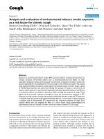
Since our newly synthesized products generally showed
high selectivity against HCT116 cancer cell proliferation, the next experiment was performed to determine
whether the inhibitory response resulted from acute cellular toxicity. Compounds 7 and 13–15 were then chosen to subject to acute cytotoxicity test on HCT116 cells
through the WST-8 cell viability assay. Figure 2 shows the
experimental results, which confirmed that neither our
compounds nor sunitinib had acute cytotoxicity in the
two tested cell lines.
Our previous works apparently showed that
C(5)-halogen substituents of 2-pyrrolidone-fused
Yang et al. Chemistry Central Journal (2017) 11:72
Page 6 of 17
derivatives of 2-pyrrolidone-fused (2-oxoindolin3-ylidene)methylpyrrole. Additionally, hydrogen bond
donor substituents at C(5) significantly affected the
potency and selectivity of anti-proliferation activity.
Kinase inhibitory assays
Fig. 2 Acute cytotoxicity assay of a HCT116; b Detroit 551 incubated
with DMSO (1%), sunitinib, 7, and 13–15 (10 μM)
(2-oxoindolin-3-ylidene)methylpyrroles affected the
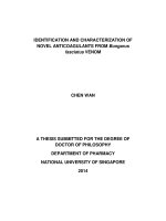
potency and cell cycle profiles of HCT116 cell [19]. For
an improved understanding of these effects, this study
performed further cell cycle analyses of 7, 13–15, and
sunitinib (Fig. 3). The preliminary results showed that
the cell cycle profiles of HCT116 cells incubated with 14
and sunitinib for 24 h caused G0/G1 cell cycle arrest. In
contrast, the cell cycle profile of HCT116 cells incubated
with 7 and 13 for 24 h displayed an increase in polyploid
cells. Surprisingly, the cell cycle profile of HCT116 cells
treated with 15 for 24 h showed an increase in tetraploid cells. Previous works had established that Inhibiting Aurora kinase obtained a polyploidal cell cycle profile
[40–42]. Our previous studies proved that (2-oxoindolin3-ylidene)methylpyrroles had great in vitro Aurora A
kinase inhibition at 1.0 μM, and some of them revealed
the inhibition of HCT116 cells proliferation via Aurora
kinase inhibition. Our experiments again revealed a similar trend, i.e., 92.9% for 7, 94.4% for 13, and 93.6% for 15,
and 50.7% for sunitinib at 1.0 μM, respectively (Table 2).
Therefore, we hypothesized that using compounds 7, 13
and 15 to inhibit HCT116 cell proliferation might also
inhibit Aurora kinase.
In summary, the experiments in this study suggested that substituents at C(5) markedly influenced the
anti-proliferation activity and selectivity of synthetic
Next, the VEGFR-2 phosphorylation inhibitory activities of the newly synthesized compounds were evaluated. The experimental results in Table 1 show that the
VEGFR-2 inhibitory activities of compounds 4, 8, 9, and
11 at concentrations of 80 nM did not differ from that of
the 1% DMSO (control). However, compounds 5, 6, and
12 at the same concentration revealed 13–20% inhibition; 7 and 13 demonstrated approximately equal inhibition percentage to sunitinib; and 14 and 15 exhibited the
most potent inhibitory activity. Therefore, IC50 values of
compounds 7 and 13–15 were further evaluated to assess
their activities against VEGFR-2, PDGFRβ, and Aurora A
kinase.
Sun et al. showed that C(5)-SO2NH2 at (2-oxoindolin3-ylidene)methylpyrroles improved VEGFR-2 inhibition
[21]. A pharmacophore model of oxindole analog binding
at the FGFR1 binding site generated from virtual screen
results in a study by Kammasud then revealed that introduction of a phenyl hydrazide motif to C(5) of oxindoles
proved to be the best possible to allow additional hydrogen bonding interactions with ATP site of receptor tyrosine kinases (RTKs), such as FGFR-1, VEGFR-2, PDGFRβ,
and EGFR [20]. In our investigation, compounds 4
(C(5)-SO2NH2), 5 (C(5)-SO2NMe2), 6 (C(5)-SO2NEt2),
8 (C(5)-SO2NHPh), and 9 (C(5)-SO2NH(4-CF3-Ph))
showed disappointing activities or only mediocre
improvement in VEGFR-2 inhibition; however, 7 (C(5)SO2N(CH2CH2Cl)2) displayed evident improvement.
These experimental results suggest that ligand–protein
binding affinity between VEGFR-2 and 2-pyrrolidonefused (2-oxoindolin-3-ylidene)methylpyrroles is probably
not be enhanced by either C(5)-SO2NH2, C(5)-SO2NHPh
or C(5)-SO2N(alkyl)2, with the exception of 7 (C(5)SO2N(CH2CH2Cl)2), the discrepancy of which already
discussed.
Since our previously reported C(5)-halogen substituted
2-pyrrolidone-fused (2-oxoindolin-3-ylidene)methylpyrrole derivatives showed fairly potent inhibiting effects
on VEGFR-2 (35–64% inhibition at 50 nM) [19], our
next objective was bioisosteric replacement of the C(5)halogens with an electron-withdrawing C(5)-CF3. Unfortunately, 11 (C(5)-CF3) had no inhibitory activity against
VEGFR-2 at 80 nM.
The effect of a C(5)-OMe substituent of indoline2-one scaffold on kinase inhibitory activity and selectivity is highly dependent on the C(3) substituents of
indoline-2-one [22, 26]. Interestingly, our study showed
Yang et al. Chemistry Central Journal (2017) 11:72
Page 7 of 17
Fig. 3 Cell cycle profiles of HCT-116 cells treated with a 1% DMSO (control); b sunitinib (5.0 μM); c 7 (5.0 μM); d 13 (3.0 μM); e 14 (3.0 μM); f 15
(3.0 μM) for 24 h. M1 G0/G1phase, M2 S phase, M3 G2/M phase
Yang et al. Chemistry Central Journal (2017) 11:72
Page 8 of 17
Table 2 In-vitro kinase inhibitory activities of 7, 13–15, and sunitinib
Compound
R
IC50 (nM)
VEGFR-2
PDGFRβ
% inhibition of Aurora
A at 1.0 μM
Sunitinib
–
151.8
94.5
7
–SO2N(CH2CH2Cl)2
23.7
63.2
50.7
94.4
13
–OMe
47.8
76.2
92.9
14
–OH
25.9
28.5
93.6
15
–SH
14.6
12.1
96.4
that compound 13 (C(5)-OMe) substantially improved
VEGFR-2 inhibition but not so noticeable in PDGFRβ
inhibition. A more or less similar effect could be observed
in 7 (C(5)-SO2N(CH2CH2Cl)2).
As Table 2 shows, in comparison to sunitinib, compounds 7 and 13 had six- and threefold lower I C50 values
for VEGFR-2, respectively. Moreover, compared to their
C(5)-OMe analog 13, compounds 14 (C(5)-OH) and 15
(C(5)-SH) even showed a two- and a fourfold decrease in
IC50 values, respectively. On the other hand, the inhibiting activities of 7 and 13 in PDGFRβ were slightly more
potent than those of sunitinib; however, 14 and 15 had a
three- and an eightfold decrease in IC50 values for inhibiting PDGFRβ, respectively. Thus, both C(5)-OH and C(5)SH substituents could significantly improve the activity
of 2-pyrrolidone-fused (2-oxoindolin-3-ylidene)methylpyrroles in the inhibition of both VEGFR-2 and PDGFRβ.
In accordance with Kammasud, we hypothesized that
groups C(5)-OH and C(5)-SH probably produced favorable potency of 14 and 15 by providing additional hydrogen bonding interactions with ATP site of RTKs.
The results once again revealed a similar trend, i.e., different C(5) substitutions markedly affect the biochemical
activities against VEGFR-2 and PDGFRβ. In summary,
hydrogen-bond-donating (HBD) substituent at C(5)
could greatly enhance inhibitory potency against both
VEGFR-2 and PDGFRβ. These experimental results suggest that the influence of C(3) substituent to the C(5)HBD substituted indoline-2-one scaffold needs further
study.
In‑vitro tube formation assay
In-vitro VEGF-induced tube formation inhibitory activity of 7, 13–15, and sunitinib were tested by Matrigel
tube formation assay using ibidi μ-Slide angiogenesis
kit. Figures 4 and 5 shows the photographs of Matrigel
tube formation assays of control and tested compounds
at 2.0, 1.0, 0.50 and 0.10 μM. Under these conditions, the
density of tube-like structures was substantially reduced.
Compounds 7 and 13–15 showed distinctly higher tube
formation inhibitory activity than the reference drug
sunitinib. Compared to sunitinib, 7 and 13 had twofold
higher potency in inhibiting in vitro tube formation than
sunitinib in terms of IC50 (Table 3). The more potent 14
and 15 were with IC50 roughly 2.5- and threefold stronger
than sunitinib, respectively. These experimental results
agree well with those for VEGFR-2 kinase inhibitory
assays, suggesting that our synthetic compounds inhibited the in vitro tube formation via VEGFR-2 inhibition.
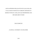
Molecular modeling
The kinase inhibitory assays revealed that the VEGFR-2
and in vitro tube formation inhibitory activities of the
synthetic compounds 7 and 13–15 exceeded that of sunitinib, and the C(5) substituents of 2-pyrrolidone-fused
(2-oxoindolin-3-ylidene)methylpyrrole were critical for
VEGFR-2 inhibitory activity. For further clarification
of these results, sunitinib, 7, and 13–15 were examined
and compared by docking into the ATP-binding site of
VEGFR-2 (PDB ID: 4AGD) using Discovery Studio LibDock [43]. LibDock is a method placing the generated
ligand conformations into the protein active site based
on polar and apolar interaction sites (hotspot). Figure 6
shows the predicted binding modes of sunitinib, 7, and
13–15. Interestingly, the modeling results for compound
7 differed from those of compounds 13–15 in that 7
formed four hydrogen bonds with VEGFR-2: the Cl of
C(5)-SO2N(CH2CH2Cl)2 and the NH of the oxindole
Yang et al. Chemistry Central Journal (2017) 11:72
Page 9 of 17
Fig. 4 Compounds 7, 13–15, and sunitinib inhibited tube formation induced by VEGF. a Solvent control; b VEGF (10 ng/ml) and 0.1 μM sunitinib; c
VEGF (10 ng/ml) and 0.10 μM 7; d VEGF (10 ng/ml) and 0.10 μM 13; e VEGF (10 ng/ml) and 0.10 μM 14; f VEGF (10 ng/ml) and 0.10 μM 15; g VEGF
(10 ng/ml) and 0.50 μM sunitinib; h VEGF (10 ng/ml) and 0.50 μM 7; i VEGF (10 ng/ml) and 0.50 μM 13; j VEGF (10 ng/ml) and 0.50 μM 14; k VEGF
(10 ng/ml) and 0.50 μM 15; l VEGF (10 ng/ml) and 1.0 μM sunitinib; m VEGF (10 ng/ml) and 1.0 μM 7; n VEGF (10 ng/ml) and 1.0 μM 13; o VEGF
(10 ng/ml) and 1.0 μM 14
Yang et al. Chemistry Central Journal (2017) 11:72
Page 10 of 17
Fig. 5 Compounds 7, 13–15, and sunitinib inhibited tube formation induced by VEGF. a VEGF (10 ng/ml) and 1.0 μM 15; b VEGF (10 ng/ml) and
2.0 μM sunitinib; c VEGF (10 ng/ml) and 2.0 μM 7; d VEGF (10 ng/ml) and 2.0 μM 13; e VEGF (10 ng/ml) and 2.0 μM 14; f VEGF (10 ng/ml) and 2.0 μM
15
scaffold of 7 formed hydrogen bonds with the same
Cys919, the oxygen atom of C(5)-SO2N(CH2CH2Cl)2 with
Cys1045, and the oxygen atom of pyrrolidone (C(4′)) with
Asn923 (Fig. 6b). The docking results further showed that
C(5)-SO2N(CH2CH2Cl)2 of 7 was laid in the hydrophobic pocket of the VEGFR-2 active site (Fig. 6c). The above
experimental results might explain why compound 7 had
the most potent VEGFR-2 inhibiting effects among 4–9.
Table 3 Inhibition activities of 7, 13–15, and sunitinib
against in vitro tube formation
Compound
R
IC50 (μM)
Area
Sunitinib
–
7
–SO2N(CH2CH2Cl)2
1.54 ± 0.08
0.76 ± 0.11
13
–OMe
0.74 ± 0.16
14
–OH
0.62 ± 0.07
15
–SH
0.53 ± 0.11
In Fig. 6d–f, the predicted binding modes of highly active
compounds 13–15 reveal that each of them formed
three hydrogen bonds with Lys868, Glu917, and Cys919,
respectively. Additionally, compounds 13–15 all formed
pi–pi interactions between their pyrrole-scaffolds and
Phe918 of VEGFR-2 (Fig. 6d–f ). Most notably, C(5)-OH
of 14 and C(5)-SH of 15 formed hydrogen bonds with
Lys686 of VEGFR-2 (Fig. 6e, f ) while C(5)-OMe of 13, the
methyl ether of 14 but with much lower activity, did not
show any interaction with Lys868 due to the blockade of
–OMe to hydrogen bond formation. These experimental
results indicate that C(5)-HBD of 2-pyrrolidone-fused
(2-oxoindolin-3-ylidene)methylpyrrole derivatives have
important inhibiting effects on VEGFR-2 activity, and
compounds 14 and 15 proved to be the case.
Conclusions
The novel series of 2-pyrrolidone-fused (2-oxoindolin3-ylidene)methylpyrrole derivatives with various C(5)
substitutions synthesized in our laboratory showed notable cellular and enzymatic anti-tumor activities. Several of these derivatives had superior inhibitory activity
against VEGFR-2 and PDGFRβ compared to sunitinib.
Among them, 14 (C(5)-OH) and 15 (C(5)-SH) possessed the highest potency and the highest selectivity in
HCT116 cells. The preliminary results in further pharmacokinetic studies of compounds 14 and 15 were satisfactory. Detailed pharmacological and pharmacokinetic
Yang et al. Chemistry Central Journal (2017) 11:72
Page 11 of 17
Fig. 6 Sunitinib, 7, and 13–15 are docked into the active site of VEGFR-2 (PDB ID: 4AGD) in 3-dimentional structure. a Sunitinib in VEGFR-2; b
7 in VEGFR-2; c 7 in VEGFR-2 (the active site of VEGFR-2 was shown in hydrophobicity maps); d 13 in VEGFR-2; e 14 in VEGFR-2, f 15 in VEGFR-2.
Compounds are showed in sticks; hydrogen bonds are shown as dashed yellow line; pi–pi interaction is shown in orange line; shades of brown indicate
regions of high hydrophobicity; shades of white indicate regions of neutral, shades of blue indicate regions of low hydrophobicity
studies are in progress and will be reported in future
works.
Experiment section
Chemistry
All the chemicals were purchased from Aldrich-Sigma
Chemical Company (St. Louis, MO,
USA) and AlfaAesar Chemical Company (Lancashire, Heysham, England) and used without further purification. All reactions
were routinely monitored by TLC on Merck F254 silica gel
plates. Silica gel (70–230 mesh, Silicacycle) was used for
column chromatography. The 1H- and 13C-NMR spectra
were determined on an Agilent Varian-400 NMR (Agilent Technologies, Santa Clara, CA, USA) instrument in
CDCl3, acetone-d6, methanol-d4, or acetic acid-d6 unless
otherwise noted. Chemical shifts (δ) were expressed as
parts per million (ppm) downfield from tetramethylsilane
(TMS) as the internal standard (σ 0.00), and coupling
constants (J) were given in hertz (Hz). High-resolution
mass spectra (HRMS) using a Bruker Impact HD (ESI)
were performed in the Instrument Center of the Ministry
of Science and Technology at the National Chiao-Tung
University, Taiwan. Dry tetrahydrofuran (THF) was
freshly distilled from lithium aluminum hydride (LAH)
before use. All the other solvents were obtained from
commercial sources and purified before use if necessary.
Images were acquired with a Leica DM1000 LED microscope (Leica Microsystems, Wetzlar, Hessen, Germany).
UV–VIS spectra were recorded on a Thermo Multiskan
Go Microplate spectrophotometer (Thermo Fisher Scientific, Waltham, MA, USA). IR spectra were registered
on a Thermo Nicolet iS5 FT-IR spectrometer (Thermo
Fisher Scientific, Waltham, MA, USA) with attenuated
total reflection (ATR) method. The purities of the final
compounds were all greater than 95% as determined by
analytical reverse-phase HPLC.
General procedure for the preparation of compounds 4–15
The key intermediate 3 for preparation of target compounds was synthesized according to the method
reported previously [19]. To each stirred solution containing 3 (263 mg, 1.00 mmol) in EtOH (15 ml) was
Yang et al. Chemistry Central Journal (2017) 11:72
added dropwise a solution of 5-substituted oxindole
(1.00 mmol) in EtOH (2 ml) and then piperidine (0.1 ml)
was added. After stirring at room temperature for 6 h, the
precipitate formed was filtrated, washed with EtOH, and
purified by column chromatography (silica gel, 90:10:1
EtOAc–MeOH–TEA).
(Z)‑3‑((5‑(2‑(diethylamino)ethyl)‑3‑methyl‑4‑oxo‑1,4,5,
6‑tetrahydropyrrolo[3,4‑b]pyrrol‑2‑yl)methylene)‑2‑ox‑
oindoline‑5‑sulfonamide (4) The requisite oxindole5-sulfonamide, for condensation with 3 to form target
compound 4, was prepared in 60% yield by a modified
method of amidation of 2-oxoindoline-5-sulfonyl chloride with ammonium hydroxide solution [32].
Yield of 4: 58%, orange solids. Mp: 252–255 °C, UV λmax
(MeOH), nm (logɛ): 418 (4.21). IR (ATR), cm−1: 3239,
2976, 1600, 1562. 1H-NMR (400 MHz, acetic acid-d4) δ,
ppm: 8.26 (d, 1H, J = 1.2 Hz ArH), 7.74–7.77 (m, 2H, –
C=CH–, ArH), 7.12 (d, 1H, J = 8.4 Hz, ArH), 4.56 (s, 2H,
Ar–CH2), 4.00 (t, 2H, J = 5.6 Hz, –NCH2CH2–), 3.50 (t,
2H, J = 5.6 Hz, –NCH2CH2–), 3.37 (q, 4H, J = 7.2 Hz,
–N(CH2CH3)2), 2.51 (s, 3H, Ar–CH3), 1.31 (t, 6H,
J = 7.2 Hz, –N(CH2CH3)2). 13C-NMR (100 MHz, acetic
acid-d4) δ, ppm: 171.5, 169.5, 150.2, 142.0, 137.7, 133.8,
128.1, 127.4, 126.9, 126.5, 121.3, 117.9, 116.5, 111.2,
51.8, 48.1, 45.7, 39.5, 9.9, 8.7. HRMS m/z (ESI): calcd.,
458.1869.1744 [M + H]+; found, 458.1857 [M + H]+.
( Z ) ‑ 3 ‑ ( ( 5 ‑ ( 2 ‑ (di e t hyl amin o) e t hyl) ‑ 3 ‑ m e t hyl ‑ 4 ‑ o
x o ‑ 1 , 4 , 5 , 6 ‑ t e t rahy dr o p y r r o l o [ 3 , 4 ‑ b] p y r r o l ‑ 2 ‑ yl)
methylene)‑N,N‑dimethyl‑2‑oxoindoline‑5‑sulfona‑
mide (5) The requisite N,N-dimethyl-2-oxoindoline5-sulfonamide, for condensation with 3 to form target
compound 5, was prepared in 74% yield by a modified
method of amidation of 2-oxoindoline-5-sulfonyl chloride with dimethylamine in methanol [32].
Yield of 5: 56%, orange solids. Mp: 244–247 °C, UV
λmax (MeOH), nm (logɛ): 428 (4.53). IR (ATR), cm−1:
2971, 1657, 1586. 1H-NMR (400 MHz, acetic acid-d4)
δ, ppm: 8.19 (d, 1H, J = 1.6 Hz, ArH), 7.86 (s, 1H, –
C=CH–), 7.63 (dd, 1H, J = 8.0, 1.6 Hz, ArH), 7.18 (d,
1H, J = 1.6 Hz, ArH), 4.57 (s, 2H, Ar–CH2), 3.96 (t,
2H, J = 6.0 Hz, –NCH2CH2–), 3.50 (t, 2H, J = 6.0 Hz,
–NCH2CH2–), 3.39 (q, 4H, J = 7.2 Hz, –N(CH2CH3)2),
2.68 (s, 6H, –N(CH3)2), 2.50 (s, 3H, Ar–CH3), 1.33 (t, 6H,
J = 7.2 Hz, –N(CH2CH3)2). 13C-NMR (100 MHz, acetic
acid-d4) δ, ppm: 171.5, 169.5, 150.2, 142.4, 133.8, 129.7,
128.2, 128.1, 127.7, 127.1, 121.3, 119.5, 116.4, 111.3, 51.7,
48.5, 48.0, 39.4, 38.3, 10.0, 8.7. HRMS m/z (ESI): calcd.,
486.2172 [M + H]+; found, 486.2170 [M + H]+.
( Z ) ‑ 3 ‑ ( ( 5 ‑ ( 2 ‑ (di e t hyl amin o) e t hyl) ‑ 3 ‑ m e t hyl ‑ 4 ‑ o
x o ‑ 1 , 4 , 5 , 6 ‑ t e t rahy dr o p y r r o l o [ 3 , 4 ‑ b] p y r r o l ‑ 2 ‑ yl)
Page 12 of 17
methylene)‑N,N‑diethyl‑2‑oxoindoline‑5‑sulfona‑
mide (6) 2-Oxoindoline-5-sulfonyl chloride (2.32 g,
10.0 mmol), prepared by a modified method [32], was suspended in dichloromethane (20 ml). The resulting mixture
was added dropwise a solution of triethylamine (1.21 g,
12.0 mmol) in dichloromethane (5 ml) and then a solution of diethylamine (0.90 g, 12.0 mmol) in dichloromethane (5 ml) was added. After stirring at room temperature
for 8 h, the precipitate formed was filtrated, washed with
dichloromethane, and purified by column chromatography
(silica gel, 1:4 EtOAc–Hexane) to yield 1.90 g (71%) of N,Ndiethyl-2-oxoindoline5-sulfonamide (16) as pale yellow
crystals. Mp: 155–156 °C, UV λmax (MeOH), nm (logɛ):
289 (4.64). IR (ATR), cm−1: 3159, 1707, 1615. 1H NMR
(400 MHz, methanol-d4) δ, ppm: 7.70 (d, 1H, J = 6.8 Hz,
ArH), 7.69 (s, 1H, ArH), 7.02 (d, 1H, J = 6.8 Hz, ArH) 3.62
(s, 2H, ArCH2), 3.21 (q, 4H, J = 7.2 Hz, –N(CH2CH3)2),
1.13 (t, 6H, J = 7.2 Hz, –N(CH2CH3)2). 13C-NMR
(100 MHz, methanol-d4) δ, ppm: 179.6, 148.9, 134.7, 129.1,
128.2, 124.6, 110.6, 47.8, 43.4, 14.7. HRMS m/z (EI): calcd.,
268.0882 [M]+; found, 268.0872 [ M]+.
N,N-Diethyl-2-oxoindoline5-sulfonamide (16) obtained
from above was used to condense with 3 to afford target
compound 6 as described in general synthesis. Yield of 6:
57%, orange solids. Mp: 245–247 °C, UV λmax (MeOH),
nm (logɛ): 429 (4.54). IR (ATR), c m−1: 2968, 1659, 1589.
1
H-NMR (400 MHz, acetic acid-d4) δ, ppm: 8.20 (d, 1H,
J = 2.0 Hz, ArH), 7.79 (s, 1H, –C=CH–), 7.63 (dd, 1H,
J = 8.0, 2.0 Hz, ArH), 7.11 (d, 1H, J = 8.0 Hz, ArH), 4.52 (s,
2H, Ar–CH2), 3.92 (t, 2H, J = 7.2 Hz, –NCH2CH2–), 3.47
(t, 2H, J = 7.2 Hz, –NCH2CH2–), 3.36 (q, 4H, J = 7.2 Hz, –
N(CH2CH3)2), 3.21 (q, 4H, J = 7.2 Hz, –N(CH2CH3)2),
2.47 (s, 3H, Ar–CH3), 1.31 (t, 6H, J = 7.2 Hz, –
N(CH2CH3)2), 1.10 (t, 6H, J = 7.2 Hz, –N(CH2CH3)2).
13
C-NMR (100 MHz, acetic acid-d4) δ, ppm: 171.4, 169.4,
150.1, 142.1, 134.7, 133.8, 128.1, 127.5, 127.2, 127.0, 121.3,
118.8, 116.5, 111.4, 51.7, 48.5, 48.1, 43.2, 39.4, 14.7, 10.0,
8.7. HRMS m/z (ESI): calcd., 514.2497 [M + H]+; found,
514.2483 [M + H]+.
(Z)‑N,N‑Bis(2‑chloroethyl)‑3‑((5‑(2‑(diethylamino)
ethyl)‑3‑methyl‑4‑oxo‑1,4,5,6‑tetrahydropyrrolo[3,4
‑b]pyrrol‑2‑yl)methylene)‑2‑oxoindoline‑5‑sulfona‑
mide (7) 2-Oxoindoline-5-sulfonyl chloride (2.32 g,
10.0 mmol), prepared by a modified method [32],
and bis(2-choroethyl)amine hydrochloride (2.14 g,
12.0 mmol) were suspended in dichloromethane
(20 ml) and then was added dropwise a solution of triethylamine (2.23 g, 22.0 mmol) in dichloromethane
(10 ml). After stirring at room temperature for 8 h, the
precipitate formed was filtrated, washed with dichloromethane, and purified by column chromatography (silica gel, 1:4 EtOAc–Hexane). 2.13 g (63%) of
Yang et al. Chemistry Central Journal (2017) 11:72
N,N-bis(2-chloroethyl)-2-oxoindoline-5-sulfonamide
(17) as pale yellow solids. Mp: 193–195 °C, UV λmax
(MeOH), nm (logɛ): 289 (4.60). IR (ATR), cm−1: 3157,
1704, 1615. 1H NMR (400 MHz, CDCl3) δ, ppm: 7.76 (d,
1H, J = 8.4, ArH), 7.71 (s, 1H, ArH), 6.98 (d, 1H, J = 8.4,
ArH), 3.70 (t, 4H, J = 7.0 Hz, –N(CH2CH2Cl)2), 3.62 (s,
2H, ArCH2) 3.49 (t, 4H, J = 7.0 Hz, –N(CH2CH2Cl)2).
13
C-NMR (100 MHz, dmso-d6) δ, ppm: 176.4, 148.2,
130.3, 128.0, 123.3, 117.5, 113.5, 109.2, 50.3, 42.3, 39.5.
N,N-Bis(2-chloroethyl)-2-oxoindoline-5-sulfonamide (17) obtained from above was used to condense
with 3 to afford target compound 7 as described in
general synthesis. Purification of 7 was performed by
recrystallization from THF. Yield of 7: 54%, orange
solids. Mp: 236–237 °C, UV λmax (MeOH), nm (logɛ):
428 (4.58). IR (ATR), cm−1: 2917, 1651, 1574. 1HNMR (400 MHz, acetic acid-d4) δ, ppm: 8.29 (s, 1H, –
C=CH–), 7.87 (s, 1H, ArH), 7.73 (d, 1H, J = 8.4 Hz,
ArH,), 7.18 (d, 1H, J = 8.4, ArH), 4.60 (s, 2H, Ar–
CH2), 3.98 (2H, J = 7.2 Hz, –NCH2CH2–) 3.73 (t, 4H,
J = 7.2 Hz, –N(CH2CH2Cl)2), 3.53 (t, 6H, J = 7.2 Hz, –
NCH2CH2–, –N(CH2CH2Cl)2), 3.39 (q, 4H, J = 7.2 Hz, –
N(CH2CH3)2), 2.50 (s, 3H, Ar–CH3), 1.31 (t, 6H,
J = 7.2 Hz, –N(CH2CH3)2). 13C-NMR (100 MHz, acetic acid-d4) δ, ppm: 171.5, 169.5, 150.4, 142.6, 133.8,
133.4, 128.4, 127.9, 127.6, 127.3, 121.4, 119.0, 116.3,
111.5, 52.2, 51.8, 48.5, 48.1, 43.1, 39.4, 10.0, 8.7. HRMS
m/z (ESI): calcd., 582.1718 [M + H]+; found, 582.1703
[M + H]+.
( Z )‑3‑((5‑(2‑( D iethyl amino)ethyl)‑3‑methyl‑4‑ o
x o ‑ 1 , 4 , 5 , 6 ‑ t e t rahy dr o p y r r o l o [ 3 , 4 ‑ b] p y r r o l ‑ 2 ‑ yl)
methylene)‑2‑oxo‑N‑phenylindoline‑5‑sulfonamide
(8) The requisite 2-oxo-N-phenylindoline-5-sulfonamide, for condensation with 3 to form target compound
8, was prepared in 83% yield by a modified method of
amidation of 2-oxoindoline-5-sulfonyl chloride with aniline [32].
Yield of 8: 46%, orange solids. Mp: 225–228 °C, UV
λmax (MeOH), nm (logɛ): 429 (4.47). IR (ATR), cm−1:
3213, 2963, 1667, 1557. 1H-NMR (400 MHz, acetic
acid-d4) δ, ppm: 8.12 (d, 1H, J = 1.6, ArH), 7.71 (s, 1H,
–C=CH–), 7.59 (dd, 1H, J = 8.4, 1.6 Hz, ArH), 7.22–7.14,
(m, 5H, ArH), 7.02 (d, 1H, J = 8.4 Hz, ArH), 4.56 (s, 2H,
Ar–CH2), 3.95 (t, 2H, J = 6.0 Hz, –NCH2CH2–), 3.48 (t,
2H, J = 6.0 Hz, –NCH2CH2–), 3.37 (q, 4H, J = 7.0 Hz,
–N(CH2CH3)2), 2.48 (s, 3H, Ar–CH3), 1.31 (t, 6H,
J = 7.0 Hz, –N(CH2CH3)2). 13C-NMR (100 MHz, acetic
acid-d4) δ, ppm: 171.3, 169.4, 150.2, 142.3, 138.4, 133.9,
133.7, 130.2, 128.1, 127.5, 127.3, 126.8, 125.9, 122.2,
121.3, 118.8, 116.2, 111.1, 51.7, 48.5, 48.1, 39.4, 10.0, 8.7.
HRMS m/z (ESI): calcd., 534.2182 [M + H]+; found,
534.2181 [M + H]+.
Page 13 of 17
(Z)‑3‑((5‑(2‑(Diethylamino)ethyl)‑3‑methyl‑4‑oxo‑1,4,5,6‑
tetrahydropyrrolo[3,4‑b]pyrrol‑2‑yl)methylene)‑2‑
oxo‑N‑(4‑(trifluoromethyl)phenyl)indoline‑5‑sulfonamide
(9) The requisite 2-oxo-N-(4-(trifluoromethyl)phenyl)
indoline-5-sulfonamide, for condensation with 3 to form
target compound 9, was prepared in 77% yield by a modified method of amidation of 2-oxoindoline-5-sulfonyl
chloride with 4-(trifluoromethyl)aniline [32].
Yield of 9: 46%, orange solids. Mp: 231–234 °C, UV
λmax (MeOH), nm (logɛ): 429 (4.32). IR (ATR), cm−1:
3162, 1634, 1582. 1H-NMR (400 MHz, acetic acid-d4) δ,
ppm: 1H-NMR (400 MHz, acetic acid-d4) δ, ppm: 8.21 (s,
1H, ArH), 7.75 (s, 1H, –C=CH–), 7.68 (dd, 1H, J = 8.0,
1.6 Hz, ArH), 7.52 (d, 2H, J = 8.4 Hz, ArH), 7.34 (d, 2H,
J = 8.4 Hz, ArH), 7.05 (d, 1H, J = 8.0 Hz, ArH), 4.58 (s,
2H, Ar-CH2), 3.97 (t, 2H, J = 7.2 Hz, –NCH2CH2–), 3.50
(t, 2H, J = 7.2 Hz, –NCH2CH2–), 3.38 (q, 4H, J = 7.2 Hz,
–N(CH2CH3)2), 2.50 (s, 3H, Ar–CH3), 1.32 (t, 6H,
J = 7.2 Hz, –N(CH2CH3)2).
13
C-NMR (100 MHz, acetic acid-d4) δ, ppm: 171.4,
169.5, 150.4, 142.7, 142.3, 133.7, 128.3, 127.6, 127.5,
127.4, 127.4, 127.1, 126.9, 126.6, 126.6, 124.0, 121.4,
120.6, 118.8, 111.3, 51.8, 48.2, 48.1, 39.4, 39.5, 10.0, 8.7.
HRMS m/z (ESI): calcd., 602.2060 [M + H]+; found,
602.2043 [M + H]+.
(Z)‑N‑(3‑((5‑(2‑(Diethylamino)ethyl)‑3‑methyl‑4‑oxo‑1,4,
5,6‑tetrahydropyrrolo[3,4‑b]pyrrol‑2‑yl)methylene)‑2‑ox‑
oindolin‑5‑yl)methanesulfonamide (10) The requisite
N-(2-oxoindolin-5-yl)methanesulfonamide, for condensation with 3 to form target compound 10, was prepared in 86% yield by a modified method of mesylation
of 5-aminooxindole with methanesulfonyl chloride [33].
Yield of 10: 54%, orange solids. Mp: 245–247 °C, UV
λmax (MeOH), nm (logɛ): 395 (4.43). IR (ATR), cm−1:
3539, 3260, 1681, 1586. 1H-NMR (400 MHz, acetic
acid-d4) δ, ppm: 7.65 (s, 1H, –C=CH–), 7.61 (d, 1H,
J = 2.0 Hz, ArH), 7.16 (dd, 1H, J = 8.4, 2.0 Hz, ArH), 6.99
(d, 1H, J = 8.4 Hz, ArH), 4.58 (s, 2H, Ar–CH2), 3.98 (t,
2H, J = 6.0 Hz, –NCH2CH2-), 3.51 (t, 2H, J = 6.0 Hz,
–NCH2CH2–), 3.89 (q, 4H, J = 7.2 Hz, –N(CH2CH3)2),
3.00 (s, 3H, –SO2CH3), 2.49 (s, 3H, Ar-CH3), 1.33 (t, 6H,
J = 7.2 Hz, –N(CH2CH3)2). 13C-NMR (100 MHz, acetic
acid-d4) δ, ppm: 171.5, 169.8, 149.7, 137.2, 133.6, 133.1,
127.3, 126.9, 126.3, 123.4, 121.0, 117.9, 115.1, 111.9,
52.0, 48.6, 48.2, 39.5, 10.0, 8.8. HRMS m/z (ESI): calcd.,
472.2028 [M + H]+; found, 472.2013 [M + H]+.
( Z )‑3‑((5‑(2‑( D iethyl amino)ethyl)‑3‑methyl‑4‑ o
x o ‑ 1 , 4 , 5 , 6 ‑ t e t rahy dr o p y r r o l o [ 3 , 4 ‑ b] p y r r o l ‑ 2 ‑ yl)
methylene)‑5‑(trifluoromethyl)indolin‑2‑one (11) Commercially available 5-trifluoromethyl-2-oxindoe was condensed with 7 to afford target compound 11 in a manner
Yang et al. Chemistry Central Journal (2017) 11:72
described above. Yield of 11: 60%, orange solids. Mp:
212–215 °C, UV λmax (MeOH), nm (logɛ): 415 (4.47). IR
(ATR), cm−1: 3180, 1667, 1583. 1H-NMR (400 MHz, acetic acid-d4) δ, ppm: 7.71 (d, 1H, J = 2.0 Hz, ArH), 7.57
(s, 1H, –C=CH–), 7.11 (dd, 1H, J = 8.4, 2.0 Hz, ArH),
7.04 (d, 1H, J = 8.4 Hz, ArH), 4.60 (s, 2H, Ar–CH2), 3.97
(t, 2H, J = 6.4 Hz, –NCH2CH2–), 3.49 (t, 2H, J = 6.4 Hz,
–NCH2CH2–), 3.37 (q, 4H, J = 7.2 Hz, –N(CH2CH3)2),
2.48 (s, 3H, Ar–CH3), 1.31 (t, 6H, J = 7.2 Hz,
–N(CH2CH3)2). 13C-NMR (100 MHz, acetic acid-d4)
δ, ppm: 171.6, 169.7, 150.1, 145.8, 137.9, 133.6, 127.8,
127.7, 127.0, 123.1, 121.3, 117.3, 113.3, 112.0, 51.8, 48.6,
48.1, 39.5, 9.9, 8.7. HRMS m/z (ESI): calcd., 464.2241
[M + NH4]+; found, 464.1999 [M + NH4]+.
(Z)‑3‑((5‑(2‑(diethylamino)ethyl)‑3‑methyl‑4‑oxo‑1,4,5,6‑
tetrahydropyrrolo[3,4‑b]pyrrol‑2‑yl)methylene)‑5‑nitroin‑
dolin‑2‑one (12) The requisite 5-nitrooxindole, for condensation with 3 to form target compound 12, was prepared in 96% yield by a modified method of nitration of
oxindole with H
NO3/H2SO4 [33].
Yield of 12: 62%, light yellow solids. Mp: 229–230 °C,
UV λmax (MeOH), nm (logɛ): 249 (4.62). IR (ATR), cm−1:
2971, 1671, 1553, 1551. 1H-NMR (400 MHz, acetic
acid-d4) δ, ppm: 8.52 (d, 1H, J = 2.0 Hz, ArH), 8.11 (dd,
1H, J = 8.4, 2.0 Hz, ArH), 7.82 (s, 1H, –C=CH–), 7.11
(d, 1H, J = 8.4 Hz, ArH), 4.60 (s, 2H, Ar–CH2), 3.99 (t,
2H, J = 6.0 Hz, –NCH2CH2–), 3.52 (t, 2H, J = 6.0 Hz,
–NCH2CH2–), 3.40 (q, 4H, J = 7.2 Hz, –N(CH2CH3)2),
2.51 (s, 3H, Ar–CH3), 1.34 (t, 6H, J = 7.2 Hz,
–N(CH2CH3)2). 13C-NMR (100 MHz, acetic acid-d4)
δ, ppm: 171.6, 169.4, 150.5, 144.6, 143.8, 133.7, 128.7,
127.9, 127.1, 124.1, 121.5, 115.9, 115.2, 111.0, 51.8, 48.6,
48.1, 39.4, 10.0, 8.7. HRMS m/z (ESI): calcd., 424.1979
[M + H]+; found, 424.1992 [M + H]+.
(Z)‑3‑((5‑(2‑(diethylamino)ethyl)‑3‑methyl‑4‑oxo‑1,4,5,6
‑tetrahydropyrrolo[3,4‑b]pyrrol‑2‑yl)methylene)‑5‑meth‑
oxyindolin‑2‑one (13) The requisite 5-methoxyoxindole, for condensation with 3 to form target compound
13, was prepared in 78% yield by a modified method of
Wolff-Kishner reduction of 5-methoxyisatin in the presence of N2H4 under basic conditions [34].
Yield of 13: 59%, orange solids. Mp: 214–216 °C, UV
λmax (MeOH), nm (logɛ): 396 (4.69). IR (ATR), cm−1:
3028, 1672, 1577. 1H-NMR (400 MHz, acetic acid-d4)
δ, ppm: 7.62 (s, 1H, –C=CH–), 7.25 (d, 1H, J = 2.0 Hz,
ArH), 6.91 (d, 1H, J = 8.4 Hz, ArH), 6.79 (dd, 1H,
J = 8.4, 2.0 Hz, ArH), 4.59 (s, 2H, Ar–CH2), 3.98 (t, 2H,
J = 6.0 Hz, –NCH2CH2–), 3.82 (s, 3H, –OCH3), 3.51 (t,
2H, J = 6.0 Hz, –NCH2CH2–), 3.39 (q, 4H, J = 7.2 Hz,
–N(CH2CH3)2), 2.47 (s, 3H, Ar–CH3), 1.33 (t, 6H,
J = 7.2 Hz, –N(CH2CH3)2). 13C-NMR (100 MHz, acetic
Page 14 of 17
acid-d4) δ, ppm: 171.5, 169.9, 157.1, 149.4, 133.5, 133.2,
127.3, 126.2, 125.5, 120.9, 118.9, 114.3, 112.0, 105.8, 55.6,
51.9, 49.2, 48.3, 39.5, 9.9, 8.8. HRMS m/z (ESI): calcd.,
409.2234 [M + H]+; found, 409.2241 [M + H]+.
(Z)‑3‑((5‑(2‑(diethylamino)ethyl)‑3‑methyl‑4‑oxo‑1,4,5,
6‑tetrahydropyrrolo[3,4‑b]pyrrol‑2‑yl)methylene)‑5‑hy‑
droxyindolin‑2‑one (14) The requisite 5-hydroxyoxindole, for condensation with 3 to form target compound
14, was prepared in 56% yield by a modified method of
demethylation of 5-methoxyoxindole with a solution of
hydrobromic acid in acetic acid [30].
Yield of 14: 59%, orange solids. Mp: 240–242 °C, UV
λmax (MeOH), nm (logɛ): 359 (4.70). IR (ATR), cm−1:
3368, 3171, 1657, 1581. 1H-NMR (400 MHz, acetic
acid -d4) δ, ppm: 7.50 (s, 1H, –C=CH–), 7.11 (d, 1H,
J = 2.0 Hz, ArH), 6.82 (d, 1H, J = 8.4 Hz, ArH), 6.72 (dd,
1H, J = 8.4, 2.0 Hz, ArH), 4.53 (s, 2H, Ar–CH2), 3.96 (t,
2H, J = 6.0 Hz, –NCH2CH2–), 3.50 (t, 2H, J = 6.0 Hz,
–NCH2CH2–), 3.38 (q, 4H, J = 7.2 Hz, –N(CH2CH3)2),
2.45 (s, 3H, Ar–CH3), 1.33 (t, 6H, J = 7.2 Hz,
–N(CH2CH3)2). 13C-NMR (100 MHz, acetic acid-d4) δ,
ppm: 171.4, 169.9, 153.4, 149.3, 133.5, 132.6, 127.4, 125.9,
125.2, 120.7, 118.9, 115.4, 111.9, 106.9, 51.9, 48.6, 48.5,
48.2, 39.5, 9.9, 8.8. HRMS m/z (ESI): calcd., 395.2067
[M + H]+; found, 395.2078 [M + H]+.
(Z)‑3‑((5‑(2‑(diethylamino)ethyl)‑3‑methyl‑4‑oxo‑1,4,5,6‑
tetrahydropyrrolo[3,4‑b]pyrrol‑2‑yl)methylene)‑5‑mer‑
captoindolin‑2‑one (15) The requisite 5-mercaptooxindole, for condensation with 3 to form target compound
15, was prepared in 90% yield by treatment of 2-oxoindoline-5-sulfonyl chloride with triphenylphosphine [29].
Yield: 62%, orange solids. Mp: 257–260 °C, UV λmax
(MeOH), nm (logɛ): 394 (4.43). IR (ATR), cm−1: 3368,
3171, 1657, 1581. 1H-NMR (400 MHz, acetic acid-d4) δ,
ppm: 7.66 (s, 1H, –C=CH–), 7.51–7.48 (m, 2H, ArH),
7.06 (d, 1H, J = 8.4 Hz, ArH), 4.67 (s, 2H, Ar–CH2), 4.00
(t, 2H, J = 5.6 Hz, –NCH2CH2–), 3.49 (t, 2H, J = 5.6 Hz, –
NCH2CH2–), 3.39 (q, 4H, J = 7.0 Hz, –N(CH2CH3)2), 2.31
(s, 3H, Ar–CH3), 1.33 (t, 6H, J = 7.0 Hz, –N(CH2CH3)2).
13
C-NMR (100 MHz, acetic acid-d4) δ, ppm: 171.3, 169.4,
149.8, 140.0, 133.5, 132.9, 131.8, 127.2, 127.0, 125.9, 123.9,
120.9, 117.2, 112.2, 51.8, 48.6, 48.1, 39.5, 9.9, 8.7. HRMS
m/z (ESI): calcd., 410.1779 [M]+; found, 410.1771 [ M]+.
Biology
Cell culture
The HCT116 (human colon cancer cells, BCRC 60349)
and Detroit 551 (human normal fibroblast cells, BCRC
60118) were maintained in DMEM (Gibco, Grand
Island, NY, USA) containing 10% FBS (HyClone, Logan,
UT, USA). NCI-H460 (BCRC 60373) and 786-O (BCRC
Yang et al. Chemistry Central Journal (2017) 11:72
60243) cells were maintained in RPMI 1640 (Gibco,
Grand Island, NY, USA) containing 10% FBS (HyClone,
Logan, UT, USA). HUVEC (BCRC, H-UV001) was maintained in medium 199 (Sigma, St. Louis MO, USA) with
25 U/ml heparin (Sigma, St. Louis MO, USA), 30 μg/
ml endothelial cell growth supplement (ECGS, Sigma,
St. Louis MO, USA) containing 10% FBS (HyClone,
Logan, UT, USA), and incubated at 37 °C in a 5% C
O2
atmosphere.
Cell proliferation assay
The cells incubated as above were plated at a density of
2000 cells/well (cancer cells) [41, 44, 45] or 10,000 cells/
well (Detroit 551) [46] on a 96-well plate for 24 h. Serial
dilutions of indicated compounds were added and incubated for additional 72 h. At the end of the incubation,
cell viability was determined by the 3-(4,5-dimethylthiazol-2-yl)-2,5-diphenyl tetrazolium bromide (MTT)
assay. The MTT formazan crystals formed were dissolved
in DMSO, and the absorbance at 570 nm was recorded
using a microplate spectrophotometer (Thermo Fisher
Scientific, Waltham, MA, USA) [46].
Acute cytotoxicity
The acute cytotoxicity effect of compounds 7 and 13–15,
and sunitinib was determined by Cell-Counting-Kit-8
(Dojindo, Rockville, MD, USA) assay on HCT116, NCI460, 786-O, and Detroit 551 cells according to the manufacturer’s protocol. Cells were seeded at 5000 cells/well
on a 96-well plate for 24 h. The indicated compounds
in different concentrations (100 μl) were added to cells.
After 6 h, old medium was aspirated, and the cells were
washed three times with PBS. WST-8 (Dojindo, Rockville, MD, USA) (10 µl) was added to each well, and the
absorbance of the plate was recorded at 450 nm on a
microplate spectrophotometer (Thermo Fisher Scientific,
Waltham, MA, USA).
Image cytometry
Cell cycle profiles of HCT116 cells were determined with
an NC-3000 image cytometer (ChemoMetec, Allerod,
Denmark) in accordance with manufacturer’s protocol.
Briefly, cells were seeded at 200,000 cells/well on a 6-well
plate for 24 h. Two ml of indicated compounds (5.0 μM
for sunitinib and 7, 3.0 μM for 13–15) were added to
cells. After incubation for 24 h, 100,000 cells were harvested and centrifuged at 400 g at room temperature for
5 min, washed once with PBS (50 μM), and resuspended
in lysis buffer (50 μl) (ChemoMetec, Allerod, Denmark)
containing 10 μg/ml of 2-(4-amidinophenyl)-1H-indole6-carboxamidine (DAPI, ChemoMetec, Allerod, Denmark). The cells were incubated at 37 °C for 5 min and
then stabilization buffer (50 μl) (ChemoMetec, Allerod,
Page 15 of 17
Denmark) was added to the mixture. The cellular fluorescence was measured with an NC-3000 image cytometer
using NC-SlideA8 (ChemoMetec, Allerod, Denmark).
The NC-3000 software (ChemoMetec, Allerod, Denmark) was used for image acquisition, image analysis and
quantification, and data visualization.
In‑vitro tube formation assay
The in vitro tube formation assay was assessed using ibidi
μ-Slides (15-well, ibidi GmbH, Martinsried, Germany) in
accordance with manufacturer’s protocol. Briefly, growth
factor reduced Matrigel (10 μl) (Sigma, St. Louis MO)
was added to the inner well of ibidi μ-Slides, and incubated at 37 °C for 1 h. HUVEC cells were harvested by
centrifugation, and the cell suspension was adjusted to
200,000 cells/ml by 10 ng/ml VEGF contained growth
medium (M199) with or without indicated compounds
7, 13, 14, 15, or sunitinib in different concentrations
(1.0, 0.50 and 0.10 μM). 10,000 HUVEC cells in 50 μl of
above growth medium was added to Matrigel (Sigma, St.
Louis MO, USA) coated ibidi μ-Slides. After 6 h of incubation at 37 °C, the supernatant was discarded, and 50 μl
of serum-free medium with diluted calcein AM (6.25 μg/
ml) was added to above ibidi μ-Slides. After incubation
in the dark at room temperature for 30 min, the μ-Slides
were washed with PBS (50 μl) and fluorescence pictures
were taken at 485 nm with a Leica DM1000 LED microscope (Leica Microsystems, Wetzlar, Hessen, Germany).
In‑vitro kinase assay
The Reaction Biology Corporation () HotSpot assay platform was used to determine the inhibitory activity of 7, 13, 14, 15, and sunitinib
against VEGFR-2, PDGFRβ, and Aurora A, measured by
quantifying the amount of 33P incorporated into the substrate in the presence of the test compound [47]. Briefly,
specific kinase and substrate and required cofactors were
prepared in reaction buffer. Test compounds were added
to the reaction and after 20 min a mixture of ATP (Sigma,
St. Louis MO, USA) and 33P ATP (Perkin Elmer, Waltham
MA, USA) was added to make a final concentration of
10.0 μM. Reactions were stood at room temperature for
120 min, and then the reactions were spotted onto a P81
ion exchange filter paper (Whatman Inc., Piscataway, NJ,
USA). Unbound phosphate was removed by extensive
washing of filters in 0.1% phosphoric acid. Kinase activity
data was reported as the percent remaining kinase activity in test compounds compared to the solvent control
dimethyl sulfoxide (DMSO).
Molecular modeling
Ligands-receptor docking calculation was carried out in
accordance with the LibDock protocol. Briefly, receptor
Yang et al. Chemistry Central Journal (2017) 11:72
active site and ligands were characterized into polar and
apolar hotspots. The ligand poses were placed into the
receptor site in accordance with hotspots map. In this
study CHARMm force field was used for energy minimization of the ligand molecules and ligand–receptor binding. The binding sphere was defined based on the protein
data bank (PDB) definition. Conformations of ligands
were generated by the BEST method.
Page 16 of 17
Funding
The research was funded by the Ministry of Science and Technology.
Publisher’s Note
Springer Nature remains neutral with regard to jurisdictional claims in pub‑
lished maps and institutional affiliations.
Received: 21 June 2017 Accepted: 20 July 2017
Statistical analysis
Statistical calculations were carried out with GraphPad
Prism vision 5. Results are reported as the mean ± SD.
Statistical significance was determined by the unpaired
student’s t test.
Additional file
Additional file 1. Additional figures.
Abbreviations
EGF: epidermal growth factor; FGF: fibroblast growth factor; VEGF: vascular
endothelial growth factor; PDGF: platelet-derived growth factor; RCC: renal cell
carcinoma; GIST: gastrointestinal stromal tumor; pNET: pancreatic neuroendo‑
crine tumor; PFS: progression free survival; SAR: Structure-activity relationship;
EWG: electron-withdrawing group; HBD: hydrogen bond donating; NRP-1:
neuropilin-1; SI: selectivity index; RTK: receptor tyrosine kinase; TMS: tetra‑
methylsilane; LAH: lithium aluminum hydride; ATR: attenuated total reflection;
TEA: triethylamine; MTT: 3-(4,5-dimethylthiazol-2-yl)-2,5-diphenyl tetrazolium
bromide; DMSO: dimethyl sulfoxide; PDB: protein data bank; ESI: electrospray
ionization; THF: tetrahydrofuran.
Authors’ contributions
T-HY, C-IL, W-HH, and A-RL conceived, designed, performed the experiments;
analyzed the data; contributed the reagents/materials/analysis tools; wrote. All
authors read and approved the final manuscript.
Author details
Graduate Institute of Medical Sciences, National Defense Medical Center, No.
161, Section 6, Mingchuan East Road, Taipei 11490, Taiwan. 2 School of Phar‑
macy, National Defense Medical Center, No. 161, Section 6, Mingchuan East
Road, Taipei 11490, Taiwan.
1
Acknowledgements
The authors would like to thank the Ministry of Science and Technol‑
ogy, R.O.C. for financially supporting this research under Contract No.
MOST104-2320-B-016-004.
Competing interests
The authors declare that they have no competing interests. The founding
sponsors had no role in the design of the study; in the collection, analyses, or
interpretation of data; in the writing of the manuscript, and in the decision to
publish the results.
Availability of data and materials
All the main experimental data have been presented in the form of tables and
figures. The datasets supporting the conclusions of this article are included
within the article and in an additional file.
Consent for publication
Not applicable.
Ethics approval and consent to participate
Not applicable.
References
1. Paleolog EM (2002) Angiogenesis in rheumatoid arthritis. Arthritis Res
4(Suppl 3):S81–S90
2. Hanahan D, Weinberg RA (2011) Hallmarks of cancer: the next generation.
Cell 144:646–674
3. Folkman J (1971) Tumor angiogenesis: therapeutic implications. N Engl J
Med 285:1182–1186
4. Holmgren L, O’Reilly MS, Folkman J (1995) Dormancy of micrometastases:
balanced proliferation and apoptosis in the presence of angiogenesis
suppression. Nat Med 1:149–153
5. Parangi S, O’Reilly M, Christofori G, Holmgren L, Grosfeld J, Folkman J et al
(1996) Antiangiogenic therapy of transgenic mice impairs de novo tumor
growth. Proc Natl Acad Sci USA 93:2002–2007
6. Tonini T, Rossi F, Claudio PP (2003) Molecular basis of angiogenesis and
cancer. Oncogene 22:6549–6556
7. Roskoski R Jr (2007) Sunitinib: a VEGF and PDGF receptor protein kinase
and angiogenesis inhibitor. Biochem Biophys Res Commun 356:323–328
8. Holmes K, Roberts OL, Thomas AM, Cross MJ (2007) Vascular endothelial
growth factor receptor-2: structure, function, intracellular signalling and
therapeutic inhibition. Cell Signal 19:2003–2012
9. Board R, Jayson GC (2005) Platelet-derived growth factor receptor
(PDGFR): a target for anticancer therapeutics. Drug Resist Update 8:75–83
10. Östman A, Heldin CH (2007) PDGF Receptors as targets in tumor treat‑
ment. Adv Cancer Res 97:247–274
11. Pietras K, Sjöblom T, Rubin K, Heldin C-H, Östman A (2003) PDGF recep‑
tors as cancer drug targets. Cancer Cell 3:439–443
12. Sun L, Liang C, Shirazian S, Zhou Y, Miller T, Cui J et al (2003) Discovery
of 5-[5-fluoro-2-oxo-1,2-dihydroindol-(3Z)-ylidenemethyl]-2,4-dimethyl1H-pyrrole-3-carboxylic acid (2-diethylaminoethyl)amide, a novel tyrosine
kinase inhibitor targeting vascular endothelial and platelet-derived
growth factor receptor tyrosine kinase. J Med Chem 46:1116–1119
13. Bergers G, Song S, Meyer-Morse N, Bergsland E, Hanahan D (2003)
Benefits of targeting both pericytes and endothelial cells in the tumor
vasculature with kinase inhibitors. J Clin Invest 111:1287–1295
14. Valle JW, Faivre S, Hubner RA, Grande E, Raymond E (2014) Practical man‑
agement of sunitinib toxicities in the treatment of pancreatic neuroendo‑
crine tumors. Cancer Treat Rev 40:1230–1238
15. Cho TP, Dong SY, Jun F, Hong FJ, Liang YJ, Lu X et al (2010) Novel potent
orally active multitargeted receptor tyrosine kinase inhibitors: synthesis,
structure-activity relationships, and antitumor activities of 2-indolinone
derivatives. J Med Chem 53:8140–8149
16. ClinicalTrials.gov. Safety and efficacy study of famitinib in patients with
advanced colorectal adenocarcinoma (FACT). nicaltrials.
gov/ct2/show/NCT02390947. Accessed 6 Apr 2017
17. ClinicalTrials.gov. A study of famitinib in patients with advanced nonsquamous and non-small cell lung cancer (NSCLC). https://clinicaltrials.
gov/ct2/show/study/NCT02356991. Accessed 6 Apr 2017
18. ClinicalTrials.gov. Famitinib in treating patients with recurrent and/or
metastatic nasopharyngeal carcinoma (NPC) />show/study/NCT01392235. Accessed 16 Apr 2017
19. Yang TH, Lee CI, Huang WH, Lee AR (2017) Synthesis and evaluation
of novel 2-pyrrolidone-fused (2-oxoindolin-3-ylidene)methylpyrrole
derivatives as potential multi-target tyrosine kinase receptor inhibitors.
Molecules 22:913
20. Kammasud N, Boonyarat C, Sanphanya K, Utsintong M, Tsunoda S, Sakurai
H et al (2009) 5-Substituted pyrido[2,3-d]pyrimidine, an inhibitor against
three receptor tyrosine kinases. Bioorg Med Chem Lett 19:745–750
Yang et al. Chemistry Central Journal (2017) 11:72
21. Sun L, Tran N, Liang C, Hubbard S, Tang F, Lipson K et al (2000) Identifica‑
tion of substituted 3-[(4,5,6,7-tetrahydro-1H-indol-2-yl)methylene]-1,3-di‑
hydroindol-2-ones as growth factor receptor inhibitors for VEGF-R2 (Flk-1/
KDR), FGF-R1, and PDGF-Rβ tyrosine kinases. J Med Chem 43:2655–2663
22. Troxler T, Greenidge P, Zimmermann K, Desrayaud S, Druckes P, Schweizer
T et al (2013) Discovery of novel indolinone-based, potent, selective and
brain penetrant inhibitors of LRRK2. Bioorg Med Chem Lett 23:4085–4090
23. Henise JC, Taunton J (2011) Irreversible Nek2 kinase inhibitors with cel‑
lular activity. J Med Chem 54:4133–4146
24. Luo Y, Xiao F, Qian S, He Q, Lu W, Yang B (2011) Synthesis and evaluation
of novel 5-sulfonyl-indolin-2-ones as potent cytotoxic agents. MedChem‑
Comm 2:1054–1057
25. Sestito S, Nesi G, Daniele S, Martelli A, Digiacomo M, Borghini A et al
(2015) Design and synthesis of 2-oxindole based multi-targeted inhibi‑
tors of PDK1/Akt signaling pathway for the treatment of glioblastoma
multiforme. Eur J Med Chem 105:274–288
26. Li X, Huang P, Cui JJ, Zhang J, Tang C (2003) Novel pyrrolyllactone and
pyrrolyllactam indolinones as potent cyclin-dependent kinase 2 inhibi‑
tors. Bioorg Med Chem Lett 13:1939–1942
27. Zhang J, Yang PL, Gray NS (2009) Targeting cancer with small molecule
kinase inhibitors. Nat Rev Cancer 9:28–39
28. Wu P, Nielsen TE, Clausen MH (2015) FDA-approved small-molecule
kinase inhibitors. Trends Pharmacol Sci 36:422–439
29. Swahn BM, Inventor (2004) Novel substituted benzimidazole derivatives.
WO patent 2004/099190 A1
30. Shenoy N, Sorasuchart W, Koparkar A, Inventors, Sugen, Inc., assignee
(2005) Formulations for pharmaceutical agents ionizable as free acids or
free bases. United States patent 6878733 B1
31. Bounaud PY, Nienaber V, Steensma RW, Lowe JA, Inventors; Zenobia
Therapeutics Inc., assignee (2014) Lrrk2 inhibitors. United States patent
20140205537 A1
32. Guan H, Laird AD, Blake RA, Tang C, Liang C (2004) Design and synthesis
of aminopropyl tetrahydroindole-based indolin-2-ones as selective and
potent inhibitors of Src and Yes tyrosine kinase. Bioorg Med Chem Lett
14:187–190
33. Patel G, Roncal NE, Lee PJ, Leed SE, Erath J, Rodriguez A et al (2014) Repur‑
posing human Aurora kinase inhibitors as leads for anti-protozoan drug
discovery. MedChemComm 5:655–658
34. Pedras MS, Jha M (2005) Concise syntheses of the cruciferous phytoalex‑
ins brassilexin, sinalexin, wasalexins, and analogues: expanding the scope
of the vilsmeier formylation. J Org Chem 70:1828–1834
35. Sun L, Tran N, Liang C, Tang F, Rice A, Schreck R et al (1999) Design,
synthesis, and evaluations of substituted 3-[(3- or 4-carboxyethylpyrrol2-yl)methylidenyl]indolin-2-ones as inhibitors of VEGF, FGF, and PDGF
receptor tyrosine kinases. J Med Chem 42:5120–5130
Page 17 of 17
36. Ahluwalia A, Jones MK, Szabo S, Tarnawski AS (2013) Aberrant, ectopic
expression of VEGF and VEGF receptors 1 and 2 in malignant colonic
epithelial cells. Implications for these cells growth via an autocrine
mechanism. Biochem Biophys Res Commun 437:515–520
37. Barr MP, Gray SG, Gately K, Hams E, Fallon PG, Davies AM et al (2015) Vas‑
cular endothelial growth factor is an autocrine growth factor, signaling
through neuropilin-1 in non-small cell lung cancer. Mol Cancer. 14:45
38. Huang D, Ding Y, Li Y, Luo WM, Zhang ZF, Snider J et al (2010) Sunitinib
acts primarily on tumor endothelium rather than tumor cells to inhibit
the growth of renal cell carcinoma. Cancer Res 70:1053–1062
39. Cao Y, Guangqi E, Wang E, Pal K, Dutta SK, Bar-Sagi D et al (2012) VEGF
exerts an angiogenesis-independent function in cancer cells to promote
their malignant progression. Cancer Res 72:3912–3918
40. Fancelli D, Berta D, Bindi S, Cameron A, Cappella P, Carpinelli P et al (2005)
Potent and selective Aurora inhibitors identified by the expansion of a
novel scaffold for protein kinase inhibition. J Med Chem 48:3080–3084
41. Chiang CC, Lin YH, Lin SF, Lai CL, Liu C, Wei WY et al (2010) Discovery of
pyrrole-indoline-2-ones as Aurora kinase inhibitors with a different inhibi‑
tion profile. J Med Chem 53:5929–5941
42. Pitts TM, Bradshaw-Pierce EL, Bagby SM, Hyatt SL, Selby HM, Spreafico A
et al (2016) Antitumor activity of the aurora a selective kinase inhibitor,
alisertib, against preclinical models of colorectal cancer. Oncotarget
7:50290–50301
43. McTigue M, Murray BW, Chen JH, Deng YL, Solowiej J, Kania RS (2012)
Molecular conformations, interactions, and properties associated with
drug efficiency and clinical performance among VEGFR TK inhibitors. Proc
Natl Acad Sci USA 109:18281–18289
44. Zhao Y, Yu D, Wu H, Liu H, Zhou H, Gu R et al (2014) Anticancer activity of
SAHA, a potent histone deacetylase inhibitor, in NCI-H460 human largecell lung carcinoma cells in vitro and in vivo. Int J Oncol 44:451–458
45. Tomita S, Ishibashi K, Hashimoto K, Sugino T, Yanagida T, Kushida N et al
(2011) Suppression of SOCS3 increases susceptibility of renal cell carci‑
noma to interferon-α. Cancer Sci 102:57–63
46. Lee SM, Chiang SH, Wang HY, Wu PS, Lin CC (2015) Curcumin enhances
the production of major structural components of elastic fibers, elastin,
and fibrillin-1, in normal human fibroblast cells. Biosci Biotechnol Bio‑
chem 79:247–252
47. Anastassiadis T, Deacon SW, Devarajan K, Ma H, Peterson JR (2011)
Comprehensive assay of kinase catalytic activity reveals features of kinase
inhibitor selectivity. Nat Biotechnol 29:1039–1045