Prevalence and antibiotic resistant pattern of Pseudomonas aeruginosa at a tertiary care centre of north India
Bạn đang xem bản rút gọn của tài liệu. Xem và tải ngay bản đầy đủ của tài liệu tại đây (282.03 KB, 9 trang )
Int.J.Curr.Microbiol.App.Sci (2018) 7(9): 1061-1069
International Journal of Current Microbiology and Applied Sciences
ISSN: 2319-7706 Volume 7 Number 09 (2018)
Journal homepage:
Original Research Article
/>
Prevalence and Antibiotic Resistant Pattern of Pseudomonas aeruginosa at a
Tertiary Care Centre of North India
Trinain Kumar Chakraverti1 and Purti C. Tripathi2*
1
Department of Microbiology, Patna Medical College, Patna – 800004, India
2
Department of Microbiology, Government Medical College, Chhindwara,
Madhya Pradesh – 480001, India
*Corresponding author
ABSTRACT
Keywords
Pseudomonas
aeruginosa, Multi-drug
resistance, Extended
spectrum of β lactamase
(ESBL), Metallo β
lactamase (MBL)
Article Info
Accepted:
08 August 2018
Available Online:
10 September 2018
The aim of this study was to analyze the extended spectrum of β lactamase (ESBL),
metallo β lactamase (MBL) and AmpC production in Pseudomonas aeruginosa in various
clinical samples. A Total of 100 clinical isolates of P. aeruginosa were collected from
different clinical specimen and confirmed by standard tests. Antibiotic susceptibility was
determined by the Kirby-Bauer disc diffusion method. ESBL screening was done using 3rd
generation cephalosporins and confirmatory combined double disc test, imipenem-EDTA
double disc synergy test for MBL enzyme and AmpC test using Cefoxitin disc. Out of 100
clinical P.aeruginosa isolates, 33% were ESBL producer, 18 % MBL producer both ESB
and MBL 9% and none were AmpC producer. Imipenem (81%), meropenem (82%),
aminoglycosides (amikacin (72%), tobramycin (74%), netilmycin (71%) and Polymyxin
B(100%) and colistin (100%) has got the better antipseudomonal activity. 28 (28%)
P.aeruginosa was found to be Multi Drug Resistant (MDR). This study highlights the
prevalence of ESBL, MBL and MDR P.aeruginosa. In our study Carbapenems and
aminoglycosides are promising drugs with antipseudomonal activity while polymyxin b
and colstin use as reserved drug.
Introduction
Pseudomonas aeruginosa belongs to a large
group of aerobic, non-fermenting saprophytic,
gram-negative bacilli widespread in nature,
particularly in moist environment. (Govan,
2008; Du Bois et al., 2001) However, its
profound ability to survive on inert materials,
minimal nutritional requirement, tolerance to a
wide variety of physical conditions and its
relative resistance to several unrelated
antimicrobial
agents
and
antiseptics,
contributes enormously to its ecological
success and its role as an effective
opportunistic pathogen. (Gales et al., 2001)
Pseudomonas aeruginosa has emerged as a
major cause of infection in the last few
decades. It is an increasingly prevalent
opportunistic pathogen and is the fourth most
frequently isolated nosocomial pathogen
accounting for 10% of all hospital acquired
infections. (Pathi et al.,) The organism has
been incriminated in cases of meningitis,
septicemia, pneumonia, ocular and burn
1061
Int.J.Curr.Microbiol.App.Sci (2018) 7(9): 1061-1069
infection, osteomyelitis, cystic fibrosis related
lung infection, malignant external otitis and
urinary tract infections with colonized patients
being an important reservoir (Hernandez et al.,
1997) Pseudomonas aeruginosa shows innate
resistance to many disinfectants and
antibiotics. (Syed Arshi et al., 2007)
Nosocomial infections mainly caused by
ESBL, MBL, MDR and PDR P.aeruginosa
strains creates enormous burden of morbidity,
mortality and high health care cost.
The aims and objectives of this study is to
determine the prevalence of (i) Pseudomonas
aeruginosa strains from various clinical
samples and their antibiotic resistance pattern.
(ii) Prevalence of ESBL, MBL and AmpC
production in Pseudomonas aeruginosa from
various clinical samples in our tertiary care
hospital PMCH Patna, Bihar, India.
antibiotic susceptibility test of identified
P.aeruginosa strains were performed by
modified Kirby Bauer disk diffusion technique
(Govan, 2006). The final bacterium
inoculation concentration was approximately
108 cfu/ml that was equal to 0.5 McFarland
prepared. Commercially available Muller
Hinton Agar with HiMedia discs of using
ceftazidime (30mcg), ceftriaxone (30mcg),
cefotaxime (30mcg), cefepime (30mcg),
gentamicin (10mcg), amikacin (30mcg),
tobramycin (30 mcg), ciprofloxacin (5mcg),
levofloxacin
(Le,
5µg),
piperacillin/
tazobactam (100/10mcg), imipenem (10mcg),
meropenem (10mcg), polymyxinB (300 µg),
colistin (10mcg), norfloxacin (10 mcg- for
urinary isolates). According to CLSI
guidelines on Muller Hinton agar plates.
(Govan, 2006; Srinivas et al., 2012)
Detection of various phenotypic resistance
mechanisms
Materials and Methods
The study was carried out in Department of
Microbiology, Patna Medical College, Patna
during the period from October 2017 till
March 2018. All the samples were obtained
from PMCH hospital, to Microbiology
department were processed as per standard
protocol. The Pseudomonas aeruginosa
strains were isolated and identified from
various clinical sample including urine,
sputum, pus, wound swab, endo tracheal tube
secretions (ETTsec.), blood and cerebrospinal
fluid (CSF) etc. The specimens on receipt in
the laboratory were inoculated on nutrient
agar, blood agar and MacConkey agar. The
plates were then incubated at 37°C for 24
hours, the growth on above media were then
picked up and processed for further
identification using standard procedures.
P.aeruginosa was identified by colony
character with peculiar diffusible pigment
production, Gram staining, motility test and
biochemical tests like- oxidase test, O/F test
and growth at 420C. (Govan, 2006) The
ESBL Screening (Clinical and Laboratory
Standards Institute, 2016)
Screening of P.aeruginosa for ESBLs
production was performed according to the
procedures as recommended by the CLSI,
using indicator cephalosporins, ceftriaxone
(30μg), ceftazidime (30μg), and cefotaxime
(30μg). Isolates exhibiting zone size ≤ 25 mm
with ceftriaxone ≤ 22 mm for ceftazidime and
≤ 27mm with cefotaxime were considered as
ESBLs producer.
Phenotypic Confirmatory Test for ESBL:
(Combined
Disc
Diffusion
Method)
(Clinical and Laboratory Standards
Institute, 2016)
A turbidity standard 0.5 McFarland
suspension in peptone water was made from
the colonies of P.aeruginosa isolate. By using
this inoculum, lawn culture was made on
Muller Hinton Agar plate. Discs of
1062
Int.J.Curr.Microbiol.App.Sci (2018) 7(9): 1061-1069
ceftazidime and ceftazidime + clavulanic acid
(30 mcg/10 mcg) and cephotaxime (30g) and
cephotaxime + clavulanic acid (30 mcg/10
mcg) were placed separately aseptically on the
surface of MHA at a distance of 15 mm apart.
Overnight incubation was done at 37°C. An
increase of ≥ 5 mm in zone diameter of
ceftazidime
+
clavulanic
acid
and
cephotaxime + clavulanic acid in comparison
to the zone diameter of ceftazidime and
cephotaxime alone confirmed the ESBL
production by the organisms.
Methods of Phenotypic Detection of MBL
(Clinical and Laboratory Standards
Institute, 2016)
Isolates resistant to Imipenem were tested for
metallo β lactamase production by Imipenem
EDTA double disc synergy test (DDST).
EDTA Double Disc Synergy Test (DDST)
(Clinical and Laboratory Standards
Institute, 2016)
Lawn culture of the test organism was made
onto MHA plates and imipenum disc (10 μg)
was placed 10 mm edge to edge from a blank
disc contained 10 μl of 0.5 M EDTA (750 μg).
Plates were incubated at 37°C overnight.
Enhancement of zone of inhibition in the area
between imipenem and EDTA disc in
comparison with the zone of inhibition on the
far side (other side) of the drug is interpreted
as a Positive test.
AmpC β lactamase detection methods
(Clinical and Laboratory Standards
Institute, 2016)
Organisms showing resistance to cefoxitin
(zone size <18mm) should be considered as
probable AmpC producer and should be
confirmed by other methods. ceftazidime
(30μg), cefotaxime (30 μg) were placed at a
distance of 20 mm from cefoxitin (30μg) on a
MHA plate inoculated with test organism.
Isolates showing blunting of zone of inhibition
of ceftazidime or cefotaxime adjacent to
cefoxitin
disc
or
showing
reduced
susceptibility to either of the above drugs and
cefoxitin are considered as AmpC producer.
Results and Discussion
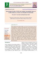
In our study, among the 1151 culture positive
clinical samples, 100 isolates of P.aeruginosa
were isolated (8.68%). The predominant
sample of isolation was pus/wound swab
(17.59%), followed by ETT Secretion
(12.5%), Ear swab (9.79%), sputum (7.66%)
urine (5.74%), Blood (1.96%) and CSF
(1.02%) (Table 1).
In our study, among the used β lactam other
than carbapenems, ceftazidime (61%),
cefepime (53%) and fluroquinolones like
cipofloxacin (63%) and levofloxacin(49%)
showed highest resistant. Among the
aminoglycosides, gentamicin (41%) showed
highest resistant while tobramicin (26%) and
amikacin (28%) exhibit less resistant.
Among the β-lactam combination (β-lactam
combined with β latamase inhibitor) by
Piperacillin/ tazobactam showed 42%
resistance. The resistant pattern of Aztreonam
is 51%. The urine isolates of P.aeruginosa
shown 50% resistant to Norfloxacin. The
carbapenems, Imipenem (18%), Meropenem
(19%), and Doripenem (16%) showed less
resistant. Most of isolates were found to be
highly sensitive to Colistin (100%),
Polymyxin B (100%),
Among 100 strains of P.aeruginosa, which
were screened phenotypically for ESBL
(33%), MBL (18%) and AMP C(0%), the
prevalence of ESBL, MBL and Both ESBL
and MBL is 33%, 18%, and 9% respectively.
No strain was positive for AMP C (Table 2
and 3). Isolates from ETT. Sec (100%), Pus
1063
Int.J.Curr.Microbiol.App.Sci (2018) 7(9): 1061-1069
(48.1%), Urine (75.0%) and wound swab
(64.2%) showed maximum resistant to
levofloxacin (Le). Among the combined drug
Piperacillin/Tazobactam (25.0%) shown less
resistant.
P.aeruginosa has emerged as a significant
pathogen, due to its intrinsic ability to resist
many classes of antibiotics as well as its
ability to acquire resistance, its virulence,
ability to resist killing by various antibiotics
and disinfectant, it presents a serious
therapeutic challenge for treatment of both
community
acquired
and
nosocomial
infections. This affects mortility, morbidity
and financial implication in therapy of
infected patients.
In India, prevalence rate of P.aeruginosa
infection varies from 10.5% to 30%. It ranged
from 3 to 16%, in a multicentric study
conducted by Ling JM et al., (1995) In other
Indian study Pathi et al., reported 8.43%.
(Pathi et al.,) The prevalence in our study was
found to be 8.68% which is comparable to
above study.
Wound infection and respiratory tract
infections were found to be commonly
affected by P.aerugiosa. In this study the
predominant sample of isolation was
pus/wound swab (17.59%), followed by ETT
secretion (12.5%), ear swab (9.79%), sputum
(7.66%) urine (5.74%), Blood (1.96%) and
CSF (1.02%). S. Senthamarai et al., (47.11%)
(Senthamarai et al., 2014) and Vijaya
Chaudhari et al., (35.3%) also reported highest
rate of isolation in pus. (Vijaya Chaudhari et
al., 2013)
In a study conducted in Punjab, India, Arora et
al., found highest recovery rates were from
urine (36%), followed by wound discharge
(20%), tracheal aspirate (8%), ear discharge
(5%) and sputum (4%). (Arora et al., 2011)
Another study by Javiya et al., from Gujarat,
India, reported higher isolation rates from
urine, pus and sputum which accounts to 27%
each, followed by ET secretion 14%. (Javiya
et al., 2008) This variation among these
studies could be due to the difference in study
period and sample size, geographical location
and patient population.
Fig.1
1
12
5 2
Pus/Wound swab
41
Sputum
Ear swab
14
Urine
ETT Secr.
25
Blood
CSF
1064
Int.J.Curr.Microbiol.App.Sci (2018) 7(9): 1061-1069
Table.1 Isolation rate of P. aeruginosa from different clinical Sample (N=1151)
Sample
Pus/ Wound swab
Sputum
Ear Swab
Urine
ET T Sec.
Blood
CSF
TOTAL
N
233
326
143
209
40
102
98
1151
No. (%)
41 (17.59)
25 (7.66)
14 (9.79)
12 (5.74)
5 (12.5)
2 (1.96)
1 (1.02)
100 (100)
Table.2 Antibiotic susceptibility pattern of P. aeruginosa in different clinical specimen
ANTIBIOTICS
Ceftriaxone (30)
Ceftazidime (30)
Cefipime
Piperacillin-Tazobactam
Gentamicin
Amikacin
Tobramycin
Ciprofloxacin
levofloxacin
Imipenem
Meropenem
Colistin
Polymyxin b
Aztreonam
Norfloxacin
SENSITIVE
28
39
47
58
59
72
74
37
51
82
81
100
100
49
48
RESISTANT
62
61
53
42
41
28
26
63
49
18
19
00
00
51
52
Table.3 Prevalence of ESBL, MBL, Amp c and from different clinical isolates (n=100)
N=100
MDR
ESBL
MBL
BOTH ESBL AND MBL
AMP C
No of isolates
28
33
18
09
0
Most of isolates were found to be highly
sensitive to colistin (100%), polymyxin B
(100%), doripenem (89.0%) imipenem (84
%), amikacin (76.0%) and piperacillin +
Percentage
28
33
18
9
0
tazobactum (75%). As the bacterial strains
that show resistance to three or more
categories of antibiotics are defined as
multidrug
resistant
(MDR)
strains,
1065
Int.J.Curr.Microbiol.App.Sci (2018) 7(9): 1061-1069
(Senthamarai et al., 2014) MDR strains of
P.aeruginosa isolated in this study were 28%.
In our study P.aeruginosa showed highest
resistant to β-lactum antibiotics and
fluroquinolones. Among the β lactam drugs,
ceftazidime (61%) and cefepime (53%)
showed the highest resistance in this present
study. K.M Mohanasundaram et al., (84.6%),
(Mohanasundaram, 2011) Yapar et al., (84%)
(Ayse Yüce et al., 2009) and Ibukun et al.,
(79.4%), (Ibukun et al., 2007) reported more
resistance against ceftazidime in their study.
Our study is in line with the reports of
Diwivedi et al., (63%) (Diwivedi et al., 2009)
& Arya et al., (55.4%). (Arya et al., 2005)
The reason for high resistance of third and
fourth generation cephalosporin may be due
to indiscriminate use of third and fourth
generation cephalosporin as broad spectrum
empirical therapy and the secretion of ESBL
enzymes mediate the resistance by hydrolysis
of β-lactam ring of β-lactam antibiotics. Other
mechanisms of drug resistance to β-lactam
group of antibiotics in Pseudomonas
aeruginosa are due to loss of outer membrane
protein, production of class C AmpC βlactamase and altered target sites.
Our study showed 33 (33%) isolates were
ESBL producer. 42.30% ESBL producer were
observed in the study of (Varun Goel et al.,
2013) Lower ESBL producer were seen in the
studies by (Prashant et al., 2011) and Agarwal
et al., which were 22.22% & 20.27%
respectively (Aggarwal et al., 2008)
The ESBL enzymes are inhibited by βlactamase inhibitors, viz., clavulanic acid.
Hence the use of β-lactam/β-lactamase
inhibitor combination may be an alternative to
3rd generation cephalosporin, but the effect of
this combination varies depending on the
subtype of ESBL present. In our study βlactamase inhibitor resistance was ranged
from 42% to 57%. Similar resistance also
observed by Senthamarai et al., (37.5% to
56.73%) (Senthamarai et al., 2014) and K.M
Mohanasundaram
et
al.,
(40.3%).
(Mohanasundaram, 2011) In therapeutic part,
increasing resistance to β lactam inhibitors is
a major problem which makes them less
reliable for therapeutic purposes. Though
imipenem was found unaffected by the action
of the enzymes in many studies, MBL
production in our study was (18%) which is
comparable with the studies of Ibukun et al.,
and
Senthamarai
et
al.,
(15.38%).
(Senthamarai et al., 2014; Ibukun et al.,
2007), (Prashant et al., 2011; Agarwal et al.,
2008; Jayakumar and Appalraju, 2007;
Navneeth et al., 2002) and slightly raised
level of carbapenem resistance were reported
by Variya et al., (25%). (Variya et al., 2008)
The percentage variation in the resistance
mechanism could be due to the study
environment where the study was done. These
carbapenem agents may be of benefit in the
treatment of ESBL infection; however,
indiscriminate use of these agents may
promote increased resistance to carbapenems.
None of our isolates showed AmpC β
lactamase.
P.aeruginosa showed higher resistance to
many other classes of antibiotics, including
fluoroquinolones (49% to 63%) and
aminoglycosides (26% to 46%). This is due to
the coexistence of genes encoding drug
resistance to other antibiotics on the plasmids
which encode ESBL. This fact has also been
observed in our study. Among the
aminoglycoside group, gentamycin showed
highest resistance (41%). Minimal resistance
was observed with other aminoglycoside such
as tobramycin (26%) and amikacin (28%)
which is shows promising effect in treatment.
Ciprofloxacin showed (63%) 61.53%
resistance to P.aeruginosa in our study. In
various reports on ciprofloxacin resistance to
P.aeruginosa was ranged between 0-89%
(Algun et al., 2004).
1066
Int.J.Curr.Microbiol.App.Sci (2018) 7(9): 1061-1069
It is evident from the study that nowadays
P.aeruginosa is becoming resistant to
cephalosporins, aminoglycosides and even
beta lactam (BL) – beta lactamase inhibitor
(BLI) combinations and Carbapenems.
Furthermore, infections with such strains may
result in poor or untoward clinical outcomes
that may increase morbidity, mortality and
economic burden. Proper use of antibiotics
following a proper antibiotic policy is the best
way to control spreading of this superbug. To
prevent the spread of the resistant bacteria it
is critically important to have strict antibiotic
policies. To minimize the resistance to in use
routine antibiotics, it is desirable that the
antibiotic susceptibility pattern of bacterial
pathogens like P.aeruginosa in clinical units
should be continuously monitored. As there
are few studies available in our locality,
studies like this would help to formulate the
antibiotic guidelines to the physician in
treatment part which in turn has a great
impact in preventing the mortality and
morbidity associated with Pseudomonas
aeruginosa infections.
References
Aggarwal R, Chaudhary U, Bala K. Detection
of extended-spectrum beta-lactamase in
Pseudomonas aeruginosa. Indian J
Pathol Microbiol. 2008; 51: 222-4.
Algun A, Arisoy, Gunduz T, Ozbakkaloglu B.
the
resistance
of
Pseudomonas
aeruginosa strains to fluoroquinolones
group of antibiotics. Ind J Med Micro.
2004; 22(2); 112-14.
Arora D, Jindal N, Romit RK. Emerging
antibiotic resistance in Pseudomonas: A
Challenge. International Journal of
Pharmacy and Pharmaceutical Science.
2011; 3(2):82–84.
Arya M, Arya P, Biswas D, Prasad R. The
antimicrobial susceptibility pattern of
the bacterial isolates from post-
operative wound infections. Indian J
Pathol Microbiol. 2005; 48(2): 266-69.
Ayse Yüce, Nur Yapar, Oya Eren Kutsoylu.
Evaluation of antibiotic resistance
patterns of pseudomonas aeruginosa and
Acinetobacter spp. strains isolated from
intensive care patients between 20002002 and 2003-2006 periods in Dokuz
Eylul University Hospital, Izmir
Mikrobiyol Bul. 2009; 43(2):195-202.
Clinical and Laboratory Standards Institute.
Performance
Standards
for
Antimicrobial Susceptibility Testing;
th
26
Informational
Supplement
(M100-S21). Wayne, PA: Clinical and
Laboratory Standards Institute; 2016.
Diwivedi M, Mishra A, Singh RK, Azim A,
Baronia AK, Prasad KN. The
nosocomial cross – transmission of
Pseudomonas
aeruginosa
between
patients in a tertiary intensive care unit.
Indian J Pathol Microbiol. 2009; 52(4):
509-13.
Du Bois V, Arpin C, Melon M et al.,
Nosocomial outbreak due to a multiresistance strain of Pseudomonas
aeruginosa P12: efficacy of cefepimeamikacin therapy and analysis of βlactam resistance. J. Clin. Microbiol.
2001; 39: 2072–2078.
Gales AC, Jones RN, Turnidge J, Rennie, R
Ramphal R. Characterisation of
Pseudomonas aeruginosa isolates:
occurrence,
rate
antimicrobial
susceptibility pattern and molecular
typing
in
the
Global
Sentry
antimicrobial surveillance program
1997–1999. Clin. Infect. Dis. 2001; 32:
146 –155.
Govan J. R. W. Pseudomonads and nonfermenters. In Medical microbiology A
Guide
to
Microbial
Infections:
Pathogenesis, Immunity, Laboratory
Diagnosis and Control. Eds. Greenwood
David, Slack Richard C.B, Peutherer
1067
Int.J.Curr.Microbiol.App.Sci (2018) 7(9): 1061-1069
John F. 6th edn. Edinburg: Churchill
Livingstone; 2008; p. 282-287.
Govan,
J.
R.
W.
Pseudomonas,
Stenotrophomonas, Burkholderia. In:
Mackie and Mc Cartney Practical
Medical Microbiology. Eds. Collee JG,
Fraser AG, Marmion BP & Simmons A.
14th
ed.
Edinburg:
Churchill
Livingstone; 2006; p 413-424.
Hernandez J, Ferrs MA, Hernandez M, Owen
RJ. Arbitrary primed PCR fingerprint
and serotyping of clinical Pseudomonas
aeruginosa strains. FEMS Immunology
and Medical Microbiology. 1997: (17);
37–47.
Ibukun A, Tochukwu N, Tolu O. Occurrence
of ESBL and MBL in clinical isolates of
Pseudomonas aeruginosa From Lagos,
Nigeria. Journal of American Science.
2007; 3(4): 81-85.
Javiya VA, Ghatak SB, Patel KR, Patel JA.
Antibiotic susceptibility patterns of
Pseudomonas aeruginosa at a tertiary
care hospital in Gujarat, India. Indian J
Pharmacol. 2008; 40(5):230-4.
Jaykumar S, Appalraju B. The prevalence of
multi
and
pan
drug
resistant
Psuedomonas aeruginosa with respect to
ESBL and MBL in a tertiary care
hospital. Indian J Pathol Microbiol.
2007; 50 (4): 922-25.
Ling J M, Cheng AF. Antimicrobial
resistance of clinical isolates from 1987
to 1993 in Hong Kong. HKMJ. 1995;
1(3):212-18.
Mohanasundaram KM. The antimicrobial
resistance pattern in the clinical isolates
of Pseudomonas aeruginosa in a tertiary
care hospital: 2008-2010(a 3 year
study). Journal of Clinical and
Diagnostic Research. 2011, Vol-5(3);
491-94.
Navneeth BV, Sridaran D, Sahay D, Belwadi
MA preliminary study on the metallo
betalactamase producing Pseudomonas
aeruginosa in hospitalised patients.
Indian J Med Res. 2002; 112: 264-67.
Pathi B, Mishra SN, Panigrahi K, Poddar N,
Lenka PR, Mallick B, Pattanik D, Jena
J. Prevalence and antibiogram pattern of
Pseudomonas aeruginosa in a tertiary
care hospital from Odisha, India.
Transworld Medical Journal. 1(3):7780.
Prashant Durwas Peshattiwar, Basavaraj
Virupaksappa Peerapur. ESBL and
MBL
mediated
resistance
in
Pseudomonas aeruginosa: an emerging
threat to clinical therapeutics. Journal of
Clinical and Diagnostic Research.
2011, Vol-5(8); 1552-554.
Senthamarai S, Suneel Kumar Reddy A, and
Sivasankari S et al., Resistance Pattern
of Pseudomonas aeruginosa in a
Tertiary Care Hospital of Kanchipuram,
Tamilnadu, Indian Journal of Clinical
and Diagnostic Research. 2014 May,
Vol-8(5): 30-32.
Srinivas B, Lalitha Devi D, Narasinga Rao B.
A prospective study of Pseudomonas
aeruginosa and its antibiogram in a
teaching hospital of Rural setup. Journal
of Pharmaceutical and Biomedical
sciences. 2012; 22:23-29.
Syed Arshi, Thakur Manzoor, Shafiq Syed,
Mr. Sheikh Ullah Assad. In-vitro
sensitivity patterns of Pseudomonas
aeruginosa strains isolated from
patients at skims – role of
antimocribials in the emergence of
multiple
resistant
strains.
JKPractitioner. 2007; 14(1):31-34.
Variya A, Kulkarni N, Kulkarni M, et al., The
incidence of metallo beta lactamase
producing Pseudomonas aeruginosa
among ICU patients. Indian J Med Res.
2008; 127: 398-402.
Varun Goel, Sumati A. Hogade, SG
Karadesai. Prevalence of extendedspectrum
beta-lactamases,
AmpC
beta-lactamase,
and
1068
Int.J.Curr.Microbiol.App.Sci (2018) 7(9): 1061-1069
metallo-beta-lactamase
producing
Pseudomonas
aeruginosa
and
Acinetobacter baumannii in an intensive
care unit in a tertiary Care Hospital
Journal of the Scientific Society. 2013
40(1), pp. 28-31.
Vijaya Chaudhari, Sandeep Gunjal, Mukesh
Mehta. Antibiotic resistance patterns of
Pseudomonas aeruginosa in a tertiary
care hospital, in Central India.
International Journal of Medical science
and Public Health. 2013; Vol 2(2) 38689.
How to cite this article:
Trinain Kumar Chakraverti and Purti C. Tripathi. 2018. Prevalence and Antibiotic Resistant
Pattern of Pseudomonas aeruginosa at a Tertiary Care Centre of North India.
Int.J.Curr.Microbiol.App.Sci. 7(09): 1061-1069. doi: />
1069