Immunotoxic effect of selenium following subacute exposure in broilers
Bạn đang xem bản rút gọn của tài liệu. Xem và tải ngay bản đầy đủ của tài liệu tại đây (455.98 KB, 7 trang )
Int.J.Curr.Microbiol.App.Sci (2018) 7(9): 1630-1636
International Journal of Current Microbiology and Applied Sciences
ISSN: 2319-7706 Volume 7 Number 09 (2018)
Journal homepage:
Original Research Article
/>
Immunotoxic Effect of Selenium Following Subacute Exposure in Broilers
Shonam Tamrakar1, M. Mondal2*, R.C. Ghosh1 and N. Sahu1
1
Department of Veterinary Pathology, College of Veterinary Science and Animal Husbandry,
Chhattisgarh Kamdhenu Vishwavidyalaya, Durg, Chhattisgarh, India
2
Department of Veterinary Clinical Complex, F/O- Veterinary and Animal Sciences, West
Bengal University of Animal and Fishery Sciences, Kolkata, India
*Corresponding author
ABSTRACT
Keywords
Broiler,
Immunotoxicity,
Selenium
Article Info
Accepted:
10 August 2018
Available Online:
10 September 2018
This study was conducted with an objective to determine the immunotoxic effect of sub
acute exposure of selenium (Se) in broilers with special reference to the
Dinitroflurobenzene (DNFB) contact skin sensitivity test and pathomorphological
alterations in histoarchitecture of lymphoid organs. The chicks were intoxicated orally
with sodium selenite @ 2, 6 and10 ppm respectively in diet, daily for 35 days. The present
study exhibited significant depression of cell mediated immunity in Se treated broilers as
measured by DNFB skin contact sensitization test. The blood picture revealed dose
dependant heterophilia and leukocytopaenia due to lymphocytopaenia. During subacute Se
toxicity, severity of pathological changes of treated broilers was dose dependent.
Lymphoid organs of selenium treated broilers revealed severe depletion of lymphocytes
from the germinal centre.
Introduction
Poultry industry is designated as major
dynamic and rapid growing segment amongst
agricultural and livestock sectors in India.
Selenium (Se) is required in the diets of
mammals and poultry, but can easily be over
supplemented due to a narrow range of safety
between ideal and toxic concentrations.
Sodium selenate and sodium selenite are used
as supplements to poultry and livestock feed
to promote growth and prevent selenium
deficiency diseases. Selenium can be toxic for
all animals, such as invertebrates, fishes,
amphibians and reptiles, birds, mammals and
humans depending on the dose and duration of
intake and also on its chemical form.
Traditionally, Se has been added to poultry
diets via inorganic sources, such as sodium
selenite (Na2SeO3). Selenium in combination
with vitamin E is used frequently in poultry
for immunomodulation.
The immunomodulatory effect and toxicity of
inorganic selenium, such as selenite and
selenate, has been heavily studied in animals,
aquatic birds and fishes. But till date work on
immunotoxicity of Se is scanty. So, present
study has designed to demonstrate the
immunotoxic effect of selenium following
subacute exposure in broilers.
1630
Int.J.Curr.Microbiol.App.Sci (2018) 7(9): 1630-1636
Materials and Methods
Chicken and diet
The experimental investigation was planned to
adjudge the toxicopathological effects of
sodium selenite on cell mediated immune
response in broilers after obtaining approval
from Institutional Animal Ethics Committee.
Clinically healthy one hundred and twenty,
day old, Cobb- 400 broiler chicks of both
sexes, weighing 45- 49 g were obtained from a
commercial hatchery and were reared on deep
litter system of housing using rice husk with
provision of artificial light at night. The chicks
were fed a standard commercial feed starter
upto14 days, thereafter a grower diet upto 28
days and finisher upto 35 days. Chickens were
allowed access to the diets and fresh and clean
drinking water ad libitum. All the
experimental chicks were kept under close
observation during entire period of study.
Experimental design
Individually weighed chicks were randomly
divided into 4 groups of 30 chicks; each group
consisting of 3 replicates of 10 chicks. Chicks
of group-I was kept as untreated control and
was given only basal diet. Chicks of groupsII,
III and IV were given diet with selenium @ 2,
6 and 10 ppm respectively from day first of
experiment for 35 days. Six broilers from each
group were used for assessing the cell
mediated immunity. The remaining broilers
from
each
group
were
kept
for
haematological, biochemical and pathological
study. When the chicks reached 35 days of
age, the feeding trial was terminated.
(1988). Featherless area was marked on both
sides of abdomen and cleaned thoroughly with
acetone and air dried. Right lateral side of
abdomen was used for DNFB application
whereas left side served as control. 2000µg of
DNFB in 0.1 ml of acetone and olive oil (4:1)
was applied on the right marked area on the
abdomen using a plastic ring to avoid spillage.
The sensitized birds were challenged with
50µg of DNFB in 0.1 ml of acetone and olive
oil (4:1) on the same area on 14th day after
initial sensitization. The response to DNFB
was assessed by measuring the skin thickness
using engineer’s micrometer on 0, 24 and 48
hours of post challenge with three readings
each and the overall mean skin thickness was
calculated.
Haematology
At the end of the experiment on day 35 blood
samples were collected in heparinised vials
from jugular veins. Thin blood smears were
prepared for differential leukocyte count
during blood collection. The total leucocytic
count (TLC) was done as per Jain (1986), by
using W.B.C. diluting fluid (Merck Limited,
Mumbai - 400018) and Haemocytometer
(Neubauer’s chamber and WBC diluting
pipette).
The differential leucocytic count (DLC) was
done as per Coles (1986), using Leishman’s
stain (Merck Limited, Mumbai- 400018). The
percentages of different leucocytes were
determined by examining the stained blood
smear under oil immersion objective lens of
light microscope.
Histopathology
Cell mediated immunity (CMI)
Cell mediated immune response was measured
by Di Nitro Fluro Benzene (DNFB) test as
described by Phanuphak et al., (1974) and
later slightly modified by Tamang et al.,
The tissue samples of spleen were collected in
10%
neutral
buffered
formalin
for
histopathological studies. The tissues were
thoroughly washed in running water;
dehydrated in ascending grades of alcohol;
1631
Int.J.Curr.Microbiol.App.Sci (2018) 7(9): 1630-1636
cleared in benzene and embedded in paraffin
at 58°C. The paraffin embedded tissue
sections of 4 to 5 μm were obtained and
stained with haematoxylin and eosin (H and
E) as per the method described by Bancroft
and Stevens (1990) with slight modifications.
The stained sections were examined under
light microscope and the lesions were
recorded.
the findings of earlier workers in alphamethrin
intoxicated broilers (Singh et al., 1999).
Statistical analysis
The epidermal Langerhans cell, a member of
the dendritic-cell family, takes up haptenated
proteins and processes them into antigenic
peptides which are transported to the cell
surface
in
association
with
major
histocompatibility complex molecules (Wang
et al., 2001).
Data obtained in different parameters were
statistically analyzed by using complete
randomized design (CRD)-single factor
analysis of variance by Snedecor and Cochran
(1968).
Contact hypersensitivity is a T- cell mediated
cutaneous immune response to reactive
haptens (Elmets and Bowen, 1986). After
exposure of the skin to contact allergens,
haptens covalently bind to discrete amino acid
residues on carrier proteins.
Matos et al., (2005) reported that DNFB
induces the activation of the extra cellular
signal-regulated kinases ERK1/2 and p38, and
also up regulates CD40 expression.
Results and Discussion
Cell mediated immune response (CMI)
In the present study the mean increment in
abdominal skin thickness of treated broilers at
different hours post challenge were depicted in
Table 1. Broilers exposed to the challenge
dose of DNFB exhibited erythema,
oedematous changes and vesicle and scab
formation.
Broilers of all Se treated groups had
pronounced changes than that of control.
Present study indicated that the contact
sensitivity to DNFB could be conveniently
applied in broilers for studying CMI response
using abdominal skin in place of comb as test
site unlike the previous method (Tiwari and
Goel, 1984).
Control broilers had significant decrease in
abdominal skin thickness as compared to
selenium treated broilers at 24 and 48 hours
post challenge which clearly indicated the
immunosuppression due to selenium toxicity.
Present findings of significant decrease CMI
response on DNFB test was in agreement with
Haematology
Results on the haematological alteration due to
subacute selenium toxicity in broilers were
given in Table 2. A significant (p≤0.05, 0.01)
decrease in leucocyte count was observed in
broilers of all intoxicated groups. The present
study also showed that selenium caused a
significant eosinopaenia and leukopaenia due
to lymphopaenia in all the intoxicated broilers.
This finding was in agreement with the
changes induced by Se in broiler chickens
earlier (Kumar et al., 2011). Marked
leucopaenia in subacute selenium toxicity in
the present study was probably due to their
cumulative
effect
following
daily
administration. Continuous exposure to Se
may then lead to lymphopaenia, which may
have an immunosuppressive effect in broilers.
The marked lymphopaenia in the present study
might have occurred due to the toxic effect of
sodium selenite on bone marrow and stress
(Goyal et al., 1986).
1632
Int.J.Curr.Microbiol.App.Sci (2018) 7(9): 1630-1636
Table.1 DNFB response (mean increase in skin thickness in mm) of broilers exposed to subacute
selenium toxicity (Left side served as vehicle control and right side treated with DNFB)
Groups
Abdominal
side
Left
Right
Left
Right
Left
Right
Left
Right
Gr I
Gr II
Gr III
Gr IV
Level of significance
Before
sensitization
0.55±0.04
0.58±0.04a
0.57±0.05
0.61±0.05a
1.25±0.07
1.33±0.08b
1.15±0.05
1.21±0.06b
**
After sensitization
24 hr
48 hr
0.63±0.08
0.61±0.08
b
3.22±0.10
3.47±0.16b
1.81±0.17
1.77±0.22
b
3.37±0.09
3.64±0.09b
1.68±0.09
1.65±0.09
a
1.90±0.14
2.71±0.18a
1.47±0.16
1.47±0.17
a
1.65±0.13
2.86±0.13a
**
**
Table.2 Effect of induced subacute toxicity of Selenium on haematological changes in broilers
PARAMETERS
TLC (*103/cu.mm)
Lymphocyte (%)
Heterophil (%)
Monocyte (%)
Eosinophil (%)
Basophil (%)
GROUPS
Group I
31.18±0.36d
74.5±1.12b
16±0.93a
5.33±0.80a
2.83±0.31b
1.33±0.21a
Group II
29.19±0.21c
64.5±2.19a
19.5±1.45b
13.33±0.67b
0.83±0.31a
1.83±0.17a
Group III
27.21±0.15b
61.83±1.42a
20.16±1.11b
14.5±0.62b
1.33±0.21a
2±0.16a
Group IV
26.08±0.27a
60.83±0.60a
21.83±0.48b
14.66±0.49b
0.83±0.31a
1.83±0.17a
Level of
significance
**
**
**
**
**
NS
Values indicate Mean ± S.E. Superscripts may read row wise for comparison of means. NS - No significance
difference. (*P≤0.05) and (**P≤0.01)
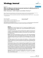
Fig.1 Section of spleen (group III) showing oedematous changes. H & E × 400
1633
Int.J.Curr.Microbiol.App.Sci (2018) 7(9): 1630-1636
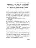
Fig.2 Section of spleen (group IV) showing severe depletion of lymphocytes and vacuolation.
H & E × 400
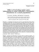
Fig.3 Section of bursa of Fabricius (group IV) showing vacuolation and severe depletion of
lymphocytes. H & E × 400
Fig.4 Section of thymus (group IV) showing severe medullary lymphocytic depletion.
H & E × 100
1634
Int.J.Curr.Microbiol.App.Sci (2018) 7(9): 1630-1636
Histopathology
Histologically the spleen of intoxicated
broilers had congestion, oedematous changes
(Fig. 1), severe depletion of the lymphocytes
and
vaccuolation
(Fig.
2).
The
histopathological changes of spleen were
closely corroborated with the findings of
Jacevic et al., (2011). In the present study the
marked lymphocytolysis of the germinal
center of the splenic follicles correlates with
the findings related to suppression of the CMI
and indicated that selenium causes
immunosuppression. Microscopically induced
broilers
revealed
serous
exudation,
vaccuolation and depletion of lymphocytes in
the follicles of bursa of Fabricius (Fig. 3).
Thymus of Se intoxicated broilers had
congestion,
haemorrhages,
oedematous
changes and severe medullary lymphocytic
depletion (Fig. 4). Histological changes of
lymphoid organs in selenium induced broilers
were in close conformity with the findings of
Narayani (2010) who reported severe
lymphocytosis in the germinal center of the
spleen, bursa of Fabricius and thymus in
alphamethrin treated broilers. So, the present
study suggested that the selenium toxicity
causes immunosuppression in the broilers.
References
Brancroft, J.D. and Stevens, A. (1990).
Theory and practice of histological
techniques. Churchill Livingstone,
Edinburgh, pp. 113-305.
Coles, E.M. (1986). Veterinary Clinical
Pathology. 4th Edn., W.B. Saunders
company, West Washington square,
Philadalphia.
Elmets,
C.A.
and
Bowen,
K.D.
(1986).Immunopathological suppression
in mice treated with hematoporphyrin
derivative
photoradiation.
Cancer
Res.46: 1608-1611.
Goyal, B.S., Garg, S.K. and Garg, B.D.
(1986). Malathion induced hyperadrenal
activity in WLH chicks. Current Sci.,
55: 526-528.
Jacevic, V., Jokic, G., Simic, V.D., Bokonjic,
D., Vucinic, S. and Vuksa, M. (2011).
Acute toxicity of sodium selenite in
rodents: Pathomorphological study.
Military
Med.
Sci.
Lett.
(Voj.Zdrav.Listy) 80: 90-96.
Jain,
N.C.
(1986).
Haematological
techniques-In
Sclams
Veterinary
th
Haematology. 4
Edn., Lea and
Febingerr. Philadelphia. pp. 20-86.
Kumar, D., Niyogi, D., Ali, I. and
Mukhopadhayay,
S.K.
(2011).
Clinicopathological study of induced
selenium
toxicity
in
broiler
chickens.Indian J. Anim. Hlth., 50 (2):
15-20.
Matos, T.J., Duarte, C.B., Goncalo, M. and
Loes, M.C. (2005). DNFB activates
MAPKs and upregulates CD40 in skinderived dendritic cells. J. Dermato.
Sci.39:113-123.
Narayani. (2010). Toxicopathological studies
on alphamethrin in broiler chicks.
M.V.Sc. Thesis submitted to Indira
Gandhi Krishi Vishwvidyalaya, Raipur,
Chhattisgarh.
Phanuphak, P., Moorhead, J.W. and Claman,
H.N. (1974). Tolerance and contact
sensitivity to DNFB in mice. In vivo
detection by ear swelling and
correlation with in vitro cell stimulation.
J. Immunol., 112: 115-123.
Singh, S.P., Sharma, L.D. and Chauhan, R.S.
(1999). Effect of single oral dose of
alphamethrin on immune response of
chicks. J. Immunol. Immunopathol.,
1(1): 63-66.
Snedecor, G.W. and Cochran, W.G.
(1968). Statistical Methods. 8 th Edn.
Iowa State University Press, Ames,
Iowa.
1635
Int.J.Curr.Microbiol.App.Sci (2018) 7(9): 1630-1636
Tamang, R.K. (1987). Comparative pathology
of pyrethroid and organophosphate
pesticides intoxication in mice and
goats. M.V.Sc. Thesis submitted to
Birsa Agricultural University, Ranchi,
Bihar.
Tiwari, B.K. and Goel, M.C. (1984). Contact
sensitivity to DNCB in normal and cell
mediated immunity deficient chickens:
In vivo detection and correlation with
lymphocyte transformation and Graft
versus
Host
reaction.
Vet.
Immunol.Immunopathol.8: 329-339.
Wang, B., Feliciani, C., Freed, I., Cai, Q. and
Saunder, D.N. (2001). Insights into
molecular mechanisms of contact
hypersensitivity gained from gene
knockout
studies.
J.
Leukoc.
Biol.70:185-191.
How to cite this article:
Shonam Tamrakar, M. Mondal, R.C. Ghosh and Sahu, N. 2018. Immunotoxic Effect of
Selenium Following Subacute Exposure in Broilers. Int.J.Curr.Microbiol.App.Sci. 7(09): 16301636. doi: />
1636