CT-based radiomics scores predict response to neoadjuvant chemotherapy and survival in patients with gastric cancer
Bạn đang xem bản rút gọn của tài liệu. Xem và tải ngay bản đầy đủ của tài liệu tại đây (2.7 MB, 11 trang )
Sun et al. BMC Cancer
(2020) 20:468
/>
RESEARCH ARTICLE
Open Access
CT-based radiomics scores predict response
to neoadjuvant chemotherapy and survival
in patients with gastric cancer
Kai-Yu Sun1†, Hang-Tong Hu2†, Shu-Ling Chen2, Jin-Ning Ye1, Guang-Hua Li1, Li-Da Chen2, Jian-Jun Peng1,
Shi-Ting Feng3, Yu-Jie Yuan1, Xun Hou1, Hui Wu1, Xin Li4, Ting-Fan Wu4, Wei Wang2* and Jian-Bo Xu1*
Abstract
Background: Neoadjuvant chemotherapy is a promising treatment option for potential resectable gastric cancer,
but patients’ responses vary. We aimed to develop and validate a radiomics score (rad_score) to predict treatment
response to neoadjuvant chemotherapy and to investigate its efficacy in survival stratification.
Methods: A total of 106 patients with neoadjuvant chemotherapy before gastrectomy were included (training
cohort: n = 74; validation cohort: n = 32). Radiomics features were extracted from the pre-treatment portal venousphase CT. After feature reduction, a rad_score was established by Randomised Tree algorithm. A rad_clinical_score
was constructed by integrating the rad_score with clinical variables, so was a clinical score by clinical variables only.
The three scores were validated regarding their discrimination and clinical usefulness. The patients were stratified
into two groups according to the score thresholds (updated with post-operative clinical variables), and their
survivals were compared.
Results: In the validation cohort, the rad_score demonstrated a good predicting performance in treatment
response to the neoadjuvant chemotherapy (AUC [95% CI] =0.82 [0.67, 0.98]), which was better than the clinical
score (based on pre-operative clinical variables) without significant difference (0.62 [0.42, 0.83], P = 0.09). The rad_
clinical_score could not further improve the performance of the rad_score (0.70 [0.51, 0.88], P = 0.16). Based on the
thresholds of these scores, the high-score groups all achieved better survivals than the low-score groups in the
whole cohort (all P < 0.001).
Conclusion: The rad_score that we developed was effective in predicting treatment response to neoadjuvant
chemotherapy and in stratifying patients with gastric cancer into different survival groups. Our proposed strategy is
useful for individualised treatment planning.
Keywords: Stomach neoplasms, Neoadjuvant therapy, Tomography, X-ray computed
* Correspondence: ;
†
Kai-Yu Sun and Hang-Tong Hu contributed equally to this work.
2
Department of Medical Ultrasonics, Institute of Diagnostic and
Interventional Ultrasound, The First Affiliated Hospital of Sun Yat-Sen
University, 58 Zhongshan Road 2, Guangzhou 510080, People’s Republic of
China
1
Department of Gastrointestinal Surgery, The First Affiliated Hospital of Sun
Yat-Sen University, 58 Zhongshan Road 2, Guangzhou 510080, People’s
Republic of China
Full list of author information is available at the end of the article
© The Author(s). 2020 Open Access This article is licensed under a Creative Commons Attribution 4.0 International License,
which permits use, sharing, adaptation, distribution and reproduction in any medium or format, as long as you give
appropriate credit to the original author(s) and the source, provide a link to the Creative Commons licence, and indicate if
changes were made. The images or other third party material in this article are included in the article's Creative Commons
licence, unless indicated otherwise in a credit line to the material. If material is not included in the article's Creative Commons
licence and your intended use is not permitted by statutory regulation or exceeds the permitted use, you will need to obtain
permission directly from the copyright holder. To view a copy of this licence, visit />The Creative Commons Public Domain Dedication waiver ( applies to the
data made available in this article, unless otherwise stated in a credit line to the data.
Sun et al. BMC Cancer
(2020) 20:468
Background
Gastric cancer remains the third most frequent cause of
cancer-related death worldwide, resulting in 782,685
deaths annually [1]. Despite the improvement in screening, a large proportion of patients in China are diagnosed at advanced stage. For locally advanced cases, the
5-year survival rate ranged from 20 to 30% after curative
resection [2–4].
Given this poor prognosis, neoadjuvant chemotherapy
has been tried for this patient population in recent years.
After the promising results obtained with “MAGIC Trial”,
“FFCD Trial”, “ACCORD Trial”, and “AIO-FLOT3 Trial”,
neoadjuvant chemotherapy has become a promising treatment option for potentially resectable or limited metastatic gastric cancer with the improved 5-year survival
rates of more than 35% [5–10]. Despite the satisfactory efficacy of neoadjuvant chemotherapy, patients’ responses
varied between 30 and 60% [11]. A good response to neoadjuvant chemotherapy was associated with good survival
outcome, while non-responding patients could suffer from
adverse events and unnecessary costs and finally risk
tumour progression and even miss the chance to undergo
curative gastrectomy. Moreover, patients who are nonresponsive to neoadjuvant chemotherapy could be waiting
longer until surgery, and this extended time to surgery
may be correlated with poorer survival of gastric cancer.
Thus, early detection of those patients who are most likely
to respond to neoadjuvant treatment is critical to provide
them a chance for a timely surgery and to optimise the
treatment plans. However, the treatment efficacy of neoadjuvant chemotherapy can only be assessed after three
cycles of treatment. Therefore, exploring the pretreatment predictors of treatment efficacy is important to
determine the need for neoadjuvant therapy and the optimal timing for surgical resection, thus improving pretreatment decision making.
Previous studies have investigated several imaging modalities such as contrast enhanced ultrasound, computed
tomography (CT), magnetic resonance imaging, and
positron emission tomography in the evaluation of patients’ response to chemotherapy for gastric cancer;
however conflicting results were obtained [12–18]. Additionally, in these studies, analyses were only based on
imaging features extracted by naked eyes or quantitative
imaging parameters, and lacked a proper validation. Although naked eyes provide valuable feature information,
some microcosmic imaging features relevant for clinical
outcomes might be lost due to the limited visual image
grey scales that can be detected by naked eyes. Radiomics is a rapidly growing discipline based on highthroughput quantitative image analysis to characterise
tumours and their microenvironment. This approach
can extract far more features than manual extraction by
acquiring two-dimensional and high-dimensional
Page 2 of 11
imaging features using computer algorithm [19]. Many
studies on other cancer types showed that radiomics features, such as texture features, filter transformed features, wavelet features, and so on, could not be visually
observed but were closely related to pathologic microscopic structures and were effective in prognostic prediction [20–23].
Computed tomography is the preferred imaging examination for gastric cancer in clinical practice, but no literature has been reported on the application of CTbased radiomics technique to predict the response to
neoadjuvant chemotherapy in gastric cancer patients.
Therefore, we aimed to develop and validate a CT-based
radiomics score to predict the response to neoadjuvant
chemotherapy and stratify the survival for patients with
gastric cancer.
Methods
Patients
Consecutive patients diagnosed with gastric cancer between January 2010 and December 2017 were identified
by reviewing the database of the Center of Gastrointestinal Surgery of the First Affiliated Hospital of Sun YatSen University. Patients were included according to the
following criteria: (1) histologically confirmed gastric
adenocarcinoma on gastroscopy; (2) potential resectable
gastric cancer at clinical stage of III, IV as determined
by pretreatment contrast-enhanced CT (patients with
M1 were those with only para-aortic lymph node metastasis without any other risk of curative resection); (3) received neoadjuvant chemotherapy of SOX regimen (S-1
plus oxaliplatin) as the initial treatment; (4) underwent
curative gastrectomy; (5) received contrast-enhanced CT
within one week before neoadjuvant chemotherapy; (6)
Eastern Cooperative Oncology Group performance status between 0 to 1; (7) a life expectancy of > 3 months;
(8) adequate bone marrow, renal, and hepatic function
[platelets > 80 × 109/L, absolute neutrophil count ≥1.5 ×
109/L, serum creatinine≤1.5 mg/dL, total bilirubin level
within 1.5 × the upper limit of normal (ULN), and serum
transaminase ≤2.5× ULN]. The following exclusion criteria were used: (1) history or presence of other malignancies; (2) presence of other uncontrolled diseases or
severe infection; (3) received other anti-tumour therapies
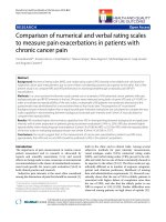
before neoadjuvant chemotherapy; (4) incomplete clinical data. The patient selection process is shown in Fig. 1.
Patients were randomly allocated to the training and validation cohorts at the ratio of 7:3. Our Institutional Ethic
Review Board has approved the current study, following
the regulations outlined in the Declaration of Helsinki.
Neoadjuvant chemotherapy
Patients received the first-line neoadjuvant chemotherapy of SOX regimen. S-1 was orally administered twice
Sun et al. BMC Cancer
(2020) 20:468
Page 3 of 11
Fig. 1 Flow diagram of study population
daily at concentrations based on body surface area (BSA):
BSA < 1.25 m2, 80 mg/d; 1.25 m2 BSA < 1.50 m2, 100 mg/
d; and BSA ≥ 1.50, 120 mg/d. On the first day, oxaliplatin
(130 mg/m2) was administered via intravenous infusion,
followed by S-1 administered for 14 consecutive days,
followed by a 1-week break for a maximum of three cycles, until tumour progression, presence of unacceptable
toxicity or treatment withdrawal by the patient or doctor.
Assessment of the response to neoadjuvant
chemotherapy
The treatment response to neoadjuvant chemotherapy
was evaluated via pathologic response. Haematoxylin
and eosin-stained slides were reviewed by two pathologists with more than 10 years of experience in gastrointestinal pathology who were blinded to the clinical
data, and they graded the specimens for pathologic response according to the Mandard tumour regression
grading (TRG) system [24]. TRG 1 was defined as
complete regression/fibrosis with no viable tumour cells,
TRG 2 was defined as fibrosis with scattered tumour
cells, TRG 3 was defined as fibrosis and tumour cells
with predominant fibrosis, TRG 4 was defined as fibrosis
and tumour cells with predominant tumour cells, and
TRG 5 was defined as tumour without evidence of regression. Disagreement was resolved by discussion with
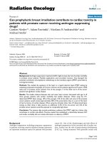
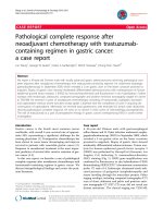
Fig. 2 A female patient was diagnosed as gastric cancer (T4aN2M0). CT before neoadjuvant chemotherapy (a) showed a mass-type tumor
measured 25 mm in maximal depth and 80 mm in maximal length. CT after neoadjuvant chemotherapy (b) showed a shrunken mass measured
14 mm in depth and 40 mm in length. CT before neoadjuvant chemotherapy (c) showed the ROI delineated manually on figure (a). Pathology
examination after surgery (d) showed residual tumor tissue (arrow) and infiltrated inflammatory cells (arrow head)
Sun et al. BMC Cancer
(2020) 20:468
Page 4 of 11
Table 1 Clinicopathological characteristics of the training and validation cohorts
Factors
Training cohort (n = 74)
Validation cohort (n = 32)
Age (years, mean ± SD)
55.15 ± 11.43
54.13 ± 12.68
Gender
48
18
Female
26
14
21 ± 3
22 ± 3
Preoperative T stage
0.12
0.39
2
1
0
3
11
2
4
62
29
Preoperative N status
0.99
0
2
1
1–3
72
31
0
47
19
1
27
13
Preoperative M status
0.85
Postoperative T stage
0.83
1–2
11
6
3–4
63
26
0
35
8
1–3
39
24
Postoperative N status
0.06
Postoperative M status
0.90
0
53
24
1
21
8
0
3
3
1
6
1
2
22
5
3
22
15
4
21
8
9.25 ± 17.79
6.48 ± 8.33
Postoperative TNM stage
AFP (ng/mL)
0.68
0.53
Male
BMI (kg/m2)
P value
0.23
CEA (IU/L)
0.40
0.23
Normal
50
26
Elevated
24
6
Normal
73
29
Elevated
1
3
CA125 (IU/L)
0.08
CA199 (IU/L)
1.00
Normal
64
27
Elevated
10
5
Operative duration (min)
349.46 ± 116.47
359.22 ± 111.98
0.69
Blood transfusion (ml)
347.92 ± 506.39
362.50 ± 458.43
0.89
Total number of dissected lymph node
40.81 ± 18.67
44.09 ± 17.77
0.40
Number of positive lymph node
5.27 ± 9.01
7.34 ± 10.42
0.30
Sun et al. BMC Cancer
(2020) 20:468
Page 5 of 11
Table 1 Clinicopathological characteristics of the training and validation cohorts (Continued)
Factors
Training cohort (n = 74)
Validation cohort (n = 32)
TRG 1
3
3
TRG 2
34
14
TRG 3
24
10
TRG 4
10
4
TRG 5
3
1
Treatment response
P value
0.25
Abbreviations: BMI body mass index, PS performance status, AFP alpha-fetoprotein, CEA carcinoembryonic antigen, TRG tumor regression grading
consensus. Responders were defined as TRG 1–2 and
non-responders were defined as TRG 3–5 [25].
CT images acquisition
The standard dynamic contrast-enhanced MDCT scan
(Aquilion 64; Toshiba Medical System, Tokyo, Japan) procedure was used. Briefly, after an unenhanced helical sequence scan from the liver dome to the symphysis pubis,
venous phase contrast-enhanced CT was performed after a
65-s delay following intravenous administration of 80–100
ml (1.5 ml/kg) of iodinated contrast agent (Ultravist 300;
Schering, Berlin, Germany) administered via the antecubital
vein at a rate of 2.0–3.0 ml/s. The following CT acquisition
parameters were used: 120 kV, 200–250 mAs, rotation time
of 0.5 s, collimation of 64 mm × 0.5 mm, slice thickness of
0.5 mm, slice increments of 0.5 mm, pitch of 0.9, field of
view of 350 × 350 mm, matrix of 512 × 512, and reconstruction thickness of 2.5 mm. CT images were retrieved from
the picture archiving and communication system (PACS)
(HP workstation XW8200, VitreaCore, version 3.7) for
image analysis. The display window width was 150–350
HU, and the window level was 50 to 80 HU. One such case
is presented in Fig. 2 with CT images before and after the
neoadjuvant chemotherapy and the image of response assessment by pathology.
Radiomics feature extraction
Portal venous phase contrast-enhanced CT images were
used for radiomics feature extraction because of the better differentiation between the tumour tissue and the adjacent normal tissue of the gastric wall in the portal
venous phase than in arterial phase. A region of interest
(ROI) was delineated around the tumour outline for the
largest cross-sectional area while excluding the air area
by two independent radiologists with more than five
years of experience in gastrointestinal imaging, and any
disagreements were resolved by the consensus with arbitration by a third author. For each ROI, a total of 1044
imaging features were extracted and analyzed by an in
house-made software: the A.K. software (Analysis-Kit,
version 2.0.0, GE healthcare), which included six kinds
of features (Supplemental Table 1): 42 histogram parameters, 10 texture parameters, 9 form factor parameters,
Table 2 Comparison of clinical variables and radiomics score in
the responding group and non-responding group in the
training cohort
Factors
Responding
group
Age (years, mean ± SD) 56.76 ± 11.42
Non-responding
group
52.85 ± 11.91
Gender
0.02
1.00
Male
24
24
Female
13
13
BMI (kg/m2)
21 ± 3
21 ± 4
AFP (ng/mL)
8.95 ± 13.51
7.86 ± 17.55
CEA (IU/L)
0.42
0.56
0.80
Normal
26
24
Elevated
11
13
Normal
32
32
Elevated
5
5
CA199 (IU/L)
1.00
CA125 (IU/L)
1.00
Normal
36
37
Elevated
1
0
2
0
1
3
8
3
4
29
33
0
2
0
1
13
8
2
18
21
3
4
8
Preoperative T stage
0.17
Preoperative N status
0.19
Preoperative M status
0.05
0
28
19
1
9
18
0.54 ± 0.22
0.41 ± 0.22
Radiomics score
P value
Abbreviations: BMI body mass index, PS performance status, AFP alphafetoprotein, CEA carcinoembryonic antigen
< 0.01
Sun et al. BMC Cancer
(2020) 20:468
Page 6 of 11
Table 3 Association of the three scores with treatment response of neoadjuvant chemotherapy for gastric cancer
Cohorts
Models
Responding group
Non-responding group
OR (95% CI)
P value
Training cohort
rad_score
0.56 ± 0.26
0.38 ± 0.25
14.51 (2.40, 98.35)
< 0.01
clinical_score
0.56 ± 0.11
0.47 ± 0.13
355.62 (7.98, 2.41*104)
< 0.01
rad_clinical score
−0.61 ± 0.29
−0.88 ± 0.34
12.22 (2.79, 64.65)
< 0.01
rad_score
0.54 ± 0.12
0.42 ± 0.08
1.21*105 (52.25, 3.07*109)
Validation cohort
4
< 0.01
clinical_score
0.52 ± 0.12
0.48 ± 0.11
33.46 (0.07, 2.98*10 )
0.28
rad_clinical score
−0.38 ± 0.23
−0.56 ± 0.27
16.90 (1.04, 422.82)
0.06
Abbreviations: OR odds ratio, CI confidence interval
432 grey level co-occurrence matrix (GLCM), 540 grey
level run-length matrix (GLRLM), and 11 grey level Size
Zone Matrix (GLSZM).
Feature reduction and model building
The included patients were divided into the training and
validation cohorts by a ratio of 7:3 using randomstratified grouping. In the training cohort, support vector
machine (SVM) and principle component analysis (PCA)
were used to select significant radiomics features in the
tumour associated with patient response to neoadjuvant
chemotherapy [26, 27]. Based on the selected radiomics
features, the Extremely Randomised Tree (Extra-Trees)
method was applied to construct the radiomics score
(rad_score) [28, 29]. The detailed Extra-Trees method is
described in the Supplemental Materials. Then, the clinical variables were selected for the univariable and multivariable logistic regression models based on the
backward selection with P-values less than 0.05 in the
training cohort. A clinical score was formulated based
on the clinical variables selected from the multivariable
model. The significant clinical variables and radiomics
score were integrated to establish the rad_clinical_score.
Model evaluation and comparison
All the three scores were applied to classify responders
and non-responders to neoadjuvant chemotherapy, and
the results were validated in the validation cohort. The
diagnostic ability of these scores was assessed with the
area under the characteristics operating curves (AUC),
accuracy, sensitivity, specificity, positive predictive value,
and negative predictive value. The comparisons of these
scores in predicting responders to neoadjuvant chemotherapy were performed using the AUCs and decision
curve analysis (DCA). DCA was conducted to determine
the clinical usefulness of these scores by quantifying the
net benefits at different threshold probabilities.
Survival analysis
In the whole cohort, the clinical score and rad_clinical_score
were updated with post-operative clinical variables. Univariable and multivariable Cox regression analyses were performed to investigate the prognostic effects of rad_score,
updated clinical score, and rad_clinical_score. According to
the thresholds obtained when the Youden index was the largest, patients were stratified into high-score and low-score
groups respectively by the above three scores. Kaplan-Meier
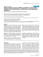
Fig. 3 Receiver operating characteristics curves of the three scores in the training and validation cohorts. a in the training cohort; b in the
validation cohort
Sun et al. BMC Cancer
(2020) 20:468
Page 7 of 11
These two cohorts were comparable in baseline characteristics (Table 1). The median time interval between the
surgery and chemotherapy was 73 days (range, 70–77
days) in the training cohort and 74 days (range, 70–77)
in the validation cohort.
Model construction
In the training cohort, SVM and PCA analysis identified
25 radiomics features significantly associated with the
response to neoadjuvant chemotherapy. These features
were histogram parameters, GLCM, and GLRLM, with
GLRLM accounting for the majority (Supplemental
Table 2). A rad_score was established based on the
above 25 radiomics features using Extra-Trees method.
Age and preoperative M status were found to be significantly different between responding group and nonresponding group (both P < 0.05) (Table 2), and thus a
clinical score was built based on them. By integrating
the rad_score and two clinical variables, a rad_clinical_
score was derived using SVM algorithm. Results showed
that the rad_score (Odds ratio [OR] = 1.21 × 10⁵, 95%
confidence interval [CI]: 52.3–3.07 × 10⁹, P < 0.01) was
significantly associated with the treatment response of
neoadjuvant chemotherapy (Table 3), and the rad_clinical_score was marginally associated with treatment response (P = 0.06), whereas the clinical score was not
(P = 0.28).
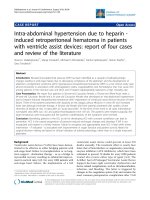
Fig. 4 Decision curve analysis for the rad_score, clinical score and
rad_clinical score
curves were plotted and survival rates were compared between two groups using log-rank tests.
Statistical analyses
The feature reduction and model building were performed
in Python (version 2.7.14), utilising ExtraTreesClassifier
from Scikit-learn. Other statistical analyses were performed
by R software version 3.2.3 (R Foundation for Statistical
Computing, Vienna, Austria, />The continuous variables were presented as mean ± standard deviation or median and quartile, and the categorical
variables were presented as frequencies and percentage. Independent sample t-test or Kruskal-Wallis (KW) nonparametric rank sum test was used to compare the baseline
characteristics between the training and validation cohorts,
and between responding group and non-responding group
for continuous variables, while Chi-square test or Fisher
exact test for categorical variables. A two-sided P-value was
considered statistically significant if less than 0.05.
Model performance in response prediction and validation
The rad_score was effective in predicting responders to
neoadjuvant chemotherapy in the training cohort (AUC:
0.77, 95% CI: 0.65–0.88) and in the validation cohort
(AUC: 0.82, 95% CI: 0.67–0.98) (Fig. 3). Compared to
the rad_score, the clinical score was poorer in predicting
accuracy without significant difference (training: 0.70,
95% CI: 0.58–0.82, P = 0.15; validation: 0.62, 95% CI:
0.42–0.83, P = 0.09), and the rad_clinical_score did not
demonstrate an improved performance (training: 0.70,
95% CI: 0.58–0.82, P = 0.12; validation: 0.70, 95% CI:
0.51–0.88, P = 0.16) (Fig. 3). The DCA showed that the
rad_score had the higher overall net benefit compared
with the rad_clinical_score and clinical score across the
majority of the risk of responders (Fig. 4). Other detailed
predicting performance is described in Table 4.
Results
Baseline characteristics
A total of 106 patients were included, with 74 patients
in the training cohort and 32 in the validation cohort.
Table 4 Predictive performance of the three scores in the treatment response of neoadjuvant chemotherapy for gastric cancer in
the validation cohort
Cut-off
rad_score
ACC
SEN
SPE
PPV
NPV
–
ACC
P
SEN
P
SPE
P
PPV
P
NPV
P
0.516
0.81
–
0.75
–
0.88
–
0.86
–
0.78
–
clinical_score
0.462
0.63
0.11
0.75
1.00
0.50
< 0.01
0.60
0.02
0.67
0.32
rad_clinical score
−0.651
0.69
0.26
0.88
0.18
0.50
< 0.01
0.64
0.04
0.80
0.84
Abbreviations: ACC accuracy, SEN sensitivity, SPE specificity, PPV positive predictive value, NPV negative predictive value
Sun et al. BMC Cancer
(2020) 20:468
Page 8 of 11
clinical score (P < 0.001) both achieved longer OS than
the low-score groups (Fig. 5b, c).
Table 5 Multivariable analysis of the three scores and
clinicopathological characteristics with overall survival
Factors
HR
95% CI
P value
Preoperative T stage
2.59
1.03–6.53
0.04
Total number of dissected lymph node
1.03
1.00–1.06
0.04
Postoperative N status
2.09
1.48–3.98
< 0.01
TNM stage
2.67
1.15–6.23
0.02
rad_score
0.22
0.11–0.42
< 0.01
clinical_score
2.65
1.07–6.54
0.03
rad_clinical_score
4.27
1.18–15.39
0.03
Abbreviations: HR hazard ratio, CI confidence interval
Survival stratification by the models
In the whole cohort, univariable and multivariable Cox
regression analyses showed that the rad_score (Hazard
Ratio [HR] = 0.22, 95% CI: 0.11–0.42, P < 0.01) was significantly associated with OS (Table 5). Univariable analysis showed that preoperative T status (HR = 2.59, 95%
CI: 1.03–6.53, P = 0.04), the total number of dissected
lymph nodes (HR = 1.03, 95% CI: 1.00–1.06, P = 0.04),
and postoperative N status (HR = 2.09, 95% CI: 1.48–
3.98, P < 0.01) were significantly associated with OS.
Based on these clinical variables, the clinical_score was
updated and also found to be significantly associated
with OS (HR = 2.65, 95% CI: 1.07–6.54, P = 0.03). Furthermore, the rad_clinical_score was also updated by integrating the rad_ score with the new selected clinical
variables, and was found to be associated with OS (HR =
2.65, 95% CI: 1.07–6.54, P = 0.03). Based on the threshold of rad_score of 0.59, patients were divided into
groups either with high-score or with low score. The OS
in patients from the high-score group was significantly
higher than that in patients from the low-score group
(P < 0.001) (Fig. 5a). Similarly, the high-score groups
stratified by the rad_clinical_score (P < 0.001) and
Discussion
Our study constructed and validated an effective CTradiomics score for predicting treatment response to
neoadjuvant chemotherapy in patients with potentially
resectable or limited metastatic gastric cancer. The rad_
clinical_score which was derived by combining clinical
variables with radiomics features, could not further improve the predicting performance when compared to the
rad_score. Moreover, the rad_score was capable to stratify patients into two groups with different survival
outcomes.
To the best of our knowledge, this is the first attempt
we develop radiomics scores to predict the response to
neoadjuvant chemotherapy in patients gastric cancer before treatment. Given the great therapeutic efficacy of
neoadjuvant chemotherapy for responding patients and
high risk of non-response in patients [11], the early identification of potentially responding patients who might
benefit from neoadjuvant chemotherapy is important to
maximise treatment efficacy and optimise personalised
therapy. Our established rad_score performed well in
this respect, indicating the possibility of radiomics in
predicting treatment response of neoadjuvant chemotherapy for gastric cancer. Several studies were conducted previously on the texture or radiomics analysis in
the evaluation of treatment response in gastric cancer.
Jiang et al. developed a radiomics signature which was
effective in predicting chemotherapy efficacy in patients
with stage II and III gastric cancer [30]. Yoon et al.
showed that texture features on CT images were correlated with the prognosis in patients with HER2-positive
advanced gastric cancer who received trastuzumabbased treatment, with heterogeneous features suggestive
Fig. 5 Comparisons of the overall survivals between high-score group and low-score group respectively stratified by rad_score, clinical score and
rad_clinical score. a stratified by rad_score; b stratified by clinical score; c stratified by rad_clinical_score
Sun et al. BMC Cancer
(2020) 20:468
of better survival outcomes [31]. Therefore, the underlying reason for our good model performance might be
the fact that intratumoural heterogeneity reflected by
radiomic features was associated with tumour biology
and even cell cycle regulating pathways, which are strong
factors influencing the efficacy of neoadjuvant chemotherapy [32–34]. The full mechanism behind the relationship
between radiomic features and neoadjuvant chemotherapy
has not been elucidated, and radiogenomics studies are
warranted to provide evidence in this issue [35]. Besides,
by integrating clinical variables with radiomics features,
the derived rad_clinical_score could not show superior
predicting performance to that of the rad_score. This indicated that radiomics features were the stronger component of this combined score while clinical data had limited
impact in elevating the performance.
In addition, our rad_score was capable to stratify patients into two groups with different risks of death,
which helped us identify the subgroup of patients with
poor prognosis for whom more intensified treatment
and closer follow-up schedule was needed. Low rad_
score was associated with poor prognosis, which made
sense because low rad_score was associated with no or
poor response to neoadjuvant chemotherapy. It was reported that patients who responded to neoadjuvant
chemotherapy had a higher likelihood to receive curative
gastrectomy, and their survival was expected to be better
than that of non-responding patients [5–9]. The finding
that the rad_score developed using the outcome of treatment response to neoadjuvant chemotherapy was effective in prognosis stratification, further confirmed its
clinical significance and usefulness. Instead of two
models, our single model could be used in both the prediction of treatment response and survival stratification.
Previous studies have found that radiomics features
were closely related to tumour biology and microscopic
structure [36–39]. Our study identified 25 radiomic features associated with treatment response to neoadjuvant
chemotherapy for gastric cancer. These were histogram
parameters, GLCM, and GLRLM with more than half of
the features being GLRLM. GLCM and GLRLM were
important markers of intra-tumour homogeneity, because they represented the level of signal heterogeneity
in a lesion in the manner of relative relationship between
the distribution and site of the gray level. These values
(GLCM and GLRLM) were higher in patients with no
response to neoadjuvant chemotherapy, which indicated
that the intratumoral heterogeneity was more apparent
in these patients than in the responding patients. Many
studies have reported that tumours with greater intratumoral heterogeneity tended to be more aggressive in
terms of proliferation, metastasis, and angiogenesis [22,
40], and thus might be more resistant to neoadjuvant
chemotherapy.
Page 9 of 11
There are several limitations in our study. First, the
sample size was small considering the relatively large
number of variables. Therefore, Extremely Randomised
Tree method was used to minimise the bias because it
used the whole training sample rather than a bootstrap
replica to build a tree, and it included a random subset
of features and split nodes by choosing cut-points at
random within each tree. Second, our models lacked the
external validation, which reduced the confirmation
strength of the model accuracy.
Conclusion
The radiomics score developed in this study was effective in predicting treatment response to neoadjuvant
chemotherapy and stratifying patients’ prognosis for gastric cancer. These findings may help clinicians in identifying potentially responding patients and providing
personalised treatment.
Supplementary information
Supplementary information accompanies this paper at />1186/s12885-020-06970-7.
Additional file 1: Table S1. A Summary of 1044 Radiomics Features,
Table S2. A summary of radiomics features significantly associated with
treatment response of neoadjuvant chemotherapy.
Abbreviations
CT: Computed tomography; MDCT: Multi-detector computed tomography;
ULN: Upper limit of normal; BSA: Body surface area; TRG: Tumor regression
grading; PACS: Picture archiving and communication system; ROI: Region of
interest; GLCM: Grey level co-occurrence matrix; GLRLM: Grey level runlength matrix; GLSZM: Gray level Size Zone Matrix; SVM: Support vector
machine; PCA: Principle component analysis; AUC: Area under the curve;
DCA: Decision curve analysis; OS: Overall survival
Acknowledgements
Not applicable.
Authors’ contributions
KYS and HTH: Original draft and Project administration; SLC, JNY, GHL and
LDC: Data curation; JJP, STF, YJY, XH and HW: Resources and Supervision; XL
and TFW: Methodology and Formal analysis; WW and JBX: Conceptualization,
Review & editing; All authors have read and approved the final manuscript.
Funding
This work is supported by the National Natural Foundation of China
(81672343 and 81871915, Recipient: Jian-Bo Xu), the Natural Science Foundation of Guangdong Province (No. 2017A030313570, Recipient: Jian-Bo Xu),
the Natural Science Foundation of Guangdong Province (No.
2018A030310326, Recipient: Kai-Yu Sun), the Natural Science Foundation of
Guangdong Province (No. 2018A030310282, Recipient: Shu-Ling Chen), the
Guangdong Medical Science and Technology Foundation (A2018280, Recipient: Kai-Yu Sun) and Science and Technology Program of Guangzhou (No.
201607010050, Recipient: Jian-Bo Xu). The funding source had no involvement in the design of the study and collection, analysis, and interpretation
of data and in writing the manuscript.
Availability of data and materials
Data would be available from the corresponding author on reasonable
request.
Sun et al. BMC Cancer
(2020) 20:468
Ethics approval and consent to participate
The ICE for Clinical Research and Animal Trials of the First Affiliated Hospital
of Sun Yat-sen University approved the study (No. [2019]103). And because
of the retrospective nature of the study, written informed consent from patients was waived.
Consent for publication
Not applicable.
Page 10 of 11
14.
15.
16.
Competing interests
The authors of this manuscript declare no relationships with any companies,
whose products or services may be related to the subject matter of the
article.
Author details
1
Department of Gastrointestinal Surgery, The First Affiliated Hospital of Sun
Yat-Sen University, 58 Zhongshan Road 2, Guangzhou 510080, People’s
Republic of China. 2Department of Medical Ultrasonics, Institute of
Diagnostic and Interventional Ultrasound, The First Affiliated Hospital of Sun
Yat-Sen University, 58 Zhongshan Road 2, Guangzhou 510080, People’s
Republic of China. 3Department of Radiology, The First Affiliated Hospital of
Sun Yat-sen University, Guangzhou 510080, China. 4Research Center of GE
Healthcare, Shanghai 200000, China.
17.
18.
19.
20.
21.
Received: 9 October 2019 Accepted: 18 May 2020
22.
References
1. Freddie B, Jacques F, Isabelle S, et al. Global cancer statistics 2018:
GLOBOCAN estimates of incidence and mortality worldwide for 36 cancers
in 185 countries. CA Cancer J Clin. 2018;68:394–424.
2. Degiuli M, Sasako M, Ponti A, Calvo F. Survival results of a multicentre phase
II study to evaluate D2 gastrectomy for gastric cancer. Br J Cancer. 2004;90:
1727–32.
3. Sano T, Sasako M, Yamamoto S, et al. Cancer surgery: morbidity and
mortality results from a prospective randomized controlled trial comparing
d2 and extended Para-aortic lymphadenectomy—Japan clinical oncology
group study 9501. J Clin Oncol. 2004;22:2767–73.
4. Takahashi T, Saikawa Y, Kitagawa Y. Gastric cancer: current status of
diagnosis and treatment. Cancers. 2013;5:48–63.
5. Cunningham D, Allum WH, Stenning SP, et al. Perioperative chemotherapy
versus surgery alone for resectable gastroesophageal cancer. N Engl J Med.
2006;355:11–20.
6. Ychou M, Boige V, Pignon JP, et al. Perioperative chemotherapy compared
with surgery alone for resectable gastroesophageal adenocarcinoma: an
FNCLCC and FFCD multicenter phase III trial. J Clin Oncol. 2011;29:1715–21.
7. Glimelius B, Ekstrom K, Hoffman K, et al. Randomized comparison between
chemotherapy plus best supportive care with best supportive care in
advanced gastric cancer. Ann Oncol. 1997;8:163–8.
8. Aoyama T, Nishikawa K, Fujitani K, et al. Early results of a randomized twoby-two factorial phase II trial comparing neoadjuvant chemotherapy with
two and four courses of cisplatin/S-1and docetaxel/cisplatom/S-1 as
neoadjuvant chemotherapy for locally advanced gastric cancer. Ann Oncol.
2017;28:1876–81.
9. Xue K, Ying XJ, Bu ZD, et al. Oxaliplatin plus S-1 or capecitabine as
neoadjuvant or adjuvant chemotherapy for locally advanced gastric cancer
with D2 lymphadenectomy: 5-year follow-up results of a phase II-III
randomized trial. Chin J Cancer Res. 2018;30:516–25.
10. AI-Batran SE, Homann N, Pauligk C, et al. Effect of neoadjuvant
chemotherapy followed by surgical resection on survival in patients with
limited metastatic gastric or gastroesophageal junction cancer: the AIOFLOT3 trial. JAMA Oncol. 2017;3:1237–44.
11. Xiong BH, Cheng Y, Ma L, Shang CQ. An updated meta-analysis of
randomized controlled trial assessing the effect of preoperative
chemotherapy in advanced gastric Cancer. Cancer Investig. 2014;32:272–84.
12. Weber WA, Ott K, Becker K, et al. Prediction of response to preoperative
chemotherapy in adenocarcinomas of the esophagogastric junction by
metabolic imaging. J Clin Oncol. 2001;19:3058–65.
13. Wieder HA, Ott K, Lordick F, et al. Prediction of tumor response by FDG-PET:
comparison of the accuracy of single and sequential studies in patients
23.
24.
25.
26.
27.
28.
29.
30.
31.
32.
33.
34.
35.
36.
37.
38.
with adenocarcinomas of the esophagogastric junction. Eur J Nucl Med Mol
Imaging. 2007;34:1925–32.
Hansen ML, Fallentin E, Lauridsen C, et al. Computed tomography (CT)
perfusion as an early predictive marker for treatment response to
neoadjuvant chemotherapy in gastroesophageal junction cancer and gastric
cancer-a prospective study. PLoS One. 2014;9:e97605.
Lee SM, Kim SH, Lee JM, et al. Usefulness of CT volumetry for primary
gastric lesions in predicting pathologic response to neoadjuvant
chemotherapy in advanced gastric cancer. Abdom Imaging. 2009;34:430–40.
Ang J, Hu L, Huang PT, et al. Contrast-enhanced ultrasonography
assessment of gastric cancer response to neoadjuvant chemotherapy. World
J Gastroenterol. 2012;18:7026–32.
Giganti F, De Cobelli F, Canevari C, et al. Response to chemotherapy in
gastric adenocarcinoma with diffusion-weighted MRI and (18) F-FDG-PET/
CT: correlation of apparent diffusion coefficient and partial volume
corrected standardized uptake value with histological tumor regression
grade. J Magn Reson Imaging. 2014;40:1147–57.
Schneider PM, Eshmuminov D, Rordorf T, et al. 18FDG-PET-CT identifies
histopathological non-responders after neoadjuvant chemotherapy in locally
advanced gastric and cardia cancer: cohort study. BMC Cancer. 2018;18:548.
Kumar V, Gu Y, Basu S, et al. Radiomics: the process and the challenges.
Magn Reson Imaging. 2012;30:1234–48.
Li Y, Liu X, Xu K, et al. MRI features can predict EGFR expression in lower
grade gliomas: a voxel-based radiomic analysis. Eur Radiol. 2018;28:356–62.
Esteva A, Kuprel B, Novoa RA, et al. Dermatologist-level classification of skin
cancer with deep neural networks. Nature. 2017;542:115–8.
Braman NM, Etesami M, Prasanna P, et al. Intratumoral and peritumoral
radiomics for the pretreatment prediction of pathological complete
response to neoadjuvant chemotherapy based on breast DCE-MRI. Breast
Cancer Res. 2017;19:57.
Yang L, Dong D, Fang MJ, et al. Can CT-based radiomics signature predict
KRAS/NRAS/BRAF mutations in colorectal cancer? Eur Radiol. 2018;28:2058–67.
Mandard AM, Dalibard F, Mandard JC, et al. Pathologic assessment of tumor
regression after preoperative chemoradiotherapy of esophageal carcinoma.
Clinicopathologic correlations. Cancer. 1994;73:2680–6.
Noble F, Lloyd MA, Turkington R, et al. Multicentre cohort study to define
and validate pathological assessment of response to neoadjuvant therapy in
oesophagogastric adenocarcinoma. Br J Surg. 2017;104:1816–28.
Qiao X, Jiao H. Data mining techniques in analyzing process data: a didactic.
Front Psychol. 2018;9:2231.
Laster L. Statistical background of methods of principle component analysis.
J Periodontol. 1967;38(Suppl):649–66.
Geurts P, Ernst D, Wehenkel L. Extremely randomized trees. Machine Learn.
2006;63:3–42.
Maree R, Geurts P, Wehenkel L. Random subwindows and extremely
randomized trees for image classification in cell biology. BMC Cell Biol.
2007;8 Suppl 1:S2.
Jiang YM, Chen CL, Xie JJ, et al. Radiomics signature of computed
tomography imaging for prediction of survival and chemotheapeutic
benefits in gastric cancer. EbioMedicine. 2018;36:171–82.
Yoon SH, Kim YH, Lee YJ, et al. Tumor heterogeneity in human epidermal
growth factor receptor 2 (HER2)-positive advanced gastric cancer assessed
by CT texture analysis: association with survival after trastuzumab treatment.
PLoS One. 2016;11:e0161278.
Aerts HJ, Velazquez ER, Leijenaar RT, et al. Decoding tumour phenotype by
noninvasive imaging using a quantitative radiomics approach. Nat
Commun. 2014;5:4006.
Tan P, Yeoh KG. Genetics and molecular pathogenesis of gastric
adenocarcinoma. Gastroenterology. 2015;149:e3.
O'Connor JP, Aboagye EO, Adams JE, et al. Imaging biomarker roadmap for
cancer studies. Nat Rev Clin Oncol. 2017;14:169–86.
Mazurowski MA. Radiogenomics: what it is and why it is important. J Am
College Radiol. 2015;12:862–6.
Grossmann P, Stringfield O, El-Hachem N, et al. Defining the biological basis
of radiomic phenotypes in lung cancer. Elife. 2017;6:e23421.
Fox MJ, Gibbs P, Pickles MD. Minkowski functionals: an MRI texture analysis
tool for determination of the aggressiveness of breast cancer. J Magn Reson
Imaging. 2016;43:903–10.
Ganeshan B, Goh V, Mandeville HC, Ng QS, Hoskin PJ, Miles KA. Non-small
cell lung cancer: histopathologic correlates for texture parameters at CT.
Radiology. 2013;266:326–36.
Sun et al. BMC Cancer
(2020) 20:468
39. Segal E, Sirlin CB, Ooi C, et al. Decoding global gene expression programs
in liver cancer by noninvasive imaging. Nat Biotechnol. 2007;25:675–80.
40. Wang WT, Yang L, Yang ZX, et al. Assessment of microvascular invasion of
hepatocellular carcinoma with diffusion kurtosis imaging. Radiology. 2018;
286:571–80.
Publisher’s Note
Springer Nature remains neutral with regard to jurisdictional claims in
published maps and institutional affiliations.
Page 11 of 11