The kinetic profile and clinical implication of SCC-Ag in squamous cervical cancer patients undergoing radical hysterectomy using the Simoa assay: A prospective observational study
Bạn đang xem bản rút gọn của tài liệu. Xem và tải ngay bản đầy đủ của tài liệu tại đây (1.57 MB, 11 trang )
Ye et al. BMC Cancer
(2020) 20:138
/>
RESEARCH ARTICLE
Open Access
The kinetic profile and clinical implication
of SCC-Ag in squamous cervical cancer
patients undergoing radical hysterectomy
using the Simoa assay: a prospective
observational study
Shuang Ye1,2†, Xiaohua Sun3,4†, Bin Kang3,4†, Fei Wu3,4, Zhong Zheng1,2, Libing Xiang1,2, Mylène Lesénéchal5,
Fabienne Heskia6, Ji Liang3,4* and Huijuan Yang1,2*
Abstract
Background: To study the kinetic profile and clinicopathological implications of squamous cell carcinoma antigen
(SCC-Ag) in cervical cancer patients who underwent surgery by a self-developed SCC-Ag single molecule assay
(Simoa) prototype immunoassay.
Methods: Participants were prospectively enrolled between 04/2016 and 06/2017. Consecutive serum samples
were collected at five points: day 0 (the day before surgery), postoperative day 4, weeks 2–4, months 2–4 and
months 5–7. In total, 92 patients and 352 samples were included. The kinetic change in SCC-Ag levels and their
associations with clinicopathological characteristics were studied.
Results: Simoa SCC-Ag was validated by comparison with the Architect assay. SCC-Ag levels measured by the
Simoa assay were highly correlated with the Architect assay’s levels (Pearson’s correlation coefficient = 0.979,
Passing-Bablok regression slope 0.894 (0.847 to 0.949), intercept − 0.009 (− 0.047 to 0.027)). The median values for
each time-point detected by the Simoa assay were 2.49, 0.66, 0.61, 0.72, and 0.71 ng/mL, respectively. The SCC-Ag
levels decreased dramatically after surgery and then stabilized and fluctuated to some extent within 6 months.
Patients with certain risk factors had significantly higher SCC-Ag values than their negative counterparts before
surgery and at earlier time points after surgery, while no difference existed at the end of observation. Furthermore,
although patients with positive lymph nodes had sustained higher SCC-Ag levels compared to those with negative
lymph nodes, similar kinetic patterns of SCC-Ag levels were observed after surgery. Patients who received
postoperative treatment had significantly higher SCC-Ag values than those with surgery only at diagnosis, while no
difference existed after treatment.
Conclusions: The Simoa SCC-Ag prototype was established for clinical settings. The SCC-Ag levels were higher in
patients with risk factors, whereas the kinetic trend of SCC-Ag might be mainly affected by postoperative adjuvant
therapy. These data indicate that the SCC-Ag level might be a good predictor for the status of cervical cancer,
including disease aggressiveness and treatment response.
Keywords: Squamous cervical cancer, Squamous cell carcinoma antigen, Simoa assay, Kinetic profile
* Correspondence: ;
†
Shuang Ye, Xiaohua Sun and Bin Kang contributed equally to this work.
3
Fudan University Shanghai Cancer Center – Institute Merieux Laboratory,
Cancer Institute, Fudan University Shanghai Cancer Center, Shanghai, China
1
Department of Gynecologic Oncology, Fudan University Shanghai Cancer
Center, Shanghai 200032, China
Full list of author information is available at the end of the article
© The Author(s). 2020 Open Access This article is distributed under the terms of the Creative Commons Attribution 4.0
International License ( which permits unrestricted use, distribution, and
reproduction in any medium, provided you give appropriate credit to the original author(s) and the source, provide a link to
the Creative Commons license, and indicate if changes were made. The Creative Commons Public Domain Dedication waiver
( applies to the data made available in this article, unless otherwise stated.
Ye et al. BMC Cancer
(2020) 20:138
Background
Cervical cancer is the fourth most common female malignancy worldwide [1]. Each year, more than half a
million women are diagnosed with cervical cancer, and
the disease results in over 300,000 deaths [2]. Cervical
squamous cell carcinoma (SCC), as the most common
histologic subtype, accounts for approximately 70% of
all cases [2, 3]. Squamous cell carcinoma antigen (SCCAg) is well known as the most useful marker for cervical squamous cell carcinoma [4, 5]. SCC-Ag was first
isolated by conventional protein purification methods
from a cervical squamous cell carcinoma [6]. Biochemical characterization of the original protein fraction
(TA-4) showed that it comprised a group of proteins
with a molecular weight of approximately 45 kDa. Currently, the most widely used SCC-Ag assay in clinical
settings is proposed by Abbott on the Architect instrument (Abbott Laboratories, Abbott Park, IL, USA) [7,
8]. SCC-Ag assays are also available on other wellknown platforms, such as the Elecsys® SCC assay used
on the Roche Elecsys and cobase analyzer (Roche Diagnostics, China) [9].
The role of serum SCC-Ag in squamous cervical
cancer has been extensively evaluated in previous
works [4, 7, 8, 10–24], and several reviews and metaanalyses have been published in the literature [5, 25–
27]. Most studies were of retrospective design and
only detected SCC-Ag at one time-point. They could
be roughly divided into two groups according to the
SCC-Ag measurement time: first, the clinical relevance of pretreatment SCC-Ag, which is still debated
[4, 5, 14, 15, 18, 20, 22–24], and second, the value of
SCC-Ag in the monitoring of response to treatment
and follow-up [7, 8, 17, 18, 20]. To date, few studies
have investigated the dynamic change in serum SCCAg levels during treatment, from surgery to adjuvant
therapies.
The single molecular array (Simoa) platform is a
new ultrasensitive technology that allows for the measurement of very small amounts of proteins using a
fully automated instrument to perform ELISA immunoassays [28, 29]. The fundamental theory of Simoa
has been published by Chang and coworkers [30]. In
cancer diagnostics, by utilizing Simoa, prostate-specific
antigen (PSA) has a thousand-fold lower limit of
quantification (< 0.01 pg/mL) than conventional ultrasensitive PSA assays, which allows for monitoring recurrence of prostate cancer after radical prostatectomy
[31–33].
In the present study, we aim to develop and validate a
Simoa SCC-Ag assay. Furthermore, we prospectively
monitored serial SCC-Ag levels in patients during treatment and follow-up to determine the SCC-Ag profiles
and clinicopathological implications.
Page 2 of 11
Methods
Preparation of beads with capture and detection
antibodies
Capture antibody (rabbit anti-human SerpinB3, 13,218RP01) and detection antibody (rabbit anti-human SerpinB3, 13,218-T52) for the development of the Simoa
SCC-Ag sandwich immunoassay were purchased from
Sino Biological (Beijing, China). The preparation of
beads with capture and detection antibodies followed
the manufacturer’s protocol (Quanterix). The capture
antibody concentration was adjusted to 0.2 mg/mL with
Bead Conjugation Buffer (Quanterix), and then paramagnetic carboxylated microparticles (Quanterix) were
activated with 0.3 mg/mL 1-ethyl-3-(3-dimethylaminopropyl) carbodiimide hydrochloride (EDC) (Thermo
Fisher Scientific, Waltham, MA, USA). To start the biotinylation reaction, 3 μL of the biotin solution (2 mg of
NHS-PEG4-Biotin dissolved in 383 μL of ddH2O) was
added to 100 μL of the detection antibody solution (1.0
mg/mL). The concentration of the recovered antibody
was adjusted to 0.2 mg/mL, and beads were stored at
4 °C.
Simoa assay setup and reagent preparation
All Simoa measurements were performed on a fully automated Simoa HD-1 Analyzer (Quanterix). The beads
coated with SCC-Ag capture antibody were diluted in
Diluent Buffer to 500,000 per test. The SCC-Ag detection antibody was diluted in diluent buffer to a working
concentration of 0.3 μg/mL. Streptavidin-β-galactosidase
concentrate was diluted to a working concentration of
100 pmol/L. The assay configuration protocol was a twostep assay. In the first step, 25 μL of the microparticle
solution, 20 μL of detection antibody and a 100-μL
serum sample (two-fold manual dilution by sample diluent (Quanterix)) or calibrator were incubated for 35 min
and 15 s (45 cadences) in a reaction cuvette (Quanterix),
followed by several wash steps. In the second step,
100 μL of SBG was added and incubated for 5 min and
15 s (7 cadences), followed by several wash steps.
Simoa assay validation procedure
During assay validation, the following basic assay parameters were addressed: calibration curve model, limit of
quantification, sensitivity (lower limit of quantification,
LLOQ), reproducibility (intra-assay, inter-assay), linearity, and calibrator stability.
To generate the calibration curve, recombinant human
SCC-Ag (TP302683, Origene, USA) was serially diluted in
the sample diluent, and the final concentrations of the 8
calibrators (calibrators A- H) in the assay were 0.049 to 50
ng/mL. The validation of the calibration curve model was
performed by running six independent measurements, including the 8 calibrators in quadruplicates. The coefficient
Ye et al. BMC Cancer
(2020) 20:138
of variation (CV) was determined over all assay runs using
the recalculated concentration values. Acceptance criteria
were a recovery of initial values within 80–120% and a CV
below 20% of all back-calculated calibrator samples. The reproducibility was assessed by two controls and four native
human serum samples shared in the whole calibration
curve. All samples were tested in quadruplicates over 6
runs on three different days (n = 24). The intra-assay and
inter-assay CV% for each sample were lower than 20%. To
determine the lower limit of quantification (LLoQ), 6 samples diluted in sample diluent to reach concentrations between zero and 0.1 ng/mL were measured over six
independent measurements in quadruplicates, and the
%CV was plotted as a function of SCC-Ag concentration to
graphically determine the concentration when 20% CV was
reached. Sixty-four replicates of the zero calibrator (sample
diluent) were tested in several assay runs to assess the limit
of blank (LoB). By ranking the concentrations of the 64 replicates of the zero calibrator in an increasing way, the LoB
corresponding to the 95th percentile was the mean of the
concentration between the 60th and the 61st. The limit of
detection (LoD) concentration was 2.5 SD from the LoB.
Linearity was evaluated by triplicate measurement within
one run: three different human serum samples with high
concentrations of SCC-Ag were mixed with a human
serum sample with low concentrations of SCC-Ag at different proportions. Acceptance criteria were a recovery of the
measured concentration within 80–120% by the nominal
concentration. Calibrator stability was addressed by performing freeze-thaw and short-term stability tests. Aliquoted sets of calibrators were stored at 4 °C, − 20 °C, and
room temperature for 1 week. In addition, one set of calibrators was frozen at − 20 °C and thawed at room
temperature to obtain calibrators with one additional
freeze-thaw cycle and then stored at − 20 °C for 1 week. To
determine the short-term temperature stability, the prepared four sets of calibrators were tested in parallel 1 week
later. Acceptance criteria were a recovery of initial values
within 80 and 120%.
Patients and treatment
After obtaining approval from the Institutional Review
Board at Fudan University Shanghai Cancer Center
(1703170–5), we prospectively enrolled the patients with
SCC scheduled for radical hysterectomy surgery in Professor Huijuan Yang’s team (Department of Gynecologic
Oncology, Fudan University Shanghai Cancer Center,
FUSCC) from April 2016 to June 2017. The inclusion
criteria were as follows: 1) preoperative confirmation of
squamous histology; 2) International Federation of
Gynecology and Obstetrics (FIGO 2009) stage IB1-IIA2;
3) no preoperative treatment including chemotherapy
and radiation; and 4) the ability to have strict follow-up
visits in our center. Patients with any skin disorder or
Page 3 of 11
past cancer history were excluded. Written informed
consent was acquired from all the participants included
in the study. Each patient was supposed to have five
consecutive blood samples collected at different points:
day 0 (the day before surgery), day 4 (postoperative day
4) and follow-up periods (weeks 2–4, months 2–4 and
months 5–7). In our clinical practice, the stage of patients’ cervical cancer was determined by two gynecologic oncologists by pelvic examination, according to the
FIGO 2009 guideline [34]. Radical hysterectomy was
performed according to Querle & Morrow (type C). Regarding postoperative adjuvant treatment, pelvic external
beam radiotherapy (EBRT) and concurrent platinumbased chemotherapy were given to patients with intermediate- and high-risk factors according to the Sedlis
criteria [35]. Intermediate-risk factors include lymphovascular space invasion (LVSI), deep stromal invasion
and tumor size, while high-risk factors refer to positive
margin, lymph node metastasis and positive parametria.
Extended-field EBRT was delivered to those with positive common iliac lymph node or para-aortic lymph
node. Systematic chemotherapy (carboplatin + paclitaxel) was administered to patients with more than two
lymph node metastases after radiation. Routinely, the patients with cervical cancer in our institution are required
to have regular follow-up visits after operation: every 3
months for the first 2 years, every 6 months in the next
3 years, and annually thereafter.
Statistical analysis
Regression analysis was used to determine the correlation between the Simoa SCC-Ag assay and the Architect SCC-Ag assay (R package mcr, version 1.2.1) [36].
The Kruskal-Wallis test was used to test whether SCCAg measurements at different time points were different
between patient groups defined by categorical clinical
factors (lymph node metastasis, LVSI), stromal invasion
and FIGO stage). Associations between categorical clinical factors were analyzed using the chi-squared test. All
statistical tests were performed using the R package
compareGroups (version 3.4.0) [37].
Relationships between important clinical factors and
the overall SCC-Ag profile were studied with the generalized additive modeling (GAM) method to accommodate the nonlinear trend of SCCA-Ag over time. GAM
allows for approximating nonlinear processes with
smoothing functions as follows:
hðyÞ ¼ β0 þ f ðt Þ þ β1 x1 þ β2 x2 þ … þ βp xp þ ε
where function f represents a nonlinear function of time t
and can be of any form. xi and βi, i = 1, 2, …, p, represent
other clinicopathological covariates and corresponding coefficients. In this study, nonparametric splines were used
Ye et al. BMC Cancer
(2020) 20:138
to approximate the nonlinear SCC-Ag profile over time.
Interactions between clinicopathological factors and the
smooth function are allowed to evaluate the association
between clinicopathological factors (lymph node metastasis and adjuvant treatment) and the SCCA profile. GAM
was performed in the R statistical environment with the
package mgcv (version 1.8.17) [38].
Results
Assay development and validation
A bead-based immunoassay was developed for the measurement of human SCC-Ag using Simoa technology
(Quanterix). The immunoassay development process included the evaluation of a suitable antibody pair and the
Page 4 of 11
optimization of assay conditions, such as the assay buffer
composition, incubation times, and applied reagent
concentrations.
Various antigens and antibodies were tested for the selection of a suitable calibrator protein and antibody pair
with high affinity for SCC-Ag in sandwich immunoassays (results not shown). The best assay performance
was achieved when 13,218-RP01 (Sino Biological) and
13,218-T52 (Sino Biological) were used as the capture
antibody and detection antibody, respectively. Assay
conditions were optimized (results not shown), and
evaluation was based on the calibration curve and human SCC-Ag serum samples. The best performances
were obtained in a two-step assay.
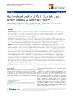
Fig. 1 Simoa SCC-Ag assay calibration curve and validation. a Typical Simoa SCC-Ag assay calibration curve. Recombinant human SCC-Ag was
serially diluted, and the calibrator range was 0.049 to 50 ng/mL with a recovery of all back-calculated concentrations between 80 and 120%. The
fitting model for the calibration curve was a weighted four-parameter logistics (1/Y2). AEB: Average enzyme per bead (measured signal). b
Validation results and acceptance criteria
Ye et al. BMC Cancer
(2020) 20:138
Page 5 of 11
During assay validation, the following basic assay parameters were addressed: calibration curve model, detection capability (LoB, LoD, and LoQ), reproducibility
(intra-assay and inter-assay), linearity, and calibrator stability. The best fitting model for the calibration curve
was the 1/Y2 weighted four-parameter logistics model.
The recovery of all back-calculated concentrations of the
individual calibrator points was between 93 and 113%. A
typical Simoa SCC-Ag immunoassay calibration curve is
given in Fig. 1a.
The Simoa SCC-Ag assay’s LoD and LoQ were calculated, and they achieved 0.029 and 0.057 ng/ml, respectively, according to the method of precision
profile (Fig. 1b). The Simoa SCC-Ag assay’s LoD was
approximately 4-fold lower than 0.1 ng/ml, the Architect SCC-Ag assay’s sensitivity. Inter-assay reproducibility for 6 samples resulted in CVs between 3.7 and
8.8%, and intra-assay repeatability for these samples
resulted in CVs between 5.1 and 13.7% (Fig. 1b). The
linearity of human serum samples with high and low
concentrations of SCC-Ag showed a recovery between 83.5 and 116.9% over the working range
(Fig. 1b). Under the optional standard curve, the dynamic range of the Simoa SCC-Ag assay was up to
0.029–100 ng/mL. Calibrator stability tests showed
that they would be stable at − 20 °C with a recovery
between 106 and 119%.
The Simoa SCC-Ag assay fulfilled acceptance criteria for all addressed validation parameters considered in the commonly used guidelines from the
Clinical and Laboratory Standards Institute (CLSI).
The method validation demonstrated that the required reproducibility and reliability for the measurement of complex matrices, such as human serum,
were met by the Simoa SCC-Ag assay.
Table 1 Clinicopathological features of the participants (n = 92)
Median age(range), years
51(32–71)
Pre-surgery SCC-Ag (ng/ml)
2.49(0.31–71.75)
FIGO stage
IB1 (%)
28(30.4%)
IB2 (%)
10(10.9%)
IIA1 (%)
30(32.6%)
IIA2 (%)
24(26.1%)
Tumor size > 4 cm (%)
33(35.9%)
Stromal invasion > 1/2 depth (%)
71(77.2%)
LVSI positive (%)
54(58.7%)
Lymph node metastasis (%)
33(35.9%)
Adjuvant treatment (%)
45(48.9%)
Abbreviations: SCC-Ag Squamous cell carcinoma antigen, FIGO International
Federation of Gynecology and Obstetrics, LVSI Lymph-vascular space invasion
Patient characteristics
During the study period, we enrolled 92 patients undergoing radical hysterectomy after receiving informed consent. For different reasons, some participants missed one
or more points’ blood collection (please refer to Fig. 3
patient numbers for specific details). Therefore, a total
of 352 blood samples from the 92 enrolled patients were
measured and analyzed.
Table 1 presents the clinicopathological characteristics
of the participants. The median age was 51 years old
(range 32–71). The median level of presurgery SCC-Ag
detected by the Simoa assay was 2.49 ng/mL (range
0.31–71.75). The FIGO stage (2009) of the patients is
listed as follows: IB1 30.4%, IB2 10.9%, IIA1 32.6%, and
IIA2 26.1%. Approximately 37% (34/92) of the patients
presented with bulky tumors (> 4 cm). Deep stromal invasion, positive LVSI and lymph node metastasis
accounted for 77.2, 58.7, and 35.9%, respectively. For the
entire cohort, 45 (48.9%) patients received postoperative
adjuvant treatment.
Method comparison between the Simoa and Architect
SCC-Ag assays
Among the 352 samples tested on the Simoa platform,
all were also tested with the Architect SCC-Ag assay. A
comparison between the two methods was conducted to
estimate the difference between the Simoa and Architect
assays. SCC-Ag levels measured by the Simoa assay were
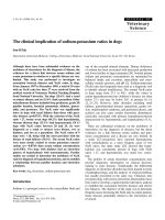
highly correlated with the Architect assay’s levels (Pearson’s correlation coefficient = 0.979) (Fig. 2). The slope
and intercept for the Passing-Bablok regression were
0.894 (0.847 to 0.949) and − 0.009 (− 0.047 to 0.027), respectively. The minimum values of SCC-Ag in the
Architect and Simoa platform were 0.17 and 0.16 ng/mL,
respectively (data not shown), and no sample had an
SCC-Ag value lower than the sensitivity of both assays.
Kinetic SCC-Ag data after surgery
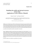
The median SCC-Ag values and ranges for each timepoint (day 0, day 4, weeks 2–4, months 2–4, months 5–
7) are summarized in Fig. 3. The median SCC-Ag values
for each time-point using Simoa were 2.49 ng/mL, 0.66
ng/mL, 0.61 ng/mL, 0.72 ng/mL, and 0.71 ng/mL. As
shown in Fig. 3, the SCC-Ag values decreased dramatically after surgery and then stabilized.
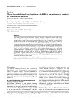
Among 92 patients, 32 patients succeeded in collecting
samples at all time points. Figure 4a depicts the profiles
of the SCC-Ag values for the 32 patients. All patients
showed a sharp decrease in the SCC-Ag level after radical surgery, and then the SCC-Ag level began to change
relatively slowly. In some patients, the SCC-Ag level
began to increase within 1 month after surgery. In other
patients, the SCC-Ag level reverted and increases during
Ye et al. BMC Cancer
(2020) 20:138
Page 6 of 11
Fig. 2 Passing–Bablok regression analysis of the SCC-Ag concentration of 352 samples obtained with the Architect and the Simoa SCC-Ag assay.
Scatter diagram with regression line (blue line) and 95% confidence bands (light blue) for the regression line. Pearson correlation coefficient (R) of
0.979 (p < 0.001). Passing–Bablok regression line equation: y = 0.89x − 0.01 (intercept 95% confidence interval (CI): − 0.05 to 0.03; slope 95% CI: 0.85
to 0.95
1–3 months. In some patients, the SCC-Ag level
remained decreased or stabilized until 3 months.
Association between the SCC-Ag level and the
Clinicopathological features
We evaluated the association between the SCC-Ag values
and clinicopathological characteristics. As shown in
Table 2, the presurgery SCC-Ag level was related to the
FIGO stage, stromal invasion, and lymph node metastasis
with statistical significance, but not to LVSI (P = 0.074).
After surgery, we noted that patients with positive LVSI
and lymph node had higher SCC-Ag levels at the time
points of day 4 and weeks 2–4 than those without. The
different levels of SCC-Ag between these groups did not
reach statistical significance at months 2–4 and months
5–7. These results indicate that the presurgery SCC-Ag
level might reflect the tumor burden and that the postoperative SCC-Ag level mainly indicates tumor metastasis
through lymph drainage. Interestingly, the SCC-Ag levels
reached the same level between the low-risk group,
intermediate-group and high-risk group after completion
of treatment.
SCC-Ag kinetic trends according to lymph node
metastasis and postoperative treatment
The above results reveal that patients with positive and
negative lymph nodes had significantly different SCC-Ag
levels within 1 month after surgery but not after 2–4
months. In our cohort, all patients with lymph node metastasis received postoperative adjuvant therapy. Thus,
the SCC-Ag kinetic trend was further evaluated based
on lymph node metastasis and postoperative treatment
using the generalized additive modeling (GAM) technique. After controlling for age, tumor size, and adjuvant
treatment, significantly elevated SCC-Ag levels were associated with positive lymph node (P < 0.001) (Fig. 4b).
Moreover, a trend analysis showed that the kinetic
trends of SCC-Ag over time were similar for patients
with or without lymph node metastasis (P = 0.62). Moreover, higher SCC-Ag levels were associated with postoperative adjuvant treatment after controlling for age,
tumor size, and lymph node status. However, the kinetic
trends of SCC-Ag levels were significantly changed (P =
0.005) between postoperative adjuvant-treated and nontreated patients (Fig. 4c). The trend-altering effect of
postoperative treatment was further demonstrated by
Ye et al. BMC Cancer
(2020) 20:138
Page 7 of 11
Fig. 3 Simoa SCC-Ag median values and range at each time point (day 0, day 4, weeks 2–4, months 2–4, months 5–7)
ANOVA. Although patients with postoperative adjuvant
treatment had significantly higher SCC-Ag levels than
patients without at the beginning of the treatment, the
difference between the two groups disappeared after
completion of adjuvant treatment (two-way ANOVA
p = 0.56). All these data indicated that the SCC-Ag levels
detected by the Simoa assay are a good predictor of disease aggressiveness and the treatment response of cervical cancers.
Discussion
In recent years, the Simoa platform has been proven to
be an ideal tool for clinical implementation due to its
simple and fully automated manipulation and ultrasensitive detection limit [28, 29]. In the current study, a
new prototype of sensitive SCC-Ag immunoassay was
developed using Simoa technology. This assay fulfilled
the acceptance criteria for all addressed analytical parameters and demonstrated improved sensitivity compared to that of the Architect assay, the most commonly
used method. Molecular cloning has demonstrated that
SCC-Ag is produced by two almost identical genes
named SCCA1 (SerpinB3) and SCCA2 (SerpinB4) [39].
In spite of controversy, most studies agreed that SCCA1
is more relevant for SCC diagnosis, and the Architect
assay only detects SCCA1 but not SCCA2 [40–42]. In
our study, our Simoa prototype also detected SCCA1
antigen.
Researchers have investigated the clinical significance
of consecutively monitoring the level of serum SCC-Ag
in cervical cancer patients during radiation/chemoradiation therapy [11, 17, 21]. Hashimoto et al. evaluated the
value of SCC-Ag as a predicator of chemotherapy response in patients with metastatic cervical cancer and
Ye et al. BMC Cancer
(2020) 20:138
Page 8 of 11
Fig. 4 SCC-Ag profile analysis. a Profiles of SCC-Ag values for the 32 patients with all five time points. Each curve represents the SCC-Ag profile
for one patient. b GAM analysis of the effect of lymph node metastasis on the SCC-Ag profile. The black arrow points to the start of adjuvant
treatment, while the red arrow indicates the end of adjuvant treatment. SCC-Ag intercept p < 0.001, SCC-Ag trend p = 0.62. c GAM analysis of the
effect of postoperative adjuvant treatment on the SCC-Ag profile. The black arrow indicates the start of adjuvant treatment, and the red arrow
indicates the end of adjuvant treatment. SCC-Ag intercept p = 0.01, SCC-Ag trend p = 0.005
concluded that a response to chemotherapy was possible
for patients in whom SCC-Ag levels declined between
the second and third cycles of chemotherapy [17]. Markovina et al. found that persistently elevated serum
SCC-Ag during definitive chemoradiation therapy was
an independent predictor of positive posttherapy FDGPET/CT, recurrence and death [11]. However, until
now, few studies have addressed the dynamic change in
Ye et al. BMC Cancer
(2020) 20:138
Page 9 of 11
Table 2 Correlation of clinical information with Simoa SCC-Ag values at different time points
LVSI
Lymph node metastasis
Negative
Positive
N = 38
N = 54
p
Negative
valuea
N = 59
FIGO stage
N = 33
p
IB
valuea
N = 38
Positive
Stromal invasion
N = 54
p
<= 1/2
valuea
N = 21
N = 71
IIA
> 1/2
p
valuea
SCC-Ag (ng/ml), median[range]
Pre-surgery
1.98 [0.34;
71.7]
4.22 [0.31;
69.6]
0.074
1.76 [0.31;
71.7]
8.72 [0.61;
69.6]
<
1.40 [0.31;
0.001 69.6]
3.88 [0.36;
71.7]
0.003 1.10 [0.34;
13.9]
3.49 [0.31;
71.7]
<
0.001
Day 4 postsurgery
0.55 [0.17;
1.72]
0.72 [0.21;
5.10]
0.048 0.57 [0.17;
5.10]
0.93 [0.33;
5.06]
<
0.65 [0.17;
0.001 5.06]
0.68 [0.32;
5.10]
0.353
0.57 [0.17;
1.19]
0.68 [0.21;
5.10]
0.041
Week 2–4 postsurgery
0.47 [0.27;
2.64]
0.69 [0.23;
2.34]
0.013 0.52 [0.23;
2.64]
0.78 [0.29;
2.34]
0.003 0.57 [0.23;
2.34]
0.63 [0.27;
2.64]
0.371
0.69 [0.27;
2.12]
0.61 [0.23;
2.64]
0.858
Month 2–4 post- 0.59 [0.25;
surgery
2.77]
0.85 [0.30;
24.2]
0.019 0.67 [0.25;
2.77]
0.85 [0.30;
24.2]
0.123
0.68 [0.25;
24.2]
0.75 [0.35;
2.77]
0.336
0.68 [0.30;
1.45]
0.75 [0.25;
24.2]
0.539
Month 5–7 post- 0.54 [0.36;
surgery
3.36]
0.77 [0.52;
2.60]
0.117
0.72 [0.52;
2.60]
0.273
0.71 [0.37;
1.00]
0.71 [0.36;
3.36]
0.477
0.58 [0.37;
1.14]
0.71 [0.36;
3.36]
0.415
0.66 [0.36;
3.36]
Abbreviations: SCC-Ag Squamous cell carcinoma antigen, FIGO International Federation of Gynecology and Obstetrics, LVSI Lymph-vascular space invasion
a
Kruskal-Wallis test
SCC-Ag value in patients receiving radical surgery. To
our knowledge, we are the first to describe the kinetic
change in SCC-Ag levels before and after radical hysterectomy surgery within a six-month duration. We found
that the SCC-Ag values stabilized after the dramatic
drop in the first few immediately after surgery. In the
dot plot graph (Fig. 3), the lowest SCC-Ag median value
was observed at the time point of weeks 2–4, although
significance was not achieved. After that nadir point,
some patients exhibited fluctuations, while others
reached a plateau. It deserves further investigation
whether different patterns correlated with treatment and
survival outcome.
In the second part of our work, we examined the relationship between the SCC-Ag values and clinicopathologic
features. Not surprisingly, the pretreatment SCC-Ag level
was related to tumor aggressiveness as indicated by advanced stage, deep stromal invasion and lymph node metastasis, which was consistent with previous works [5, 19].
Most published studies focused on the clinical value of one
time-point of SCC-Ag and both the pretreatment level
[10–12, 14, 15, 20, 22, 23], and posttreatment level [7, 8,
13–16, 18, 24]. Here, we monitored SCC-Ag values in a
longitudinal way to try to understand the possible clinical
meaning of the SCC-Ag levels. Our new finding was that
patients with intermediate- and high-risk factors had higher
SCC-Ag levels postoperatively, while the difference became
insignificant 6 months after surgery. As patients with risk
factors received adjuvant treatment after surgery, we further
evaluated the impact of postoperative treatment on the
SCC-Ag pattern. Patients with positive lymph nodes before
surgery showed sustained elevated levels of SCC-Ag compared to those negative counterparts, while the two groups
had similar overall SCC-Ag tendencies. In contrast, although patients who received adjuvant therapy had raised
baseline SCC-Ag levels, no difference existed at the
completion of treatment. In short, we postulated that the
absolute levels of SCC-Ag might be determined by the disease severity, while the dynamic change was possibly influenced by postoperative adjuvant treatment.
Given the short follow-up time, we did not evaluate
the prognostic value of the SCC-Ag level in cervical cancer patients. A recent retrospective study with a large
sample size from our institution demonstrated that a
preoperative serum SCC-Ag level > 2.75 ng/mL is an independent prognostic factor for progression-free survival
in cervical squamous cell carcinoma patients with highrisk factors [23]. In addition, a recent study investigated
the association between posttreatment SCC-Ag levels
and survival in patients treated with concurrent chemoradiation [24]. Patients with posttreatment SCC-Ag ≥ 1.8
ng/mL had significantly poor survival [24].
The study has several limitations. First, not all patients
completed the five-point blood collection for various reasons. Second, we prospectively enrolled 92 participants,
which was not a large sample size. Finally, given the shortterm follow-up, no survival outcome was analyzed in the
current work, which deserves further assessment.
Conclusion
The Simoa SCC-Ag assay exhibited competitive analytical
performances when compared with the Architect SCC-Ag
assay. The profile of SCC-Ag after radical surgery was illustrated for the first time. Both pre- and postoperative
SCC-Ag values are good predictors for tumor aggressiveness with different clinical applications. In addition, postoperative SCC-Ag is an effective response factor for
adjuvant treatments following radical surgery.
Acknowledgments
This work was supported by bioMerieux SA. We thank all of the patients
who participated in this study and all of the staff members from the
Ye et al. BMC Cancer
(2020) 20:138
Department of Gynecologic Oncology in Fudan University Shanghai Cancer
Center for collaborating in collecting serum samples.
Authors’ contributions
SY, XS, BK, FW, ZZ, LX, JL and HY contributed to the conception and design
of the study. SY, XS, BK, ZZ, LX and HYcollected and analyzed the patients’
clinicopathological data. XS, FW, ML, FH and JL performed the laboratory
work. SY, XS, BK, FH, JL and HY were major contributors in writing the
manuscript. All authors read and approved the final manuscript.
Funding
The study was supported by grants from the Key Research Project of
Shanghai Municipal Health Commission (201640010). The funding body
didn’t participate in the design of the study and collection, analysis, and
interpretation of data and in writing the manuscript.
Availability of data and materials
The dataset supporting the conclusions of this article is available upon
request. Please contact Prof. Huijuan Yang ().
Ethics approval and consent to participate
The study was approved by the Fudan University Shanghai Cancer Center
review board. Written informed consent was acquired from all the
participants to participate in the study.
Page 10 of 11
9.
10.
11.
12.
13.
14.
15.
Consent for publication
Not applicable.
16.
Competing interests
The authors declare that they have no competing interests.
17.
Author details
1
Department of Gynecologic Oncology, Fudan University Shanghai Cancer
Center, Shanghai 200032, China. 2Department of Oncology, Shanghai
Medical College, Fudan University, Shanghai, China. 3Fudan University
Shanghai Cancer Center – Institute Merieux Laboratory, Cancer Institute,
Fudan University Shanghai Cancer Center, Shanghai, China. 4bioMerieux
(Shanghai) Company Limited, Shanghai 200032, China. 5R&D Immunoassay
Department, bioMerieux SA, Marcy l’Etoile, France. 6Global Medical Affairs
Department, bioMerieux SA, Marcy l’Etoile, France.
18.
19.
20.
Received: 10 July 2019 Accepted: 13 February 2020
21.
References
1. Bray F, Ferlay J, Soerjomataram I, Siegel RL, Torre LA, Jemal A. Global cancer
statistics 2018: GLOBOCAN estimates of incidence and mortality worldwide
for 36 cancers in 185 countries. CA Cancer J Clin. 2018;68(6):394–424.
2. Cohen PA, Jhingran A, Oaknin A, Denny L. Cervical cancer. Lancet (London,
England). 2019;393(10167):169–82.
3. Small W Jr, Bacon MA, Bajaj A, Chuang LT, Fisher BJ, Harkenrider MM,
Jhingran A, Kitchener HC, Mileshkin LR, Viswanathan AN, et al. Cervical
cancer: a global health crisis. Cancer. 2017;123(13):2404–12.
4. Duk JM, Groenier KH, de Bruijn HW, Hollema H, ten Hoor KA, van der Zee
AG, Aalders JG. Pretreatment serum squamous cell carcinoma antigen: a
newly identified prognostic factor in early-stage cervical carcinoma. J Clin
Oncol. 1996;14(1):111–8.
5. Gadducci A, Tana R, Cosio S, Genazzani AR. The serum assay of tumour
markers in the prognostic evaluation, treatment monitoring and follow-up
of patients with cervical cancer: a review of the literature. Crit Rev Oncol
Hematol. 2008;66(1):10–20.
6. Kato H, Torigoe T. Radioimmunoassay for tumor antigen of human cervical
squamous cell carcinoma. Cancer. 1977;40(4):1621–8.
7. Salvatici M, Achilarre MT, Sandri MT, Boveri S, Vanna Z, Landoni F.
Squamous cell carcinoma antigen (SCC-Ag) during follow-up of cervical
cancer patients: role in the early diagnosis of recurrence. Gynecol Oncol.
2016;142(1):115–9.
8. Oh J, Bae JY. Optimal cutoff level of serum squamous cell carcinoma
antigen to detect recurrent cervical squamous cell carcinoma during posttreatment surveillance. Obstet Gynecol Sci. 2018;61(3):337–43.
22.
23.
24.
25.
26.
27.
Holdenrieder S, Molina R, Qiu L, Zhi X, Rutz S, Engel C, Kasper-Sauer P,
Dayyani F, Korse CM. Technical and clinical performance of a new assay to
detect squamous cell carcinoma antigen levels for the differential diagnosis
of cervical, lung, and head and neck cancer. Tumour Biol. 2018;40(4):
1010428318772202.
Xu D, Wang D, Wang S, Tian Y, Long Z, Ren X. Correlation between
squamous cell carcinoma antigen level and the clinicopathological features
of early-stage cervical squamous cell carcinoma and the predictive value of
squamous cell carcinoma antigen combined with computed tomography
scan for lymph node metastasis. Int J Gynecol Cancer. 2017;27(9):1935–42.
Markovina S, Wang S, Henke LE, Luke CJ, Pak SC, DeWees T, Pfeifer JD,
Schwarz JK, Liu W, Chen S, et al. Serum squamous cell carcinoma antigen as
an early indicator of response during therapy of cervical cancer. Br J Cancer.
2018;118(1):72–8.
Han X, Wen H, Ju X, Chen X, Ke G, Zhou Y, Li J, Xia L, Tang J, Liang S, et al.
Predictive factors of para-aortic lymph nodes metastasis in cervical cancer
patients: a retrospective analysis based on 723 para-aortic
lymphadenectomy cases. Oncotarget. 2017;8(31):51840–7.
Bolli JA, Doering DL, Bosscher JR, Day TG Jr, Rao CV, Owens K, Kelly B,
Goldsmith J. Squamous cell carcinoma antigen: clinical utility in squamous
cell carcinoma of the uterine cervix. Gynecol Oncol. 1994;55(2):169–73.
Bolger BS, Dabbas M, Lopes A, Monaghan JM. Prognostic value of
preoperative squamous cell carcinoma antigen level in patients surgically
treated for cervical carcinoma. Gynecol Oncol. 1997;65(2):309–13.
Strauss HG, Laban C, Lautenschlager C, Buchmann J, Schneider I, Koelbl H.
SCC antigen in the serum as an independent prognostic factor in operable
squamous cell carcinoma of the cervix. Eur J Cancer. 2002;38(15):1987–91.
Micke O, Bruns F, Schafer U, Prott FJ, Willich N. The impact of squamous cell
carcinoma (SCC) antigen in patients with advanced cancer of uterine cervix
treated with (chemo-)radiotherapy. Anticancer Res. 2005;25(3a):1663–6.
Hashimoto K, Yonemori K, Katsumata N, Hirakawa A, Hirata T, Yamamoto H,
Shimizu C, Tamura K, Ando M, Fujiwara Y. Use of squamous cell carcinoma
antigen as a biomarker of chemotherapy response in patients with
metastatic cervical carcinoma. Eur J Obstet Gynecol Reprod Biol. 2011;
159(2):394–8.
Kawaguchi R, Furukawa N, Kobayashi H, Asakawa I. Posttreatment cut-off
levels of squamous cell carcinoma antigen as a prognostic factor in patients
with locally advanced cervical cancer treated with radiotherapy. J Gynecol
Oncol. 2013;24(4):313–20.
Kim BG. Squamous cell carcinoma antigen in cervical cancer and beyond. J
Gynecol Oncol. 2013;24(4):291–2.
Ryu HK, Baek JS, Kang WD, Kim SM. The prognostic value of squamous cell
carcinoma antigen for predicting tumor recurrence in cervical squamous
cell carcinoma patients. Obstet Gynecol Sci. 2015;58(5):368–76.
Lee JH, Lee SW, Kim JR, Kim YS, Yoon MS, Jeong S, Kim JH, Lee JY, Eom KY,
Jeong BK, et al. Tumour size, volume, and marker expression during
radiation therapy can predict survival of cervical cancer patients: a multiinstitutional retrospective analysis of KROG 16-01. Gynecol Oncol. 2017;
147(3):577–84.
Choi KH, Lee SW, Yu M, Jeong S, Lee JW, Lee JH. Significance of elevated SCCAg level on tumor recurrence and patient survival in patients with squamouscell carcinoma of uterine cervix following definitive chemoradiotherapy: a
multi-institutional analysis. J Gynecol Oncol. 2019;30(1):e1.
Guo Q, Zhu J, Wu Y, Wen H, Xia L, Wu X, Ju X. Predictive value of preoperative
serum squamous cell carcinoma antigen (SCC-Ag) level on tumor recurrence
in cervical squamous cell carcinoma patients treated with radical surgery: a
single-institution study. Eur J Surg Oncol. 2020;46(1):131–38.
Wang W, Liu X, Hou X, Lian X, Liu Z, Shen J, Sun S, Yan J, Miao Z, Wang D,
et al. Posttreatment squamous cell carcinoma antigen predicts treatment
failure in patients with cervical squamous cell carcinoma treated with
concurrent chemoradiotherapy. Gynecol Oncol. 2019;155(2):224–8.
Zhou Z, Li W, Zhang F, Hu K. The value of squamous cell carcinoma antigen
(SCCa) to determine the lymph nodal metastasis in cervical cancer: a metaanalysis and literature review. PLoS One. 2017;12(12):e0186165.
Charakorn C, Thadanipon K, Chaijindaratana S, Rattanasiri S, Numthavaj P,
Thakkinstian A. The association between serum squamous cell carcinoma
antigen and recurrence and survival of patients with cervical squamous cell
carcinoma: a systematic review and meta-analysis. Gynecol Oncol. 2018;
150(1):190–200.
Fu J, Wang W, Wang Y, Liu C, Wang P. The role of squamous cell carcinoma
antigen (SCC Ag) in outcome prediction after concurrent
Ye et al. BMC Cancer
28.
29.
30.
31.
32.
33.
34.
35.
36.
37.
38.
39.
40.
41.
42.
(2020) 20:138
chemoradiotherapy and treatment decisions for patients with cervical
cancer. Radiat Oncol. 2019;14(1):146.
Rivnak AJ, Rissin DM, Kan CW, Song L, Fishburn MW, Piech T, Campbell TG,
DuPont DR, Gardel M, Sullivan S, et al. A fully-automated, six-plex single
molecule immunoassay for measuring cytokines in blood. J Immunol
Methods. 2015;424:20–7.
Wilson DH, Rissin DM, Kan CW, Fournier DR, Piech T, Campbell TG, Meyer
RE, Fishburn MW, Cabrera C, Patel PP, et al. The Simoa HD-1 analyzer: a
novel fully automated digital immunoassay analyzer with single-molecule
sensitivity and multiplexing. J Lab Autom. 2016;21(4):533–47.
Chang L, Rissin DM, Fournier DR, Piech T, Patel PP, Wilson DH, Duffy DC.
Single molecule enzyme-linked immunosorbent assays: theoretical
considerations. J Immunol Methods. 2012;378(1–2):102–15.
Rissin DM, Fournier DR, Piech T, Kan CW, Campbell TG, Song L, Chang L,
Rivnak AJ, Patel PP, Provuncher GK, et al. Simultaneous detection of single
molecules and singulated ensembles of molecules enables immunoassays
with broad dynamic range. Anal Chem. 2011;83(6):2279–85.
Rissin DM, Kan CW, Campbell TG, Howes SC, Fournier DR, Song L, Piech T,
Patel PP, Chang L, Rivnak AJ, et al. Single-molecule enzyme-linked
immunosorbent assay detects serum proteins at subfemtomolar
concentrations. Nat Biotechnol. 2010;28(6):595–9.
Lepor H, Cheli CD, Thiel RP, Taneja SS, Laze J, Chan DW, Sokoll LJ, Mangold
L, Partin AW. Clinical evaluation of a novel method for the measurement of
prostate-specific antigen, AccuPSA(TM), as a predictor of 5-year biochemical
recurrence-free survival after radical prostatectomy: results of a pilot study.
BJU Int. 2012;109(12):1770–5.
Pecorelli S, Zigliani L, Odicino F. Revised FIGO staging for carcinoma of the
cervix. Int J Gynaecol Obstet. 2009;105(2):107–8.
Sedlis A, Bundy BN, Rotman MZ, Lentz SS, Muderspach LI, Zaino RJ. A
randomized trial of pelvic radiation therapy versus no further therapy in
selected patients with stage IB carcinoma of the cervix after radical
hysterectomy and pelvic lymphadenectomy: a gynecologic oncology group
study. Gynecol Oncol. 1999;73(2):177–83.
Schuetzenmeister EMAMF. mcr: Method comparison regression. R package
version 1.2.1; 2014.
Subirana ISH, Vila J. Building bivariate tables: the compare groups package
for R. J Stat Softw. 2014;57(12):16.
SN W. Fast stable restricted maximum likelihood and marginal likelihood
estimation of semiparametric generalized linear models. J Royal Stat Soc B
Stat Methodol. 2011;73(1):36.
Izuhara K, Ohta S, Kanaji S, Shiraishi H, Arima K. Recent progress in
understanding the diversity of the human ov-serpin/clade B serpin family.
Cell Mol Life Sci. 2008;65(16):2541–53.
Roijer E, de Bruijn HW, Dahlen U, ten Hoor K, Lundin M, Nilsson K,
Soderstrom K, Nilsson O. Squamous cell carcinoma antigen isoforms in
serum from cervical cancer patients. Tumour Biol. 2006;27(3):142–52.
Ohta S, Shibata R, Nakao Y, Azuma Y, Taniguchi K, Arima K, Suzuki S,
Shiraishi H, Iwasaka T, Izuhara K. The usefulness of combined measurements
of squamous cell carcinoma antigens 1 and 2 in diagnosing atopic
dermatitis. Ann Clin Biochem. 2012;49(Pt 3):277–84.
Cataltepe S, Gornstein ER, Schick C, Kamachi Y, Chatson K, Fries J, Silverman
GA, Upton MP. Co-expression of the squamous cell carcinoma antigens 1
and 2 in normal adult human tissues and squamous cell carcinomas. J
Histochem Cytochem. 2000;48(1):113–22.
Publisher’s Note
Springer Nature remains neutral with regard to jurisdictional claims in
published maps and institutional affiliations.
Page 11 of 11