Relationship of lymphovascular invasion with lymph node metastasis and prognosis in superficial esophageal carcinoma: Systematic review and meta-analysis
Bạn đang xem bản rút gọn của tài liệu. Xem và tải ngay bản đầy đủ của tài liệu tại đây (1.03 MB, 8 trang )
Yang et al. BMC Cancer
(2020) 20:176
/>
RESEARCH ARTICLE
Open Access
Relationship of lymphovascular invasion
with lymph node metastasis and prognosis
in superficial esophageal carcinoma:
systematic review and meta-analysis
Jinxin Yang1†, Zhouyi Lu2†, Lintao Li1, Yong Li1, Yulong Tan2, Dekang Zhang1* and An Wang2*
Abstract
Background: The development of tumor cells inside the lymphatics or blood vessels is known as lymphovascular
invasion (LVI). The correlation between LVI, lymph node metastasis (LNM), and the diagnosis of superficial
esophageal carcinoma (SEC) remains unclear.
Methods: We searched Embase, PubMed, Web of Science, and Cochrane Library databases for prospective articles
to better understand the relationship between LVI, LNM, and SEC diagnosis.
Results: We included 23 articles containing data for 4749 patients (range: 54–598) in our meta-analysis. The hazard
ratio between LVI and overall survival (OS) was 1.85 with 95% confidence interval (CI) (1.10–3.11, P = 0.02). LNM rate
was higher in SEC patients with LVI than SEC patients without LVI (univariate: OR = 4.94, 95% CI: 3.74–6.53, P <
0.0001; multivariate: OR = 5.72, 95%CI: 4.38–7.4, P < 0.0001). No obvious publication was found.
Conclusions: The results indicate that LVI plays a dominant role in the prognosis of LNM in SEC and in the
prognostic prediction for SEC.
Keywords: Lymphovascular invasion, Lymph node metastasis, Prognosis, Superficial esophageal carcinoma
Background
Superficial esophageal carcinoma (SEC) can be classified
as submucosal (T1b), mucosal (T1a), or intraepithelial
(Tis) irrespective of lymph node metastasis (LNM). Patients suffering from SEC have a better chance of survival after esophagectomy compared to those with
advanced esophageal carcinoma (EC). According to the
Japanese criteria, the depth of tumor invasion is subclassified into six layers. The mucosa is subdivided into the
* Correspondence: ;
†
Jinxin Yang and Zhouyi Lu contributed equally to this work.
1
Department of Radiation Oncology, Sichuan Cancer Hospital and Institute,
Sichuan Cancer Center, School of Medicine, University of Electronic Science
and Technology of China, Chengdu, Sichuan, China
2
Department of Thoracic Surgery, Huashan Hospital, Fudan University,
Shanghai, China
intraepithelial (m1) region, lamina propria (m2), and
muscularis mucosa (m3) while the submucosa is homogeneously classified into three sections: inner (sm1),
middle (sm2), and deep submucosa (sm3) [1]. The prognostic factors for EC include the histology type, tumor
size, grade category, invasion depth, blood vessel and
lymphatic vessel permeation, as well as LNM and distant
metastasis [2]. EC patients with LNM frequently have an
adverse prognosis. Therefore, the impact of LVI on
LNM and prognosis requires attention.
The development of tumor cells inside the lymphatics
or blood vessels is known as lymphovascular invasion
(LVI). Lymphatic vessels are believed to play a crucial
role in LNM and their presence increases the micrometastatic risk in locoregional malignancy [3]. Though
© The Author(s). 2020 Open Access This article is licensed under a Creative Commons Attribution 4.0 International License,
which permits use, sharing, adaptation, distribution and reproduction in any medium or format, as long as you give
appropriate credit to the original author(s) and the source, provide a link to the Creative Commons licence, and indicate if
changes were made. The images or other third party material in this article are included in the article's Creative Commons
licence, unless indicated otherwise in a credit line to the material. If material is not included in the article's Creative Commons
licence and your intended use is not permitted by statutory regulation or exceeds the permitted use, you will need to obtain
permission directly from the copyright holder. To view a copy of this licence, visit />The Creative Commons Public Domain Dedication waiver ( applies to the
data made available in this article, unless otherwise stated in a credit line to the data.
Yang et al. BMC Cancer
(2020) 20:176
Page 2 of 8
lymph node metastasis via LVI or lymphatic vessels has
not been confirmed [4], lymphatic vessels are known to
provide entry for the penetration of tumor cells [5].
Some studies have provided evidence of an association
between LVI and LNM in SEC. Nonetheless, the impact
of LVI on OS and LNM in SEC requires investigation.
Thus, we conducted a meta-analysis to obtain additional
insight into the correlation between LVI, LNM, and
prognosis in SEC.
above. When a discrepancy arose, a third author was involved to resolve the differences. Quality assessment was
performed using the Newcastle-Ottawa Scale (NOS) [6]
and all articles included scored a minimum of five points
on the NOS. Researches about prognosis were assessed
by critical appraisal of prognostic studies (https://www.
cebm.net/wp-content/uploads/2018/11/Prognosis.pdf).
The detailed quality assessment of these studies was displayed in a Table 1.
Methods
Search strategy
Data extraction
We searched the Embase, PubMed, Web of Science, and
Cochrane Library databases for prospective articles. The
search terms used were (lymphovascular invasion (LVI)
OR lymph vessel invasion OR angiolymphatic invasion
OR lymphatic invasion) AND (superficial esophageal cancer (SEC) OR submucosal esophageal carcinoma OR mucosal esophageal cancer OR T1 esophageal carcinoma).
We conducted a manual search of the results to identify
the prospective studies relevant to our investigation. We
then performed preliminary screening by checking the titles followed by the abstracts. Relevant studies were confirmed after reviewing the full text. In the present study,
we regarded lymphatic invasion as LVI.
Two independent authors collected data from the studies. The following information was extracted: surname
of the first author, follow-up years, region, sample size
for the research, treatment characteristics, histology
type, depth of invasion, staining methods, the percentage
of patients with LVI, information about OS, and LNM
and NOS scores. All of the collected information is listed
in Table 2. Discrepancies among authors were resolved.
Exclusion and inclusion criteria
Studies were considered eligible based on the following
criteria: (1) SEC; (2) hazard ratio (HR) for prognosis and
odds ratio (OR) for LNM; (3) papers published in English; (4) the latest or most relevant articles published by
the same group/author.
The exclusion criteria were as follows: (1) duplicate conference papers, reviews, reports, abstracts, and letters; (2) studies about other cancer types, animal models, esophageal
cancer cell lines, and treatment methods; (3) lack of data on
prognosis or LNM; (4) studies published in languages other
than English; (5) esophagogastric junction cancer (EJC).
Preliminary review of studies and quality assessment
Each selected article was reviewed by two independent
authors based on the exclusion and inclusion criteria
Statistical analysis
We investigated the correlation between LVI, prognosis,
and LNM in SEC patients. HR and OR were effective for
the prognosis and LNM with 95% CI individually. Worse
prognosis for SEC was indicated by an HR value > 1.
Cochrane’s Q test (Chi-squared test; Chi2) and the I2
metric were used to test the heterogeneity of the pooled
results. I2 < 25% indicated no heterogeneity; I2 = 25–50%,
moderate heterogeneity; I2 = 50–75%, medium heterogeneity; and I2 > 75%, extreme heterogeneity. We used a
fixed-effect model (the Mantele Haenszel method) for
I2 < 50% with P > 0.05 in this meta-analysis. If not, a
random-effect model was appropriate for our analysis.
We used meta regression and subgroup analysis to explore heterogeneity when necessary [18]. Begg’s test was
used to assess publication bias. Two-tailed tests were
used to calculate the P value and P ≤ 0.05 was considered
statistically significant. Statistical analysis was performed
using the Stata/SE version 12.0 for Windows (Stata Corporation, College Station, TX, USA).
Table 1 The detailed quality assessment of prognostic studies
Author
Years
Included
Region Comment
1
Comment
2
Comment
3
Comment
4
What are the results
Leggett (2015) [7]
1995-2011
USA
Yes
Yes
Yes
Yes
Survival curve, CI is narrow, conclusion is
promotable
Yamashina (2013)
[8]
1995-2010
Japan
Yes
Yes
Yes
Yes
CI is relative marrow, conclusion is promotable
Tanaka (2014) [9]
1988-2010
Japan
Yes
Yes
Yes
Yes
CI is narrow, conclusion is promotable
Xue (2018) [10]
1990-2004
China
Yes
Yes
Yes
Yes
CI is relative marrow, conclusion is relative
promotable
CI Confidence interval
Yang et al. BMC Cancer
(2020) 20:176
Page 3 of 8
Table 2 Characteristics of studies included in out meta-analysis
Author
Years
Region
Included
No. Treatment Characteristic
Pathology Depth
Staining Indicator
of
(No.)
Invasion
Including
Statistics
NOS
Scores
Jia (2016)
[11]
20102015
China
93
Esophagectomy and lymphadenectomy
SCC/
Others
M1-SM3 NM
LVI(28)
LNM
5
Sepesi
(2010) [12]
20002008
USA
54
Esophagectomy and lymphadenectomy
AD
SM
NM
LVI(7)
LNM
5
Leggett
(2015) [7]
19952011
USA
269 EMR followed by ablative techniques
AD
LP-SM
H&E
LVI(53)
OS
6
Huh (2017) 1996[13]
2015
Korea
275 187 Esophagectomy and 88 ER
(Esophagectomy or ER)
SCC
M-SM
H&E
LVI(36)
LNM
6
Zhou
(2016) [14]
20082015
China
498 Esophagectomy with lymphadenectomy
SCC
M1-SM3 H&E/
IHC
LI(16/
412)
LNM
7
Moon
(2014) [15]
20092012
Korea
104 Esophagectomy with lymphadenectomy
SCC
M1-SM3 H&E
LVI(13)
LNM
6
Mitobe
(2013) [16]
19902009
Japan
110 106 Esophagectomy with lymphadenectomy,
4 esophagectomy follwed ER and
lymphadenectomy
SCC
LP-SM3
IHC
LI(42)
LNM
6
Nentwich
(2014) [17]
19942009
Germany 67
Esophagectomy
SCC/AD
SM
NM
LI(16/61)
LNM
5
Raja (2011) 1983[18]
2010
USA
120 Esophagectomy
SCC/AD
SM
NM
LVI(26)
LNM/OS
5
Nakajima
(2002) [19]
19851995
Japan
84
Esophagectomy with lymphadenectomy
SCC
SM
IHC
LI(60)
LNM
6
Choi (2011) 1991[20]
2009
Korea
190 Esophagectomy with lymphadenectomy
SCC
M1-SM3 H&E
LVI(39)
LNM
7
Tajima
(2000) [21]
19681996
Japan
240 Esophagectomy with lymphadenectomy
SCC
LP-SM
H&E
LI(39/
186)
LNM
6
Chiba
(2010) [22]
19922008
Japan
110 107 underwent esophagectomy, 3 patients
underwent ER followed esophagectomy
SCC
M-SM
IHC
LI(46)
LNM
6
Yamashina
(2013) [8]
19952010
Japan
402 EMR or ESD, some patients received surgery
after ER
SCC
EP-SM2
NM
LVI(33)
OS
5
Xue (2012)
[23]
19902004
China
271 Esophagectomy
SCC
M2-SM3 IHC
LI(51)
LNM
7
Ancona
(2008) [24]
19802006
Italy
98
SCC/AD
M1-SM3 NM
LI(34)
LNM
5
Li (2013)
[25]
20062011
China
189 Esophagectomy with lymphadenectomy
SCC
M1-SM3 NM
LVI(22)
LNM
5
Qi (2016)
[26]
20092014
China
258 Esophagectomy with lymphadenectomy
SCC
SM
H&E
LVI(18)
LNM/OS
6
Wang
(2016) [27]
20022014
Japan
598 Esophagectomy with lymphadenectomy
SCC
M-SM
H&E/
IHC
LI(62/
228)
LNM
6
Kim (2008)
[28]
19942006
Korea
200 Esophagectomy with lymphadenectomy
SCC/AD
M-SM
NM
LI(33)
LNM
5
Tanaka
(2014) [9]
19882010
Japan
145 Esophagectomy with lymphadenectomy
SCC
SM1SM3
NM
LVI(84)
OS
5
Zhuge
(2018) [29]
20062016
China
175 Esophagectomy with lymphadenectomy
SCC
SM1SM3
NM
LVI(32)
LNM
6
Xue (2018)
[10]
19902004
China
199 Esophagectomy with lymphadenectomy
SCC
M2-SM3 IHC
LVI(27)
OS
6
Esophagectomy with lymphadenectomy
LVI Lymphovascular Invasion, LI Lymphatic invasion
ER Endoscopic resection, EMR Endoscopic mucosal resection, ESD Endoscopic submucosal dissection
SCC Squamous cell carcinoma, AD Adenocarcinoma, OS Overall survival
EP Epithelium, M Mucosa, SM Submucosa, LP Lamina propria, NM Not mentioned
H&E Hematoxylin-eosin, IHC Immunohistochemical
Yang et al. BMC Cancer
(2020) 20:176
Results
Characteristics of studies
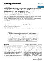
We retrieved 603 articles after removing duplicates but
excluded 487 articles that were either case reports or only
abstracts. A few of the excluded articles were review articles and others contained information about other cancer
conditions. Articles published in languages other than
English were also excluded. We identified 116 potential
articles for full-text review. We excluded 93 articles for
the following reasons: 25 were about EJC; 67 lacked data
relevant to LVI, prognosis, or LNM; and retrieval of the
full text was not possible for six articles; one was excluded
due to the same author and institution. The remaining 23
articles, which included information for 4749 patients
(range: 54–598), were included in the meta-analysis
(Fig. 1). Table 2 shows detailed information about the
studies. All studies included in this meta-analysis were
rated with a minimum of five stars based on the NOS.
Six studies provided survival information between LVI
and prognosis. Two studies reported the association between LVI and prognosis with univariate Cox proportional
hazards analysis in included studies [18, 26]. Four of included studies suggested the association between LVI and
prognosis was not significant in SEC patients [8, 9, 18, 26].
The rest two studies showed LVI was a poor prognostic
indicator in SEC patients [7, 10].
Sixteen studies provided information on LVI from
multivariate analysis of LNM cases. Eight studies provided
information on LVI from univariate analysis. One study
using univariate analysis reported a p value of 0.049 [12].
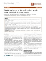
LVI impact on OS
2We included 4 eligible studies containing 1005 patients
from multivariate analysis in our meta-analysis. The
Page 4 of 8
pooled HR was 1.85 with 95% CI (1.10–3.11, P = 0.02)
and the pooled OS showed medium heterogeneity based
on random effect model (I2 = 54.6%, P = 0.085, Fig. 2).
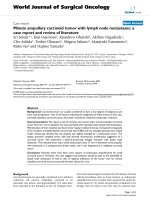
Association between LVI and LNM
The pooled results showed that patients in the LNMpositive group had an advanced LVI detection rate
(OR = 4.94, 95% CI: 3.74–6.53, P < 0.0001, Fig. 3) in univariate analysis. The combined results exhibited no heterogeneity (I2 = 0.9%, P = 0.422). The pooled results from
20 studies in multivariate analysis suggested that LVI
significantly increased the risk for LNM (OR = 5.72, 95%
CI: 4.38–7.48, P < 0.0001, Fig. 4) with no heterogeneity
(I2 = 0%, P = 0.926).
Publication bias of included studiessl
There was no evidence of publication bias for OS as
demonstrated by Begg’s test (P = 1) or for LNM (multivariate: P = 0.961; univariate: P = 0.805). The funnel plots
were displayed in Fig. 5.
Discussion
Our study demonstrated that SEC patients with LVI
have a poor OS (HR = 1.85, 95% CI: 1.10–3.11, P = 0.02;
I2 = 54.6%, P = 0.085). LVI significantly reduces OS in
patients with SEC. This conclusion should be clarified
with caution due to medium heterogeneity. Additionally,
LVI and LNM are strongly correlated (univariate: OR =
4.94, 95% CI: 3.74–6.53, P < 0.0001, I2 = 0.9%, P = 0.422;
multivariate: OR = 5.72, 95% CI: 4.38–7.4, P < 0.0001;
I2 = 0%, P = 0.926) in patients suffering from SEC. These
results suggest that LVI is an important prognostic factor for patients with SEC with regard to predicting LNM
and survival.
Fig. 1 Flow chart showing the literature collection procedure for included studies
Yang et al. BMC Cancer
(2020) 20:176
Page 5 of 8
Fig. 2 Forrest plot showing pooled HR for OS in patients with LVI
SEC is similar to the esophageal tumors, which are
limited to the mucosal layer (T1, T0) and include highgrade dysplasia, intramucosal cancer (T1a), and tumors
infiltrating the submucosa (T1b) [30]. .Reports state that
patients with T0 (0% chance) or T1a (1–2% chance)
esophageal cancer have a minimal risk of local LNM
[31]. There is no specific standard available for the detection of LVI. However, the identification of tumor cells
in the lymphatic vessels, arteries, or veins during pathological evaluation of specimens indicates LVI. The condition is an independent prognostic factor of LNM in
malignant tumors causing lung, prostate, breast, and
esophageal cancer. However, the role of LVI in SEC has
not been clarified to date. Additionally, the impact of
LVI in SEC on OS and LNM has not been assessed
using meta-analysis in the past. Therefore, we conducted
this study by analyzing data for 4854 patients reported
in 24 eligible articles retrieved from PubMed and other
relevant sources. We demonstrated LVI relevance in
LNM and the prognosis for patients with SEC. According to a literature review, our work is the first systematic
review and meta-analysis on LVI relevance in LNM and
prognosis in patients with SEC.
During the early stage of esophageal cancer, LVI is
regarded as a potential prognostic factor in predicting LNM. Current research has demonstrated that
patients with T1b esophageal cancers without LVI
have a significantly higher survival rate up to 5 years
higher those with LVI [32]. A larger cohort study revealed that LVI has a significant effect on the prognosis after resection for ESCC [33]. Our study shows
that SEC patients with LVI have a poor OS (HR =
1.62, 95% CI: 1.17–2.26, P = 0.004, I2 = 0.0%), and
LVI significantly increases the risk of LNM in SEC
(univariate: OR = 5.26, 95% CI: 4–6.91, P < 0.0001,
I2 = 30.2%; multivariate: OR = 5.7, 95% CI:4.43–7.33,
Fig. 3 Forrest plot showing pooled OR for LNM in patients with LVI from univariate analysis
Yang et al. BMC Cancer
(2020) 20:176
Page 6 of 8
Fig. 4 Forrest plot showing pooled OR for LNM in patients with LVI from multivariate analysis
P < 0.0001; I2 = 16%). Reports describing the relationship between LVI, LNM, and OS in SEC indicate
that LVI raises the possibility of LNM, leading to a
poor OS.
Esophagectomy and other non-surgical options including chemotherapy and radiotherapy are the mainstream
treatments for esophageal cancer. However, endoscopic
resection (ER) is the diagnostic and radical choice for the
treatment of SEC with a low possibility of LNM. The
Japan Esophageal Society published a guideline in 2014
recommending ER as the best treatment option for T0
and T1a lesions located within the limits of the mucosal
layer and not associated with LNM. The treatment can
still be applied for lesions that infiltrate the muscularis
mucosae or the inner submucosa (T1b-SM1) but the risk
of LNM exists for these cases. Hence, other classifications
for superficial carcinomas (T1b-SM2 and T1b-SM3)
should not be treated with endoscopy alone due to the
high rates of metastasis [34]. ER can be classified as endoscopic mucosal resection (EMR) or endoscopic submucosal dissection (ESD). All visible neoplasms are removed by
EMR for definitive histopathological staging. However,
EMR is ineffective compared to ESD in terms of en bloc
resection of large lesions. The largest lesion amenable to
en bloc resection with the EMR device is approximately
15 mm [35, 36] whereas en bloc resection can be achieved
with ESD regardless of the size of neoplastic lesions [36].
Furthermore, several studies have reported that ESD has a
higher R0 resection rate and a lower local recurrence rate
compared to EMR. Therefore, ESD is considered the
standard for ER treatment of ESCC [37–39]. Esophagectomy, the main surgical treatment for EC, was compared
Fig. 5 The funnel plots of publication bias, a OS publication bias; b Bias of LNM on univariate; c Bias of LNM on multivariate
Yang et al. BMC Cancer
(2020) 20:176
with ER treatment and the results revealed that T1b lesions were managed endoscopically with no impact on
survival [40–42]. Therefore, ER is preferable to surgery
and also appears to be an optimal first-line treatment for
early esophageal cancer.
This study does have some limitations. First, we used
only studies published in English for our meta-analysis.
Consequently, studies reporting negative results may
have been overlooked. Next, the stages, treatment, staining method, and adjuvant therapy differed for each
study. In addition, the heterogeneity of OS was medium.
The subgroup analysis was unable to carry out due to
limited studies. Few studies provided Kaplan-Meier
curves and we calculated the HR and 95% CI where necessary. Therefore, we strongly recommend interpreting
the results with caution.
Conclusions
SEC patients with positive LVI indicated poor prognosis
compared with patients without LVI. Therefore, the association between LVI and LNM in SEC patients was close.
Abbreviations
EC: Esophageal carcinoma; EJC: Esophagogastric junction cancer;
EMR: Endoscopic mucosal resection; ER: Endoscopic resection;
ESCC: Esophageal squamous cell carcinoma; ESD: Endoscopic submucosal
dissection; HR: Hazard ratio; LNM: Lymph node metastasis;
LVI: Lymphovascular invasion; NOS: Newcastle-Ottawa Scale; OS: Overall
survival; SEC: Superficial esophageal carcinoma
Acknowledgements
None.
Authors’ contributions
JY and ZL contributed equally to this work. JY and DZ designed this project.
JY, ZL, LL, YL and YT did the data collection. JY, ZL, DZ and AW did the data
analysis. JY and ZL wrote the manuscript. All authors read and approved the
final manuscript.
Funding
Not applicable.
Page 7 of 8
3.
4.
5.
6.
7.
8.
9.
10.
11.
12.
13.
14.
15.
16.
17.
18.
Availability of data and materials
The data sets used and analyzed during the current study available from the
corresponding author on reasonable request.
19.
Ethics approval and consent to participate
Not applicable.
20.
Consent for publication
Not applicable.
21.
Competing interests
The authors declare that they have no competing interests.
22.
Received: 22 August 2019 Accepted: 18 February 2020
References
1. Japan Esophageal S. Japanese classification of Esophageal Cancer, 11th
edition: part I. Esophagus. 2017;14(1):1–36.
2. Rice TW, Patil DT, Blackstone EH. 8th edition Ajcc/Uicc staging of cancers of
the esophagus and Esophagogastric junction: application to clinical
practice. Ann Cardiothorac Surg. 2017;6(2):119–30.
23.
24.
Huang Q, Luo K, Chen C, Wang G, Jin J, Kong M, et al. Identification and validation
of Lymphovascular invasion as a prognostic and staging factor in node-negative
Esophageal squamous cell carcinoma. J Thorac Oncol. 2016;11(4):583–92.
Karaman S, Detmar M. Mechanisms of lymphatic metastasis. J Clin Invest.
2014;124(3):922–8.
Sleeman JP, Thiele W. Tumor metastasis and the lymphatic vasculature. Int J
Cancer. 2009;125(12):2747–56.
Stang A. Critical evaluation of the Newcastle-Ottawa scale for the
assessment of the quality of nonrandomized studies in Meta-analyses. Eur J
Epidemiol. 2010;25(9):603–5.
Leggett CL, Lewis JT, Wu TT, Schleck CD, Zinsmeister AR, Dunagan KT, et al.
Clinical and Histologic Determinants of Mortality for Patients with Barrett's
Esophagus-Related T1 Esophageal Adenocarcinoma. Clin Gastroenterol
Hepatol. 2015;13(4):658–64 e1 3.
Yamashina T, Ishihara R, Nagai K, Matsuura N, Matsui F, Ito T, et al. Long-term
outcome and metastatic risk after endoscopic resection of superficial
Esophageal squamous cell carcinoma. Am J Gastroenterol. 2013;108(4):544–51.
Tanaka T, Matono S, Mori N, Shirouzu K, Fujita H. T1 squamous cell
carcinoma of the esophagus: long-term outcomes and prognostic factors
after Esophagectomy. Ann Surg Oncol. 2014;21(3):932–8.
Xue LY, Qin XM, Liu Y, Liang J, Lin H, Xue XM, et al. Clinicopathological
parameters predicting recurrence of Pt1n0 Esophageal squamous cell
carcinoma. World J Gastroenterol. 2018;24(45):5154–66.
Jia R, Luan Q, Wang J, Hou D, Zhao S. Analysis of predictors for lymph node
metastasis in patients with superficial Esophageal carcinoma. Gastroenterol
Res Pract. 2016;2016:3797615.
Sepesi B, Watson TJ, Zhou D, Polomsky M, Litle VR, Jones CE, et al. Are
endoscopic therapies appropriate for superficial submucosal Esophageal
adenocarcinoma? An analysis of Esophagectomy specimens. J Am Coll Surg.
2010;210(4):418–27.
Huh CW, Jung DH, Kim JH, Ma DW, Youn YH, Park H. Clinical implication of
endoscopic gross appearance in superficial Esophageal squamous
carcinoma: revisited. Surg Endosc. 2018;32(1):367–75.
Zhou Y, Du J, Li H, Luo J, Chen L, Wang W. Clinicopathologic analysis of
lymph node status in superficial Esophageal squamous carcinoma. World J
Surg Oncol. 2016;14(1):259.
Moon JY, Kim GH, Kim JH, Kim HH, Ryu KD, Park SO, et al. Clinicopathologic
factors predicting lymph node metastasis in superficial Esophageal
squamous cell carcinoma. Scand J Gastroenterol. 2014;49(5):589–94.
Mitobe J, Ikegami M, Urashima M, Takahashi H, Goda K, Tajiri H.
Clinicopathological investigation of lymph node metastasis predictors in
superficial Esophageal squamous cell carcinoma with a focus on evaluation
of Lympho-vascular invasion. Scand J Gastroenterol. 2013;48(10):1173–82.
Nentwich MF, von Loga K, Reeh M, Uzunoglu FG, Marx A, Izbicki JR, et al.
Depth of submucosal tumor infiltration and its relevance in lymphatic
metastasis formation for T1b squamous cell and adenocarcinomas of the
esophagus. J Gastrointest Surg. 2014;18(2):242–9 discussion 9.
Raja S, Rice TW, Goldblum JR, Rybicki LA, Murthy SC, Mason DP, et al.
Esophageal submucosa: the watershed for Esophageal Cancer. J Thorac
Cardiovasc Surg. 2011;142(6):1403–11 e1.
Nakajima Y, Nagai K, Miyake S, Ohashi K, Kawano T, Iwai T. Evaluation of an
Indicator for lymph node metastasis of Esophageal squamous cell carcinoma
invading the submucosal layer. Jpn J Cancer Res. 2002;93(3):305–12.
Choi JY, Park YS, Jung HY, Ahn JY, Kim MY, Lee JH, et al. Feasibility of
Endoscopic Resection in Superficial Esophageal Squamous Carcinoma.
Gastrointest Endosc. 2011;73(5):881–9 9 e1–2.
Tajima Y, Nakanishi Y, Ochiai A, Tachimori Y, Kato H, Watanabe H, et al.
Histopathologic findings predicting lymph node metastasis and prognosis
of patients with superficial Esophageal carcinoma: analysis of 240 surgically
resected tumors. Cancer. 2000;88(6):1285–93.
Chiba T, Kawachi H, Kawano T, Kumagai J, Kitagaki K, Sekine M, et al.
Independent histological risk factors for lymph node metastasis of
superficial Esophageal squamous cell carcinoma; implication of Claudin-5
immunohistochemistry for expanding the indications of endoscopic
resection. Dis Esophagus. 2010;23(5):398–407.
Xue L, Ren L, Zou S, Shan L, Liu X, Xie Y, et al. Parameters predicting lymph
node metastasis in patients with superficial Esophageal squamous cell
carcinoma. Mod Pathol. 2012;25(10):1364–77.
Ancona E, Rampado S, Cassaro M, Battaglia G, Ruol A, Castoro C, et al.
Prediction of lymph node status in superficial Esophageal carcinoma. Ann
Surg Oncol. 2008;15(11):3278–88.
Yang et al. BMC Cancer
(2020) 20:176
25. Li B, Chen H, Xiang J, Zhang Y, Kong Y, Garfield DH, et al. Prevalence of
lymph node metastases in superficial Esophageal squamous cell carcinoma.
J Thorac Cardiovasc Surg. 2013;146(5):1198–203.
26. Qi X, Li M, Zhao S, Luo J, Shao Y, Zhang Z, et al. Prevalence of metastasis in
T1b Esophageal squamous cell carcinoma: a retrospective analysis of 258
Chinese patients. J Thorac Dis. 2016;8(5):966–76.
27. Wang S, Chen X, Fan J, Lu L. Prognostic significance of Lymphovascular
invasion for thoracic Esophageal squamous cell carcinoma. Ann Surg Oncol.
2016;23(12):4101–9.
28. Kim DU, Lee JH, Min B-H, Shim SG, Chang DK, Kim Y-H, et al. Risk factors of
lymph node metastasis in T1 Esophageal squamous cell carcinoma. J
Gastroenterol Hepatol. 2008;23(4):619–25.
29. Zhuge L, Wang S, Xie J, Huang B, Zheng D, Zheng S, et al. A model based
on endoscopic morphology of submucosal Esophageal squamous cell
carcinoma for determining risk of metastasis on lymph nodes. J Thorac Dis.
2018;10(12):6846–53.
30. Barret M, Prat F. Diagnosis and treatment of superficial Esophageal Cancer.
Ann Gastroenterol. 2018;31(3):256–65.
31. Dunbar KB, Spechler SJ. The risk of lymph-node metastases in patients with
high-grade dysplasia or Intramucosal carcinoma in Barrett's esophagus: a
systematic review. Am J Gastroenterol. 2012;107(6):850–62 quiz 63.
32. Cen P, Hofstetter WL, Correa AM, Wu TT, Lee JH, Ross WA, et al.
Lymphovascular invasion as a tool to further subclassify T1b Esophageal
adenocarcinoma. Cancer. 2008;112(5):1020–7.
33. Yang YS, Wang WP, Chen LQ. The effect of interaction between Lymphovascular
invasion and lymph node metastasis. Surgery. 2017;161(5):1466–7.
34. Kuwano H, Nishimura Y, Oyama T, Kato H, Kitagawa Y, Kusano M, et al.
Guidelines for Diagnosis and Treatment of Carcinoma of the Esophagus April
2012 Edited by the Japan Esophageal society. Esophagus. 2015;12:1–30.
35. Othman MO, Wallace MB. Endoscopic mucosal resection (Emr) and
endoscopic submucosal dissection (Esd) in 2011, a Western perspective. Clin
Res Hepatol Gastroenterol. 2011;35(4):288–94.
36. Yamamoto H, Kawata H, Sunada K, Sasaki A, Nakazawa K, Miyata T, et al.
Successful en-bloc resection of large superficial tumors in the stomach and
Colon using sodium hyaluronate and small-caliber-tip transparent Hood.
Endoscopy. 2003;35(8):690–4.
37. Pimentel-Nunes P, Dinis-Ribeiro M, Ponchon T, Repici A, Vieth M, De Ceglie
A, et al. Endoscopic submucosal dissection: European Society of
Gastrointestinal Endoscopy (Esge) guideline. Endoscopy. 2015;47(9):829–54.
38. Takahashi H, Arimura Y, Masao H, Okahara S, Tanuma T, Kodaira J, et al.
Endoscopic Submucosal Dissection Is Superior to Conventional Endoscopic
Resection as a Curative Treatment for Early Squamous Cell Carcinoma of the
Esophagus (with Video). Gastrointest Endosc. 2010;72(2):255–64 64 e1–2.
39. Cao Y, Liao C, Tan A, Gao Y, Mo Z, Gao F. Meta-analysis of endoscopic
submucosal dissection versus endoscopic mucosal resection for tumors of
the gastrointestinal tract. Endoscopy. 2009;41(9):751–7.
40. Pech O, Bollschweiler E, Manner H, Leers J, Ell C, Holscher AH. Comparison
between endoscopic and surgical resection of mucosal Esophageal
adenocarcinoma in Barrett's esophagus at two high-volume centers. Ann
Surg. 2011;254(1):67–72.
41. Das A, Singh V, Fleischer DE, Sharma VK. A comparison of endoscopic
treatment and surgery in early Esophageal Cancer: an analysis of
surveillance epidemiology and end results data. Am J Gastroenterol. 2008;
103(6):1340–5.
42. Prasad GA, Wu TT, Wigle DA, Buttar NS, Wongkeesong LM, Dunagan KT, et al.
Endoscopic and surgical treatment of mucosal (T1a) Esophageal
adenocarcinoma in Barrett's esophagus. Gastroenterology. 2009;137(3):815–23.
Publisher’s Note
Springer Nature remains neutral with regard to jurisdictional claims in
published maps and institutional affiliations.
Page 8 of 8