Prevalence and molecular characterisation of Listeria spp. in retail and mastitic milk of Punjab, India
Bạn đang xem bản rút gọn của tài liệu. Xem và tải ngay bản đầy đủ của tài liệu tại đây (380.87 KB, 12 trang )
Int.J.Curr.Microbiol.App.Sci (2018) 7(10): 2484-2495
International Journal of Current Microbiology and Applied Sciences
ISSN: 2319-7706 Volume 7 Number 10 (2018)
Journal homepage:
Original Research Article
/>
Prevalence and Molecular Characterisation of Listeria spp. in
Retail and Mastitic Milk of Punjab, India
Richa Tiwari*, Randhir Singh Saini, Simranpreet Kaur and R.S. Aulakh
School of Public Health and Zoonoses, Guru Angad Dev Veterinary and Animal Sciences
University, Ludhiana- 141004, Punjab, India
*Corresponding author
ABSTRACT
Keywords
Listeria spp, Retail
milk, Mastitic milk,
Prevalence,
PCR
Article Info
Accepted:
18 September 2018
Available Online:
10 October 2018
Listeriosis caused by Listeria monocytogenes is an important foodborne infectious disease
and is associated with severe diseases in humans and animals, prevalent worldwide. In the
present study, a total of 1018 retail milk samples and 250 mastitic milk samples were
collected from different districts of Punjab for isolation and molecular characterization of
Listeria spp. The isolates were phenotypically and genotypically characterised by
biochemical tests, in-vitro pathogenicity assay followed by detection of genus specific
gene and different virulence-associated genes viz. hlyA, actA, iapA, plcA and prfA using
PCR along with multiplex PCR for geno-serotyping of L. monocytogenes. A total of seven
samples were found positive for Listeria spp by biochemical and molecular tests thereby
resulting in an overall Listeria spp. prevalence of (0.68%) in retail milk samples. These
seven Listeria isolates belonged to L. seeligeri (2 isolates) and L. grayi (5 isolates).
Further, analysis of the results based on the zones revealed prevalence of 1.30% and 0.82%
from the central plain zone and sub-mountain zone of Punjab, respectively. Taking into
consideration the districts, then Ludhiana, Patiala, Tarantaran and Pathankot yielded 3%,
1.66%, 3.33%, and 3.33% prevalence of Listeria spp respectively. From the total 7 isolates
varying degree of hemolysis was exhibited by the 2 isolates on SBA. The two isolates of L.
seeligeri were haemolytic in nature. The remaining five isolates of L. grayi were nonhaemolytic. All the seven isolates were not pathogenic based on in-vitro pathogenicity
assay. None of the mastitic milk sample was positive for Listeria spp. Retail milk samples
in our study meet the food safety guidelines of zero tolerance of L. monocytogenes, but the
presence of other non-pathogenic Listeria spp require further scaling of hygiene measures
during production, processing and retailing.
Introduction
Listeriosis caused by Listeria monocytogenes
is an important foodborne infectious disease of
humans and animals, prevalent worldwide. In
the past few years listeriosis has turn out to be
one of the most dangerous foodborne diseases
with a high mortality rate of 20-30%
(Dmowska and Osek, 2010). It is the third
main cause of death due to food-borne
bacterial pathogens, with the fatality rates
exceeding that of Salmonella and Clostridium
botulinum (Ramaswamy et al., 2007). There
2484
Int.J.Curr.Microbiol.App.Sci (2018) 7(10): 2484-2495
are thirteen serotypes of L. monocytogenes. It
is characterized by invasive and non-invasive
illness and has a tendency to cause severe
complications especially in pregnant women,
neonates, elderly, and the immunosuppressed
individuals, specifically in pregnant women it
leads to abortion, septicaemia or infections of
the central nervous system (Rebagliati et al.,
2009).
Farm animals and their environment act as an
important source of food contamination and
infections for humans (Jemmi and Stephen,
2006). Milk and other dairy products such as
cheese and ice cream which are produced
from unpasteurised milk (Brooks et al., 2012)
are often contaminated with this pathogen and
have been reported as source of listeriosis in
numerous widely publicized incidents. There
are several documented studies which have
found milk from infected animal i.e. raw milk
(Rahimi et al., 2010) or mastitic milk or milk
available in the retail market contaminated
with L. monocytogenes (Fretz et al., 2010).
Besides milk, meat and meat products have
also been found to be contaminated with L.
monocytogenes (Schwartz et al., 1988).
to 2017-18, has increased from 17 million
tonnes to 176.4 million tonnes. During the
year 2016–17, the estimates of milk
production revealed the milk production in
Punjab as 11.28 million tonnes. (Source:
Department of Animal Husbandry, Dairying
and Fisheries, Ministry of Agriculture
Government of India).
Several molecular genotyping techniques such
as DNA restriction endonuclease analysis,
ribotyping, multilocus enzyme electrophoresis
and PFGE have been established for molecular
epidemiological studies (Borucki et al 2003).
The expansion of PCR-based serotyping
procedures has provided further benefits for
the identification and grouping of L.
monocytogenes (Doumith et al., 2004). The
present study, therefore is undertaken with a
prime objective to assess the prevalence of
Listeria species, with particular reference to L.
monocytogenes in retail and mastitic milk in
Punjab state of Northern India and molecular
characterization
of
pathogenic
L.
monocytogenes isolates using virulence
associated genes based PCR.
Materials and Methods
However not phenomenal, there has been an
upswing in the number of human Listeriosis
cases in India, with the reports on sporadic
cases and incidence in clinical samples which
has been quoted as an emerging foodborne
disease in by Chug, 2008. Similarly, Aurora et
al., (2006) also reported the incidence of L.
monocytogenes in milk based foods from Agra
region. Moreover a study conducted by
Sawant et al., (2016) in bovine raw milk
samples from Punjab resulted in the isolation
and recovery of four L. monocytogenes out of
total 350 samples studied.
India ranks first among the world’s milk
producing nations since 1998 and has the
largest bovine population in the world. Milk
production in India during the period 1950-51
Bacterial strains
The standard strains of L. monocytogenes
(ATCC 19115), Staphylococcus aureus
(ATCC 11632) and Rhodococcus equi (ATCC
6939), used in this study were procured from
Hi Media Labs Mumbai.
The standard strain of Listeria monocytogenes
with serotype 1/2c was procured from
Division of Veterinary Public Health, ICAR
Research Complex, Goa. All the strains were
stored in brain heart infusion (BHI) broth with
20% v/v glycerol at -20 ◦C. The cultures were
periodically sub cultured in BHI broth and
agar.
2485
Int.J.Curr.Microbiol.App.Sci (2018) 7(10): 2484-2495
Collection of milk samples
Polymerase chain reaction
A total of 1018 retail milk samples were
collected from retail outlets and shops of the
local market of different districts of Punjab
and 250 mastitic milk samples were collected
from Mastitis laboratory, Department of
Veterinary Medicine, Guru Angad Dev
Veterinary and Animal Sciences University,
Ludhiana.
DNA extraction of Listeria spp. isolates was
done by snap-chill method and used as
template in multiplex PCR.
The samples were collected aseptically in the
sterilized 50 ml sampling tubes with proper
labelling and were transported to the
laboratory in an insulated ice box.
Processing and isolation of Listeria spp.
from milk samples
Isolation of Listeria spp. from the milk
samples collected from retail shops was
attempted as per the study conducted by
Sawant et al., (2016).
Confirmation of the isolates
After a two-step selective enrichment of
samples and selective plating on to polymixin
acriflavin lithium chloride ceftazidime
aesculin mannitol (PALCAM) agar, the
typical morphological colonies of Listeria
were picked and verified by
Gram’s staining, catalase reaction, tumbling
motility at 25°C, methyl red-Voges Proskauer
(MR-VP)
reactions,
nitrate
reduction,
fermentation of sugars (rhamnose, xylose,
mannitol, lactose, glucose and α-methyl- dmannoside),
followed
by
in-vitro
pathogenicity
tests
such
as
phosphatidylinositol-specific phospholipase C
(PI-PLC) activity on Agar Listeria according
to Ottaviani and Agosti (ALOA), haemolysis
on 7% sheep blood agar and CAMP test with
Staphylococcus aureus and Rhodococcus equi
(Seeliger and Jones, 1986).
Molecular confirmation of Listeria isolates
After conventional biochemical and sugar
fermentation tests, employed for the detection
of Listeria spp., further confirmation was done
by molecular technique especially, PCR.
The PCR was employed for the detection of
Listeria genus targeting genus specific,
putative
phosphoribosyl
pyrophosphate
synthetase (prs) gene and for L.
monocytogenes, specific haemolysin (hlyA)
gene. All the biochemically confirmed isolates
were additionally confirmed by the molecular
technique based PCR assay.
Initially the gradient PCR was standardized
using ATCC standard strains with published
set of primers.
Genus of the presumptive isolates was
confirmed by standardizing PCR for genus
specific prs gene whereas confirmation of the
species L. monocytogenes was done by PCR
targeting hly gene of Listeria.
PCR for genus Listeria and species L.
monocytogenes
All DNA amplification reactions in
conventional PCR for detection of genus
Listeria and the species L. monocytogenes
were performed in Mastercycler Gradient
Thermocycler (Eppendorf, Germany) with a
pre-heated lid as per protocol optimized in
department by Sawant et al., (2013). The
amplified PCR products were analysed by
using agarose gel electrophoresis and the
bands in the gel were visualized by Gel
Documentation System (Syngene, USA).
2486
Int.J.Curr.Microbiol.App.Sci (2018) 7(10): 2484-2495
Multiplex PCR for the detection
virulence genes of L. monocytogenes
of
The multiplex PCR was standardized using L.
monocytogenes ATCC (19115) culture strain
for the detection of virulence associated genes
namely, haemolysin (hlyA), PI-PLC (plcA),
actin (actA), p60 (iapA) and regulatory (PrfA)
as per the method described by Rawool et al.,
(2007). The annealing temperature varied
from 53oC to 56oC depending on gene to be
amplified. Amplification of PrfA was
standardized at 56oC, whereas plcA was
standardized at 53oC, hlyA at 53oC or 56oC,
actA at 53oC or 56oC and iapA at 53oC or
56oC. Hence, depending on their annealing
temperature two sets of multiplex reactions
were carried out.
In one set virulent genes namely, plcA, hlyA,
actA and iapA were amplified with annealing
temperature of 53oC, while in another set,
virulent genes namely, PrfA, hlyA, actA and
iapA were amplified with annealing
temperature of 56oC. Rest of the cycling
conditions of PCR were same as described
above for the genus specific PCR
Results and Discussion
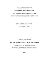
In this study out of 1268 (both retail and
mastitic) milk samples processed for the
isolation, only 120 milk samples developed
the typical greyish green colonies on
PALCAM agar with visible esculin hydrolysis
(Fig. 1), the colonies of 82 samples only were
found to be Gram positive coco-bacillary rods.
On biochemical examination only seven
isolates showed typical reaction. For further
characterization to species level they were
subjected to sugar fermentation test and based
on these test five isolates fermented α-methylD-mannoside, mannitol, glucose and lactose
hence were designated as L. grayi (Table 1).
The two isolates produced acid from xylose,
lactose and dextrose and were designated as L.
seeligeri. Summarizing the results on
prevalence of Listeria as obtained by
conventional culture and molecular based
method, an overall positivity of 0.68%was
reported in Punjab (Table 2). Among the five
zones 1.3% and 0.8% prevalence were
recorded in central and sub-mountain zone
respectively. Taking into consideration the
districts, then Ludhiana, Patiala, Tarantaran
and Pathankot yielded 3%, 1.66%, 3.33%, and
3.33% prevalence of Listeria spp. respectively
(Table 3). None of the retail milk samples
from other three zones and mastitic milk
samples were found positive. Therefore this
study reported a very low prevalence of
Listeria species in retail milk samples in
Punjab.
The results of this study can be supported with
the studies conducted in Mysore city,
Karnataka wherein authors reported 0.76%
prevalence of Listeria in milk and none of the
sample was positive for L. monocytogenes
(Shantha and Gopal, 2014). Whereas in
another study from, Mohali Punjab
documented zero prevalence of Listeria spp.
in the milk samples (Agarwal et al., 2013).
Khan et al., (2012) also specified the presence
of L. monocytogenes from only two samples
out of total 250 raw milk samples and milk
products in Bareilly. In another study from
Odissa by Sarangi et al., (2009), three samples
revealed the presence of L.monocytogenes
(2.01%) out of the total 137 raw milk samples
examined. Nayak et al., (2015) screened a
total of 200 milk samples and milk products,
of these 18 (9%) were found positive for the
Listeria spp. whereas L.monocytogenes was
isolated from the three milk samples only with
the prevalence 1.5%.
On the contrary some of the studies stated
higher prevalence of Listeria. Kalorey et al.,
(2008) conducted a large survey of central
India and reported 5.1% prevalence of L.
monocytogenes from 2060 raw milk samples.
2487
Int.J.Curr.Microbiol.App.Sci (2018) 7(10): 2484-2495
Similarly, Soni et al., (2013) reported 5.8%
prevalence of L. monocytogenes in raw cow
milk samples collected from Varanasi, Uttar
Pradesh. Another study carried out in Tamil
Nadu by Marry and Shrinithivihahshini (2017)
reported
52.7%
prevalence
of
L.
monocytogenes. The results from these studies
concluded that the high prevalence of L.
monocytogenes in the milk was indication of
the direct faecal contamination of milk, poor
sanitary practices during collection and
transportation of milk and further faulty
handling process which leads to low standards
of the milk and milk products sold at shops.
In the study, all seven isolates of Listeria (two
L. seelegiri and five L. grayi) were subjected
to haemolysin test on 7% sheep blood agar
(SBA). Accordingly, the 2 isolates (L.
seeligeri) turned out to be haemolytic in
nature. The 5 isolates characterized as L. grayi
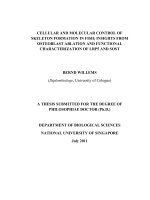
were non-haemolytic. Listeria isolates were
tested for their pathogenicity by plating them
on ALOA, all the 7 isolates produced typical
blue green colonies on ALOA but failed to
produce a halo even after one week of
incubation (Fig. 2).
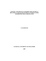
Listeria spp. isolates when subjected to genus
specific PCR for genus level confirmation
were found to amplify the DNA fragments of
370 bp respectively. All the isolates were
confirmed molecularly as Listeria spp (Fig. 3).
The findings in the present study are in
agreement with the work carried out by
Shantha and Gopal (2014) who reported
isolates recovered from raw cattle milk when
subjected to the molecular identification by
PCR for determining the genus Listeria, were
found to be positive for the genus specific
gene prs.
Another study carried out in Malaysia where
raw milk was assessed for the presence of
Listeria spp. (Chye et al., 2004), reported
4.4% of the raw milk sample was positive for
the Listeria spp. Among this, 1.9% were L.
monocytogenes, 2.1% were L. innocua and
0.6% were L. welshimeri.
Table.1 Biochemical characterization and sugar fermentation test of
Listeria spp. from raw retail milk
District
Isolates
Catalase
Nitrate
reduction
Pathankot
Ludhiana
PK5
L28
L10
L24
TT11
TT35
Pat14
+
+
+
+
+
+
+
-
Tarantaran
Patiala
VogesProskauer’s
test
+
+
+
+
+
+
+
Methyl
red
test
+
+
+
+
+
+
+
Xylose
Lactose
+
+
+
-
+
+
+
+
+
+
+
α-MethylDMannoside
+
+
+
+
+
Rham
nose
Dextrose
Mannitol
Designated
spp.
V
V
V
-
+
+
+
+
+
+
+
+
+
+
+
+
L. grayi
L. grayi
L. seeligeri
L. seeligeri
L. grayi
L. grayi
L. grayi
Table.2 Prevalence of Listeria spp. in retail and mastitic milk samples
Sr. Sample type
No.
1
2
Retail milk
Mastitic milk
Sample
size
1018
250
Conventional methods
Positive Per
cent
samples positivity (%)
7
0.68
Nil
Nil
2488
Molecular methods
Positive
Per
cent
samples positivity (%)
7
0.68
Nil
Nil
Int.J.Curr.Microbiol.App.Sci (2018) 7(10): 2484-2495
Table.3 Districts wise prevalence of Listeria spp. in retail milk samples in Punjab
Sr. No.
District
Sample
Positive sample
Per cent positivity
1
Ludhiana
100
3
3
2
Taran taran
60
2
3.33
3
Patiala
60
1
1.66
4
Pathankot
30
1
3.33
Fig.1 Typical greenish-yellow, glistening, lustrous and pointed colonies surrounded by a diffuse
black zone of aesculin hydrolysis on PALCAM agar
2489
Int.J.Curr.Microbiol.App.Sci (2018) 7(10): 2484-2495
Fig.2 ALOA plate showing the typical green colour of Listeria isolates
Fig.3 Genus specific PCR of Listeria
Lane L: Marker (100 bp)
Lane 1: Standard
Lane 2, 3, 6, 10: Negative isolates
Lane 3: Negative isolate
Lane 4, 5, 7-9, 11, 12: prs
Lane 13: NTC (Negative control template)
2490
Int.J.Curr.Microbiol.App.Sci (2018) 7(10): 2484-2495
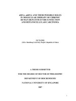
Fig.4 Virulence marker genes of Listeria monocytogenes
Lane L: Marker (100 bp)
Lane 1: HlyA/ORF2110
Lane 2: Negative control
Lane 3: actA
Lane 4: Negative control
Lane 5: prs
Lane 6: iap
Lane 7: plcA
Lane 8: prfA
Lane 9: NTC (Negative control template)
The PCR for the hlyA gene employed in the
study could not detect the gene in any of the
seven isolates except the standard strains of L.
monocytogenes which amplified the DNA
fragment of 456 bp in the PCR (Fig. 4). As
none of the isolates were found to be positive
for the species L. monocytogenes, therefore
multiplex PCR and PCR targeting the genes
for L. monocytogenes serovars was not
accomplished further.
Based on the documented scientific reports
we can say that prevalence or contamination
of Listeria spp, and especially L.
monocytogenes varies in different studies
done in different regions. Listeria spp,
2491
Int.J.Curr.Microbiol.App.Sci (2018) 7(10): 2484-2495
including L. monocytogenes are ubiquitous
microorganism that survive at different places
in farm environment and serve as a source of
contamination (Sunitha et al., 2016). In some
studies it has been found to be excreted
intermittently in the faeces of apparently
healthy animals at farm, more in animals that
are kept indoor and in cooler parts of the year
such as December (Husu, 1990). Excretion of
these organisms in the faeces and their
widespread presence in the farm environment
make milk samples highly prone to
contamination
from
these
organisms.
Contaminated dairy equipments and hands of
milkers can also add to the contamination of
raw milk with Listeria spp. (Tahoun et al.,
2017; Sunitha et al., 2016)
Therefore strict hygiene of the farm and
during milking is necessary to prevent
contamination. Dairy farming in farm is either
semi-organised or organised and farmers are
aware of the importance of clean and hygienic
milk production.
Although Listeria spp, especially L.
monocytogenes have not been frequently
associated with mastitis but some studies have
documented their presence in the mastitic
milk as well. Yadav et al., (2010) reported L.
monocytogenes from the mastitic milk
samples of buffalo and cow. In another study,
Rawool et al., (2007) also detected L.
monocytogenes from subclinical mastitic milk
samples, indicating that even if the farm
hygiene is good direct excretion of these
organisms in milk samples also act as another
source of milk contamination. None of our
milk samples from the mastitis cases were
positive for Listeria spp.
The other milk samples (raw retail milk
samples) of our study which reported
presence of Listeria spp, were however not
examined for their mastitis status.
Milk samples collected in the study were from
retail milk shops and from the milk sellers,
which usually collect milk from different
household and pool them all. This pooling
effect results in dilution of the organisms and
become difficult to detect.
There are also different isolation methods
available for the isolation of Listeria spp.
along with different amount of sample
processed for Listeria isolation by different
research workers. This difference in
processing also adds to the variation in
prevalence level reported from different
studies. Absence of L. monocytogenes in
retail milk samples in this study meets the
zero tolerance criteria set by food safety
agency. However, the detection of Listeria
spp. in the milk samples raises the food safety
and public health concerns in Punjab and
highlights the need to further strengthen
production, processing, packaging, storage
and distribution system into the retail market
to safeguard consumer health.
Acknowledgement
The authors are thankful to Department of
Health Research, Ministry of Health and
Family Welfare, GOI for providing funds for
this research work under Grant-in-aid Scheme
for
'Inter-sectoral
Convergence
&
Coordination for Promotion and Guidance on
Health Research.
References
Agarwal, A., Awasthi, V., Dua, A., Ganguly,
S., Garg, V., and Marwaha, S. S. 2013.
Microbiological profile of milk:
impact of household practices. Indian
Journal of Public. 56(1): 88-94.
Aurora, R., Prakash, A., Prakash, S. 2006.
Occurrence of pathogenic Listeria
monocytogenes in raw milk and readyto-eat milk products in Agra city,
2492
Int.J.Curr.Microbiol.App.Sci (2018) 7(10): 2484-2495
India. Indian Journal of Comparative
Microbiology,
Immunology
and
Infectious Diseases. 27: 87–93.
Borucki, M.K., Peppin, J.D., White, D., Loge
F., and Call, D.R. 2003. Variation in
biofilm formation among strains of
Listeria monocytogenes. Applied and
Environmental Microbiology. 69:
7336-7342.
Brooks, J. C., Martinez, B., Stratton, J.,
Bianchini, A., Krokstrom, R., &
Hutkins, R. 2012. Survey of raw milk
cheeses for microbiological quality
and
prevalence
of
foodborne
pathogens. Food Microbiology. 31(2):
154-158.
Chugh, T.D. 2008. Emerging and re-emerging
bacterial diseases in India. Journal of
Biosciences. 4:549–555.
Chye, F. Y., Abdullah, A., and Ayob, M. K.
2004. Bacteriological quality and
safety of raw milk in Malaysia. Food
Microbiology. 21(5): 535-541.
Dmowska, K., and Osek, J. 2010. Molecular
aspects of Listeria monocytogenes
pathogenicity.
Medycyna
Weterynaryjna. 66(4): 236-241.
Doumith, M., Buchrieser, C., Glaser, P.,
Jacquet, C., and Martin, P. 2004.
Differentiation of the major Listeria
monocytogenes serovars by multiplex
PCR.
Journal
of
Clinical
Microbiology. 42(8): 3819-22.
Fretz, R., Pichler, J., Sagel, U., Much, P.,
Ruppitsch, W., Pietzka, A. T., and
Werber,
D.
2010.
Update:
Multinational listeriosis outbreak due
to ‘Quargel’, a sour milk curd cheese,
caused
by
two
different
L.
monocytogenes serotype 1/2a strains,
2009-2010. European Surveillance.
32: 19543.
Husu, J. R. 1990. Epidemiological studies on
the
occurrence
of
Listeria
monocytogenes in the feces of dairy
cattle. Journal of Veterinary Medicine.
37(1-10): 276-282.
Jemmi, T., and Stephan, R. 2006. Listeria
monocytogenes: food-borne pathogen
and
hygiene
indicator. Revue
Scientifique et Technique. 25(2): 57180.
Kalorey, D. R., Warke, S. R., Kurkure, N. V.,
Rawool, D, B., and Barbuddhe, S. B.
2008. Listeria species in bovine raw
milk: A large survey of Central India.
Food Control. 19: 109–12.
Khan, J. A., Rathore, R. S., Khan, S., and
Ahmad, I. 2013. In vitro detection of
pathogenic Listeria monocytogenes
from food sources by conventional,
molecular
and
cell
culture
method. Brazilian
Journal
of
Microbiology. 44(3): 751-758.
Mary, M. S., Shrinithivihahshini, N.D. 2017.
Pervasiveness
of
Listeria
monocytogenes in Milk and Dairy
Products.
Journal
of
Food
Microbiology, Safety and Hygiene. 2:
125.
McClain, D., and Lee, W. H. 1988.
Development of USDA-FSIS method
for
isolation
of
Listeria
monocytogenes from raw meat and
poultry. Journal of Association of
Official Analytical Chemists. 71 (3):
660–63.
Nayak, D. N., Savalia, C. V., Kalyani, I. H.,
Kumar, R., Kshirsagar, D. P. 2015.
Isolation,
identification
and
characterization of Listeria spp. from
various
animal
origin
foods.
Veterinary World. 8(6): 695-701.
Rahimi, E., Ameri, M., and Momtaz, H.
2010. Prevalence and antimicrobial
resistance of Listeria species isolated
from milk and dairy products in Iran.
Food Control. 21(11): 1448-52.
Ramaswamy, V., Cresence, V., Rejitha, J. S.,
Lekshmi, M. U., Dharsana, K. S.,
Prasad, S. P., and Vijila, H. M. 2007.
2493
Int.J.Curr.Microbiol.App.Sci (2018) 7(10): 2484-2495
Listeria: review of epidemiology and
pathogenesis. Journal of Microbiology
and Immunology and Infection. 40(1):
4-13.
Rawool, D. B., Malik, S. V. S., Barbuddhe, S.
B., Shakuntla, I., and Aurora, R. 2007.
A multiplex PCR for detection of
virulence associated genes in Listeria
monocytogenes. International Journal
of Food Safety. 9: 56-62.
Rebagliati, V., Philippi, R., Rossi, M., and
Troncoso, A. 2009. Prevention of
foodborne listeriosis. Indian Journal of
Pathology and Microbiology. 52:1459.
Sarangi, L. N., Panda, H. K., Priyadarshini,
A., Sahoo, S., Palai, T. K., Ranabijuli,
S., Senapati, S., and Mohanty, D. N.
2009. Prevalence of Listeria species in
milk sample of cattle of Odisha.
Immunology and Infectious Diseases.
2: 135-35.
Sawant, L., Kaur, S., Aulakh, R., & Gill, J.
2016. Molecular characterization of
Listeria monocytogenes in bovine
milk and evaluating the sensitivity of
PCR for direct detection in milk.
Indian Journal of Animal Sciences. 86:
512-517.
Schwartz, B., Broome, C., Brown, G.,
Hightower,
A.,
Ciesielski,
C.,
Gaventa, S., and Listeriosis Study
Group. 1988. Association of sporadic
listeriosis with consumption of
uncooked hot dogs and undercooked
chicken. The Lancet. 332(8614): 779782.
Seeliger, H. P. R., and Jones, D. 1986.
Listeria.
Bergey’s
Manual
of
Systematic Bacteriology. 2: 1235–45.
Shantha, S., and Gopal, S. 2014. Prevalence
of Listeria species in environment and
milk samples. Advances in Animal
and Veterinary Sciences. 2(5): 1-4.
Soni, D. K., Singh, R. K., Singh, D. V., and
Dubey, S. K. 2013. Characterization of
Listeria monocytogenes isolated from
Ganges water, human clinical and
milk samples at Varanasi, India.
Infection, Genetics and Evolution. 14:
83–91.
Statistics, BAHS-Basic Animal Husbandry.
"Department of Animal Husbandry,
Dairying & Fisheries, Ministry of
Agriculture, Government of India."
KrishiBhavan, New Delhi (2012).
Sunitha, R., Raghunath, R. R., Vinod, V.K.,
Sunil, B., Prejit, Asha, K., Samitha
and Vergheese, J. 2016. Prevalence of
Listeria Monocytogenes in bovine
mastitic milk and dairy farm
environment. International Journal of
Science, Environment. 5(2): 638-643.
Tahoun, A. B., Elez, R. M. A., Abdelfatah, E.
N., Elsohaby, I., El-Gedawy, A. A.,
and Elmoslemany, A. M. 2017.
Listeria monocytogenes in raw milk,
milking equipment and dairy workers:
molecular
characterization
and
antimicrobial
resistance
patterns. Journal
of
global
antimicrobial resistance. 10: 264-270.
Yadav, M. M., Roy, A., Bhanderi, B., and
Joshi, C. 2010. Pheno- genotypic
characterization
of
Listeria
monocytogenes from bovine clinical
mastitis. Buffalo Bulletin. 29(1): 29–
38.
2494
Int.J.Curr.Microbiol.App.Sci (2018) 7(10): 2484-2495
How to cite this article:
Richa Tiwari, Randhir Singh Saini, Simranpreet Kaur and Aulakh, R.S. 2018. Prevalence and
Molecular Characterisation of Listeria spp. in Retail and Mastitic Milk of Punjab, India.
Int.J.Curr.Microbiol.App.Sci. 7(10): 2484-2495. doi: />
2495