Clinical, epidemiological and laboratory profile of dengue fever at tertiary care hospital in Hyderabad, India
Bạn đang xem bản rút gọn của tài liệu. Xem và tải ngay bản đầy đủ của tài liệu tại đây (276.29 KB, 9 trang )
Int.J.Curr.Microbiol.App.Sci (2018) 7(10): 2496-2504
International Journal of Current Microbiology and Applied Sciences
ISSN: 2319-7706 Volume 7 Number 10 (2018)
Journal homepage:
Original Research Article
/>
Clinical, Epidemiological and Laboratory Profile of Dengue Fever at
Tertiary Care Hospital in Hyderabad, India
Qursheed Sultana1*, Mohammed Abdur Rab Ansari2, Mahwish Jawaid1,
Mohd Ahmed1, Khaled Ahmed1 and Maimoona Mustafa1
1
Deccan College of Medical Sciences, Hyderabad, Telangana, India
2
MBBS Deccan, College of Medical Sciences, India
*Corresponding author
ABSTRACT
Keywords
Epidemiological,
Laboratory profile,
Dengue fever, Tertiary
care hospital
Article Info
Accepted:
18 September 2018
Available Online:
10 October 2018
Dengue Fever (DF), Dengue hemorrhagic fever (DHF) and Dengue Shock Syndrome (DSS) are
topmost public health concerns today, especially in tropical and subtropical countries, mainly
involving urban and semi-urban areas. In India, Dengue is hyperendemic (Category A) and is a
notifiable infectious disease. Though gaps exist in terms of public health measures and health
education among public, microbiological laboratory plays a crucial role in confirmation of
dengue infection and estimation of burden of disease. The aim of the study was to investigate
and assess the epidemiology and prevalence of dengue infection in patients attending our
tertiary care hospital in Hyderabad. A total of 1381 patients of suspected Dengue presented
during the study period of January – December 2017 were included in the study. All the
samples were processed using Dengue NS1 microlisa, Dengue MAC IgM ELISA and Dengue
GAC IgG ELISA. Out of these 1381 samples, 395 were serologically positive; 240 (61%) for
NS1 antigen and 82 (21%) for anti-dengue IgM and IgG antibodies. Maximum number of
dengue positive serum samples was observed in the month of November 421(31%) followed by
October 310 (23%). Out of 395 positive dengue cases, 309 were adult and 86 were of pediatric
age group. Majority of positive cases falls under age group of 11-20 years 149(38%) followed
by 21-30 years of age group 104 (26%). The lowest age of positive case was 4 months and
highest was 71 years. Most common clinical presentation was fever followed by
thrombocytopenia and arthralgia. To look for platelet and leucocyte counts, blood samples were
drawn for routine hemogram at the time of admission. This was evaluated by fully automated
haematology analyser-5 part differential. Thrombocytopenia was defined as platelet counts less
than 1,50,000/microliter and confirmed on peripheral smear examination. Leucocyte counts of
dengue patients were recorded and in majority of patients they were in normal range (400011000 per microliter). From the above study, it is concluded that prompt surveillance and
regular awareness are the two major steps to be taken for early detection and prevention of
dengue virus infection.
Introduction
Dengue (DEN) is the most rapidly spreading
viral disease in the world with a 30‑ fold
increase in incidence in the last 50 years. An
estimated 50 million DEN infections occur
annually and approximately 2.5 billion people
live in DEN endemic countries (World Health
Organization, 2009). In India, Dengue is
hyperendemic (Category A) and is a notifiable
2496
Int.J.Curr.Microbiol.App.Sci (2018) 7(10): 2496-2504
infectious disease (Park, 2013). Since DEN
produces a broad-spectrum of symptoms, a
diagnosis based only on clinical symptoms is
unreliable. Early laboratory confirmation is
valuable because some patients may
deteriorate rapidly resulting in death (World
Health Organization, 2009). During primary
infection, IgM appears after 5–6 days and IgG
after 7–10 days. During a secondary infection,
high levels of IgG are detectable even during
the acute phase and rise over the next 2 weeks,
whereas IgM are low or even absent in some
cases of secondary infection. IgM antibodies
suggest a recent infection; however, they can
persist for 2–3 months (Guzmán and Kourí,
2002; De Paula and Fonseca, 2004). High
titres of IgG are a criterion of secondary
infection. Viral nonstructural 1 (NS1) antigen
is abundant in the serum of patients in the
early stages of DEN infection, lasting from 1
to 9 days; therefore, NS1 antigen ELISA,
especially when used together with a IgM
capture ELISA, is sufficiently informative in
an endemic setting (Blacksell et al., 2008).
Materials and Methods
This prospective study was carried out in the
microbiology laboratory between January
2017 and December 2017 and was approved
by the Institutional Ethics Committee of
Deccan College of Medical Sciences. Case
inclusion criteria included presentation of
sudden high fever (39-40˚C) of 2-7 days
duration, intense headache, myalgia, arthralgia
or rash. The case was excluded if there was
evidence of bacterial or other viral illness
based on laboratory testing, duplicate samples
were excluded. All cases were managed
according to WHO Guidelines (National
Guidelines for Clinical Management of
Dengue Fever, 2015). After consent of the
patient, blood was taken by venipuncture
according to standard guidelines. Serum was
refrigerated (2-8ºC) if not tested within two
days. Sera showing haemolysis, icterus,
lipaemia or microbial growth were excluded
because they may cause false positive /
negative interpretation. Before beginning
assay, all reagents were brought to room
temperature (20-25ºC). Blood (serum) samples
collected from the patients were processed
using IgM and IgG capture ELISA and NS1
ELISA
according
to
manufacturer’s
instructions (J. Mitra and Co. Pvt. Ltd.). In
this kit, wells were coated with purified
dengue virus type 2 antigen cultured in Vero
cells. The test was executed according to the
manufacturer’s instructions. Dengue NS1
antigen, if present in serum, binds to anti-NS1
antibodies attached to the polystyrene surface
of the micro-wells which is followed by
chromogenic reaction. Development of colour
was indicative of the presence of dengue NS1
antigen in the test sample. An active primary
dengue infection was considered when
positive result came (>11 Panbio Units). IgM
antibodies in the patient’s blood was captured
by Antihuman IgM (μ chain specific) coated
on the wells. In the next step, DEN antigen
was added which could capture IgM, if IgM
and antigen were homologous. Unbound
antigen was removed during the washing step.
In the subsequent step Biotinylated flavivirus
cross-reactive monoclonal antibody (Hx-B)
was added followed by Avidin –HRP.
Subsequently, substrate/chromogen was added
and noted for development of colour. The
reaction was stopped by 1N H2SO4. Intensity
of colour/optical density was monitored at 450
nm. For quality control, each kit had a positive
and a negative control. Calculations and
interpretation were done as per the kit
literature.
Results and Discussion
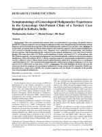
A total of 1381 patients of suspected Dengue
presented during the study period. Out of these
1381 samples, 395 were serologically positive
by at least one of the following tests-IgM
ELISA, IgG ELISA, NS1 ELISA (Figure 1).
2497
Int.J.Curr.Microbiol.App.Sci (2018) 7(10): 2496-2504
Maximum number of dengue positive serum
samples were observed in the month of
November 421(31%) followed by October
310(23%) (Table 1).
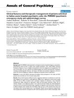
Out of 395 positive dengue cases, 309 were
adult and 86 were of pediatric age group
(Figure 2). Majority of positive cases falls
under age group of 11-20 years n=149(38%)
followed by 21-30 years of age group n=104
(26%) (Figure 3). The lowest age of positive
case was 4 months and highest was 71 years.
Most common clinical presentation was fever
followed by thrombocytopenia and arthralgia.
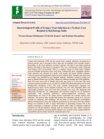
To look for platelet and leucocyte counts,
blood samples were drawn for routine
hemogram at the time of admission. This was
evaluated by fully automated haematology
analyser-5 part differential. Thrombocytopenia
was defined as platelet counts less than
1,50,000/microliter
and
confirmed
on
peripheral smear examination (Figure 4).
Leucocyte counts of dengue patients were
recorded and in majority of patients they were
in normal range (4000-11000 per microliter)
(Figure 5). Male preponderance was seen
among positive cases (Figure 6).
DEN is an emerging vector‑ borne disease.
Rapid urbanisation, globalisation, poor solid
waste and water management and increasing
population have given rise to new habitats for
mosquito breeding thereby increasing the
number of cases.
Dengue viruses are arboviruses capable of
infecting humans and causing outbreaks.
Dengue infection can be caused by any one or
more of the four different but closely related
serotypes; DEN1, DEN2, DEN3 or DEN4
dengue virus of the genus Flavivirus (WHO,
2011). Dengue fever (DF) is a self-limiting
disease in majority of cases, rarely it may
cause Dengue Hemorrhagic Fever (DHF) and
Dengue Shock Syndrome (DSS) (National
Guidelines for Clinical Management of
Dengue Fever, 2015). DF has been defined as
fever with two or more symptoms including
headache, retro-orbital pain, myalgia or
arthralgia and leucopenia, thrombocytopenia.
There are four grades of severity of DHF.
DHF -I include above criteria for DF plus
positive tourniquet test, evidence of plasma
leakage with platelet count less than 100,000/
cu.mm and haematocrit rise 20% or more.
DHF II includes DHF I plus some evidence of
spontaneous bleeding in skin or other organs
(black tarry stools, epistaxis, bleeding from
gums, etc.) and abdominal pain. DHF III
includes DHF II plus circulating failure (weak
rapid pulse, pulse pressure <20 mm Hg or
high diastolic pressure, hypotension with the
presence of cold clammy skin and
restlessness). DHF IV incudes profound shock
with undetectable blood pressure or pulse and
haematocrit rise more than 20%. DHF III and
IV are the criteria for labelling as DSS
(National Guidelines for Clinical Management
of Dengue Fever, 2015).
Early diagnosis may be challenging as dengue
infection presents with non-specific signs and
symptoms which may not be easily
differentiated from other febrile illnesses. In
our study, peak incidence of dengue occurred
in the month of October and November which
correlates with other Indian studies (Chandy et
al., 2013; Neeraja et al., 2006). This may be
supported by scientific evidence of
temperature and rainfall influencing dengue
incidence. According to IDSP data, dengue
fever cases peak during the post monsoon
period between mid-September to November
(National Centre for Disease Control
Newsletter, 2013). The density of Aedes
aegypti in an area is also linked with rainfall
and water storage. Life span of this vector is
influenced by temperature and humidity.
2498
Int.J.Curr.Microbiol.App.Sci (2018) 7(10): 2496-2504
Table.1 Shows the serological distribution of these positive cases. Out of 395 positive samples,
240 (61%) were positive for NS1 antigen and 82 (21%) for anti-dengue IgM & IgG antibodies
Month
Jan
Feb
Mar
Apr
May
June
July
Aug
Sep
Oct
Nov
Dec
NS1
NS1+
IgM
NS1 +
IgG
IgM
4
1
1
1
IgG
IgM + IgG
NS1+IgG+IgM
1
1
1
1
1
1
5
13
36
65
80
15
1
3
2
2
1
1
3
1
1
3
6
10
2
4
2
7
3
8
2
4
9
8
18
30
12
Month
Positive
Negative
Jan (12)
5
37
Feb (15)
3
12
Mar (18)
2
16
Apr (13)
1
12
May (14)
1
13
June (13)
1
12
July (34)
14
20
Aug (97)
34
63
Sep (263)
61
202
Oct (310)
94
216
Nov (421)
134
287
Dec (141)
48
93
2499
4
5
5
1
Int.J.Curr.Microbiol.App.Sci (2018) 7(10): 2496-2504
Distribution of positive samples
Positive
Negative
395
986
160
149
140
120
100
104
86
80
60
30
40
14
20
10
0
0 - 10 11 20
21 30
31 40
2500
41 50
51 60
1
1
61 70
71 80
Int.J.Curr.Microbiol.App.Sci (2018) 7(10): 2496-2504
180
160
140
120
100
80
60
40
20
0
156
88
68
42
41
<10000
10000 50000
50000 - 100000 - >150000
100000 150000
350
292
300
250
200
150
100
62
41
50
0
<4000
4000 - 11000
2501
>11000
Int.J.Curr.Microbiol.App.Sci (2018) 7(10): 2496-2504
It survives best between 16˚C-30˚C and a
relative humidity of 60-80%; breeds in
containers in and around the house. It has
been observed that even a 2˚C rise in
temperature can reduce the extrinsic
incubation period of DENV, hence an
increase in mosquito density. The extent of
dengue transmission depends on interplay of
multiple factors including host population
density, vector density and proportion of nonimmunized people in a community (Park,
2013; National Guidelines for Clinical
Management of Dengue Fever, 2015; WHO,
2011; Chandy et al., 2013; Neeraja et al.,
2006; National Centre for Disease Control
Newsletter, 2013; Karim et al., 2012).
The association of the DEN positivity with
thrombocytopaenia was in concordance with
the findings of other recent Indian studies
(Neeraja et al., 2013; Kulkarni et al., 2011).
Majority of the cases in our study belong to
the age group 20–40 years, with a male
preponderance as seen in other studies
(Kashinkunti et al., 2013; Chandralekha et al.,
2008). Studies done by Halsey et al., and
Kashinkunti et al., to correlate DEN with its
clinical manifestations revealed that headache
and myalgia/arthralgia were the most
common clinical manifestation of DEN
infection which was also seen in our study
(Chandralekha et al., 2008; Halsey et al.,
2012).
Diagnosis of dengue infection is confirmed by
the detection of virus, viral genome or NS1
Antigen, or seroconversion of IgM or IgG
(from negative to positive IgM/IgG or fourfold increase in the specific antibody titre) in
paired sera. Of all the methods available for
dengue diagnosis, virus isolation provides the
most specific test result. However, facilities
that can support viral culture are not always
available. The detection of the viral genome
or viral antigens also provides evidence of
infection. A large window of opportunity for
Dengue diagnosis is provided by NS1 Ag
which is highly conserved glycoprotein.
Single IgM ELISA test positivity is probable
of dengue and definitive diagnosis requires
use of paired sera to demonstrate rising titre
(Halsey et al., 2012).
Majority of our cases (61%) were detected
exclusively by the presence of viral NS1
antigen compared to IgM (7%) antibodies in
patient’s sera. It is known that early detection
of DEN cases by NS1 assay helps in
diagnostic detection and confirmation of cases
(Khan et al., 2014; Datta and Wattal, 2010).
NS1 antigen detection is particularly useful
during the first 5 days of illness and
significantly more sensitive for primary than
secondary DEN infection as was also seen in
our study (Khan et al., 2014; Dussart et al.,
2008; Lima Mda et al., 2010; Tricou et al.,
2010). Five percent of NS1‑ positive samples
were also IgG positive. These patients
provided serological evidence of previous
exposure. Fifteen patients who were
simultaneously positive for NS1 antigen, IgM
and IgG antibodies were probably in the late
stage of either a primary or a secondary
infection (Khan et al., 2014; Arya et al.,
2011). Seven of these 15 patients had
serological evidence of secondary infection. It
is a known fact that during a primary
infection, individuals develop IgM after 5–6
days and IgG antibodies after 7–10 days
(Guzmán and Kourí, 2002; De Paula and
Fonseca, 2004). Majority of the patients
(71%) presenting with fever of >5 days in our
study were positive for IgM ELISA as
compared to other serological parameters. In
2007, Kumarasamy et al., compared the use
of NS1 ELISA with viral isolation in cell
cultures and RT‑ PCR assay and achieved
great results in patients in the early stages of
infection concluding that NS1 antigen
2502
Int.J.Curr.Microbiol.App.Sci (2018) 7(10): 2496-2504
detection may be an appropriate marker of
acute DENV infection (Kumarasamy et al.,
2007). Thus, it can be stated that supporting
clinical symptoms along with early detection
of viral NS1 antigen can help speedup
diagnosis of DEN cases during the first 5 days
of fever and fever beyond that can be
diagnosed by IgM ELISA alone.
Limitations
Molecular testing to know the serotype of
dengue virus could not be done.
Dengue has become widespread and repeated
attacks due to different serotypes are
becoming common. Early diagnosis of
dengue allows institution of appropriate
supportive therapy and decreases risk of
complications. In this regard, the role of the
laboratory for diagnosis of Dengue, through
serological, molecular and virological
methods remains crucial to understand the
exact burden of disease. Monitoring of
climatic and environmental factors has
important association with high vector
density. Understanding of local epidemiology,
risk factors and disease burden in dengue is
essential to initiate preventive measures in
time so that the outbreaks and epidemics can
be avoided or controlled.
References
Arya SC, Agarwal N, Parikh SC, Agarwal S.
Simultaneous detection of dengue NS1
antigen, IgM plus IgG and platelet
enumeration during an outbreak. Sultan
Qaboos Univ Med J 2011; 11: 470‑ 6.
Blacksell
SD,
Mammen
MP
Jr.,
Thongpaseuth S, Gibbons RV, Jarman
RG, Jenjaroen K, et al., Evaluation of
the Panbio dengue virus nonstructural 1
antigen detection and immunoglobulin
M
antibody
enzyme‑ linked
immunosorbent assays for the diagnosis
of acute dengue infections in Laos.
Diagn Microbiol Infect Dis 2008; 60:
43‑ 9.
Chandralekha, Gupta P, Trikha A. The North
Indian dengue outbreak 2006: A
retrospective analysis of Intensive Care
Unit admissions in a tertiary care
hospital. Trans R Soc Trop Med Hyg
2008; 102: 143‑ 7.
Chandy S, Ramanathan K, Manoharan A,
Mathai D, Baruah K (2013) Assessing
effect of climate on the incidence of
dengue in Tamil Nadu. Indian J Med
Microbiol 31: 283-286.
Datta S, and Wattal C. Dengue NS1 antigen
detection: A useful tool in early
diagnosis of dengue virus infection.
Indian J Med Microbiol 2010; 28:
107‑ 10.
De Paula SO, and Fonseca BA. Dengue: A
review of the laboratory tests a clinician
must know to achieve a correct
diagnosis. Braz J Infect Dis 2004; 8:
390‑ 8.
Dussart P, Petit L, Labeau B, Bremand L,
Leduc A, Moua D, et al., Evaluation of
two new commercial tests for the
diagnosis of acute dengue virus
infection using NS1 antigen detection in
human serum. PLoS Negl Trop Dis
2008; 2: e280.
Guzmán MG, and Kourí G. Dengue: An
update. Lancet Infect Dis 2002; 2:
33‑ 42.
Halsey ES, Marks MA, Gotuzzo E, Fiestas V,
Suarez L, Vargas J, et al., Correlation of
serotype‑ specific
dengue
virus
infection with clinical manifestations.
PLoS Negl Trop Dis 2012; 6: e1638.
Karim MN, Munshi SU, Anwar N, Alam MS
(2012) Climatic factors influencing
dengue cases in Dhaka city: A model
for dengue prediction. Indian J Med Res
136: 32-39.
Kashinkunti MD, Shiddappa, Dhananjaya M.
A study of clinical profile of dengue
2503
Int.J.Curr.Microbiol.App.Sci (2018) 7(10): 2496-2504
fever in a tertiary care teaching hospital.
Sch J Appl Med Sci 2013; 1: 280‑ 2.
Khan SA, Dutta P, Topno R, Soni M,
Mahanta J. Dengue outbreak in a hilly
state of Arunachal Pradesh in Northeast
India. Scientific World Journal 2014;
2014: 584093.
Kulkarni RD, Patil SS, Ajantha GS, Upadhya
AK, Kalabhavi AS, Shubhada RM, et
al., Association of platelet count and
serological markers of dengue infection
‑ .Importance of NS1 antigen. Indian J
Med Microbiol 2011; 29: 359‑ 62.
Kumarasamy V, Wahab AH, Chua SK,
Hassan Z, Chem YK, et al., Evaluation
of a commercial dengue NS1 antigencapture ELISA for laboratory diagnosis
of acute dengue virus infection. J Virol
Methods 2007; 140: 75–79.
Lima Mda R, Nogueira RM, Schatzmayr HG,
dos Santos FB. Comparison of three
commercially available dengue NS1
antigen capture assays for acute
diagnosis of dengue in Brazil. PLoS
Negl Trop Dis 2010; 4: e738.
National Centre for Disease Control
Newsletter (2013) Quarterly newsletter
from the national centre for disease
control (NCDC).
National Guidelines for Clinical Management
of Dengue Fever (2015) National
Vector
Borne
Disease
Control
Programme,
World
Health
Organization, India.
Neeraja M, Lakshmi V, Dash PK, Parida
MM, Rao PV. The clinical, serological
and molecular diagnosis of emerging
dengue infection at a tertiary care
institute in Southern, India. J Clin Diagn
Res 2013; 7: 457‑ 61.
Neeraja M, Lakshmi V, Teja VD, Umabala P,
Subbalakshmi
MV
(2006)
Serodiagnosis of dengue virus infection
in patients presenting to a tertiary care
hospital. Indian J Med Microbiol 24:
280-282.
Park K (2013) Preventive and social
medicine: Dengue syndrome (22nd
edn). Banarasidas Bhanot, Jabalpur.
Tricou V, Vu HT, Quynh NV, Nguyen CV,
Tran HT, Farrar J, et al., Comparison of
two dengue NS1 rapid tests for
sensitivity, specificity and relationship
to viraemia and antibody responses.
BMC Infect Dis2010; 10: 142.
WHO (2011) Comprehensive guidelines for
prevention and control of dengue and
dengue hemorrhagic fever, revised and
expanded edition, SEAR.
World
Health
Organization.
Dengue:
Guidelines for Diagnosis, Treatment,
Prevention and Control. 2nd ed.
Geneva: World Health Organization;
2009. p. 1‑ 144.
How to cite this article:
Qursheed Sultana, Mohammed Abdur Rab Ansari, Mahwish Jawaid, Mohd Ahmed, Khaled
Ahmed and Maimoona Mustafa. 2018. Clinical, Epidemiological and Laboratory Profile of
Dengue Fever at Tertiary Care Hospital in Hyderabad, India. Int.J.Curr.Microbiol.App.Sci.
7(10): 2496-2504. doi: />
2504