SET domain containing protein 5 (SETD5) enhances tumor cell invasion and is associated with a poor prognosis in nonsmall cell lung cancer patients
Bạn đang xem bản rút gọn của tài liệu. Xem và tải ngay bản đầy đủ của tài liệu tại đây (6.18 MB, 10 trang )
Yu et al. BMC Cancer
(2019) 19:736
/>
RESEARCH ARTICLE
Open Access
SET domain containing protein 5 (SETD5)
enhances tumor cell invasion and is
associated with a poor prognosis in nonsmall cell lung cancer patients
Hairu Yu1,2, Jiayi Sun1,2, Congxuan Zhao1,2, Haotian Wang3, Yeqiu Liu1,2, Jiajia Xiong1,2, Jing Chang1,2,
Mixue Wang1,2, Wenhui Wang1,2, Dongman Ye1,2, Hongyan Zhou1,2 and Tao Yu1,2*
Abstract
Background: SET domain containing 5 (SETD5) is related to the aggressiveness of prostate and mammary cancers,
but its association with non-small cell lung cancer (NSCLC) is unknown. Therefore, the purpose of this research was
to determine the expression pattern and function of SETD5 in NSCLC.
Methods: SETD5 was detected by immunohistochemical analysis in 147 patients with non-small cell lung cancer.
SETD5 was overexpressed in A549 cells or suppressed with siRNA in H1299 cells. Wound healing and transwell
assays were performed. The expression levels of SETD5, p-AKT/AKT, Snail, p-JNK/JNK, Slug, E-cadherin, Zo-1, p-P38/
P38, occludin, α-catenin, p-ERK/ERK, and p-P90RSK/ P90RSK were assessed by western blot.
Results: Online analysis of overall survival in 1928 patients with NSCLC showed that the SETD5 gene was related to
worse overall survival (OS)(P < 0.001). The positive expression rate of SETD5 in noncancerous tissues was lower than
that in cancerous tissues (16.7% vs. 44.2%, P < 0.001). SETD5 was significantly correlated with advanced TNM stage
(P < 0.001), lymph node metastasis (P < 0.001) and overall survival rate (P < 0.001). Overexpression of SETD5 in A549
cells increased migration and invasion, while deletion of SETD5 in H1299 cells decreased migration and invasion.
After overexpression of SETD5, the expression of ZO-1 was downregulated, and that of Snail was upregulated. After
overexpression of SETD5, the levels of p-ERK and its downstream factor p-p90rsk increased.
Conclusion: These results suggest that SETD5 could regulate p-P90RSK and facilitate the migration and invasion of
NSCLC and may be related to the poor prognosis of patients with NSCLC.
Keywords: SET domain containing 5 (SETD5), Non-small cell lung cancer, Invasion, ERK signaling, Prognosis
Background
Non-small cell lung cancer (NSCLC) is a malignant
tumor of the lung accounting for 85–90% of all lung
cancers [1]. It affects mainly adults > 65 years of age,
men, and tobacco smokers [1, 2]. In the USA, the incidence of NSCLC is 75 per 100,000 men and 53.5 per
* Correspondence:
1
Department of Medical Imaging, Cancer Hospital of China Medical
University, No. 44 Xiaoheyan Road, Dadong District, Shenyang 110042,
Liaoning Province, China
2
Department of Medical Imaging, Liaoning Cancer Hospital and Institute, No.
44 Xiaoheyan Road, Dadong District, Shenyang 110042, Liaoning Province,
China
Full list of author information is available at the end of the article
100,000 women [3]. Mortality is high, with 55.9 per 100,
000 men and 36.3 per 100,000 women [3]. The treatment for NSCLC is multidisciplinary and includes surgery, chemotherapy, and radiation therapy [2]. Despite
great advances in techniques, regimens, and targeted
therapies, the 5-year survival for patients with NSCLC
(all stages together) is only 18% [4], highlighting the
need to better understand the disease to further improve
the treatment strategies.
SETD5 (SET domain containing 5), localized on chromosome 3p25.3, is a member of the SET domain protein family. These proteins play pivotal roles in histone lysine
methylation, thus inducing numerous cellular processes,
© The Author(s). 2019 Open Access This article is distributed under the terms of the Creative Commons Attribution 4.0
International License ( which permits unrestricted use, distribution, and
reproduction in any medium, provided you give appropriate credit to the original author(s) and the source, provide a link to
the Creative Commons license, and indicate if changes were made. The Creative Commons Public Domain Dedication waiver
( applies to the data made available in this article, unless otherwise stated.
Yu et al. BMC Cancer
(2019) 19:736
including heterochromatin formation, X-chromosome inactivation, and transcription regulation [5, 6]. Osipovich et
al. [7] also found that SETD5 plays an important role in the
co-transcriptional regulation of mammalian development
and histone acetylation. Previous studies demonstrated that
SET domain family proteins exhibited diverse biological
roles in cancer progression [8–17]. Nevertheless, the expression pattern and biological roles of SETD5 in human
malignant cancers remain unclear. Kuechler et al. [18] confirmed that loss of function of SETD5 was associated with
intellectual disability and was the critical driver of the
phenotype of 3p25.3 microdeletion syndrome [18–20].
Poissonnier et al. [21] showed that miR126-5p abolished
leukocyte transendothelial migration by suppressing
SETD5, indicating that SETD5 may participate in the
process of migration and invasion. A microarray analysis
suggested that the SETD5 locus was associated with prostate cancer aggressiveness [22]. A transcriptomics study
also showed that SETD5 was associated with the treatment
reaction in metastatic prostate tumors [23]. High mRNA
levels of SETD5 were related to poor prognosis in patients
with breast tumors [24]. Nevertheless, studies directly
assessing the mechanistic role of SETD5 in tumors are
lacking.
Therefore, the objective of this research was to determine the expression pattern and function of SETD5 in
NSCLC. The results showed that SETD5 enhanced the
invasion of NSCLC cells by activating the ERK signaling
pathway, suggesting that SETD5 may be a therapeutic
target for NSCLC patients.
Page 2 of 10
containing two-drug regimen should be applied. The
chemotherapy cycle was generally 4–6 cycles. Of the
147 patients, 48 had corresponding non-cancerous
tissues available. All patients were followed up.
NSCLC-specific survival was defined as the time from
surgery to the end of follow-up or death due to relapse
or transfer [19]. Histological diagnosis and grading
were assessed according to the World Health
Organization (WHO) classification of lung tumors from
2015 [26]. Tumor staging was based on the seventh edition of the International Union against Cancer (UICC)
TNM Staging System for Lung Cancer [27]. The characteristics of the cases and cancers are presented in
Table 1. The research was approved by the Institutional
Review Committee of China Medical University. Informed consent was obtained from each patient to use
their specimens for research purposes. Written consent
was provided in the ethics approval and consent to participate section.
Immunohistochemistry (IHC)
Samples were fixed in 10% neutral formalin, embedded in paraffin (Shanghai Shenggong Biological
Engineering Co., Ltd., Shanghai, China), and sectioned
Table 1 Correlations between SETD5 expression and
clinicopathological features in non-small cell lung cancer
(NSCLC)
Clinical parameters
Number SETD5 expression χ2
(N =
Positive Negative
147)
Age (years)
Methods
Online analysis of the total survival rate in patients with
NSCLC
To assess the relationship between the expression of
SETD5 and patient clinical results, we used the KM
Plotter Online Tool for NSCLC patients (http://www.
kmplot.com). This is a public database with information
about 1928 patients that allows us to examine the relevance of genes with overall survival (OS). The clinical
features of all specimens have been described [25].
Patients and clinical specimens
Tissue samples were obtained from 147 patients who
underwent complete surgical excision at the Cancer
Hospital of China Medical University from 2009 to
2011. All specimens were diagnosed as lung squamous
cell carcinoma or lung adenocarcinoma. No patients
had received chemotherapy or neoadjuvant radiotherapy, and all patients received chemotherapy after surgery. Adjuvant chemotherapy was started from 3 to 4
weeks after the operation. The chemotherapy regimen
was as follows: NP, GP regimen or according to drug
sensitive gene test results. In principle, a platinum-
< 59
65
35
30
≥ 59
82
39
43
Gender
Male
88
48
40
Female
59
26
33
Squamous cell
carcinoma
54
27
27
Adenocarcinoma
92
46
46
Large cell carcinoma
1
1
0
Histological type
Differentiation
Well
57
27
30
Moderate + Poor
90
47
43
I + II
103
42
61
III
44
32
12
TNM stages
0.573
0.449
1.551
0.213
0.993
0.609
0.329
0.566
12.590 < 0.001
Lymph node metastasis
15.252 < 0.001
Positive
66
45
21
Negative
81
29
52
TNM tumor node metastasis
P
Yu et al. BMC Cancer
(2019) 19:736
at 4 μm. IHC was performed using the streptavidinperoxidase method. Tissue slices were incubated with
a polyclonal rabbit anti-SETD5 antibody (1,100,
ab139987; Abcam, Cambridge, UK) at 4 °C overnight;
then, we used a biotin goat anti-mouse IgG secondary
antibody (Ultrasensitive; MaiXin, Fuzhou, China).
After washing, the tissue slices were incubated with
horseradish peroxidase binding streptomycin biotin
(Ultrasensitive; MaiXin, Fuzhou, China), and 3,3-diaminobenzidine tetrachloride (MaiXin, Fuzhou, China)
was used for development. Finally, the samples were
lightly re-dyed with hematoxylin (Shanghai Shenggong
Biological Engineering Co., Ltd., Shanghai, China), dehydrated and fixed in alcohol. Without considering the clinical data, the two researchers semi-quantitatively scored
the slides by assessing the staining intensity and percentage of stained cells in representative areas. The staining
intensity was scored as 0 (not stained), 1 (weak), 2
(moderate), or 3 (strong). The percentage of stained
cells was scored as 1 (1–25%), 2 (26–50%), 3 (51–75%),
or 4 (76–100%). Finally, the intensity and percentage
scores were multiplied to obtain 0–12 points. A score ≥
4 proved that the tumors were positive for SETD5
Page 3 of 10
expression. Tumor specimens scoring between 1 and 3
were classified as having weak expression, while those
scoring 0 were considered to have no expression; both
weak expression and no expression were defined as
negative SETD5 expression.
Cell culture
The HBE cell line was obtained from the American
Type Culture Collection (ATCC; Manassas, VA, USA).
The H1299, H460, A549, H292, and SK-MES-1 cell
lines were purchased from the Shanghai Cell Bank
(Shanghai, China). All of these cells were cultured in
RPMI 1640 (Invitrogen, Carlsbad, CA, USA) containing
10% fetal bovine serum (Invitrogen, Carlsbad, CA,
USA), 100 μg/ml streptomycin (Sigma, St Louis, MO,
USA), and 100 IU/ml penicillin (Sigma, St Louis, MO,
USA). Cells were passaged every other day using 0.25%
trypsin (Invitrogen, Carlsbad, CA, USA).
Plasmid transfection and small interfering RNA treatment
We bought the pCMV6-ddk-myc-SETD5 and pCMV6ddk-myc plasmids from Origene (RC240118, Rockville,
MD, USA). SETD5-siRNA (sc-78478) and NC-siRNA
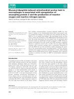
Fig. 1 Online analysis of the overall survival of 1928 patients with NSCLC. The relationship between SETD5 expression and overall survival was
evaluated using the KM Plotter Online Tool in 1928 patients with NSCLC. NSCLC, non-small cell lung cancer; HR, hazard ratio
Yu et al. BMC Cancer
(2019) 19:736
(sc-37007) were obtained from Santa Cruz Biotechnology (Santa Cruz, CA, USA). Transfection was carried
out using the Lipofectamine 3000 reagent (Invitrogen,
Carlsbad, CA, USA) according to the manufacturer’s
instructions.
Wound healing assay
Wounds were created in confluent areas of cell monolayers with < 90% confluence 48 h after transfection
using a 200-μl pipette tip. Cell migration into the wound
areas at different time points was observed. ImageJ software (National Institutes of Health, Bethesda, MD, USA)
was used to measure the distance the cells traveled into
the wound areas. Representative images were captured.
Page 4 of 10
Each specimen was analyzed twice, and three independent experiments were carried out.
Matrigel invasion assay
Cell invasion assays were carried out in 24-well Transwell
chambers with 8-μm pores (Costar, Cambridge, MA,
USA). The inserts were coated with 20 μl of Matrigel in
RPMI 1640 medium (1:3; BD Bioscience, San Jose, CA,
USA). Cells were trypsinized 48 h after transfection, resuspended at 3 × 10⁵ cells in 100 μl of serum-free medium,
and transferred to the upper transwell chamber; 10% FBS
was added to the lower chamber as a chemoattractant.
After incubation for 18 h, cells that passed through the filter were fixed with 4% paraformaldehyde and stained with
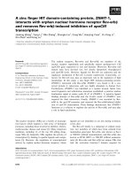
Fig. 2 SETD5 expression in NSCLC specimens and cell lines. a-f Representative SETD5 expression in adjacent normal tissues, squamous cell
carcinoma tissues, and adenocarcinoma tissues detected by immunohistochemistry. a Normal bronchial tissue, b alveolar epithelial tissue, c
squamous cell carcinoma, and d adenocarcinoma, only localized in the cytoplasm (e) or the nuclei (f) in some cases. Scale bar = 50 μm. g KaplanMeier analysis of the association between SETD5 expression and overall survival in patients with NSCLC. h SETD5 expression in different NSCLC
cell lines detected by western blot. GAPDH was used as an internal control
Yu et al. BMC Cancer
(2019) 19:736
hematoxylin (Zhongshan Jinqiao Biotechnology Co., Ltd.,
Beijing, China). Next, we randomly selected 10 visual
fields at 40× magnification under a microscope (Leica
Microsystems, Wetzlar, Germany) and counted the number of cells that invaded the subventricular space.
Western blotting
Protein was extracted with a lysis buffer (Pierce, Rockford,
IL, USA) and quantified with the Bradford method [28].
We used 10% sodium dodecyl sulfate-polyacrylamide gel
electrophoresis to isolate the proteins (50 μg) and transferred them to polyvinylidene fluoride (PVDF; Millipore,
Billerica, MA, USA) membranes. We incubated the
membranes overnight at 4 °C with the following primary
antibodies: SETD5 (1:100, ab139987; Abcam, Cambridge,
UK); GAPDH (1:5000, Sigma, St Louis, MO, USA); Myctag, Snail, Slug, p-P38, P38, p-ERK, ERK, p-AKT, AKT, pJNK, JNK, p-P90RSK, P90RSK (1:1000; Cell Signaling
Technology, Danvers, MA, USA); α-catenin (1:500; BD
Transduction Laboratories, Lexington, KY, USA); Zo-1, Ecadherin (1:1000; BD Transduction Laboratories,
Lexington, KY, USA); and occludin (1:500; Proteintech,
Chicago, IL, USA). Next, we washed the membranes and
incubated them with peroxidase-bound anti-rat or antirabbit IgG (Santa Cruz Biotechnology, Santa Cruz, CA,
USA) at 37 °C for 2 h. We visualized the proteins by electrochemiluminescence (Pierce, Rockford, IL, USA) and
detected them with a bio-imaging system (DNR Bio-Imaging Systems, Jerusalem, Israel).
Page 5 of 10
SETD5 was upregulated in NSCLC and is related to poor
prognosis in NSCLC patients
Next, to prove the results from the KM plotter tool, we
performed IHC on 147 specimens of NSCLC and 48
specimens of corresponding normal lung tissues to detect the expression and subcellular localization of
SETD5. The expression of SETD5 was low in peritumoral lung tissues (Fig. 2a-b) but high in the cytoplasm
and nuclei of NSCLC specimens (Fig. 2c-d). The positive
expression rate of SETD5 in peritumoral normal tissues
(8/48) was lower than that in cancerous tissues (65/147)
(16.7% vs. 44.2%, P < 0.001). In a few cases, we found
that SETD5 was localized only in the cytoplasm (5.4%,
8/147, Fig. 2e) or the nuclei (3.4%, 5/147, Fig. 2f ).
Positive expression of SETD5 was significantly associated with advanced TNM stage (P < 0.001) and lymph
node metastasis (P < 0.001) but not with age, sex, histological type, or differentiation (all P > 0.05, Table 1). A
Kaplan-Meier analysis showed that the OS was shorter
in patients with positive SETD5 expression than in those
with negative SETD5 expression (46.8 ± 3.1 vs. 64.9 ± 1.8
months, P < 0.001, Fig. 2g). Through univariate analysis
(UA) and multivariate analysis (MA), we concluded that
along with positive lymph node metastasis (P < 0.001 for
UA and P = 0.012 for MA), the independent prognostic
factors of OS in NSCLC patients may be related to the
SETD5 overexpression (P < 0.001 for UA and P = 0.013
for MA, Table 2). Then, we assessed the SETD5 protein
levels in various NSCLC cell lines and the human bronchial epithelial cell line HBE by western blot. The results
showed that the expression of SETD5 in HBE cells was
lower than that in NSCLC cell lines (Fig. 2h). Therefore,
Statistical analysis
All our data analyses were performed using SPSS22.0 for
Windows (IBM, Armonk, NY, USA). To evaluate the correlations between SETD5 and clinicopathological factors, the
Pearson Chi-square test was used. Kaplan-Meier survival
analyses were performed, and curves were compared using
the log-rank test. To estimate prognostic factors, we used
the Cox regression model for univariate and multivariate
analysis. We used the Mann-Whitney U test to analyze the
results of the invasion assay. P < 0.05 was considered to
have statistical significance.
Results
SETD5 is related to worse overall survival in 1928 NSCLC
patients from a public database
To preliminarily examine the potential role of SETD5
in NSCLC, the online tool KM plotter was used to predict the effect of SETD5 gene expression on OS in 1928
patients with NSCLC. As shown in Fig. 1, the SETD5
gene was related to worse OS in patients with NSCLC
(p < 0.001).
Table 2 Univariate and multivariate analyses of the associations
between clinicopathological features and overall survival in
NSCLC patients
Variables
Hazard ratio
P
(95% CI)
Univariate analysis
Age
0.795 (0.458–1.378)
0.413
Gender
0.997 (0.571–1.742)
0.992
Histological type
1.539 (0.852–2.778)
0.153
Differentiation
1.989 (1.075–3.682)
0.029
TNM stages
5.274 (2.983–9.324)
< 0.001
Lymph node metastasis
6.415 (3.338–12.326)
< 0.001
SETD5 expression
3.493 (1.886–6.473)
< 0.001
Differentiation
1.425 (0.757–2.683)
0.273
TNM stages
1.981 (0.953–4.116)
0.067
Lymph node metastasis
3.034 (1.272–7.233)
0.012
SETD5 expression
2.267 (1.192–4.311)
0.013
Multivariate analysis
Yu et al. BMC Cancer
(2019) 19:736
Fig. 3 (See legend on next page.)
Page 6 of 10
Yu et al. BMC Cancer
(2019) 19:736
Page 7 of 10
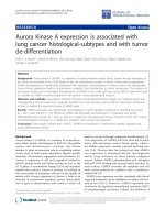
(See figure on previous page.)
Fig. 3 SETD5 promoted the migration and invasion of NSCLC cells. a Western blot analysis of SETD5 protein levels after SETD5 overexpression in
A549 cells or SETD5 silencing in H1299 cells. b Cell migration was assessed by wound healing assay after SETD5 overexpression in A549 cells or
SETD5 knockdown in H1299 cells. c Invasion was detected using transwell assays after SETD5 overexpression in A549 cells or SETD5 knockdown
in H1299 cells. Scale bar = 50 μm. The data are shown as the mean ± standard deviation (SD) from three independent experiments. *P < 0.05;
**P < 0.01; ***P < 0.001
we can conclude that SETD5 is likely to play an important role in NSCLC.
SETD5 enhanced NSCLC cell migration and invasion
To better understand the role of SETD5 in NSCLC aggressiveness, we overexpressed or suppressed SETD5 in
A549 or H1299 cells, respectively (Fig. 3a). Through
wound healing and transwell assays, we revealed that
migration (Fig. 3b) and invasion (Fig. 3c) increased after
overexpressing SETD5 in A549 cells. Migration (Fig. 3b)
and invasion (Fig. 3c) were decreased after depleting
SETD5 in H1299 cells. Hence, these results suggest that
SETD5 expression plays a role in the aggressiveness of
NSCLC.
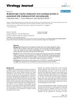
SETD5 promoted ERK and P90RSK phosphorylation,
upregulated snail and downregulated zo-1
Finally, to explore the possible mechanisms involved in
the regulation of NSCLC aggressiveness by SETD5, we
screened epithelial-mesenchymal transition (EMT)-related
proteins and key signaling pathway proteins. Regarding
EMT-related proteins, western blot results suggested that
Snail was upregulated and that Zo-1 was downregulated
when SETD5 was overexpressed in A549 cells. Snail and
Zo-1 were downregulated after silencing SETD5 with
siRNA (Fig. 4a). Slug, E-cadherin, α-catenin, and occludin
were unchanged (Fig. 4a).
Regarding key cell proliferation pathways, western blot
results indicated that p-ERK and its downstream factor pP90RSK were enhanced after overexpressing SETD5 in
A549 cells, while p-ERK and P90RSK were decreased after
SETD5 inhibition via siRNA in H1299 cells (Fig. 4b). The
levels of p-P38, P38, p-AKT, AKT, p-JNK, and JNK
showed no obvious alterations (Fig. 4b). These results suggest that SETD5 may facilitate NSCLC cell invasion by
promoting the phosphorylation of ERK and P90RSK and
then upregulating Snail and downregulating Zo-1.
Discussion
SETD5 plays a key role in mammalian development and
histone acetylation co-transcription. SETD5 is a member
of the SET domain protein family [5–7]. SETD5 is related to the aggressiveness of prostate and mammary
cancers [22–24], but the mechanism of its role in nonsmall cell lung cancer remains unclear. This study
showed that SETD5 was significantly correlated with
lymph node metastasis, advanced TNM stage and OS in
NSCLC patients. SETD5 may promote the migration
and invasion of NSCLC. SETD5 may be an upstream
regulator of the ERK-P90RSK signaling pathway.
This research showed that SETD5 was clearly expressed
in both the cytoplasm and nuclei of NSCLC specimens,
while SETD5 expression in normal lung tissues was low.
The expression of SETD5 was related to clinicopathological factors and poor OS. Taken together, these results indicated that SETD5 may be an oncogenic factor; this finding
is supported by the oncogenic role of other SET domain
protein family members [8, 10, 11], except SETD2, which
was demonstrated to be a tumor suppressor in renal and
breast carcinomas [12, 14–16, 29]. SETD4 is an oncoprotein that is localized to both the cytoplasm and nuclei
[10], similar to SETD5 in the present study. Previous studies indicated that SETD5 expression was related to the
prognosis of prostate and breast cancers [22–24], but this
research is the first to indicate a correlation between
SETD5 expression and NSCLC prognosis.
We found that SETD5 overexpression enhanced invasion and migration in NSCLC cells, while SETD5 suppression led to decreased invasion and migration.
Poissonnier et al. [21] showed that miR126-5p abolished
leukocyte transendothelial migration by suppressing
SETD5. These studies indicated that SETD5 may be involved in the process of migration and invasion. This hypothesis is supported by the subsequent observation that
SETD5 overexpression upregulated Snail and downregulated Zo-1. Indeed, Snail and Zo-1 are involved in EMT
[30, 31]. EMT is the process by which epithelial cells
lose their epithelial features and gain mesenchymal characteristics, leading to higher migratory abilities. High expression of Snail will lead to EMT and chemotherapy
resistance [30]. Zo-1 is a tight junction protein that is involved in cell-cell interactions. Therefore, loss of Zo-1
will be associated with nonadherent cells that are free to
migrate [31]. Snail upregulation could be responsible for
the decrease in Zo-1 and the induction of EMT [32, 33].
Snail levels are modulated by numerous signaling pathway factors [33–36], and the exact molecular mechanisms responsible for the upregulation of Snail by
SETD5 in the present study require additional study.
Nevertheless, the present study strongly suggests that
SETD5 may upregulate Snail and downregulate Zo-1 by
promoting the phosphorylation of ERK, which is supported by previous studies [34, 37–39]. SETD5 possesses
Yu et al. BMC Cancer
(2019) 19:736
Page 8 of 10
Fig. 4 Overexpression of SETD5 upregulated p-ERK, p-P90RSK, and Snail and downregulated Zo-1 in NSCLC cells. SETD5 was overexpressed in
A549 cells or suppressed with siRNA in H1299 cells. a EMT-related proteins were measured by western blot. b MAPK-related proteins were
measured by western blot. GAPDH was used as an internal control. EMT, epithelial-mesenchymal transition; MAPKs, mitogen-activated
protein kinases
Yu et al. BMC Cancer
(2019) 19:736
a conserved SET domain and a PH domain. Previous
studies showed that the SET domain was responsible for
histone lysine methylation [5, 6, 13]. Lu et al. [40] demonstrated that the PH domain of MKK1 is responsible
for modulating ERK expression [40]. The role of the
SET and PH domains of SETD5 in the activation of pERK remains to be further explored in NSCLC.
Conclusions
In conclusion, we found that the overexpression of
SETD5 was associated with lymph node metastasis, advanced TNM stage, and poor prognosis in patients with
NSCLC. SETD5 may promote the migration and invasion of NSCLC by enhancing the expression of Snail and
inhibiting that of ZO-1. SETD5 may be an upstream
regulator of the ERK-P90RSK signaling pathway. These
results indicate that SETD5 could be a factor involved in
the aggressiveness of NSCLC and a potential target for
improving the prognosis of NSCLC patients. The limitations of this study include the limited number of patients and follow-up time. However, the study of SETD5
is not complete. We will continue to explore the molecular and biological functions of SETD5.
Abbreviations
IHC: Immunohistochemistry; NSCLC: Non-small cell lung cancer; OS: Overall
survival; SETD5: SET domain containing 5; WHO: World Health Organization
Acknowledgments
Not applicable.
Authors’ contributions
HRY, JYS and CXZ conceived and supervised the study; HRY and TY designed
the experiments; HRY, HTW, YQL, JJX, JC, HYZ and MXW performed the
experiments; WHW and DMY developed new software and performed the
simulation studies; HRY, WHW and TY analyzed the data; HRY wrote the
manuscript; TY, HRY and WHW revised the manuscript. All authors reviewed
the results and approved the final version of the manuscript.
Funding
Not applicable.
Availability of data and materials
All data generated or analyzed during this study are included in this article.
The datasets used and/or analyzed during the current study are available
from the corresponding author upon reasonable request.
Ethics approval and consent to participate
This study was approved by the Institutional Review Board of the China
Medical University. Informed consent was obtained from each patient to use
their specimens for research purposes. Written consent was provided in the
ethics approval and consent form.
Consent for publication
Not applicable.
Competing interests
The authors declare that they have no competing interests.
Author details
1
Department of Medical Imaging, Cancer Hospital of China Medical
University, No. 44 Xiaoheyan Road, Dadong District, Shenyang 110042,
Liaoning Province, China. 2Department of Medical Imaging, Liaoning Cancer
Hospital and Institute, No. 44 Xiaoheyan Road, Dadong District, Shenyang
Page 9 of 10
110042, Liaoning Province, China. 3The First Clinical College, Dalian Medical
University, No. 9 West Section of Lushun South Road, Dalian City, Liaoning
Province, China.
Received: 2 June 2018 Accepted: 16 July 2019
References
1. Novello S, Barlesi F, Califano R, Cufer T, Ekman S, Levra MG, et al. Metastatic
non-small-cell lung cancer: ESMO clinical practice guidelines for diagnosis,
treatment and follow-up. Ann Oncol. 2016;27:v1–v27.
2. NCCN. Clinical practice guidelines in oncology (NCCN guidelines). Non-small
cell lung Cancer. Versiom 3.2018. Fort Washington: National Comprehensive
Cancer Network; 2018.
3. Siegel RL, Miller KD, Jemal A. Cancer statistics, 2018. CA Cancer J Clin.
2018;68:7–30.
4. Goldstraw P, Chansky K, Crowley J, Rami-Porta R, Asamura H, Eberhardt
WE, et al. The IASLC lung Cancer staging project: proposals for revision
of the TNM stage groupings in the forthcoming (eighth) edition of the
TNM classification for lung Cancer. J Thorac Oncol. 2016;11:39–51.
5. Qian C, Zhou MM. SET domain protein lysine methyltransferases: structure,
specificity and catalysis. Cell Mol Life Sci. 2006;63:2755–63.
6. Martin C, Zhang Y. The diverse functions of histone lysine methylation. Nat
Rev Mol Cell Biol. 2005;6:838–49.
7. Osipovich AB, Gangula R, Vianna PG, Magnuson MA. Setd5 is essential for
mammalian development and the co-transcriptional regulation of histone
acetylation. Development. 2016;143:4595–607.
8. Ayton PM, Cleary ML. Molecular mechanisms of leukemogenesis mediated
by MLL fusion proteins. Oncogene. 2001;20:5695–707.
9. Chen Z, Yan CT, Dou Y, Viboolsittiseri SS, Wang JH. The role of a newly
identified SET domain-containing protein, SETD3, in oncogenesis.
Haematologica. 2013;98:739–43.
10. Faria JA, Correa NC, de Andrade C, de Angelis Campos AC, de Dos
Santos Samuel AR, Rodrigues TS, et al. SET domain-containing protein 4
(SETD4) is a newly identified cytosolic and nuclear lysine
methyltransferase involved in breast Cancer cell proliferation. J Cancer
Sci Ther. 2013;5:58–65.
11. O’Neill DJ, Williamson SC, Alkharaif D, Monteiro IC, Goudreault M,
Gaughan L, et al. SETD6 controls the expression of estrogenresponsive genes and proliferation of breast carcinoma cells.
Epigenetics. 2014;9:942–50.
12. Li J, Duns G, Westers H, Sijmons R, van den Berg A, Kok K. SETD2: an epigenetic
modifier with tumor suppressor functionality. Oncotarget. 2016;7:50719–34.
13. Wilson JR, Jing C, Walker PA, Martin SR, Howell SA, Blackburn GM, et al.
Crystal structure and functional analysis of the histone methyltransferase
SET7/9. Cell. 2002;111:105–15.
14. Liu W, Fu Q, An H, Chang Y, Zhang W, Zhu Y, et al. Decreased Expression of
SETD2 Predicts Unfavorable Prognosis in Patients With Nonmetastatic ClearCell Renal Cell Carcinoma. Medicine (Baltimore). 2015;94:e2004.
15. Newbold RF, Mokbel K. Evidence for a tumour suppressor function of SETD2
in human breast cancer: a new hypothesis. Anticancer Res. 2010;30:3309–11.
16. Al Sarakbi W, Sasi W, Jiang WG, Roberts T, Newbold RF, Mokbel K. The
mRNA expression of SETD2 in human breast cancer: correlation with clinicopathological parameters. BMC Cancer. 2009;9:290.
17. Grouse L. Translational genetic research of complex diseases. J Transl Int
Med. 2015;3:137–43.
18. Kuechler A, Zink AM, Wieland T, Ludecke HJ, Cremer K, Salviati L, et al. Loss-offunction variants of SETD5 cause intellectual disability and the core phenotype
of microdeletion 3p25.3 syndrome. Eur J Hum Genet. 2015;23:753–60.
19. Szczaluba K, Brzezinska M, Kot J, Rydzanicz M, Walczak A, Stawinski P, et al.
SETD5 loss-of-function mutation as a likely cause of a familial syndromic
intellectual disability with variable phenotypic expression. Am J Med Genet
A. 2016;170:2322–7.
20. Lossignol D. A little help from steroids in oncology. J Transl Int Med. 2016;4:52–4.
21. Poissonnier L, Villain G, Soncin F, Mattot V. miR126-5p repression of
ALCAM and SetD5 in endothelial cells regulates leucocyte adhesion and
transmigration. Cardiovasc Res. 2014;102:436–47.
22. Dmitriev AA, Rosenberg EE, Krasnov GS, Gerashchenko GV, Gordiyuk VV,
Pavlova TV, et al. Identification of novel epigenetic markers of prostate
Cancer by NotI-microarray analysis. Dis Markers. 2015;2015:241301.
Yu et al. BMC Cancer
(2019) 19:736
23. Sowalsky AG, Xia Z, Wang L, Zhao H, Chen S, Bubley GJ, et al. Whole
transcriptome sequencing reveals extensive unspliced mRNA in metastatic
castration-resistant prostate cancer. Mol Cancer Res. 2015;13:98–106.
24. Liu L, Kimball S, Liu H, Holowatyj A, Yang ZQ. Genetic alterations of histone
lysine methyltransferases and their significance in breast cancer. Oncotarget.
2015;6:2466–82.
25. Gyorffy B, Surowiak P, Budczies J, Lanczky A. Online survival analysis
software to assess the prognostic value of biomarkers using transcriptomic
data in non-small-cell lung cancer. PLoS One. 2013;8:e82241.
26. Travis WD, Brambilla E, Burke A, Marx A, Nicholson AG. WHO classification of
tumours of the lung, pleura, thymus and heart; 2015.
27. Goldstraw P. Updated staging system for lung cancer. Surg Oncol Clin N
Am. 2011;20:655–66.
28. Bradford MM. A rapid and sensitive method for the quantitation of
microgram quantities of protein utilizing the principle of protein-dye
binding. Anal Biochem. 1976;72:248–54.
29. Grigorescu A. Chemotherapy for elderly patients with advanced cancer: a
pilot study in Institute of Oncology Bucharest. J Transl Int Med. 2015;3:24–8.
30. Kaufhold S, Bonavida B. Central role of Snail1 in the regulation of EMT and
resistance in cancer: a target for therapeutic intervention. J Exp Clin Cancer
Res. 2014;33:62.
31. Kalluri R, Weinberg RA. The basics of epithelial-mesenchymal transition. J
Clin Invest. 2009;119:1420–8.
32. Banerjee P, Venkatachalam S, Mamidi MK, Bhonde R, Shankar K, Pal R.
Vitiligo patient-derived keratinocytes exhibit characteristics of normal
wound healing via epithelial to mesenchymal transition. Exp Dermatol.
2015;24:391–3.
33. Zhang X, Yu X, Jiang G, Miao Y, Wang L, Zhang Y, et al. Cytosolic TMEM88
promotes invasion and metastasis in lung cancer cells by binding DVLS.
Cancer Res. 2015;75:4527–37.
34. Pan H, Jiang T, Cheng N, Wang Q, Ren S, Li X, et al. Long non-coding RNA
BC087858 induces non-T790M mutation acquired resistance to EGFR-TKIs by
activating PI3K/AKT and MEK/ERK pathways and EMT in non-small-cell lung
cancer. Oncotarget. 2016;7:49948–60.
35. Yang S, Ji Q, Chang B, et al. STC2 promotes head and neck squamous cell
carcinoma metastasis through modulating the PI3K/AKT/Snail signaling[J].
Oncotarget. 2016;8(4):5976–91.
36. Hung TW, Tsai JP, Lin SH, Lee CH, Hsieh YH, Chang HR. Pentraxin 3 activates
JNK signaling and regulates the epithelial-to-mesenchymal transition in
renal fibrosis. Cell Physiol Biochem. 2016;40:1029–38.
37. Li S, Lu J, Chen Y, Xiong N, Li L, Zhang J, et al. MCP-1-induced ERK/GSK3beta/Snail signaling facilitates the epithelial-mesenchymal transition and
promotes the migration of MCF-7 human breast carcinoma cells. Cell Mol
Immunol. 2017;14(7):621–30.
38. Strippoli R, Loureiro J, Moreno V, Benedicto I, Perez Lozano ML, Barreiro O,
et al. Caveolin-1 deficiency induces a MEK-ERK1/2-Snail-1-dependent
epithelial-mesenchymal transition and fibrosis during peritoneal dialysis.
EMBO Mol Med. 2015;7:102–23.
39. Martinez-Estrada OM, Culleres A, Soriano FX, Peinado H, Bolos V, Martinez
FO, et al. The transcription factors slug and snail act as repressors of
Claudin-1 expression in epithelial cells. Biochem J. 2006;394:449–57.
40. Lu Z, Xu S, Joazeiro C, Cobb MH, Hunter T. The PHD domain of MEKK1 acts
as an E3 ubiquitin ligase and mediates ubiquitination and degradation of
ERK1/2. Mol Cell. 2002;9:945–56.
Publisher’s Note
Springer Nature remains neutral with regard to jurisdictional claims in
published maps and institutional affiliations.
Page 10 of 10