Metformin reverses mesenchymal phenotype of primary breast cancer cells through STAT3/NF-κB pathways
Bạn đang xem bản rút gọn của tài liệu. Xem và tải ngay bản đầy đủ của tài liệu tại đây (1.95 MB, 13 trang )
Esparza-López et al. BMC Cancer
(2019) 19:728
/>
RESEARCH ARTICLE
Open Access
Metformin reverses mesenchymal
phenotype of primary breast cancer cells
through STAT3/NF-κB pathways
José Esparza-López1,2, Juan Francisco Alvarado-Muñoz2, Elizabeth Escobar-Arriaga3, Alfredo Ulloa-Aguirre1* and
María de Jesús Ibarra-Sánchez1,2*
Abstract
Background: Breast cancer currently is the most frequently diagnosed neoplasm and the leading cause of death
from cancer in women worldwide, which is mainly due to metastatic disease. Increasing our understanding of the
molecular mechanisms leading to metastasis might thus improve the pharmacological management of the disease.
Epithelial-mesenchymal transition (EMT) is a key factor that plays a major role in tumor metastasis. Some proinflammatory cytokines, like IL-6, have been shown to stimulate phenotypes consistent with EMT in transformed
epithelial cells as well as in carcinoma cell lines. Since the EMT is one of the crucial steps for metastasis, we studied
the effects of metformin (MTF) on EMT.
Methods: Cytotoxic effect of MTF was evaluated in eight primary breast cancer cell cultures by crystal violet assay.
EMT markers and downstream signaling molecules were measured by Western blot. The effect of MTF on cell
proliferation and cell migration were analyzed by MTT and Boyden chamber assays respectively.
Results: We observed that the response of cultured breast cancer primary cells to MTF varied; mesenchymal cells
were resistant to 10 mM MTF and expressed Vimentin and SNAIL, which are associated with a mesenchymal
phenotype, whereas epithelial cells were sensitive to this MTF dose, and expressed E-cadherin but not
mesenchymal markers. Further, exposure of mesenchymal cells to MTF down-regulated both Vimentin and SNAIL as
well as cell proliferation, but not cell migration. In an in vitro IL-6-induced EMT assay, primary breast cancer cells
showing an epithelial phenotype underwent EMT upon exposure to IL-6, with concomitant activation of STAT3 and
NF-κB; addition of MTF to IL-6-induced EMT reversed the expression of the mesenchymal markers Vimentin and
SNAIL, decreased pSTAT3 Y705 and pNF-κB S536 and increased E-cadherin. In addition, downregulation of
STAT3·activation was dependent on AMPK, but not NF-κB phosphorylation. Further, MTF inhibited cell proliferation
and migration stimulated by IL-6.
Conclusion: These results suggest that MTF inhibits IL-6-induced EMT, cell proliferation, and migration of primary
breast cancer cells by preventing the activation of STAT3 and NF-κB. STAT3 inactivation occurs through AMPK, but
not NF-κB.
Keywords: Breast Cancer, Epithelial-mesenchymal transition, Metformin, STAT3, NF-κB, AMPK
* Correspondence: ; ;
1
Red de Apoyo a la Investigación (RAI), Universidad Nacional Autónoma de
México- Instituto Nacional de Ciencias Médicas y Nutrición Salvador Zubirán,
Vasco de Quiroga 15, Col. Belisario Domínguez Sección XVI, Delegación
Tlalpan, 14080 Mexico City, CP, Mexico
Full list of author information is available at the end of the article
© The Author(s). 2019 Open Access This article is distributed under the terms of the Creative Commons Attribution 4.0
International License ( which permits unrestricted use, distribution, and
reproduction in any medium, provided you give appropriate credit to the original author(s) and the source, provide a link to
the Creative Commons license, and indicate if changes were made. The Creative Commons Public Domain Dedication waiver
( applies to the data made available in this article, unless otherwise stated.
Esparza-López et al. BMC Cancer
(2019) 19:728
Background
Breast cancer is a major health problem in women
worldwide, with an estimated 1.7 million women diagnosed with this neoplasia in 2012 [1]. Approximately
30% of breast cancer patients will eventually develop
metastatic disease, which is the main cause of death,
particularly when present at distant organs. Currently,
predicting accurately the risk for metastasis in a particular patient is not yet feasible. In fact, more than 80% of
breast cancer patients receive adjuvant chemotherapy
and approximately 40% will relapse and eventually die
from metastatic disease. According to the widely held
model of metastasis, rare subpopulations of cells within
the primary tumor acquire advantageous genetic alterations over time, thereby enabling these cells to
metastasize and form new solid tumors at distant sites
[2]. Thus, increasing our understanding on the molecular mechanisms leading to metastasis might improve the
clinical and pharmacological management of the disease.
The epithelial-mesenchymal transition (EMT) plays a
major role in tumor progression by assisting invasion
and intravasation of neoplastic cells into the bloodstream and inducing proteases involved in the degradation of the extracellular matrix (ECM) [3]. During the
EMT, cell-cell junctions and cell adhesion to ECM are
lost and, concomitantly, the apical-basolateral polarity is
disrupted, enabling the cells to evolve into a mesenchymal phenotype with invasive properties [4]. Down-regulation of E-cadherin has been reported to reflect
progression and metastasis in breast cancer associated
with poor prognosis [5, 6]. In addition, both downregulation of E-cadherin and up-regulation of Vimentin
and N-cadherin are frequently observed in cancer cells
from epithelial cancers during stromal invasion [7].
Down-regulation of E-cadherin is believed to result in
loss of adhesion between epithelial breast cancer cells
and other epithelial cells, whereas N-cadherin increase
promotes adhesion and intrusion of tumor cells into
the stroma [8]. Studying EMT in vitro has facilitated
the characterization of the several signaling pathways
typically involving a series of genes proposed as “EMT
master genes”. These genes are a group of transcription
factors that include SNAIL, TWIST, ZEB and E47 [9].
Extrinsic signals from soluble mediators from the
tumor microenvironment have been implicated in the
regulation of EMT.
Some cytokines have been shown to stimulate phenotypes consistent with EMT in transformed epithelial as
well as carcinoma cell lines. One of these is IL-6, a
pleiotropic cytokine that participates in acute inflammation, and that also plays a central role in hematopoiesis,
tumor progression, and proliferation; in addition, this
cytokine has been found within the tumor microenvironment [10–12]. IL-6 signaling uses a specific IL-6
Page 2 of 13
receptor (IL-6R/CD126) as well as a common transmembrane signal transducer, gp130 (CD130) to initiate the JAK/STAT3 and NF-κB signaling pathways. In
fact, elevated serum levels of IL-6 have been associated with poor prognosis of lung and breast cancer
[13–15]. Several studies have found that IL-6 contributes to the induction of EMT in several types of
tumors including lung, head and neck, breast, and
ovarian cancers [16–19].
Since the EMT is one of the crucial steps for metastasis, there is an enormous interest to find strategies
aimed to interrupt this process and to establish new
strategies for cancer treatment. Metformin (MTF), an
anti-diabetic drug widely prescribed for treating type 2diabetes, has been associated with reduction in the risk
to develop distinct types of cancer [20–22]. Several signaling pathways have been reported as putative mechanisms involved in the anti-tumor function of MTF,
including inhibition of pro-inflammatory cytokines similar to IL-6 [23] and down-regulation of EMT markers
such as E-cadherin, TWIST, ZEB, and Slug [24]. In lung
adenocarcinoma cells, MTF has been shown to affect IL6-induced EMT, most likely through inhibition of
STAT3 phosphorylation [25]. Some anticancer effects of
MTF have been associated with activation of adenosine
monophosphate protein kinase (AMPK). AMPK is an
energy sensor that is activated under low glucose levels,
hypoxia and stress [26]. To overcome a stress condition,
AMPK limits anabolic processes and activates catabolic
processes to generate energy, thereby increasing cell survival under stress [27]. Another mechanism of action
proposed for the MTF effects on tumor cells is through
inhibition of the electron transport chain of the mitochondria, hence decreasing Complex I activity of the
respiratory chain and the oxidative phosphorylation of
cells [28, 29]. Moreover, inhibition of Complex I lowers
the ATP production, leading to increase ADP levels
that later are converted to AMP, ultimately activating
AMPK [30, 31].
In the present study, we used a model of cultured primary breast cancer cells to analyze the impact of MTF
on the EMT. We employed patient-derived breast cancer
cell models because they represent better the molecular
characteristics from the original tumors and these
models are clinically relevant. We used 2 groups of primary breast cancer cells, a group with mesenchymal
phenotype and another with epithelial phenotype. We
found that the response to MTF is different between
mesenchymal and epithelial primary breast cancer cells.
MTF can suppressed basal mesenchymal markers with
reduction of cell proliferation, but it did not modify cell
migration rate. Furthermore, in an IL-6-induced EMT
model, MTF diminished IL-6-induced cell proliferation,
and migration by reducing the phosphorylation of
Esparza-López et al. BMC Cancer
(2019) 19:728
STAT3- and NF-κB. Moreover, inhibition of STAT3 activation by MTF appeared to be dependent on AMPK
activation, but not on the reduction of NF-κB
phosphorylation.
Methods
Antibodies and reagents
Recombinant human IL-6 was purchased from PeproTech (Rocky Hill, NJ, USA). E-cadherin and Vimentin
antibodies were obtained from GeneTex (Irvine, CA,
USA). SNAIL, pNF-κB-p65 (Ser536), pAMPK (Thr172),
AMPK, GAPDH were purchased from Cell Signaling
Technology (Danvers, MA, USA). STAT3, pSTAT3
Y705, NF-κB-p65, and β-actin were obtained from Santa
Cruz Biotechnology (Dallas, TX, USA).
Cell culture
The primary cell cultures MBCDF, MBCD3, MBCD4,
MBCD17, MBCD23, MBCD25, were derived from biopsies of mastectomies performed on patients with breast
cancer. The study was approved by the Ethics and Research Committee of the Instituto Nacional de Ciencias
Médicas y Nutrición Salvador Zubirán (Ref. 1549, BQ0–
008-06 / 9–1) as described before [32, 33]. MBCDF-D5
and MBCDF-B3 are subpopulations from the primary
culture MBCDF previously characterized by EsparzaLópez et. al. [33]. Cell cultures were maintained in
RPMI-1640 medium supplemented with 10% fetal bovine serum (FBS), antibiotic and antimycotic (Invitrogen
Corporation, Camarillo, CA) at 37 °C in a humidified atmosphere with 5% CO2.
Page 3 of 13
(MET), MBCDF and MBCD17 were treated with four
different conditions: no treatment, 40 ng/mL IL-6, 10
mM MTF and the combination IL-6 + MTF. At day 0,
an initial IL-6 treatment was given for 24 h. Then, MTF
was added with an additional dose of 40 ng/mL IL-6 to
sustain EMT. These conditions were maintained for further 24 h and cells were collected for protein extraction.
For inhibition of AMPK in MBCDF and MBCD17 cells,
10 μM compound C (Dorsomorphin) was added 2 h before the addition of IL-6. To activate AMPK, MBCDF
and MBCD17 cells were treated with 1 mM AICAR 2 h
before adding IL-6.
Western blot
Stimulated cultured primary breast cancer cells were lysed
in a buffer containing 50 mM HEPES pH 7.4, 1 mM
EDTA, 250 mM NaCl, 1% Nonidet P-40, 10 mM NaF, and
1X protease inhibitors (Complete EDTA-free, Roche).
Twenty micrograms of whole cell lysate were subjected to
SDS-PAGE and transferred to an Immobilon-P PVDF
membrane (Millipore Corp. Bedford, MA). The membrane was blocked for 60 min in 5% non-fat milk in PBSTween and then incubated with the corresponding primary antibodies overnight at 4 °C and thereafter with secondary anti-mouse-HRP or anti-rabbit-HRP antibodies
(Jackson Immuno-Research, West Grove, PA, USA). Detection of the HRP signal was performed using the ECL™
Prime Western Blotting Detection Reagent (GE Healthcare, Buckinghamshire, UK). Blot images were digitized
using Chemidoc (Bio-Rad, Hercules, CA, USA).
Cell proliferation
Cytotoxicity assay
Primary breast cancer cells were seeded at a density of
7500 cells/cm2 in 48-well plates. MTF (MP Biomedicals,
Burlingame, CA) was added at increasing concentrations
(0, 0.5, 1, 5, 10, 25, 50 and 100 mM), in triplicate incubations, and incubated for 48 h. Cell viability was evaluated
using the crystal violet technique. Thereafter, cells were
fixed with 1.1% glutaraldehyde in PBS for 20 min,
followed by staining with 0.05% crystal violet and dissolved in 10% acetic acid before measuring the absorbance at 570 nm using an ELISA plate reader. The results
are expressed as the percentage of viability calculated
from the absorbance of a given MTF concentration with
respect to the untreated control.
Cell stimulation
Primary breast cancer cells (MBCDF-D5, MBCD3,
MBCDF-B3, MBCD23) were treated with 10 mM MTF
to evaluate its effect on mesenchymal markers. MBCDF,
MBCD17 were induced to EMT by adding IL-6 40 ng/
mL. Cells were collected for protein extraction at day 0,
1, and 2. To induce mesenchymal-epithelial transition
Cell proliferation of cultured primary breast cancer cells
in the presence of 10 ng/mL IL-6, 10 mM MTF or IL6 + MTF was assessed by seeding 2500 cells/cm2 (5000
cells/well) in 24-well plates in RPMI 1640 supplemented
with 10% FBS. Cell proliferation was analyzed by the
MTT assay (3-[4,5-dimethylthiazol-2-yl]-2,5-diphenyltetrazolium bromide, Sigma-Aldrich, St Louis, MO,
USA) at 0, 1, 3 and 5 days. MBCDF-D5, MBCD3,
MBCDF-B3 and MBD23 cells were plated at the same
density as above. Cell proliferation was evaluated after
addition of MTF 0, 5, 10 and 25 mM on day 0 and 5 by
MTT assay. Formazan salt was dissolved with acidulated
isopropanol. The absorbance was read at 530 nm and
630 nm in an ELISA reader. Results are expressed as the
increase in absorbance (570–630 nm) at days 1,3 and 5
over the absorbance (570–630 nm) on day 0. The experiments were repeated at least three times in triplicate
incubations.
Migration assay
Cell migration of MBCDF and MBCD17 cells was carried out using a Boyden chamber assay. The upper
Esparza-López et al. BMC Cancer
(2019) 19:728
chamber was sown with 30,000-cells/200 μl in RPMI
1640 plus 10% of FBS. The lower chamber contained the
following conditions: control (no additions), 10 ng/mL
IL-6, 10 mM MTF, or 10 ng/mL IL-6 plus 10 mM MTF.
In the case of MBCDF-D5, MBCD3, MBCDF-B3, and
MBCD23 cells were seeded at the same density as above.
MTF was added in the upper and lower chamber at 0. 5,
10, and 25 mM. In all conditions, cells were incubated
for 6 h at 37 °C and 5% CO2. Non-migrating cells were
removed from the upper chamber with a cotton swap.
The migrating cells on the Boyden chamber were fixed
with 1.1% glutaraldehyde in PBS for 20 min and then
stained with crystal violet for 20 min. Cells were then
counted from five random fields. The number of migrating cells was obtained by dividing the mean of the 5
fields counted by 0.001cm2 (viewing field area) and then
multiplied by the insert area (0.33 cm2).
Page 4 of 13
a
b
Statistical analysis
Data are presented as mean ± SEM of three independent
experiments. MTF dose-response curves were analyzed
by Student’s t-test using SPSS 22.0. ANOVA was applied
to proliferation and migration assays and multiple comparisons were then performed employing the Turkey
HSD post-hoc test using GraphPad PRISM v6.01. P <
0.05 was considered significant.
Results
Primary breast cancer cells present variable responses to
metformin
For this study, we used a model of primary breast cancer
cells derived from patients with this type of cancer. The
molecular subtype of MBCDF-D5, MBCD3, MBCD23,
MBCDF-B3, MBCD25, MBCD17, MBCDF and MBCD4
breast cancer cells was determined according to the expression of estrogen and progesterone receptors and
HER2 (epidermal growth factor receptor 2) (Additional file 1: Table S1) [33], and the response to MTF in
these primary breast cancer cell cultures was evaluated
after treatment with increasing doses of MTF (0.5, 1, 5,
10, 25, 50, and 100 mM). We found that these cells were
distributed in two groups according to their sensitivity
to MTF. At low concentrations of MTF, cell viability did
not show any significant difference among all breast cancer cells. The major change was observed at 5, 10, and
25 mM of MTF, where MBCDF-D5, MBCD3, MBCD23,
and MBCDF-B3 cells were less sensitive to MTF. Cell
viability varied from 92 to 68% at 5 and 10 mM MTF
doses respectively, whereas at 25 mM MTF cell viability
oscillated between 79 and 57%. MBCD25, MBCD17,
MBCDF, and MBCD4 cells were more sensitive to MTF;
in these cells, viability varied from 66 and 27% at the
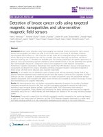
range of 5 to 25 mM MTF (Fig. 1a). To further study the
difference in the response to MTF among the primary
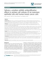
Fig. 1 Metformin-resistance correlates with mesenchymal phenotype in
primary breast cancer cells. a MBCD3, MBCD23, MBCD-D5, MBCD-B3,
MBCDF, MBCD17, MBCD25 primary breast cancer cell were treated
with increasing concentrations of MTF and cell viability was analyzed
by crystal violet after 48 h. Data represent the mean ± SEM of three
independent experiments in triplicate incubations. *P < 0.001
epithelial versus mesenchymal from 1 to 100 mM MTF. b
Representative immunoblot showing E-cadherin, Vimentin, and
SNAIL EMT markers expression. Actin was used as loading control
breast cancer cells used, we calculated the half inhibitory
concentration (IC50) for each primary culture. The IC50
of MBCDF-D5, MBCD3, MBCD23 and MBCDF-B3 cells
varied from 23.97 mM to 52.61 mM, while MBCD25,
MBCD17, MBCDF, and MBCD4 cells exhibited IC50s
from 5.31 to 11.45 mM (Table 1).
In order to analyze for differences causing MTF resistance among these breast cancer cell lines, the status of
EMT markers was measured. Interestingly, we found
that MBCDF-D5, MBCD3, MBCD23, and MBCDF-B3
cells exhibited features of mesenchymal phenotype as
disclosed by the lack of E-cadherin and presence of
Vimentin and SNAIL, while MBCD25, MBCD17,
MBCDF and MBCD4 cells expressed of E-cadherin with
a concomitant absence of Vimentin and SNAIL, both
distinctive of the epithelial phenotype (Fig. 1b). These
data indicated that the response of primary breast cancer
Esparza-López et al. BMC Cancer
(2019) 19:728
Page 5 of 13
Table 1 Metformin IC50 values
Primary breast cancer cell culture
IC50 [mM]
MBCDF-D5
44.70 ± 1.06
MBCD3
23.97 ± 1.97
MBCD23
36.55 ± 1.07
MBCDF-B3
52.61 ± 1.08
MBCD25
10.11 ± 1.20
MBCD17
5.31 ± 1.10
MBCDF
11.45 ± 1.13
MBCD4
8.17 ± 1.14
cell cultures to MTF exposure varied depending on the
EMT status.
Metformin decreases mesenchymal markers
Several studies have suggested that MTF reverses EMT
in several types of cancer [23, 24]. With this information
in mind, we examined whether MTF affected the mesenchymal markers in MBCDF-D5, MBCD3, MBCD23, and
MBCDF-B3 primary breast cancer cells. Cells were
treated with 10 mM MTF for 24 and 48 h, and expression of Vimentin and SNAIL was analyzed by Western
blot. The results showed that MTF treatment reduced
the amount of Vimentin and SNAIL in a timedependent manner (Fig. 2a). To examine the potential
role of MTF on cell proliferation and migration of mesenchymal primary breast cancer cells, we performed cell
proliferation assays in presence of MTF 0, 5, 10, and 25
mM. The effect of MTF was evaluated at day 6 by MTT
assay (Fig. 2b). MTF reduced proliferation in a dosedependent manner. The basal cell proliferation rate in
these cells fluctuated between 7 and 12-fold. We observed that MTF 5 mM had no significant impact on any
of this type of breast cancer cells. However, MTF at 10
and 25 mM had a major effect on cell proliferation, being MTF 25 mM where it was more significant (Fig. 2b,
Additional file 2). Next, mesenchymal breast cancer cells
(MBCDF-D5, MBCD3, MBCDF-B3 and MBCD23)
treated either with 10 or 25 mM MTF for 6 h were used
to evaluate cell migration by Boyden chamber assay (Fig.
2c). We found that cell migration was not affected by
MTF at any of the two concentrations used.
IL-6-induced epithelial-mesenchymal transition
Since MTF down-regulated Vimentin and SNAIL levels
in mesenchymal breast cancer primary cells, a model of
EMT induction using IL-6, which is a well-known EMT
inducer in several types of tumors including breast
cancer [34, 35], was established. MBCDF and MBCD17
cells were treated with 40 ng/mL IL-6 for 1 and 2 days.
A slight decrease in E-cadherin expression and an increase in Vimentin and SNAIL were concomitantly
observed (Fig. 3a). Further, examination of two IL-6-induced transcription factors (STAT3 and NF-κB) revealed
that IL-6 transactivated STAT3 as shown by the presence of increased STAT3Y705 phosphorylation and a
slight increase in the total amount of STAT3 in a timedependent fashion (Fig. 3b). Moreover, we found that
NF-κB phosphorylation at S536 also was increased in response to IL-6 stimulation (Fig. 3c). These results indicate that the particular primary breast cancer cells
studied can be induced to EMT by IL-6 exposure
through the activation of STAT3 and NF-κB signaling
pathways.
Metformin reverses IL-6-induced epithelial mesenchymal
transition
Once an IL-6-induced EMT model in primary breast
cancer cells was established, we investigated whether
MTF is able to reverse EMT. MBCDF and MBCD17 primary epithelial breast cancer cells were treated with 40
ng/mL IL-6; after 24 h of IL-6 exposure, 10 mM MTF
was added and cells were incubated for an additional 24
h period. As shown in Fig. 4a, IL-6 promoted EMT
through lowering E-cadherin and increasing Vimentin
and SNAIL. MTF alone did not exhibit a significant effect on EMT markers, while the addition of MTF to IL6 treatment provoked re-expression of E-cadherin and
inhibition of IL-6-stimulated Vimentin and SNAIL expression. These results indicate that MTF reverses the
EMT induced by IL-6 in primary breast cancer cells.
We next examined the effect of MTF on the activation
of IL-6-induced STAT3 and NF-κB in MBCDF and
MBCD17 primary breast cancer cells. Similar experiments to those shown in Fig. 4a were performed and activation of the STAT3 and NF-κB pathways was
analyzed. As shown in Fig. 4b, IL-6 induced phosphorylation of Y705 on STAT3 whereas MTF alone had no effect on STAT3 activation. However, addition of MTF to
IL-6 stimulation reversed the phosphorylation of STAT3
at Y705 (Fig. 4b). In addition, IL-6 provoked phosphorylation of NF-κB at S536 (Fig. 3c), and reversed this phosphorylation when MTF was combined with IL-6 (Fig.
4c). Similar results were observed in both MBCDF and
MBCD17 primary breast cancer cell cultures. These data
suggest that MTF reverses EMT by blocking activation
of the IL-6-induced transcription factors STAT3 and
NF-κB.
AMPK activation is required for decrease of pSTAT3, but
not pNF-κB
Several reports have shown that MTF anticancer effects
may be dependent- or independent of AMPK [36]. In
order to determine the role of AMPK in MTF-reduction
of STAT3 and NF-κB phosphorylation in MBCDF and
MBCD17 cells, we used two different approaches;
Esparza-López et al. BMC Cancer
(2019) 19:728
Page 6 of 13
a
b
c
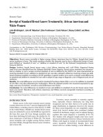
Fig. 2 Metformin reduces the expression of Vimentin and SNAIL, decreases cell proliferation but not migration in mesenchymal breast cancer cells. a
Primary breast cancer cell with mesenchymal phenotype (MBCDF-D5, MBCD3, MBCDF-B3 and MBCD23) were treated with 10 mM MTF for 0, 1
and 2 days and the effect of MTF on the mesenchymal markers Vimentin and SNAIL was analyzed by immunoblotting. Actin was used as loading
control. b For cell proliferation, primary breast cancer cells with mesenchymal phenotype (MBCDF-D5, MBCD3, MBCDF-B3 and MBCD23) were
seeded at 2500 cell/cm2 (5000 cells/well) in a 24 well-plate and incubated in the absence (control) or presence of 0, 5,10, and 25 mM MTF. Cell
proliferation was evaluated by MTT at days 0, and 6. Data represents the mean ± SEM of three independent experiments performed in triplicate
incubations. *P < 0.05. c Migration assays were performed using Boyden chambers. Thirty thousand cells with mesenchymal phenotype (MBCDFD5, MBCD3, MBCDF-B3 and MBCD23) were seeded in the upper chamber in presence of MTF 0, 10 and 25 mM, the same concentrations of MTF
were added in the in-bottom chamber, and then incubated for 6 h at 37 °C. After this time the cells that did not migrate were removed from the
upper chamber. Cells that migrated were fixed and stained with Cristal Violet. Five fields were counted under the microscope at 20X. Migration
assays were performed three independent times in triplicate
AMPK inhibition with compound C (Dorsomorphin), or
AMPK activation using an activator, 5-aminoimidazole4-carboxamide-1-β-D-ribofuranoside
(AICAR).
For
AMPK inhibition, 10 μM compound C was added alone
or 2 h before IL-6 addition and incubated for 24 h. After
this time, compound C alone and compound C + IL-6
conditions both were treated with MTF, incubation was
extended further 24 h. Activation of STAT3 and NF-κB
Esparza-López et al. BMC Cancer
(2019) 19:728
a
b
Page 7 of 13
a
b
c
c
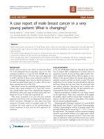
Fig. 3 Primary epithelial breast cancer cells undergo IL-6-induced EMT
through STAT3 and NF-κB activation. Primary breast cancer cells with
epithelial phenotype (MBCDF and MBCD17) were treated with 40
ng/mL of IL-6 during 0, 1 and 2 days. a Induction of EMT was
analyzed by assessing the expression of E-cadherin, Vimentin, and SNAIL by
Western blots. b The activation of STAT3 was measured by
phosphorylation of STAT3 on tyrosine 705 using a phospho-specific antipSTAT3 Y705 antibody. c Activation of NF-κB was assessed by analyzing
phosphorylation of NF-κB/p65 on serine 536 employing a phospho-specific
anti- pNF-κB S536 antibody. Actin was used as loading control in all cases
was evaluated as in Fig. 4. We observed that MTF reduced phosphorylation of both STAT3 and NF-κB as
demonstrated before. Compound C alone did not have a
significant effect on STAT3 phosphorylation (Fig. 5a,
lane 5). Compound C added before IL-6 increased
STAT3 phosphorylation (Fig. 5a, lane 6). Combination of
compound C + MTF did not affect pSTAT3 Y705 (Fig.
5a, line 7) and treatment with compound C + IL-6 +
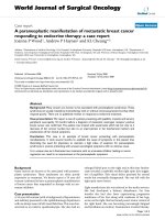
Fig. 4 Metformin reverses IL-6-induced EMT in primary epithelial breast
cancer cells by inhibiting STAT3 and NF-κB phosphorylation. MBCDF and
MBCD17 cells were treated with 40 ng/mL IL-6. At day 1, MTF was
added to cells incubated in the presence or absence of IL-6. After 2
days of incubation all conditions were collected for protein
extraction and Western blot analysis. a Effect of MTF on IL-6-induced
EMT markers (E-cadherin, Vimentin, and SNAIL). b Effect of MTF on
IL-6-induced activation of STAT3 was assessed as in Fig. 3b. c Effect
of MTF on IL-6-induced NF-κB phosphorylation was assessed as in
Fig. 3c. Actin was used as loading control
MTF partially prevented the reduction of pSTAT3 Y705
(Fig. 5a, lane 8). These results suggest that AMPK inhibition with compound C partially interferes with the
MTF-reduced STAT3 activation. In the case of NF-κB,
we observed again that IL-6 induced NF-κB phosphorylation whereas co-treatment with MTF reduced IL-6-induced phosphorylation. Compound C alone exhibited
opposite effects on pNF-κB S536, in MBCD17 increased
phosphorylation while in MBCDF had no effect (Fig. 5a,
Esparza-López et al. BMC Cancer
(2019) 19:728
Page 8 of 13
Fig. 5 Metformin effects on primary epithelial breast cancer cells are dependent of AMPK activation. a MBCDF and MBCD17 primary breast cancer
cell lines were treated with 40 ng/mL IL-6. At day 1, MTF was added to cells incubated with or without IL-6. Same experiment was repeated in
the presence of 10 μM Compound C (COMP C) that was added 2 h before IL-6. The activation of STAT3 and NF-κB was measured by phosphospecific antibodies anti-pSTAT3 Y705 and anti-pNF-κB S536 antibody respectively. b MBCDF and MBCD17 cells were treated with 40 ng/mL IL-6,
previous addition of 1 mM 5-aminoimidazole-4-carboxamide-1-β-D-ribofuranoside (AICAR), an activator of AMPK kinase. The activation of STAT3,
NF-κB was evaluated as in Fig. 5a. AMPK activation was measured by a phospho-specific anti-pAMPK-T172
lane 5). Both IL-6 + compound C and the combination
of compound C + IL-6 + MTF presented similar levels of
pNF-κBS536 similar to IL-6 treatment. These data suggest that the reduction in NF-κB activation induced by
MTF is not dependent on AMPK.
Next, we examined whether AICAR-induced AMPK activation could mimic MTF reduction of IL-6-induced phosphorylation of STAT3 and NF-κB in MBCDF and
MBCD17 breast cancer cells. Breast cancer cells treated
with 1 mM AICAR alone or added 2 h before IL-6 were
collected for protein extraction 2 days after treatment. We
analyzed phosphorylation of STAT3 Y705 and NF-κB S536
(Fig. 5b). IL-6 induced phosphorylation of STAT3 Y705
whereas AICAR alone did not affect this phosphorylation;
but when it was added before IL-6, IL-6-induced pSTAT3
Y705 was reduced (Fig. 5b). Next, we evaluated the effect of
AICAR on the IL-6-induced NF-κB phosphorylation. We
found that AICAR did not interfere with IL-6-induced
phosphorylation of NF-κB. These data suggest that activation of AMPK can mimic reduction of pSTAT3 Y705
similar to that observed with MTF + IL-6. However, IL-6induced pNF-κB S536 was not affected by AICAR (Fig. 5b).
We confirm that AICAR induced AMPK activation by
phosphorylation on T142 that indeed was increased by
treatment (Fig. 5b). Together these results suggest that
MTF-reduced phosphorylation of STAT3, but not NF-κB
phosphorylation is dependent on AMPK activation.
Metformin inhibits IL-6-induced cell proliferation and cell
migration
Since MTF interfered with IL-6-induced EMT of primary breast cancer cells, we then analyzed whether
Esparza-López et al. BMC Cancer
(2019) 19:728
MTF had an effect on cell proliferation and migration.
MBCDF and MBCD17 breast cancer cells were treated
with IL-6 and MTF alone or in combination and cell proliferation was assessed by MTT assay at 0, 1, 3 and 5 days
of stimulation. The basal rate of proliferation for MBCDF
and MBCD17 cells reached 13- and 9-fold on day 6 respectively. IL-6 exposure increased cell proliferation up to
18-fold in MBCDF cells and 14-fold in MBCD17 cells.
MBCDF and MBCD17 cells treated with MTF or with
both IL-6 plus MTF showed a trend towards less proliferation than control cells, suggesting an inhibitory effect of
MTF on IL-6-induced cell proliferation (Fig. 6a, Additional file 3). We next investigated the effect of MTF on
IL-6-induced cell migration employing the Boyden chamber assay. The basal cell migration in the primary breast
cancer cells studied showed different patterns, with
MBCDF cells migrating more than MBCD17 cells. IL-6
treatment increased basal cell migration of both MBCDF
and MBCD17 cells, whereas MTF-treated cells showed a
downward trend migration when compared with control,
unexposed cells. Migration in the presence of both IL-6
and MTF was similar to that exhibited by the control cells
(Fig. 6b), suggesting that MTF interferes with the migration stimulated by IL-6.
Page 9 of 13
Discussion
In the present study, we analyzed the effects of MTF on
the mesenchymal phenotype and IL-6-induced EMT in
cultured primary breast cancer cells. EMT is a key
process in metastasis development and the major cause
of mortality among breast cancer patients and evidence
has been accumulated over the past decade suggesting a
potential role of MTF in suppressing the progression of
several types of cancer [37]. We here demonstrate that
MTF displays different effects associated with the EMT
status of cultured primary breast cancer cells. Mesenchymal cells were resistant to MTF and epithelial cells were
sensitive to MTF. Further analysis showed that high
MTF doses reduced expression of mesenchymal markers
as well as IL-6-induced EMT by blocking STAT3 and
NF-κB phosphorylation. Reduction of STAT3 phosphorylation, but not that of NF-κB is dependent on AMPK
activation. Additionally, MTF inhibited cell proliferation
of mesenchymal breast cancer cells, but not cell migration. Moreover, MTF overturned IL-6-stimulated cell
proliferation and migration of cultured primary breast
cancer cells.
A number of studies have suggested a potential role of
MTF on the prevention and improvement of overall
a
b
Fig. 6 Metformin inhibits IL-6-induced cell proliferation and migration. a MBCDF and MBCD17 primary breast cancer cell lines were seeded at 15000
cells/cm2 in a 24-well plate and incubated under the absence (control) or presence of 10 ng/mL IL-6, 10 mM MTF or the combination of IL-6 and
MTF. Cell proliferation was studied at days 0, 1, 3, and 5 by MTT. Data represent the mean ± SEM of three independent experiments performed in
triplicate incubations. *P < 0.05, **P < 0.001. b Migration assays were tested using Boyden chambers. MBCDF and MBCD17 were seeded at 30000
cells/transwell in triplicate in the upper chamber. In the bottom chamber the same conditions were maintained as in Fig. 5a. Cells were allowed
to migrate for 6 h. Migrating cells were fixed and stained with crystal violet. Data are presented as the mean ± SEM of three independent
experiments. **P < 0.001
Esparza-López et al. BMC Cancer
(2019) 19:728
survival in breast cancer [38, 39], and proposed potential
mechanisms on how MTF may affect cell survival, proliferation, migration, and inflammation [40–42]. Many of
these studies have been performed using immortalized
breast cancer cell lines, which have been the standard
experimental paradigm employed for many years. Nevertheless, cell lines may present several drawbacks including the effects of long time in culture on the potential
development of new mutations and phenotypes [43, 44].
Thus, they frequently do not fully reflect what actually
occurs in in vivo conditions. In this and other studies,
we have used a model of cultured primary breast cancer
cells that retain most of the biochemical features of the
original tumor [32, 33]. Using this experimental model,
we here demonstrate that the sensitivity to MTF depends on the EMT status: a mesenchymal phenotype
correlated with resistance to MTF, whereas on the contrary, an epithelial phenotype was associated with sensitivity to MTF. In fact, differences in the IC50 for MTF
indicated that mesenchymal cells required 4 to 10 times
more MTF than epithelial cells to decrease 50% cell viability. Nonetheless, other studies have shown different
effects of MTF on breast cancer cell lines. One study
showed that MTF induced cell cycle arrest in estrogen
receptor-positive but not in estrogen receptor-negative
cells [45], while another study found that cells without
expression of hormonal receptors were more responsive
to this drug [46–48]. These studies proposed that differences in the response to MTF may be associated with
particular breast cancer molecular subtypes [41, 47–49].
Here we demonstrate that the response to MTF in primary breast cancer cells is associated with the EMT status rather than with the molecular subtype.
During the EMT, cancer cells go through biochemical
and morphological changes that allow them to acquire
and enhance their invasive capacity [7]. We here show
that Vimentin, SNAIL and cell proliferation decreased
by MTF treatment in breast cancer cells with a mesenchymal phenotype, although these particular cells required higher doses of MTF to provoke an inhibitory
effect. SNAIL expression has been associated with the
repression of E-cadherin, invasion and metastases in several types of malignancies like breast, lung, hepatocellular and ovarian carcinomas [50–52]. SNAIL also has
been associated as negative regulator of cell growth in
lung and prostate cancer [53, 54]. Our results of the
MTF-treated mesenchymal breast cancer cell, the reduction of SNAIL expression correlates with decreasing of
cell proliferation. Consequently, MTF might reduce the
invasive capacity of mesenchymal primary breast cancer
cells by lowering SNAIL and Vimentin, which are also
important factors involved in the structural changes of
the cytoskeleton and thus in cell motility and invasiveness creating a phenotypic switch [55]. In fact, several
Page 10 of 13
studies have found that MTF represses EMT in several
tumors including cervical cancer cells [56], thyroid cancer cells [57], hepatocellular carcinoma [58] and lung
adenocarcinoma [25] by reducing the levels of these
factors.
It has been shown that resumption of EMT promoted
by growth factors and pro-inflammatory cytokines
present in the tumor microenvironment is closely linked
to this epithelial cell transformation and the acquisition
of a metastatic phenotype [59, 60]. Factors involved in
EMT in cancer include TNF, IL-1, and IL-4, which in
turn activate several transcription factors that promote
EMT [59]. Zinc finger protein SNAI1 or SNAIL is one
of the transcription factors that regulate EMT and whose
expression is governed by STAT3 [18]. In the present
study, we tested whether primary cultures of breast cancer epithelial cells develop EMT when exposed to IL-6, a
well-known pro-inflammatory cytokine that promotes
EMT in several cancers via the JAK-STAT3-SNAIL signaling pathway [16–19]. We found that in cells exposed
to IL-6, levels of Vimentin and SNAIL increased, albeit
the changes observed in E-cadherin were subtle when
compared to those previously detected in cell lines derived from lobular breast cancer tumors [61]. Nevertheless, our results correlate with previous studies in triple
negative breast cancer cells, in which EMT induction
was not associated to E-cadherin loss; in these particular
cells, loss of E-cadherin expression was apparently an
event occurring after the morphological changes promoted by EMT [61].
In addition to analyzing changes in biomarkers of
EMT, we studied the activation of two transcription factors, STAT3 and NF-κB, both closely linked to EMT and
activated by IL-6 [19, 62, 63]. These transcription factors, which regulate expression of Vimentin and SNAIL,
increased in cultured primary breast cancer cells in response to IL-6. In this setting, we then explored the effects of MTF on IL-6-induced EMT. We found that
MTF reduced IL-6-promoted upregulation of Vimentin
and SNAIL allowing, in parallel, the recovering of E-cadherin levels from the subtle downregulation provoked by
IL-6 exposure. Further, MTF also prevented IL-6-stimulated STAT3 and NF-κB phosphorylation. Concurrently,
these data indicate that MTF inhibits EMT promoted by
IL-6 by inhibiting STAT3 and NF-κB signaling, thereby
reversing the cells to a less mesenchymal and invasive
phenotype. Anticancer activities of MTF have been associated with activation of AMPK in a dependent or independent manner. AMPK is an energy sensor that is
activated by several types of stress such as hypoxia, low
glucose levels, oxidative stress, etc. [27]. On the other
hand, AMPK has been described as a negative regulator
of inflammatory response to IL-1, IL-6 and TNF [64].
We explored the putative role of AMPK in MTF-induced
Esparza-López et al. BMC Cancer
(2019) 19:728
reduction of STAT3 and NF-κB phosphorylation. Our results show that inhibition of AMPK by using compound C
blocks the inhibition of STAT3 phosphorylation provoked
by MTF. We use another approach that consisted in the
activation of AMPK by AICAR trying to mimic the effect
of MTF; indeed, we observe that pre-treatment with
AICAR before IL-6 reduces phosphorylation of STAT3.
These data suggest that reduction of phosphorylation of
STAT3 is mediated by AMPK. However, neither inhibition
nor activation of AMPK affected MTF-mediated reduction of NF-κB phosphorylation.
It is also known that IL-6 participates in the regulation
of migration and invasiveness of several types of cancer
cells [15], including nasopharyngeal carcinoma cells, in
which blocking of the IL-6 receptor by a specific monoclonal antibody reversed both processes and also EMT
[65]. The effects of MTF on the proliferation and migration of cell lines derived from fibrosarcoma as well as
from carcinomas of the thyroid, prostate, and pancreas
also have been reported [57, 66, 67]. Considering this information, we explored the effects of this drug on the
proliferation and migration of breast cancer cells with
an initial epithelial phenotype and that were transformed
to a mesenchymal phenotype by the exposure to IL-6.
We found that MTF consistently inhibited both proliferation and migration of these cells, most probably by the
reduction of IL-6-induced SNAIL and by antagonizing
the effects of IL-6 on STAT3 and NF-κB phosphorylation. These results are in line with data from studies in
cholangiocarcinoma cells suggesting that MTF inhibits
migration and invasion through inactivation of the
STAT3-mediated signaling pathway [68]. Our study additionally demonstrated that NF-κB activation may also
be affected by MTF.
Conclusions
In summary, the data presented herein indicate that the inhibitory effect of MTF on primary breast cancer cells depends on the EMT status. MTF efficiently decreases
Vimentin, SNAIL and cell proliferation in mesenchymal
breast cancer cells and also reverses IL-6-induced EMT by
blocking STAT3 and NF-κB phosphorylation. Inhibition of
STAT3 activation depends on AMPK activity. Further,
MTF inhibits cell proliferation and cell migration induced
by IL-6. These data suggest that MTF may represent a useful therapeutic strategy to reverse the metastatic phenotype,
supporting its potential application as an add-on treatment
associated to chemotherapy in breast cancer patients.
Additional files
Additional file 1: Table S1. Molecular classification of primary breast
cancer cells. (PDF 21 kb)
Page 11 of 13
Additional file 2: Effect of MTF on primary breast cancer cells with
mesenchymal phenotype. Cell proliferation of primary breast cancer cells
with mesenchymal phenotype (MBCDF-D5, MBCD3, MBCDF-B3 and
MBCD23) was assessed in a 24 well plate, were 2500 cell/cm2 were
seeded (5000 cells/well) and incubated under the absence (control) or
presence 5, 10, and 25 mM of MTF for 6 days. Phase-contrast images
show the density of cells in a representative field of the well at day 6.
Magnification 10X. (PDF 96 kb)
Additional file 3: Effect of MTF on primary breast cancer cells with
epithelial phenotype incubated with IL-6 and MTF. MBCDF and MBCD17
primary breast cancer cell lines were seeded at 15000 cells/cm2 in a 24well plate and incubated under the absence (control) or presence of IL-6
10 ng/mL, MTF 10 mM or the combination of IL-6 and MTF. Phasecontrast images show the density of cells in a representative field of the
well at days 0, 1, 3, and 5. Magnification 10X. (PDF 89 kb)
Abbreviations
AMPK: Adenosine monophosphate protein kinase; EMT: Epithelial
mesenchymal transition; ER: Estrogen receptor; HER2: Epidermal growth
factor receptor 2; IL-1: Interleukin-1; IL-4: Interleukin-4; IL-6 : Interleukin-6;
MTF: Metformin; NF-κB: Nuclear factor-κB; PR: Progesterone receptor;
STAT3: Signal transducer and activator of transcription 3; TNF: Tumor necrosis
factor
Acknowledgments
We are grateful to Dr. Alberto Huberman and Dr. Leticia Rocha-Zavaleta for
their critical review of the manuscript. We are grateful to Dr. Juan Francisco
Martínez-Aguilar who kindly provided the AMPK antibodies and to Dr. Gabriela Aleman Escondrillas for kindly donating AICAR and Compound C. JEL,
AU-A, and MJIS belong to the Sistema Nacional de Investigadores (SNI), CONACyT, Mexico.
Authors’ contributions
JEL established the primary breast cancer cells, performed all Western blots,
and participated in data analyses and writing of the manuscript. JFAM
performed the proliferation and migration experiments. EEA participated in
data and statistical analyses. AUA participated in data analysis, manuscript
review and writing of the final document. MJIS designed and coordinated
the whole study, reviewed data, and wrote the manuscript. all authors have
read and approved the final version of the manuscript.
Funding
This study was supported by funds from the Instituto Nacional de Ciencias
Médicas y Nutrición Salvador Zubirán (INCMNSZ) to the Unidad de
Bioquímica and from the Universidad Nacional Autónoma de México to the
Red de Apoyo a la Investigación (RAI), Mexico City, Mexico. Sponsors did not
play any role in the design, data collection, analysis, interpretation, writing
and decision to publish the manuscript.
Availability of data and materials
The datasets used and/or analyzed during the current study are available
from the corresponding author on reasonable request.
Ethics approval and consent to participate
To generate the primary breast cancer cell cultures a small tumor tissue was
taken during surgery from a patient with breast cancer. Patients signed a
written informed consent for protocol approved by the Ethics and Research
Committee of the Instituto Nacional de Ciencias Médicas y Nutrición
Salvador Zubirán (Ref. 1549, BQ0–008-06/9–1).
Consent for publication
Not applicable.
Competing interests
The authors declare that they have no competing interests.
Author details
1
Red de Apoyo a la Investigación (RAI), Universidad Nacional Autónoma de
México- Instituto Nacional de Ciencias Médicas y Nutrición Salvador Zubirán,
Vasco de Quiroga 15, Col. Belisario Domínguez Sección XVI, Delegación
Esparza-López et al. BMC Cancer
(2019) 19:728
Tlalpan, 14080 Mexico City, CP, Mexico. 2Unidad de Bioquímica, Instituto
Nacional de Ciencias Médicas y Nutrición, Salvador Zubirán Vasco de
Quiroga 15, Col. Belisario Domínguez Sección XVI, Delegación Tlalpan, 14080
Mexico City, CP, Mexico. 3Hospital Ángeles del Pedregal, Camino a Santa
Teresa # 1055, Col. Héroes de Padierna, 10700 Mexico City, CP, Mexico.
Received: 7 December 2018 Accepted: 16 July 2019
References
1. Ferlay J, Soerjomataram I, Dikshit R, Eser S, Mathers C, Rebelo M, et al.
Cancer incidence and mortality worldwide: sources, methods and major
patterns in GLOBOCAN 2012. Int J Cancer J Int Cancer. 2015;136(5):E359–86.
2. Celia-Terrassa T, Kang Y. Distinctive properties of metastasis-initiating cells.
Genes Dev. 2016;30(8):892–908.
3. Ota I, Li XY, Hu Y, Weiss SJ. Induction of a MT1-MMP and MT2-MMPdependent basement membrane transmigration program in cancer cells by
Snail1. Proc Natl Acad Sci U S A. 2009;106(48):20318–23.
4. Wendt MK, Taylor MA, Schiemann BJ, Schiemann WP. Down-regulation of
epithelial cadherin is required to initiate metastatic outgrowth of breast
cancer. Mol Biol Cell. 2011;22(14):2423–35.
5. Gould Rothberg BE, Bracken MB. E-cadherin immunohistochemical
expression as a prognostic factor in infiltrating ductal carcinoma of the
breast: a systematic review and meta-analysis. Breast Cancer Res Treat. 2006;
100(2):139–48.
6. Kowalski PJ, Rubin MA, Kleer CG. E-cadherin expression in primary
carcinomas of the breast and its distant metastases. Breast Cancer Res. 2003;
5(6):R217–22.
7. Yilmaz M, Christofori G. Mechanisms of motility in metastasizing cells. Mol
Cancer Res. 2010;8(5):629–42.
8. Cavallaro U, Christofori G. Cell adhesion and signalling by cadherins and IgCAMs in cancer. Nat Rev Cancer. 2004;4(2):118–32.
9. Thiery JP. Epithelial-mesenchymal transitions in tumour progression. Nat Rev
Cancer. 2002;2(6):442–54.
10. Yeh HH, Lai WW, Chen HH, Liu HS, Su WC. Autocrine IL-6-induced Stat3
activation contributes to the pathogenesis of lung adenocarcinoma and
malignant pleural effusion. Oncogene. 2006;25(31):4300–9.
11. Park EJ, Lee JH, Yu GY, He G, Ali SR, Holzer RG, et al. Dietary and genetic
obesity promote liver inflammation and tumorigenesis by enhancing IL-6
and TNF expression. Cell. 2010;140(2):197–208.
12. Sasser AK, Mundy BL, Smith KM, Studebaker AW, Axel AE, Haidet AM, et al.
Human bone marrow stromal cells enhance breast cancer cell growth rates
in a cell line-dependent manner when evaluated in 3D tumor
environments. Cancer Lett. 2007;254(2):255–64.
13. Koh E, Iizasa T, Yamaji H, Sekine Y, Hiroshima K, Yoshino I, et al. Significance
of the correlation between the expression of interleukin 6 and clinical
features in patients with non-small cell lung cancer. Int J Surg Pathol. 2012;
20(3):233–9.
14. Kozlowski L, Zakrzewska I, Tokajuk P, Wojtukiewicz MZ. Concentration of
interleukin-6 (IL-6), interleukin-8 (IL-8) and interleukin-10 (IL-10) in blood
serum of breast cancer patients. Rocz Akad Med Bialymst. 2003;48:82–4.
15. Lippitz BE, Harris RA. Cytokine patterns in cancer patients: a review of the
correlation between interleukin 6 and prognosis. Oncoimmunology. 2016;
5(5):e1093722.
16. Colomiere M, Ward AC, Riley C, Trenerry MK, Cameron-Smith D, Findlay J, et
al. Cross talk of signals between EGFR and IL-6R through JAK2/STAT3
mediate epithelial-mesenchymal transition in ovarian carcinomas. Br J
Cancer. 2009;100(1):134–44.
17. Lee SO, Yang X, Duan S, Tsai Y, Strojny LR, Keng P, et al. IL-6 promotes
growth and epithelial-mesenchymal transition of CD133+ cells of non-small
cell lung cancer. Oncotarget. 2016;7(6):6626–38.
18. Sullivan NJ, Sasser AK, Axel AE, Vesuna F, Raman V, Ramirez N, et al.
Interleukin-6 induces an epithelial-mesenchymal transition phenotype in
human breast cancer cells. Oncogene. 2009;28(33):2940–7.
19. Yadav A, Kumar B, Datta J, Teknos TN, Kumar P. IL-6 promotes head
and neck tumor metastasis by inducing epithelial-mesenchymal
transition via the JAK-STAT3-SNAIL signaling pathway. Mol Cancer Res.
2011;9(12):1658–67.
20. Berstein LM, Boyarkina MP, Teslenko SY. Familial diabetes is associated with
reduced risk of cancer in diabetic patients: a possible role for metformin.
Med Oncol. 2012;29(2):1308–13.
Page 12 of 13
21. Evans JM, Donnelly LA, Emslie-Smith AM, Alessi DR, Morris AD. Metformin
and reduced risk of cancer in diabetic patients. Bmj. 2005;330(7503):1304–5.
22. Kourelis TV, Siegel RD. Metformin and cancer: new applications for an old
drug. Med Oncol. 2012;29(2):1314–27.
23. Isoda K, Young JL, Zirlik A, MacFarlane LA, Tsuboi N, Gerdes N, et al.
Metformin inhibits proinflammatory responses and nuclear factor-kappaB in
human vascular wall cells. Arterioscler Thromb Vasc Biol. 2006;26(3):611–7.
24. Vazquez-Martin A, Oliveras-Ferraros C, Cufi S, Del Barco S, Martin-Castillo B,
Menendez JA. Metformin regulates breast cancer stem cell ontogeny by
transcriptional regulation of the epithelial-mesenchymal transition (EMT)
status. Cell Cycle. 2010;9(18):3807–14.
25. Zhao Z, Cheng X, Wang Y, Han R, Li L, Xiang T, et al. Metformin inhibits the
IL-6-induced epithelial-mesenchymal transition and lung adenocarcinoma
growth and metastasis. PLoS One. 2014;9(4):e95884.
26. Kahn BB, Alquier T, Carling D, Hardie DG. AMP-activated protein kinase:
ancient energy gauge provides clues to modern understanding of
metabolism. Cell Metab. 2005;1(1):15–25.
27. Jeon SM, Chandel NS, Hay N. AMPK regulates NADPH homeostasis to promote
tumour cell survival during energy stress. Nature. 2012;485(7400):661–5.
28. Andrzejewski S, Gravel SP, Pollak M, St-Pierre J. Metformin directly acts on
mitochondria to alter cellular bioenergetics. Cancer Metab. 2014;2:12.
29. Griss T, Vincent EE, Egnatchik R, Chen J, Ma EH, Faubert B, et al. Metformin
antagonizes Cancer cell proliferation by suppressing mitochondrialdependent biosynthesis. PLoS Biol. 2015;13(12):e1002309.
30. Xiao B, Sanders MJ, Underwood E, Heath R, Mayer FV, Carmena D, et al. Structure
of mammalian AMPK and its regulation by ADP. Nature. 2011;472(7342):230–3.
31. Miller RA, Chu Q, Xie J, Foretz M, Viollet B, Birnbaum MJ. Biguanides
suppress hepatic glucagon signalling by decreasing production of cyclic
AMP. Nature. 2013;494(7436):256–60.
32. Esparza-Lopez J, Medina-Franco H, Escobar-Arriaga E, Leon-Rodriguez E,
Zentella-Dehesa A, Ibarra-Sanchez MJ. Doxorubicin induces atypical NFkappaB activation through c-Abl kinase activity in breast cancer cells. J
Cancer Res Clin Oncol. 2013;139(10):1625–35.
33. Esparza-Lopez J, Ramos-Elias PA, Castro-Sanchez A, Rocha-Zavaleta L,
Escobar-Arriaga E, Zentella-Dehesa A, et al. Primary breast cancer cell culture
yields intra-tumor heterogeneous subpopulations expressing exclusive
patterns of receptor tyrosine kinases. BMC Cancer. 2016;16(1):740.
34. Miao JW, Liu LJ, Huang J. Interleukin-6-induced epithelial-mesenchymal
transition through signal transducer and activator of transcription 3 in
human cervical carcinoma. Int J Oncol. 2014;45(1):165–76.
35. Saglam O, Unal ZS, Subasi C, Ulukaya E, Karaoz E. IL-6 originated from breast
cancer tissue-derived mesenchymal stromal cells may contribute to
carcinogenesis. Tumour Biol. 2015;36(7):5667–77.
36. Ikhlas S, Ahmad M. Metformin: insights into its anticancer potential with
special reference to AMPK dependent and independent pathways. Life Sci.
2017;185:53–62.
37. Pernicova I, Korbonits M. Metformin--mode of action and clinical
implications for diabetes and cancer. Nat Rev Endocrinol. 2014;10(3):143–56.
38. Tsilidis KK, Capothanassi D, Allen NE, Rizos EC, Lopez DS, van Veldhoven K,
et al. Metformin does not affect cancer risk: a cohort study in the U.K.
clinical practice research datalink analyzed like an intention-to-treat trial.
Diabetes Care. 2014;37(9):2522–32.
39. Tang GH, Satkunam M, Pond GR, Steinberg GR, Blandino G, Schunemann
HJ, et al. Association of metformin with breast cancer incidence and
mortality in patients with type 2 diabetes: a GRADE assessed systematic
review and meta-analysis. Cancer Epidemiol Biomark Prev. 2018.
40. Blandino G, Valerio M, Cioce M, Mori F, Casadei L, Pulito C, et al. Metformin
elicits anticancer effects through the sequential modulation of DICER and cMYC. Nat Commun. 2012;3:865.
41. Amaral MEA, Nery LR, Leite CE, de Azevedo Junior WF, Campos MM. Preclinical effects of metformin and aspirin on the cell lines of different breast
cancer subtypes. Investig New Drugs. 2018.
42. Hirsch HA, Iliopoulos D, Struhl K. Metformin inhibits the inflammatory
response associated with cellular transformation and cancer stem cell
growth. Proc Natl Acad Sci U S A. 2013;110(3):972–7.
43. Cree IA, Glaysher S, Harvey AL. Efficacy of anti-cancer agents in cell lines versus
human primary tumour tissue. Curr Opin Pharmacol. 2010;10(4):375–9.
44. Gillet JP, Calcagno AM, Varma S, Marino M, Green LJ, Vora MI, et al.
Redefining the relevance of established cancer cell lines to the study of
mechanisms of clinical anti-cancer drug resistance. Proc Natl Acad Sci U S
A. 2011;108(46):18708–13.
Esparza-López et al. BMC Cancer
(2019) 19:728
45. Hadad SM, Hardie DG, Appleyard V, Thompson AM. Effects of metformin on
breast cancer cell proliferation, the AMPK pathway and the cell cycle. Clin
Transl Oncol. 2014;16(8):746–52.
46. Liu B, Fan Z, Edgerton SM, Deng XS, Alimova IN, Lind SE, et al. Metformin
induces unique biological and molecular responses in triple negative breast
cancer cells. Cell Cycle. 2009;8(13):2031–40.
47. Alimova IN, Liu B, Fan Z, Edgerton SM, Dillon T, Lind SE, et al. Metformin
inhibits breast cancer cell growth, colony formation and induces cell cycle
arrest in vitro. Cell Cycle. 2009;8(6):909–15.
48. Scherbakov AM, Sorokin DV, Tatarskiy VV Jr, Prokhorov NS, Semina SE,
Berstein LM, et al. The phenomenon of acquired resistance to metformin in
breast cancer cells: the interaction of growth pathways and estrogen
receptor signaling. IUBMB Life. 2016;68(4):281–92.
49. Gao ZY, Liu Z, Bi MH, Zhang JJ, Han ZQ, Han X, et al. Metformin induces
apoptosis via a mitochondria-mediated pathway in human breast cancer
cells in vitro. Exp Ther Med. 2016;11(5):1700–6.
50. Cano A, Perez-Moreno MA, Rodrigo I, Locascio A, Blanco MJ, del Barrio MG, et
al. The transcription factor snail controls epithelial-mesenchymal transitions by
repressing E-cadherin expression. Nat Cell Biol. 2000;2(2):76–83.
51. de Herreros AG, Peiro S, Nassour M, Savagner P. Snail family regulation and
epithelial mesenchymal transitions in breast cancer progression. J Mammary
Gland Biol Neoplasia. 2010;15(2):135–47.
52. Yang X, Han M, Han H, Wang B, Li S, Zhang Z, et al. Silencing snail
suppresses tumor cell proliferation and invasion by reversing epithelial-tomesenchymal transition and arresting G2/M phase in non-small cell lung
cancer. Int J Oncol. 2017;50(4):1251–60.
53. Come C, Magnino F, Bibeau F, De Santa Barbara P, Becker KF, Theillet C, et
al. Snail and slug play distinct roles during breast carcinoma progression.
Clin Cancer Res. 2006;12(18):5395–402.
54. Vega S, Morales AV, Ocana OH, Valdes F, Fabregat I, Nieto MA. Snail blocks the
cell cycle and confers resistance to cell death. Genes Dev. 2004;18(10):1131–43.
55. Liu CY, Lin HH, Tang MJ, Wang YK. Vimentin contributes to epithelialmesenchymal transition cancer cell mechanics by mediating cytoskeletal
organization and focal adhesion maturation. Oncotarget. 2015;6(18):
15966–83.
56. Cheng K, Hao M. Metformin inhibits TGF-beta1-induced epithelial-tomesenchymal transition via PKM2 relative-mTOR/p70s6k signaling pathway
in cervical carcinoma cells. Int J Mol Sci. 2016;17(12).
57. Han B, Cui H, Kang L, Zhang X, Jin Z, Lu L, et al. Metformin inhibits thyroid
cancer cell growth, migration, and EMT through the mTOR pathway.
Tumour Biol. 2015;36(8):6295–304.
58. Chengye W, Yu T, Ping S, Deguang S, Keyun W, Yan W, et al. Metformin
reverses bFGF-induced epithelial-mesenchymal transition in HCC cells.
Oncotarget. 2017;8(61):104247–57.
59. Suarez-Carmona M, Lesage J, Cataldo D, Gilles C. EMT and inflammation:
inseparable actors of cancer progression. Mol Oncol. 2017;11(7):805–23.
60. Liu CY, Xu JY, Shi XY, Huang W, Ruan TY, Xie P, et al. M2-polarized tumorassociated macrophages promoted epithelial-mesenchymal transition in
pancreatic cancer cells, partially through TLR4/IL-10 signaling pathway. Lab
Investig. 2013;93(7):844–54.
61. Hollestelle A, Peeters JK, Smid M, Timmermans M, Verhoog LC,
Westenend PJ, et al. Loss of E-cadherin is not a necessity for epithelial
to mesenchymal transition in human breast cancer. Breast Cancer Res
Treat. 2013;138(1):47–57.
62. Siddiqui I, Erreni M, Kamal MA, Porta C, Marchesi F, Pesce S, et al. Differential
role of Interleukin-1 and Interleukin-6 in K-Ras-driven pancreatic carcinoma
undergoing mesenchymal transition. Oncoimmunology. 2018;7(2):e1388485.
63. Lee H, Herrmann A, Deng JH, Kujawski M, Niu G, Li Z, et al. Persistently
activated Stat3 maintains constitutive NF-kappaB activity in tumors. Cancer
Cell. 2009;15(4):283–93.
64. Nerstedt A, Johansson A, Andersson CX, Cansby E, Smith U, Mahlapuu M.
AMP-activated protein kinase inhibits IL-6-stimulated inflammatory response
in human liver cells by suppressing phosphorylation of signal transducer
and activator of transcription 3 (STAT3). Diabetologia. 2010;53(11):2406–16.
65. Sun W, Liu DB, Li WW, Zhang LL, Long GX, Wang JF, et al. Interleukin-6
promotes the migration and invasion of nasopharyngeal carcinoma cell
lines and upregulates the expression of MMP-2 and MMP-9. Int J Oncol.
2014;44(5):1551–60.
66. Liu Q, Tong D, Liu G, Xu J, Do K, Geary K, et al. Metformin reverses prostate
cancer resistance to enzalutamide by targeting TGF-beta1/STAT3 axisregulated EMT. Cell Death Dis. 2017;8(8):e3007.
Page 13 of 13
67. Duan W, Qian W, Zhou C, Cao J, Qin T, Xiao Y, et al. Metformin suppresses
the invasive ability of pancreatic cancer cells by blocking autocrine
TGFbeta1 signaling. Oncol Rep. 2018.
68. Trinh SX, Nguyen HT, Saimuang K, Prachayasittikul V, Chan On W. Metformin
inhibits migration and invasion of cholangiocarcinoma cells. Asian Pac J
Cancer Prev. 2017;18(2):473–7.
Publisher’s Note
Springer Nature remains neutral with regard to jurisdictional claims in
published maps and institutional affiliations.