Outcomes of orbital malignancies treated with eye-sparing surgery and adjuvant particle radiotherapy: A retrospective study
Bạn đang xem bản rút gọn của tài liệu. Xem và tải ngay bản đầy đủ của tài liệu tại đây (2.32 MB, 10 trang )
Hu et al. BMC Cancer
(2019) 19:776
/>
RESEARCH ARTICLE
Open Access
Outcomes of orbital malignancies treated
with eye-sparing surgery and adjuvant
particle radiotherapy: a retrospective study
Weixu Hu1,2, Jiyi Hu1,2, Jing Gao1,2, Jing Yang1,2, Xianxin Qiu1,2, Lin Kong2,3* and Jiade J. Lu1,2*
Abstract
Background: To report the clinical experience of eye sparing surgery (ESS) and adjuvant carbon-ion or proton
radiotherapy (CIRT or PRT) for orbital malignancies.
Methods: An analysis of the retrospective data registry from the Shanghai Proton and Heavy Ion Center for
patients with orbital tumors was conducted. The 2-year local progression-free, regional recurrence-free, distant
metastasis-free, progression-free, and overall survival (LPFS, RRFS, DMFS, PFS, OS) rates as well as associated
prognostic indicators were analyzed. Radiotherapy-induced acute and late toxicities were summarized.
Results: Between 7/2014 to 5/2018, 22 patients with orbital malignancies of various pathologies received ESS
followed by CIRT (18), PRT (1), or PRT + CIRT boost (3). With a median follow-up of 20.25 (range 3.8–38.8) months,
the 2-year OS, PFS, LPFS, RRFS, and DMFS rates were 100, 57.9, 92.9, 93.3, and 72.8%, respectively. No acute severe
(i.e., ≥grade 3) toxicity was observed. Two patients experienced severe visual impairment as late toxicities.
Conclusion: With few observed acute and late toxicities, particle radiotherapy following ESS provided effective local
control with infrequent severe toxicities for patients with orbital malignancies.
Keywords: Particle radiotherapy, Orbital malignancies, Eye-sparing surgery
Background
Orbital tumors are relatively rare with an incidence of
3.4/106 person-years [1]; however, its management poses
a major challenge to oncologists due to the complexities
in the pathologies of the tumors and their proximity to
the critical organs at risk (OARs).
Orbital malignancies can arise from any of the orbital
structures such as extra-ocular muscles, fat, glands, vessels, nerves, and ocular adnexa. Extensive resection inevitably causes vision damage and disfigurement. Eye-sparing
surgery (ESS) is the current preferred primary treatment
for nearly all types of neoplasm of epithelial or mesenchymal origin [2]; nevertheless, sufficient margins are difficult
to achieve especially for locally advanced diseases. Limited
resection poses a high risk of local recurrence.
* Correspondence: ;
2
Shanghai Engineering Research Center of Proton and Heavy Ion Radiation
Therapy, 4365 Kangxin Road, Pudong, Shanghai 201321, China
1
Department of Radiation Oncology, Shanghai Proton and Heavy Ion Center,
Shanghai, China
Full list of author information is available at the end of the article
Multidisciplinary approach including surgery followed
by adjuvant radiotherapy or chemoradiation is usually
needed for orbital malignancies. Intensity-modulated
radiotherapy (IMRT) has been used adjuvantly after surgery or in definitive settings for unresectable cases; however, radiation-induced toxicity limits the doses of IMRT
to tumor targets due to excessive entrance and exit
doses in the beam paths [3]. Lower doses are usually insufficient for controlling the more commonly diagnosed
orbital malignancies including squamous cell carcinoma,
adenoid cystic carcinoma (ACC) and soft-tissue sarcoma
(STS) [1, 2, 4–6].
There is an increasing interest in the use of particle radiotherapy such as carbon-ion or proton radiotherapy (CIRT
or PRT) in the management of head and neck malignancies, particularly for those occurred close to critical OARs,
such as orbital tumors [7]. Due to its unique physical characteristic of Bragg Peak, particle radiotherapy allows for
providing a high-dose coverage to the tumor with relatively
low entrance and minimal exit doses [8, 9]. The use of
© The Author(s). 2019 Open Access This article is distributed under the terms of the Creative Commons Attribution 4.0
International License ( which permits unrestricted use, distribution, and
reproduction in any medium, provided you give appropriate credit to the original author(s) and the source, provide a link to
the Creative Commons license, and indicate if changes were made. The Creative Commons Public Domain Dedication waiver
( applies to the data made available in this article, unless otherwise stated.
Hu et al. BMC Cancer
(2019) 19:776
intensity-modulated particle therapy (IMPT) technology
may further improve dose distribution and reduce adverseeffects without compromising efficacy in the treatment of
cancers within complex anatomical scenario thereby improves the therapeutic ratio [10, 11].
Carbon-ion beam has higher linear energy transfer
(LET) and relative biological effectiveness (RBE) as compared to those of photon or proton [12–15]. The advantages in both physical and biological characteristics of
carbon ion, in theory, make it more suitable in the management of conditions with both anatomic limitations
and the radio-resistance such as ACC, melanoma, and
sarcoma of the orbit. However, data describing clinical
outcomes after particle radiotherapy especially CIRT for
tumors of the orbit or ocular adnexa is lacking.
The Shanghai Proton and Heavy Ion Center (SPHIC)
started to provide IMPT using pencil beam scanning
(PBS) technology in 5/2015 [16]. In this article, we report the outcomes in terms of efficacy and safety of a
group of patients with orbital tumors treated with adjuvant particle radiotherapy after ESS.
Methods
Pretreatment evaluation
Pretreatment evaluations included a complete history
and physical examination (H&P), complete blood count
(CBC), serum electrolytes, and MRI or CT (if MRI was
contraindicated) of the head and neck region. PET-CT
was performed if clinically indicated.
All patients were staged with the AJCC staging system
(7th or 8th edition depend on the date of diagnosis). All
protocols were registered to the institutional review board
(IRB) of the SPHIC. All cases were discussed in the multidisciplinary tumor clinic of SPHIC to confirm the indication of adjuvant particle radiotherapy before inclusion into
the institutional cancer registry and planning.
IMPT and chemotherapy
All patients were immobilized with AlphaCradle® and
thermoplastic masks in supine position. Plain CT for simulation from the vertex to the inferior margin of clavicular
heads were performed at 1.5-mm slice thickness. MRI-CT
fusion was performed for all patients prior to target delineation. The gross tumor volume (GTV) was defined as the
tumor discovered on clinical examination or imaging studies
for patients with incomplete surgical resection. We define
clinical target volume (CTV) covering post-surgical GTV
(CTV-G) after R2 resection/biopsy to deliver prescribed
doses as GTV plus 1-3 mm margin (depend on the proximity to OARs). CTV for patients with R1 resection or achieved
complete response (CR) after chemotherapy included pretreatment tumor bed plus high-risk areas for tumor extension. An additional 3–6 mm margin was added to the CTVs
Page 2 of 10
to create the planning target volume (PTV) for uncertainty
with regard to dose distribution and potential setup errors.
Doses of particle radiotherapy were measured by Gyequivalents (GyE) to account for the RBE differences
compared to photon. Dose constraints of critical OARs
are based on TD5/5 described by Emami et al. [17] except for optic nerve (D20 < 30GyE) and temporal lobes
(V40 < 7.66 cc; V50 < 4.66 cc) set forth by the National
Institute or Radiation Science of Japan [18]. For patients
who had previous photon-based radiation, old treatment
plans were obtained. Recovery from previous radiotherapy doses was set at 70% [19]. Planning for particle
radiotherapy were performed using the Siemens Syngo®
treatment planning system.
CIRT and PRT were delivered with PBS technology.
Two-3 beams were typically delivered from the horizontal or 45o directions. Setup accuracy was confirmed
using bony landmarks on orthogonal X-ray on daily
basis. Weekly CT were required to verify tumor regression/progression and anatomic changes. Chemotherapy
was used at the discretion of the attending oncologists.
Follow-up
All patients were admitted and examined daily during
particle radiotherapy. After the discharge, all patients
were encouraged to be followed-up using the standardized institutional follow-up protocol. The first follow-up
was provided within 4–6 weeks after the completion of
treatment. Patients were then followed-up every 3
months in the first 2 years, every 6 months in the following 3 years, and annually thereafter. A complete H&P
with a focus to the eyes, orbits, head/neck region, as well
as MRI of the head area are required at each follow-up.
Other studies are ordered if clinically indicated.
Data analysis
The duration of survival was calculated from the diagnosis of the disease until death or the last follow-up. The
time to locoregional or distant failure was measured
from the initiation of any treatment until disease progression or recurrence. Freedom from failure and OS
rates were calculated using the Kaplan-Meier method.
Cox regression model as was used for both uni- and
multi-variate analyses to compare the difference of the
survival probabilities and to define significant prognostic
factors. All analyses were performed using the SPSS statistics package (Version 22.0).
Adverse events were scored by the attending radiation
oncologist(s) according to the CTCAE (version 4.03).
Acute toxicities included the adverse events occurred
during or within 3 months after the initiation of particle
radiotherapy. Late toxicity was defined as those occurred
after 3 months from or persisted for > 3 months after the
initiation of particle radiotherapy.
Hu et al. BMC Cancer
(2019) 19:776
Page 3 of 10
Results
Table 1 Characteristics of patients, their disease, and treatment
Characteristics of patients and surgery
Characteristic
No. of patients (%)
Median age (range)
46.5 (14–74)
Between 11/2015 and 6/2018, 23 consecutive patients with
orbital tumor were treated at SPHIC. All patients had ESS
before particle radiotherapy. One patient was excluded
from this analysis due to a change of diagnosis from pathology confirmation in the mid of CIRT which substantially
changed treatment. The median follow-up of the
remaining 22 patients was 20.25 (range 3.8–38.8) months.
Most patients (81.8%) presented with malignancies of
the lacrimal gland or lacrimal sac, and 77.2% had malignancies of epithelial origin. One patient presented with
locally recurrent lacrimal gland ACC had ESS twice. She
also received photon-based radiotherapy (60Gy/30Fx)
after the first surgery. The characteristics of the patients,
their diseases, and treatment were detailed in Table 1.
Sex
Male
14 (63.6)
Female
8 (36.4)
Tumor site
Lacrimal gland
13 (59.1)
Lacrimal sac
5 (22.7)
Orbital bone
1 (4.5)
Other
3 (13.6)
Tumor histology
Adenoid cystic carcinoma
11 (50.0)
Adenocarcinoma
5 (22.7)
Squamous cells carcinoma
1 (4.5)
Particle radiotherapy
Melanoma
1 (4.5)
PRT, CIRT, or their combination were used in 1, 18, and
3 patients with curative intention, respectively. The median time between surgery and particle therapy was 2.2
months (range 1.2–6.13).
Three patients who achieved R0 resection received PRT
(56 GyE/28 fractions, 1 case) or CIRT (60 GyE/20 fractions,
2 cases), respectively. For the remaining 19 patients, 3 received PRT (56 GyE/28 fractions) followed by CIRT boost
(15 GyE/3 fractions), and 16 received CIRT (60–70 GyE for
primary/residual tumor and 54-62GyE for CTVs in 18–23
fractions using simultaneous integrated boost technique).
Elective nodal irradiation was not performed for any patients.
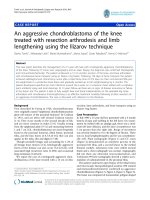
All 22 patients completed particle radiotherapy without
break. A typical treatment plan is illustrated in Fig. 1.
rhabdomyosarcoma
1 (4.5)
desmoplastic small round cell tumor
1 (4.5)
Survival outcomes
With a median follow-up of 20.25 (range 3.8–38.8) months,
all 22 patients were alive. One patient who had R2 resection
followed-by cisplatin chemotherapy for T3N0M0 rhabdomyosarcoma of the lacrimal sac developed local recurrence
at 17.8 months after CIRT. Another with R2 resected
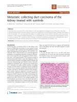
T4N0M0 ACC of the lacrimal gland developed regional recurrence after PRT + CIRT boost at 11.5 months. The 2-year
local-progression-free and regional-recurrence-free survival
(LPFS and RRFS) rates were 92.9 and 93.3%, respectively
(Fig. 2a & b). Four patients with lacrimal gland malignancies
(2 with ACC and 2 with adenocarcinoma, 2 with T2 and 2
with T4 disease) developed distant metastasis (DM) at a median time of 9.47 months (range 8.13–20.8). The 2-year
DM-free survival (DMFS) rate was 72.8%, and the 2-year
progression free survival (PFS) rate was 57.9% (Fig. 2c & d)
for the entire cohort. None of the 13 patients with lacrimal
gland malignancy developed local failure or progression (i.e.,
LPFS = 100%). The 2-year RRFS, DMFS and PFS were 88.9
and 53.0% and 39.3% for patients with lacrimal gland malignancies, respectively.
alveolar soft part sarcoma
1 (4.5)
chondrosarcoma
1 (4.5)
T category
T1
2 (9.1)
T2
7 (31.8)
T3
4 (18.2)
T4
9 (40.9)
Tumor status
Primary
21 (95.5)
Recurrence
1 (4.5)
Surgical margin
R0
3 (13.6)
R1
6 (27.3)
R2 or biopsy
13 (59.1)
Interval from surgery to radiotherapy, mo
Median (range)
2.2 (1.2–6.13)
Radiotherapy technique
PRT
1 (4.5)
CIRT
18 (81.8)
PRT + CIRT
3 (13.6)
Radiotherapy dose (Gy BED)
Median (range)
85.05 (67.2–94.5)
GTV (ml)
Median (range)
16.0 (1.9–67.6)
CTV (ml)
Median (range)
43.4 (18.8–209.9)
Concurrent chemotherapy or immunotherapy
Cisplatin
2 (9.1)
Interferon α-2b
1 (4.5)
No
19 (86.4)
Hu et al. BMC Cancer
(2019) 19:776
Page 4 of 10
Fig. 1 Axial (a) and coronal (b) views of a post eye sparing surgery CT scan of a patient with left lacrimal gland ACC. Axial (c) and coronal (d)
views of a typical intensity-modulated carbon ion radiotherapy treatment plan
Acute and chronic toxicities
The characteristics of acute and late toxicities are summarized in Table 2. Nine patients (40.9%) experienced grade 1
or 2 acute toxicities induced by particle radiotherapy. No
acute toxicity of grade 3 or above was observed. Seven patients (31.8%) experienced late toxicities of various grades
including 3 with grade 1 dry eyes, 1 with grade 1 brain injury, 1 with grade 2 retinopathy, 1 with grade 3 visual impairment and 1 with blindness in the affected eye (grade 4).
During the follow-up period, no vision impairment was observed, except for 2 patients developed grade 3 and grade 4
visual acuity reduction after CIRT. One of them experienced ipsilateral vision acuity reduction from normal to 20/
200–40/200 at 6 months accompanied by optic atrophy diagnosed by MRI; the other patient developed blindness at
3 months without changes on MR scan. One patient who
has affected eyeball fixation due to twice eye-sparing surgery and photon-based radiotherapy prior to re-irradiation
by CIRT, and no ocular movement disorder was observed
in remaining patients who received particle radiotherapy. In
addition, there was no eye injury in contralateral side in all
patients at present.
Prognostic factors
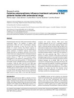
Univariate analyses using log-rank test indicated tumors
with mesenchymal origins had a trend toward a worse
LPFS (p = 0.056) (Table 3, Fig. 3a). In addition, tumors of
lacrimal gland had a trend with worse DMFS (p = 0.072)
(Table 3, Fig. 3b). When BED was taken as a continuous
variable using Cox regression analysis, higher BED had a
trend to associate with improved DMFS (hazard ratio,
0.884; 95% CI, 0.776–1.007 [P = 0.064]) (Table 4). However,
margin status, T-classification, volume of GTV or CTV did
not associate with PFS, LPFS, RRFS or DMFS in both logrank test and Cox regression analysis (Tables 3 & 4).
Multivariate analysis using Cox regression using tumor
correlation factors such as histological type, BED (continuous variable), T-classification, volume of GTV and
CTV suggested a significant relationship for higher BED
with improved PFS (hazard ratio, 0.732; 95% CI, 0.557–
0.960 [P = 0.024]) and a trend with improved DMFS
(hazard ratio, 0.717; 95% CI, 0.512–1.004 [P = 0.053]).
Furthermore, larger CTV field may improve PFS (hazard
ratio, 0.952; 95% CI, 0.906–1.001 [P = 0.054]) (Table 5).
Discussion
We analyzed 22 patients with orbital tumor after ESS
followed by PRT and/or CIRT. With a median follow-up of
20.3 months, the 2-year OS, PFS, LPFS, RRFS, and DMFS
rates were 100, 57.9, 92.9, 93.3, and 72.8%, respectively. No
acute severe (i.e., ≥grade 3) toxicity was observed. The occurrences of severe late toxicities were also infrequent.
These findings suggest that particle radiotherapy after ESS
Hu et al. BMC Cancer
(2019) 19:776
Page 5 of 10
Fig. 2 Local progression-free survival (LPFS) (a), regional recurrence-free survival (b), distant metastasis free-survival (DMFS) (c), and progressionfree survival (PFS) (d) rate curves of the entire cohort
could provide satisfactory local control with acceptable toxicities at 2 years for patients with orbit tumors. However,
DM remains a challenge for overall disease control.
Due to the complexity of the anatomy, ocular exenteration was advocated histologically; however, disease control remained suboptimal. In a report of 39 patients with
orbital malignancies received exenteration with (10 patients) or without (29 patients) adjuvant radiation, ~ 20%
experienced local recurrence after a median follow-up of
34.7 weeks. The 3-year OS and recurrence/death-free survival rates were 50.5 and 47.5%, respectively [20]. Results
from multiple retrospective studies revealed that 5-year
LPFS ranged at 20–22% after surgery (exenteration or eye
spearing) without adjuvant radiation [21, 22]. Adjuvant
radiotherapy following exenteration produced substantially improved local control as compared to surgery
alone. Three- or 5-year LPFS rates of 60~65% [21, 23],
with similar OS rates of 60% have been reported. In a
more recently published series, adjuvant IMRT after exenteration produced a 3-year LPFS and OS rates of 91 and
70%, respectively [24].
ESS provides an important opportunity for function preservation for patients with orbital tumors. Disease control
and survival rates after less aggressive (i.e., eye-sparing) surgery assimilates those from exenteration when adjuvant
radiotherapy was added. In 11 lacrimal gland tumor patients treated with ESS, only 1 patient declined adjuvant
radiotherapy and developed local recurrence [25]. A more
recently published series of 37 patients with lacrimal gland
carcinoma (> 80% with T1 or T2 diseases) reported a 2-year
recurrence free survival of approximately 95%. Of the 31
patients received adjuvant radiotherapy, 12 had PRT [22].
Although the composition of patients and pathologies in
our series differ substantially from the above-mentioned
Hu et al. BMC Cancer
(2019) 19:776
Page 6 of 10
Table 2 Characteristics of toxicities
papers, the outcomes remain encouraging. All 22 patients
were alive at the time of analysis with a 2-year LPFS rate of
92.9%, although ~ 60% of patients in our series presented
with T3/T4 diseases. Furthermore, patients with lacrimal
gland tumors achieved 2-year LPFS and RRFS rates of 100
and 88.9%, respectively, although ~ 40% had T4 disease.
Our results mimicked the most favorable 2-year outcome
in terms of survival and local control despite of a less favorable clinical presentation [21, 24, 26, 27].
With effective locoregional control, DM became the
most common mode of failure in patients with orbital
malignancies. DM rate of 27.5% and 3-year DMFS of
70% were reported for orbital carcinomas [23]. However,
DM is more challenging for lacrimal glade cancer especially adenoid cystic carcinoma [21, 23]. Skinner et al.
[21] reported a 5-year DMFS rate of 65% for lacrimal
gland carcinoma patients, and found that DM was not
correlated with histopathology, surgical margin or type
of surgery (eye-spearing vs. exenteration). These results
were in line with our finds, of which DM was seen in
30.8% of lacrimal gland patients. Furthermore, the 2-year
DMFS of our entire cohort and those with lacrimal
gland malignancies were 71.6 and 53%, respectively. Our
univariate analysis indicated a trend for DM in patients
with lacrimal gland malignancies (P = 0.072). In both the
univariate and multivariate analysis, factors such as
histopathology, surgical margin status, or T-classification
were not associated with DMFS. However, BED may
have significant impact on DMFS: The higher the BED,
the lower the risk of DM. Moreover, the overall PFS,
largely related to DMFS, is not only significantly associated with BED but also may correlated with CTV volume (P = 0.053). These findings suggested that particle
therapy may have a significant impact on disease control
due to its improved conformality secondary to its physical characteristics. Well localized and more precise dose
Table 3 Univariate analysis by the log-rank test
Characteristics
PFS
LPFS
RRFS
DMFS
Gender
0.910
0.527
0.480
0.362
Age (<46.5 y vs. >46.5 y)
0.442
0.527
0.480
0.847
Tumor site (other vs. lacrimal gland)
0.118
0.248
0.414
0.072
Origin (mesenchymal vs. epithelial histology)
0.653
0.056
0.617
0.254
T classification (T1/2 vs. T3/4)
0.524
0.386
0.414
0.784
Surgical margin (R0 + R1 vs. R2)
0.887
0.317
0.350
0.221
BED (<85.05 GyE vs. ≥85.05 GyE)
0.813
0.602
0.617
0.806
GTV (< 16 ml vs.>16 ml)
0.928
0.527
0.157
0.666
CTV (< 43.4 ml vs.>43.4 ml)
0.431
0.317
0.285
0.327
Hu et al. BMC Cancer
(2019) 19:776
Page 7 of 10
Fig. 3 Local progression-free survival (a), Distant metastasis-free survival (b) curves showing malignancies of mesenchymal origin had a trend
toward a worse LPFS and a trend that lacrimal gland tumor had worse DMFS
distribution enables higher dose as well as bigger CTV
with OAR sparing, a feature that is important in particle
radiotherapy for most head and neck cancers [10, 11].
Furthermore, CIRT not only provide advantages in dose
distribution, but also biologically due to its higher linear
energy transfer (LET). The value of the RBE of carbonion is 2–5:1 as compared with photons and protons,
which is highly relevant for radio-resistant tumors such
as STS [14, 15]. Our data suggested mesenchymal malignancies of the orbit may pose a higher risk of local recurrence (p = 0.056). And our previous experience with
CIRT for head and neck sarcomas revealed favorable disease control with acceptable toxicity profile [28].
In spite of the improved function preservation and locoregional disease control with ESS and adjuvant IMRT, radiation-induced toxicities remained a challenge in the
management of orbital tumors due to its anatomical complexity [29, 30]. Published data indicated that with doses
exceeding 50Gy, conjunctival keratinization, lacrimal gland
atrophy and fibrosis, corneal decompensation would occur.
When doses exceeded 60Gy, symblepharon, keratoconjunctivitis, permanent dry eyes became a concern.
Table 4 Univariate analysis of DMFS and PFS by Cox regression
DMFS
Variables
PFS
P
HR (95% CI)
P
HR (95% CI)
Male
Ref
Ref
Ref
Ref
Female
0.378
2.431 (0.338–17.48)
0.910
1.103 (0.202–6.036)
Age, y
0.186
1.046 (0.979–1.118)
0.752
1.001 (0.958–1.061)
Epithelium
Ref
Ref
Ref
Ref
Mesenchyme
0.488
0.031 (0.001–560.47)
0.656
0.613 (0.071–5.280)
T1 + T2
Ref
Ref
Ref
Ref
T3 + T4
0.784
0.760 (0.107–5.415)
0.529
1.731 (0.313–9.564)
R0 + R1
Ref
Ref
Ref
Ref
R2
0.254
0.268 (0.028–2.579)
0.887
0.890 (0.178–4.450)
BED
0.064
0.884 (0.776–1.007)
0.100
0.898 (0.791–1.021)
GTV, ml
0.948
1.002 (0.948–1.059)
0.928
0.998 (0.953–1.045)
CTV, ml
0.978
1.000 (0.980–1.021)
0.797
0.998 (0.980–1.016)
Gender
Histology
T classification
Surgical margin
Hu et al. BMC Cancer
(2019) 19:776
Page 8 of 10
Table 5 Multivariate analysis of DMFS and PFS by Cox regression
DMFS
Variables
PFS
P
HR (95% CI)
P
HR (95% CI)
T1 + T2
Ref
Ref
Ref
Ref
T3 + T4
0.368
6.673 (0.107–415.609)
0.189
6.28 (0.406–97.134)
R0 + R1
Ref
Ref
Ref
Ref
R2
0.233
0.042 (0–7.646)
0.969
1.059 (0.059–18.20)
Epithelium
Ref
Ref
Ref
Ref
Mesenchyme
0.976
0
0.384
0.255 (0.012–5.548)
BED
0.053
0.717 (0.512–1.004)
0.023
0.726 (0.551–0.957)
GTV, ml
0.079
1.199 (0.979–1.469)
0.104
1.117 (0.977–1.001)
CTV, ml
0.095
0.946 (0.887–1.010)
0.053
0.953 (0.907–1.278)
T classification
Surgical margin
Histology
Moreover, the probability of radiation-induced optic neuropathy may occur in 7–20% of patients [6, 17, 31, 32]. Particle radiotherapy provides distinctive advantages for
tumors close to critical OARs such as orbital malignancies
due to its distinctive physical characteristics. Patterns of
treatment-induced adverse effects were studied extensively
in a series of 20 orbital tumor patients treated with ESS
followed by PRT at M.D. Anderson Cancer Center [7]. Although disease control was not the focus of the study, the
authors reported one patient with local and another with
regional recurrence. In addition to the 35% patients who
experienced grade 3 acute dermatitis, 30% experienced
grade 3 chronic toxicities of epiphora and eyelid function
disorder. In addition, grade 2, 3, and 4 visual decrease were
observed in 2, 2, and 1 patient, respectively. The risk of severe chronic toxicity was higher when the maximum corneal dose exceeded 36Gy (BED) [7]. Higher BED was found
to associate with improved outcome in our series, indicating the advantage of particle radiotherapy for this condition
which usually occur close to dose limiting OARs. CIRT
with less penumbra as compared to proton may provide
additional physical advantage. In addition, favorable outcomes in terms of radiation-induced toxicities were observed in our series: Only grade 1/2 acute adverse-effects
were observed in 9 patients (40.9%). Approximately 22.7%
of patients developed grade 1 or 2 late effects. Nevertheless,
2 patients experienced severe decrease of vision at 3 and 6
months after CIRT. In both cases the tumors were attached
or close to the optic nerve or eye.
Several pitfalls of our study need to be discussed. First,
because of variations in certain institutional clinical trial
regimens, few patients were treated with PRT (1 cases)
or PRT + CIRT boost (3 cases) although most patients
received CIRT alone. Our analysis largely reflected the
results after CIRT for orbital malignancies. Second,
orbital tumor includes a group of heterogenous conditions from various origins with substantial different biological behaviors. Combining different pathologies would
inevitably affect the uniformity of the results. Third,
owing to the limited follow up time, these clinical results
must be considered with caution, we will continue to
follow up these patients and report clinical outcomes
with longer period. Fourth, our study suffered from the
nature of retrospective studies with a relatively small
sample size; nevertheless, we provided the outcomes of
the largest series of orbital tumors treated with particle
radiotherapy in terms of disease, control, survival, and
safety. Considering the rarity of the condition, nearly all
published literatures were retrospective in nature from
single institutions. Clearly, prospective investigations to
compare efficacies from different treatment modalities
or technologies are difficult to initiate without international collaboration among specialized academic
centers.
Conclusion
Adjuvant particle radiotherapy following ESS provided a
satisfactory OS and locoregional control at 2 years. DM
remained a major form of treatment failure. No severe
acute treatment-induced toxicity was observed, and severe late toxicities was observed in < 10% of cases. Longterm follow-up is needed to confirm the efficacy and
safety of adjuvant particle radiotherapy, in particular
CIRT, for orbital tumor after ESS.
Abbreviations
ACC: Adenoid cystic carcinoma; BED: Biological equivalent dose;
CBC: Complete blood count; CIRT: Carbon-ion radiotherapy; CR: Complete
response; CT: Computed tomography; CTCAE: Common terminology criteria
for adverse events; CTV: Clinical target volume; CTV-G: Post-surgical GTV;
DMFS: Distant metastasis free survival; ESS: Eye sparing surgery; GTV: Gross
tumor volume; GyE: Gy-equivalents; H&P: History and physical examination;
Hu et al. BMC Cancer
(2019) 19:776
IMPT: Intensity-modulated particle therapy; IMRT: Intensity-modulated
radiotherapy; IRB: Institutional review board; LET: Linear energy transfer;
LPFS: Local progression-free survival; MRI: Magnetic resonance imaging;
OARs: Organs at risk; OS: Overall survival; PBS: Pencil beam scanning; PETCT: Positron emission tomography/computed tomography; PFS: Progression
free survival; PRT: Proton radiotherapy; PTV: Planning target volume;
RBE: Relative biological effectiveness; RRFS: Regional recurrence-free survival;
SPHIC: Shanghai Proton and Heavy Ion Center; STS: Soft-tissue sarcoma
Page 9 of 10
4.
5.
6.
7.
Acknowledgements
Not applicable.
8.
Authors’ contributions
WH: Design of the study, data acquisition, statistical analysis, results interpretation,
drafting and revising the article, final approval of the version, responsible for all
aspects of the work. JH: data acquisition, data analysis, drafting and revising the
article, version approval, responsible for all aspects of the work. JG: data acquisition,
drafting and revising the article. JY: data acquisition, revising the article, final approval
of the version, responsible for all of the work. XQ: data acquisition, revising the
article, version approval, responsible for all aspects of the work. LK: Conceptualization
and design of the of study, data analysis and interpretation, drafting and revising the
article, funding acquisition, final approval of the version, responsible for all aspects of
the work. JJL: Conceptualization and design of the study, data analysis, interpretation
of the results, drafting and revising the article. All authors approved the submitted
version, agreement to be accountable for all aspects of the work.
Funding
This work was supported by Shanghai Municipal Commission of Health and
Family Planning (Project No. 20164Y0155), Pudong New Area Science and
Technology Development Foundation (Project No. PKJ2016-Y41). The funders
had no role in design of the study, in the data collection, analysis, or interpretation of data, in the decision to publish, or in the writing of the
manuscript.
Availability of data and materials
The datasets used and/or analyzed during the current study are available
from the corresponding author on reasonable request.
Ethics approval and consent to participate
This retrospective study was approved by the Ethics Committee of Shanghai
Proton and Heavy Ion Center. Written informed consent was obtained for
each participant before enrolling in this study.
9.
10.
11.
12.
13.
14.
15.
16.
17.
18.
Consent for publication
Written informed consent for publication was obtained for each patient
before enrolling in this study.
19.
Competing interests
The authors declare that they have no competing interests.
20.
Author details
1
Department of Radiation Oncology, Shanghai Proton and Heavy Ion Center,
Shanghai, China. 2Shanghai Engineering Research Center of Proton and
Heavy Ion Radiation Therapy, 4365 Kangxin Road, Pudong, Shanghai 201321,
China. 3Department of Radiation Oncology, Shanghai Proton and Heavy Ion
Center, Fudan University Shanghai Cancer Center, Shanghai, China.
21.
Received: 18 February 2019 Accepted: 22 July 2019
23.
References
1. Hassan WM, Bakry MS, Hassan HM, Alfaar AS. Incidence of orbital, conjunctival
and lacrimal gland malignant tumors in USA from surveillance, epidemiology
and end results, 1973-2009. Int J Ophthalmol. 2016;9(12):1808–13.
2. Mallen-St Clair J, Arshi A, Tajudeen B, Abemayor E, St John M. Epidemiology
and treatment of lacrimal gland tumors: a population-based cohort analysis.
JAMA Otolaryngol Head Neck Surg. 2014;140(12):1110–6.
3. Rosenthal DI, Chambers MS, Fuller CD, Rebueno NC, Garcia J, Kies MS,
Morrison WH, Ang KK, Garden AS. Beam path toxicities to non-target
structures during intensity-modulated radiation therapy for head and neck
cancer. Int J Radiat Oncol Biol Phys. 2008;72(3):747–55.
24.
22.
25.
26.
Krishna Y, Coupland SE. Lacrimal sac tumors--a review. Asia Pac J
Ophthalmol (Phila). 2017;6(2):173–8.
Sakaida H, Kobayashi M, Yuta A, Imanishi Y, Majima Y. Squamous cell
carcinoma of the nasolacrimal duct. Eur Arch Otorhinolaryngol. 2009;266(3):
455–8.
Karcioglu ZA, Hadjistilianou D, Rozans M, DeFrancesco S. Orbital
rhabdomyosarcoma. Cancer Control. 2004;11(5):328–33.
Holliday EB, Esmaeli B, Pinckard J, Garden AS, Rosenthal DI, Morrison WH,
Kies MS, Gunn GB, Fuller CD, Phan J, et al. A multidisciplinary orbit-sparing
treatment approach that includes proton therapy for epithelial tumors of
the orbit and ocular adnexa. Int J Radiat Oncol Biol Phys. 2016;95(1):344–52.
Khan FM: The physics of radiation therapy, fourth edn: Lippincott Williams &
Wilkins; 2009.
van de Water TA, Bijl HP, Schilstra C, Pijls-Johannesma M, Langendijk JA. The
potential benefit of radiotherapy with protons in head and neck cancer
with respect to normal tissue sparing: a systematic review of literature.
Oncologist. 2011;16(3):366–77.
Frank SJ, Cox JD, Gillin M, Rosenthal DI, Garden AS, Ang KK, Mohan R, Palmer
MB, Amin M, Zhu XR. Intensity modulated proton therapy for head-and-neck
cancer: the first clinical experience. Int J Radiat Oncol Biol Phys. 2012;84:S475–6.
Frank SJ, Cox JD, Gillin M, Mohan R, Garden AS, Rosenthal DI, Gunn GB,
Weber RS, Kies MS, Lewin JS, et al. Multifield optimization intensity
modulated proton therapy for head and neck tumors: a translation to
practice. Int J Radiat Oncol Biol Phys. 2014;89(4):846–53.
Tsujii H, Kamada T, Baba M, Tsuji H, Kato H, Kato S, Yamada S, Yasuda S,
Yanagi T, Kato H, et al. Clinical advantages of carbon-ion radiotherapy. New
J Phys. 2008;10:075009.
Kanai T, Endo M, Minohara S, Miyahara N, Koyama-ito H, Tomura H,
Matsufuji N, Futami Y, Fukumura A, Hiraoka T, et al. Biophysical
characteristics of HIMAC clinical irradiation system for heavy-ion radiation
therapy. Int J Radiat Oncol Biol Phys. 1999;44(1):201–10.
Elsasser T, Kramer M, Scholz M. Accuracy of the local effect model for the
prediction of biologic effects of carbon ion beams in vitro and in vivo. Int J
Radiat Oncol Biol Phys. 2008;71(3):866–72.
Jones B. A simpler energy transfer efficiency model to predict relative
biological effect for protons and heavier ions. Front Oncol. 2015;5:184.
Lu JD, Ye M, Guo Q, Fu JF, Moyers XM, Zhang ST, Mao NY, Kong YS, Hsi X,
Shahnazi XY, et al. The preliminary report of a registration clinical trial of
proton and heavy ion irradiation. Zhonghua Zhong Liu Za Zhi. 2018;40(1):52–6.
Emami B, Lyman J, Brown A, Coia L, Goitein M, Munzenrider JE, Shank B,
Solin LJ, Wesson M. Tolerance of normal tissue to therapeutic irradiation. Int
J Radiat Oncol Biol Phys. 1991;21(1):109–22.
Koto M. Skull base and upper cervical spine tumors. In: Tsujii H, Kamada T,
Shirai T, et al., editors. In Carbon-Ion Radiotherapy Principles, Practices, and
Treatment Planning. Heidelberg: Springer; 2014. p. 155–61.
Nieder C, Milas L, Ang KK. Tissue tolerance to reirradiation. Semin Radiat
Oncol. 2000;10(3):200–9.
Aryasit O, Preechawai P, Hirunpat C, Horatanaruang O, Singha P. Factors
related to survival outcomes following orbital exenteration: a retrospective,
comparative, case series. BMC Ophthalmol. 2018;18(1):186.
Skinner HD, Garden AS, Rosenthal DI, Ang KK, Morrison WH, Esmaeli B,
Pinnix CC, Frank SJ. Outcomes of malignant tumors of the lacrimal
apparatus: the University of Texas MD Anderson Cancer Center experience.
Cancer. 2011;117(12):2801–10.
Woo KI, Sagiv O, Han J, Frank SJ, Kim YD, Esmaeli B. Eye-preserving surgery
followed by adjuvant radiotherapy for lacrimal gland carcinoma: outcomes
in 37 patients. Ophthalmic Plast Reconstr Surg. 2018;34(6):570–4.
Esmaeli B, Ahmadi MA, Youssef A, Diba R, Amato M, Myers JN, Kies M, ElNaggar A. Outcomes in patients with adenoid cystic carcinoma of the
lacrimal gland. Ophthalmic Plast Reconstr Surg. 2004;20(1):22–6.
Tao R, Ma D, Takiar V, Frank SJ, Fuller CD, Gunn GB, Beadle BM,
Morrison WH, Rosenthal DI, Edson MA, et al. Orbital carcinomas treated
with adjuvant intensity-modulated radiation therapy. Head Neck. 2016;
38(Suppl 1):E580–7.
Esmaeli B, Yin VT, Hanna EY, Kies MS, William WN Jr, Bell D, Frank SJ. Eyesparing multidisciplinary approach for the management of lacrimal gland
carcinoma. Head Neck. 2016;38(8):1258–62.
Ahmad SM, Esmaeli B, Williams M, Nguyen J, Fay A, Woog J, Selvadurai D,
Rootman J, Weis E, Selva D, et al. American joint committee on Cancer
classification predicts outcome of patients with lacrimal gland adenoid
cystic carcinoma. Ophthalmology. 2009;116(6):1210–5.
Hu et al. BMC Cancer
(2019) 19:776
27. Woo KI, Yeom A, Esmaeli B. Management of Lacrimal Gland Carcinoma:
lessons from the literature in the past 40 years. Ophthalmic Plast Reconstr
Surg. 2016;32(1):1–10.
28. Yang J, Gao J, Wu X, Hu J, Hu W, Kong L, Lu JJ. Salvage carbon ion radiation
therapy for locally recurrent or radiation-induced second primary sarcoma
of the head and neck. J Cancer. 2018;9(12):2215–23.
29. Jeganathan VS, Wirth A, MacManus MP. Ocular risks from orbital and
periorbital radiation therapy: a critical review. Int J Radiat Oncol Biol Phys.
2011;79(3):650–9.
30. Petsuksiri J, Frank SJ, Garden AS, Ang KK, Morrison WH, Chao KS, Rosenthal
DI, Schwartz DL, Ahamad A, Esmaeli B. Outcomes after radiotherapy for
squamous cell carcinoma of the eyelid. Cancer. 2008;112(1):111–8.
31. Gordon KB, Char DH, Sagerman RH. Late effects of radiation on the eye and
ocular adnexa. Int J Radiat Oncol Biol Phys. 1995;31(5):1123–39.
32. Batth SS, Sreeraman R, Dienes E, Beckett LA, Daly ME, Cui J, Mathai M, Purdy
JA, Chen AM. Clinical-dosimetric relationship between lacrimal gland dose
and ocular toxicity after intensity-modulated radiotherapy for sinonasal
tumours. Br J Radiol. 2013;86(1032):20130459.
Publisher’s Note
Springer Nature remains neutral with regard to jurisdictional claims in
published maps and institutional affiliations.
Page 10 of 10