Effects of adjunct testosterone on cardiac morphology and function in advanced cancers: An ancillary analysis of a randomized controlled trial
Bạn đang xem bản rút gọn của tài liệu. Xem và tải ngay bản đầy đủ của tài liệu tại đây (856.49 KB, 9 trang )
Scott et al. BMC Cancer
(2019) 19:778
/>
RESEARCH ARTICLE
Open Access
Effects of adjunct testosterone on cardiac
morphology and function in advanced
cancers: an ancillary analysis of a
randomized controlled trial
Jessica M. Scott1, E. Lichar Dillon2, Michael Kinsky3, Albert Chamberlain2, Susan McCammon4, Daniel Jupiter5,
Maurice Willis2, Sandra Hatch6, Gwyn Richardson7, Christopher Danesi2, Kathleen Randolph2,8, William Durham2,
Traver Wright2,8, Randall Urban2 and Melinda Sheffield-Moore2,8*
Abstract
Background: Adjunct testosterone therapy improves lean body mass, quality of life, and physical activity in patients
with advanced cancers; however, the effects of testosterone on cardiac morphology and function are unknown.
Accordingly, as an ancillary analysis of a randomized, placebo-controlled trial investigating the efficacy of
testosterone supplementation on body composition in men and women with advanced cancers, we explored
whether testosterone supplementation could prevent or reverse left ventricular (LV) atrophy and dysfunction.
Methods: Men and women recently diagnosed with late stage (≥IIB) or recurrent head and neck or cervical cancer
who were scheduled to receive standard of care chemotherapy or concurrent chemoradiation were administered
an adjunct 7 week treatment of weekly intramuscular injections of either 100 mg testosterone (T, n = 1 M/5F) or
placebo (P, n = 6 M/4F) in a double-blinded randomized fashion. LV morphology (wall thickness), systolic function
(ejection fraction, EF), diastolic function (E/A; E’/E), arterial elastance (Ea), end-systolic elastance (Ees), and ventriculararterial coupling (Ea/Ees) were assessed.
Results: No significant differences were observed in LV posterior wall thickness in placebo (pre: 1.10 ± 0.1 cm; post:
1.16 ± 0.2 cm; p = 0.11) or testosterone groups (pre: 0.99 ± 0.1 cm; post: 1.14 ± 0.20 cm; p = 0.22). Compared with
placebo, testosterone significantly improved LVEF (placebo: − 1.8 ± 4.3%; testosterone: + 6.2 ± 4.3%; p < 0.05), Ea
(placebo: 0.0 ± 0.2 mmHg/mL; testosterone: − 0.3 ± 0.2 mmHg/mL; p < 0.05), and Ea/Ees (placebo: 0.0 ± 0.1;
testosterone: − 0.2 ± 0.1; p < 0.05).
Conclusions: In patients with advanced cancers, testosterone was associated with favorable changes in left
ventricular systolic function, arterial elastance, and ventricular-arterial coupling. Given the small sample size, the
promising multisystem benefits of testosterone warrants further evaluation in a definitive randomized trial.
Trial registration: This study was prospectively registered on ClinicalTrials.gov (NCT00878995; date of registration:
April 9, 2009).
Keywords: Testosterone, Cardiac function, Cachexia
* Correspondence:
2
Department of Internal Medicine, The University of Texas Medical Branch,
Galveston, TX, USA
8
Department of Health and Kinesiology, Texas A&M University, 155 Ireland
St., College Station, TX TX 77845, USA
Full list of author information is available at the end of the article
© The Author(s). 2019 Open Access This article is distributed under the terms of the Creative Commons Attribution 4.0
International License ( which permits unrestricted use, distribution, and
reproduction in any medium, provided you give appropriate credit to the original author(s) and the source, provide a link to
the Creative Commons license, and indicate if changes were made. The Creative Commons Public Domain Dedication waiver
( applies to the data made available in this article, unless otherwise stated.
Scott et al. BMC Cancer
(2019) 19:778
Background
Cancer cachexia is a complex, multifactorial syndrome
characterized by a progressive loss of skeletal muscle
mass with or without loss of fat mass that cannot be
fully reversed by conventional nutritional support [1].
Cachexia occurs in 50 to 80% of advanced cancer
patients and is associated with decreased mobility [2],
reduced response to chemotherapy [3], and is estimated
to directly account for more than 20% of cancer-related
deaths [2]. There are no established therapies for cancer
cachexia; accordingly, identification and testing of effective interventions are of major clinical importance in this
at-risk population.
Cancer cachexia involves not only the loss of skeletal
muscle, but also results in pathologic alterations within
the heart [4, 5]. The first report linking tumor burden and
cardiac atrophy was first published in 1904 [6], and was
extensively outlined using autopsies by Hellerstein and
Santlago-Stevenson in 1950 [7]. More recent preclinical
findings indicate that cardiac muscle loss occurs to a similar degree as in skeletal muscles, with concomitant impairment in systolic and diastolic function [8, 9]. Collectively,
the global nature of cachexia portends the requirement
for multifactorial treatment strategies with the capacity to
augment or reverse whole-organism atrophy.
Testosterone therapy has been used in patients
exposed to atrophic stimuli [10] to increase muscle
strength and bone mineral density [11, 12]. The heart is
also a target organ for steroids; there are receptors with
a high affinity for testosterone in cardiomyocytes [13],
suggesting that testosterone supplementation may also
improve cardiac morphology and function. In support, a
meta-analysis of randomized placebo-controlled studies
found that testosterone administered to patients with
chronic heart failure reduced systemic vascular resistance and increased both cardiac output and overall exercise capacity [14]. However, whether there are similar
salutary cardiovascular effects of testosterone in patients
with advanced cancers is not known. Accordingly, as an
ancillary analysis of a randomized, placebo controlled
trial investigating the efficacy of testosterone supplementation on body composition in men and women with advanced cancers [15], we explored whether testosterone
supplementation could prevent or reverse left ventricular
(LV) atrophy and dysfunction.
Methods
Patients and study design
Details of the design, rationale, and primary results of
study have been published elsewhere [15]. This is an ancillary analysis of a RCT (NCT00878995) among men
and women with histologically-confirmed advanced or
recurrent squamous cell carcinoma of the cervix (stages
IIB, IIIA, and IIIB) or head and neck squamous cell
Page 2 of 9
carcinoma (stage III or IV) conducted at the University
of Texas Medical Branch at Galveston, TX. Major eligibility criteria were: [1] loss of at least 5% of body mass
over the past 12 months, [2] Eastern Cooperative Oncology Group score of 0 or 1, [3] score of > 23 points on
the 30 point Mini Mental State Examination. All study
procedures were reviewed and approved by the institutional review board. Participation in both intervention
groups continued for a maximum of 7 weeks or until unacceptable toxicity or withdrawal of consent, whichever
came first. Patients were randomly allocated in blocks of
three to receive weekly injections of either 100 mg of
testosterone enanthate (n = 10) or placebo (n = 14).
Interventions were matched in terms of setting (clinicbased), and length (7 weeks). All outcomes were evaluated at pre-randomization (study treatments were initiated ≤14 days) and were repeated within ≤7 days of the
final treatment session at postintervention (month 3).
Intervention
A testosterone replacement paradigm commonly used to
treat hypogonadal men was chosen to include weekly
intramuscular injections of either 100 mg testosterone
enanthate or placebo (sterile saline) over a period of 7
weeks. Testosterone and placebo injections were given
by a nurse using an opaque syringe to obscure visual
differences between testosterone and placebo.
Cardiac structure and function
Patients underwent two-dimensional transthoracic and
pulsed Doppler imaging by use of a commercial ultrasound system (iE33, Phillips Healthcare). Images were
obtained by one experienced sonographer in the long
axis, short axis, and apical 4 chamber views according to
the American Society of Echocardiography guidelines
[16] to determine LV wall thickness, end-diastolic
volume (EDV), end-systolic volume (ESV), and LVEF.
LV volumes were calculated using the biplane Simpson
method. Pulsed Doppler recordings were employed to
assess diastolic filling; in particular, early (E) and atrial
(A) peak mitral inflow velocities were measured and the
ratio of early to late diastolic filling velocity (E:A) was
calculated. Tissue Doppler data were used to assess
mitral annular velocity (E’). The ratio of E/E’ was also
used to assess diastolic function. Images were analyzed
off-line by experienced technicians blinded to group
allocation. A minimum of three consecutive cardiac
cycles were measured and averaged.
End-systolic pressure (ESP) was calculated as 0.9 ×
brachial systolic blood pressure, a noninvasive estimate
that accurately predicts LV pressure-volume loop measurements of ESP [17]. End-systolic elastance (Ees) was
calculated as Ees = ESP/ESV, effective arterial elastance
(Ea) was calculated as Ea = ESP/SV, and ventricular-
Scott et al. BMC Cancer
(2019) 19:778
vascular coupling was determined as Ea/Ees [18].
Systemic vascular resistance (SVR) was calculated as
mean arterial pressure/CI × 80.
Statistical analysis
Repeated-measures ANOVA was initially used to compare means between groups. Because of the small sample
size and large amount of variability in the data, nonparametric tests were carried out at each level of intensity
and at each time of measurement. Comparisons among
groups were performed using the Kruskal-Wallis test.
When differences were determined to be significant,
pairwise comparisons were made using the MannWhitney method. The association between baseline
cardiac morphology and function and change with testosterone was explored with Pearson correlation coefficient. Values are means ± SD; significance level was set
at 0.05.
Page 3 of 9
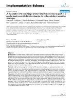
LV morphology, resting heart rate, and blood pressure
No significant differences were observed in LV posterior
wall thickness in placebo (pre: 1.10 ± 0.1 cm; post: 1.16 ±
0.2 cm; p = 0.11) or testosterone group (pre: 0.99 ± 0.1
cm; post: 1.14 ± 0.20 cm; p = 0.22); Fig. 1. No differences
between groups in change in resting heart rate (placebo:
+ 3 ± 11 bpm; testosterone: + 6 ± 11 bpm; p = 0.39) or
mean arterial pressure (placebo: + 3 ± 12.1 mmHg; testosterone: − 5 ± 12.1 mmHg; p = 0.28) were observed.
There was no significant correlation between baseline
values and change in LV morphology (r = 0.48).
LV volumes, systolic, and diastolic function
Men and women recently diagnosed with late stage (IIB
or higher) or recurrent head and neck or cervical cancer
who were scheduled to receive standard of care chemotherapy or chemoradiotherapy were recruited to participate. A total of 28 potentially eligible patients were
contacted for the study, and 24 (86%) were randomly
grouped and administered an adjunct 7 weeks regimen
of weekly intramuscular injections of either 100 mg
testosterone or placebo. Of these, 16 (67%) completed
cardiac assessments (testosterone, n = 1 M/5F; placebo,
n = 6 M/4F). No significant differences were found in
the baseline characteristics between placebo and testosterone groups (Table 1).
No differences in end diastolic volume (EDV) or end systolic volume (ESV) were observed in the placebo (EDV,
pre: 118.9 ± 16.3 mL, post: 119.3 ± 16.5 mL; p = 0.95; ESV,
pre: 46.9 ± 13.3 mL, post: 49.2 ± 8.2 mL; p = 0.62) or testosterone group, (EDV, pre: 109.5 ± 16.3 mL, post: 116.0 ±
16.5 mL; p = 0.16; ESV, pre: 46.2 ± 13.3 mL, post: 41.2 ± 8.2
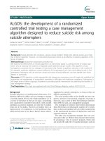
mL; p = 0.18). There was a significant difference in change
in stroke volume between the placebo (− 1.9 ± 5.3 mL) and
testosterone (+ 11.5 ± 5.3 mL) groups (Fig. 2a). There was a
significant difference in change in LV ejection fraction
(LVEF) between the placebo (− 1.8 ± 4.3%) and testosterone
(6.2 ± 4.3%) groups (p = 0.02) (Fig. 2b). There was a
significant negative association between baseline and
change in LV ejection fraction in the testosterone group
(r = 0.95; p < 0.05). Diastolic function assessed by E/A (placebo pre: 1.1 ± 0.3 cm/s; post: 1.3 ± 0.4 cm/s; p = 0.35; testosterone pre: 1.1 ± 0.3 cm/s; post: 1.0 ± 0.4 cm/s; p = 0.63)
and E/E’ (placebo pre: 6.0 ± 2.0; post: 5.7 ± 1.6; p = 0.75; testosterone pre: 7.7 ± 2.0; post: 5.7 ± 1.6; p = 0.63) (Fig. 2c)
was preserved in both groups. Absolute changes in volumes, systolic, and diastolic function are presented in
Additional file 1.
Testosterone supplementation
Ventricular-vascular coupling
Pre-study average total serum testosterone levels were
significantly different between males and females (328 ±
420 ng/dL and 17 ± 14 ng/dL respectively, p < 0.001).
Testosterone levels in females in the placebo group were
unchanged from pre- (16 ± 9 ng/dL) to post-intervention
(23 ± 24 ng/dL; p = 0.40) whereas testosterone levels
were increased in the testosterone group (pre: 19 ± 17
ng/dL; post: 644 ± 327 ng/dL; p = 0.01). Testosterone
levels in males in the placebo group decreased from
354 ± 193 ng/dL to 342 ± 174 ng/dL (p = 0.80). Only one
male was randomized into the testosterone group; serum
testosterone level increased from 177 to 885 ng/dL.
Estrogen values remained below 62 pg/mL for all
subjects and there were no changes in response to testosterone treatment.
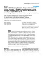
End-systolic elastance (Ees) was unchanged in both
groups (placebo pre: 2.4 ± 0.7 mmHg/mL; post: 2.4 ±
0.5 mmHg/mL; p = 0.79; testosterone pre: 2.4 ± 0.7;
post: 2.4 ± 0.5; p = 0.85). There was a significant difference between groups in change in systemic vascular
resistance (SVR, placebo: 45.7 ± 166.9 dynes/sec/cm5;
testosterone: − 359.3 ± 166.9 dynes/sec/cm5; Fig. 3a),
effective arterial elastance (Ea, placebo: 0.0 ± 0.2
mmHg/mL; testosterone: − 0.3 ± 0.2 mmHg/mL; Fig.
3b), and ventricular-vascular coupling (Ea/Ees, placebo: 0.0 ± 0.1; testosterone: − 0.2 ± 0.1; Fig. 3c). No
significant associations were observed between
baseline and change in ventricular-vascular coupling.
Absolute changes in ventricular-vascular coupling are
presented in Additional file 1.
Results
Patient characteristics
Scott et al. BMC Cancer
(2019) 19:778
Page 4 of 9
Table 1 Demographic and Treatment Characteristics of the Participants
Characteristic
All Patients
(n = 16)
Placebo
(n = 10)
Testosterone
(n = 6)
P = value
Time (mos) from diagnosis to enrollment – mean (SD)
3.1 (3.2)
2.9 (3.4)
3.6 (3.1)
0.684
Age (yrs) – mean (SD)
50.9 (9.5)
48.4 (10.9)
55.0 (5.1)
0.189
BMI (kg/m2) – mean (SD)
22.1 (6.8)
23.9 (7.3)
19.3 (5.2)
0.200
9.0 (8.0)
9.9 (9.6)
7.1 (3.6)
Exercise behavior (activity score) – mean (SD)
a
Race – no. (%)
0.588
0.330
Non-Hispanic white
11 (69)
6 (60)
5 (83)
Other group
5 (31)
4 (40)
1 (20)
Male
7 (44)
6 (60)
1 (17)
Female
9 (56)
4 (40)
5 (83)
n = 16
n = 10
n=6
Never
4 (25)
3 (30)
1 (17)
Former
7 (44)
3 (330)
4 (67)
Current
5 (31)
4 (40)
1 (17)
n = 15
n =9
n =6
IIB
1 (7)
0 (0)
1 (17)
III
0 (0)
0 (0)
0 (0)
IIIB
4 (27)
3 (33)
1 (17)
IV
0 (0)
0 (0)
0 (0)
IVA
8 (53)
5 (56)
3 (50)
IVB
2 (13)
1 (11)
1 (17)
Cervical
6 (38)
4 (40)
2 (33)
Head/neck
10 (62)
6 (60)
4 (37)
6 (38)
3 (30)
3 (50)
0.986
11 (69)
6 (60)
5 (83)
0.985
Radiotherapy
13 (81)
9 (90)
4 (67)
Other Therapy
0 (0)
0 (0)
0 (0)
Sex – no. (%)
Smoking – no. (%)
Disease stage – no. (%)
0.091
Cancer Type – no. (%)
PEG feeding tube – no. (%)
0.355
0.852
1.000
Current Therapy – no. (%)
Chemotherapy
Prior therapy – no. (%)
n = 16
n = 10
n =6
Surgery
2 (13)
2 (20)
0 (0)
Chemotherapy
0 (0)
0 (0)
0 (0)
Radiotherapy
0 (0)
0 (0)
0 (0)
0 (0)
0 (0)
0 (0)
n = 16
n = 10
n =6
Other Therapy
Current Medications – no. (%)
Beta-blocker
0 (0)
0 (0)
0 (0)
ACE inhibitor
1 (6)
1 (10)
0 (0)
ARB
1 (6)
1 (10)
0 (0)
Diuretic
1 (6)
1 (10)
0 (0)
Calcium channel blocker
1 (6)
1 (10)
0 (0)
Aspirin
3 (19)
2 (20)
1 (17)
Statin
2 (13)
2 (20)
0 (0)
n = 16
n = 10
n =6
Pre-existing conditions – no. (%)
0.927
0.728
0.586
Scott et al. BMC Cancer
(2019) 19:778
Page 5 of 9
Table 1 Demographic and Treatment Characteristics of the Participants (Continued)
Characteristic
Peripheral vascular disease
All Patients
(n = 16)
Placebo
(n = 10)
Testosterone
(n = 6)
2 (13)
1 (10)
1 (17)
Coronary artery disease
1 (6)
1 (10)
0 (0)
Osteoporosis
1 (6)
0 (0)
1 (17)
Arrhythmia
0 (0)
0 (0)
0 (0)
Arthritis
0 (0)
0 (0)
0 (0)
Type II diabetes
2 (13)
2 (20)
0 (0)
Hyperlipidemia
2 (13)
2 (20)
0 (0)
Hypertension
1 (6)
1 (10)
0 (0)
P = value
Abbreviations: SD standard deviation, BMI body mass index, ACE angiotensin converting enzyme, ARB angiotensin II receptor blockers. aExercise behavior sum of
mild, moderate, and strenuous exercise obtained from ActiGraph 3 axis accelerometry monitors available in a subset of patients (n = 8 placebo; n = 4
testosterone). No significant differences between the groups. P-values provided are from t-tests when group means were compared or chi-square tests when
comparing frequency of cases between the groups
Discussion
This is the first randomized trial to explore the potential
efficacy of testosterone to augment / reverse cardiac
morphology and function in patients with advanced cancers. The major new findings of this study were that
compared with placebo, testosterone improved LV systolic function, as well as ventricular-vascular coupling.
This may have important health implications for patients
with cachexia given that this entity has no established
evidence-based interventions that improve outcomes.
Changes in cardiac morphology and function may stem
from the cancer itself and/or the cardiotoxic effects of
cancer therapies [19]. For instance, Springer et al. [8] reported extensive loss of cardiomyocyte volume and replacement with fibrotic tissue among patients who died of
pancreatic, lung, and colorectal cancer; however, a subset
of patients with significant cancer-related weight loss and
cachexia had reduced LV wall thickness and mass compared with cancer patients without cachexia. A reduction
in LV mass following anthracycline-based chemotherapy
Fig. 1 Percent change in left ventricular posterior wall thickness
from pre to post-intervention in placebo (red) and
testosterone (blue)
has also consistently been reported [20, 21] and is associated with major adverse cardiac events (cardiovascular
death, appropriate implantable cardioverter-defibrillator
therapy, or admission for decompensated HF) [21]. Of
note, average BMI of included patients was ~ 27 kg/m2,
and whether patients with cachexia were included was not
reported [20, 21]. The present study confirms and extends
previous reports by including patients with advanced cancers, none of whom had been previously treated with
cytotoxic therapy or radiotherapy. Collectively, these
findings indicate that cardiac alterations in patients with
advanced cancers is part of a complex, systemic issue that
results in widespread muscle wasting. Accordingly,
intervention strategies with multifactorial effects will be
required to reverse whole-organism atrophy.
At least 19 studies have assessed the efficacy of
pharmacological agents in clinical trials to manage cancer cachexia [22]; however, few have explored the potential salutary effects on cardiac morphology and function.
Testosterone therapy has been used in patients exposed
to atrophic stimuli [10] to increase muscle strength and
bone mineral density [11], and we previously reported
that in patients with advanced cancer adjunct testosterone improved lean body mass and was associated with
increased quality of life, and physical activity compared
with placebo [15]. Previous findings from non-oncology
settings indicate that exogenous testosterone may also
directly induce physiological cardiac myocyte hypertrophy [23]. For instance, among men with type 1 diabetes, higher total testosterone was associated with
higher LV mass and volume [24], and Subramanya and
colleagues [25] recently reported that after a median of
9.1 years, higher free testosterone levels were independently associated with an increase in LV mass in women
and men in the Multiethnic Study of Atherosclerosis. In
RCTs, testosterone treatment improved cardiac biomarkers in patients with type II diabetes [26], and reduced systemic vascular resistance and increased both
Scott et al. BMC Cancer
(2019) 19:778
a
b
Page 6 of 9
a
b
c
c
Fig. 2 Percent change in stroke volume (a) left ventricular ejection
fraction (b), and E/E’ (c) from pre to post-intervention in placebo
(red) and testosterone (blue)
cardiac output and overall exercise capacity in heart failure patients [14]. Similar findings were observed here in
patients with advanced cancers; compared with placebo,
testosterone improved indices of LV function. In
addition, patients with the lowest LV ejection fraction at
baseline experienced the greatest improvement with
Fig. 3 Percent change in systemic vascular resistance (a), arterial
elastance (b), and ventricular-vascular coupling (c) from pre to postintervention in placebo (red) and testosterone (blue)
testosterone, suggesting that testosterone may be an important intervention for patients with poor LV ejection
fraction. Nevertheless, these findings should be interpreted with caution given the small sample size.
Collectively, these findings indicate that testosterone
supplementation may be an effective intervention to improve cardiac function; however, larger trials are needed
Scott et al. BMC Cancer
(2019) 19:778
to address whether testosterone is fully protective
against cardiac atrophic remodeling in patients with
advanced cancers.
The mechanisms underlying testosterone-induced cardioprotection are not fully known; however, may involve
both cardiac and vascular systems. Cardiomyocytes contain receptors with a high affinity for testosterone [13]
and in vitro studies of nonhuman cardiac myocytes
found that testosterone can decrease action potential
duration (thereby altering repolarization) and peak
shortening times [27]. Testosterone is also an acute
vasodilator [28] and lowers blood pressure [29]. Thus,
understanding how the heart and systemic vasculature
function independently as well as how they interact
(termed ventricular-arterial coupling) is important when
evaluating global cardiovascular function [17]. In the
present study we found that testosterone had beneficial
effects on vascular parameters (e.g., Ea, SVR), which in
turn, improved ventricular-vascular coupling compared
to placebo-treated patients. Future studies evaluating the
mechanistic underpinnings of the effects of testosterone
on cardiac and peripheral vasculature in the cachectic
setting are needed.
In current clinical practice, the discipline of cardiooncology traditionally focuses on the detection and management of cancer treatment-induced reductions in
cardiac function (i.e., LVEF), and/or development of
overt heart failure [30–32] and coronary artery disease
[33]. Intriguingly, based on conventional metrics, all patients in the current study have ‘normal’ cardiac function
(e.g., LVEF > 55%). Nevertheless, there is burgeoning
interest in detection of early and subclinical therapyrelated cardiac consequences, including changes in cardiac size and ventricular-vascular coupling. Furthermore,
techniques such as assessing the heart during exercise
has provided novel prognostic information beyond traditional resting cardiac measures in patients with breast
cancer [34]. Collectively, these findings indicate that
evaluating cardiac morphology and function in the cachectic setting, as well as evaluating other metrics such as
cardiorespiratory fitness and cardiac function during exercise will be important in the design of future intervention
trials. Given the systemic effects of cachexia, evaluation of
multimodal approaches including nutritional support,
pharmacological intervention, and exercise training will be
important for this high-risk population.
A number of study limitations should be considered.
First, the trial was designed to assess the effect of testosterone treatment on lean body mass, and changes in cardiac parameters were not predefined outcome measures.
Second, our sample size was small. Trials with larger
samples sizes are needed to definitively assess the efficacy of testosterone on cardiac morphology and function
in advanced cancers. Third, our subject population was
Page 7 of 9
predominantly female, and although androgens stimulate
skeletal muscle protein synthesis similarly between men
and women [35], potential sex differences in cardiac
androgen receptor density [36] and the mechanisms of
response to testosterone treatment may limit the
generalizability of our findings. For instance, following
exercise training the development of LV hypertrophy
and increase in cardiorespiratory fitness in females was
markedly blunted compared with males [37]; whether
females have blunted response to testosterone compared
to males should be addressed in future studies. Finally,
to fully characterize the physiological importance of
atrophic remodeling and potential efficacy of testosterone supplementation, there is a need to move beyond
the study of global measures of LV function at rest. For
example, reduced strain and strain rate revealed impaired myocardial function prior to LVEF decline [38] in
cancer patients treated with anthracycline-containing
therapy. Thus, evaluation of cardiac and vascular function with advanced imaging techniques at rest [39], as
well as responses to a peak cardiopulmonary exercise
test [40], may provide important insight into characterizing the ‘cachectic heart’.
Conclusions
In patients with advanced cancers, testosterone was
associated with favorable changes in left ventricular systolic function, arterial elastance, and ventricular-arterial
coupling. There are promising multisystem benefits of
testosterone; however, given the small sample size in the
current study, further evaluation in a larger randomized
trial is warranted.
Additional file
Additional file 1: Absolute change in cardiac outcomes. (PDF 42 kb)
Author contributions
Conceptualization, MSM; methodology, MSM, MK, RJU, WJD, TJW; formal
analysis, JMS, ELD, AC, DJ; investigation, ELD, MK, CPD, KMR, MSM, WJD, TJW;
resources, MK, SMC, MW, SH, GR, RJU, MSM; data curation, ELD, KMR;
writing—original draft preparation, JMS, ELD, AC, MSM; writing—review and
editing, all authors; funding acquisition, MSM. All authors read and approved
the manuscript.
Availability of data and materials
The datasets used and/or analysed during the current study are available
from the corresponding author on reasonable request.
Ethics approval and consent to participate
This study was conducted in accordance with the principles of the
Declaration of Helsinki and approved by the Institutional Review Board at
the University of Texas Medical Branch. Written informed consent was
obtained from all patients prior to participation.
Consent for publication
Not applicable.
Scott et al. BMC Cancer
(2019) 19:778
Page 8 of 9
Competing interests
The authors declare that they have no competing interests.
17.
Author details
1
Department of Medicine, Memorial Sloan Kettering Cancer Center, New
York, NY, USA. 2Department of Internal Medicine, The University of Texas
Medical Branch, Galveston, TX, USA. 3Department of Anesthesiology, The
University of Texas Medical Branch, Galveston, TX, USA. 4Department of
Otolaryngology, The University of Texas Medical Branch, Galveston, TX, USA.
5
Department of Preventive Medicine and Community Health, The University
of Texas Medical Branch, Galveston, TX, USA. 6Department of Radiation
Oncology, The University of Texas Medical Branch, Galveston, TX, USA.
7
Department of Gynecologic Oncology, The University of Texas Medical
Branch, Galveston, TX, USA. 8Department of Health and Kinesiology, Texas
A&M University, 155 Ireland St., College Station, TX TX 77845, USA.
18.
19.
20.
21.
Received: 15 May 2019 Accepted: 31 July 2019
22.
References
1. Fearon K, Strasser F, Anker SD, Bosaeus I, Bruera E, Fainsinger RL, et al.
Definition and classification of cancer cachexia: an international consensus.
Lancet Oncol. 2011;12(5):489–95.
2. von Haehling S, Anker SD. Prevalence, incidence and clinical impact of
cachexia: facts and numbers-update 2014. J Cachexia Sarcopenia Muscle.
2014;5(4):261–3.
3. Antoun S, Birdsell L, Sawyer MB, Venner P, Escudier B, Baracos VE.
Association of skeletal muscle wasting with treatment with sorafenib in
patients with advanced renal cell carcinoma: results from a placebocontrolled study. J Clin Oncol. 2010;28(6):1054–60.
4. Kazemi-Bajestani SM, Becher H, Fassbender K, Chu Q, Baracos VE.
Concurrent evolution of cancer cachexia and heart failure: bilateral effects
exist. J Cachexia Sarcopenia Muscle. 2014;5(2):95–104.
5. Murphy KT. The pathogenesis and treatment of cardiac atrophy in cancer
cachexia. Am J Physiol Heart Circ Physiol. 2016;310(4):H466–77.
6. Gordon W. The cardiac Dulness in cases of Cancer. Med Chir Trans. 1904;87:
327–37.
7. Hellerstein HK, Santiago-Stevenson D. Atrophy of the heart; a correlative
study of 85 proved cases. Circulation. 1950;1(1):93–126, illust.
8. Springer J, Tschirner A, Haghikia A, von Haehling S, Lal H, Grzesiak A, et al.
Prevention of liver cancer cachexia-induced cardiac wasting and heart
failure. Eur Heart J. 2014;35(14):932–41.
9. Tian M, Asp ML, Nishijima Y, Belury MA. Evidence for cardiac atrophic
remodeling in cancer-induced cachexia in mice. Int J Oncol. 2011;39(5):
1321–6.
10. Emmelot-Vonk MH, Verhaar HJ, Nakhai Pour HR, Aleman A, Lock TM, Bosch
JL, et al. Effect of testosterone supplementation on functional mobility,
cognition, and other parameters in older men: a randomized controlled
trial. JAMA. 2008;299(1):39–52.
11. Borst SE, Yarrow JF, Conover CF, Nseyo U, Meuleman JR, Lipinska JA, et al.
Musculoskeletal and prostate effects of combined testosterone and
finasteride administration in older hypogonadal men: a randomized,
controlled trial. Am J Physiol Endocrinol Metab. 2014;306(4):E433–42.
12. Sheffield-Moore M, Dillon EL, Casperson SL, Gilkison CR, Paddon-Jones D,
Durham WJ, et al. A randomized pilot study of monthly cycled testosterone
replacement or continuous testosterone replacement versus placebo in
older men. J Clin Endocrinol Metab. 2011;96(11):E1831–7.
13. Kinson GA, Layberry RA, Hebert B. Influences of anabolic androgens on
cardiac growth and metabolism in the rat. Can J Physiol Pharmacol. 1991;
69(11):1698–704.
14. Toma M, McAlister FA, Coglianese EE, Vidi V, Vasaiwala S, Bakal JA, et al.
Testosterone supplementation in heart failure: a meta-analysis. Circ Heart
Fail. 2012;5(3):315–21.
15. Wright TJ, Dillon EL, Durham WJ, Chamberlain A, Randolph KM, Danesi C, et
al. A randomized trial of adjunct testosterone for cancer-related muscle loss
in men and women. J Cachexia Sarcopenia Muscle. 2018;9(3):482–96.
16. Lang RM, Bierig M, Devereux RB, Flachskampf FA, Foster E, Pellikka PA, et al.
Recommendations for chamber quantification: a report from the American
Society of Echocardiography's guidelines and standards committee and the
chamber quantification writing group, developed in conjunction with the
23.
24.
25.
26.
27.
28.
29.
30.
31.
32.
33.
34.
35.
36.
37.
European Association of Echocardiography, a branch of the European
Society of Cardiology. J Am Soc Echocardiogr. 2005;18(12):1440–63.
Kelly RP, Ting CT, Yang TM, Liu CP, Maughan WL, Chang MS, et al. Effective
arterial elastance as index of arterial vascular load in humans. Circulation.
1992;86(2):513–21.
Scott JM, Esch BT, Haykowsky MJ, Warburton DE, Toma M, Jelani A, et al.
Cardiovascular responses to incremental and sustained submaximal exercise in
heart transplant recipients. Am J Physiol Heart Circ Physiol. 2009;296(2):H350–8.
Ewer MS, Ewer SM. Cardiotoxicity of anticancer treatments: what the
cardiologist needs to know. Nat Rev Cardiol. 2010;7(10):564–75.
Ferreira de Souza T, Quinaglia ACST, Osorio Costa F, Shah R, Neilan TG,
Velloso L, et al. Anthracycline therapy is associated with cardiomyocyte
atrophy and preclinical manifestations of heart disease. JACC Cardiovasc
Imaging. 2018;11(8):1045–55.
Neilan TG, Coelho-Filho OR, Pena-Herrera D, Shah RV, Jerosch-Herold M,
Francis SA, et al. Left ventricular mass in patients with a cardiomyopathy
after treatment with anthracyclines. Am J Cardiol. 2012;110(11):1679–86.
Advani SM, Advani PG, VonVille HM, Jafri SH. Pharmacological management
of cachexia in adult cancer patients: a systematic review of clinical trials.
BMC Cancer. 2018;18(1):1174.
Bell JR, Bernasochi GB, Varma U, Raaijmakers AJ, Delbridge LM. Sex and sex
hormones in cardiac stress--mechanistic insights. J Steroid Biochem Mol
Biol. 2013;137:124–35.
Kim C, Bebu I, Braffett B, Cleary PA, Arends V, Steffes M, et al. Testosterone
and cardiac mass and function in men with type 1 diabetes in the
epidemiology of diabetes interventions and complications study (EDIC). Clin
Endocrinol. 2016;84(5):693–9.
Subramanya V, Zhao D, Ouyang P, Ying W, Vaidya D, Ndumele CE, et al.
Association of endogenous sex hormone levels with coronary artery
calcium progression among post-menopausal women in the multi-ethnic
study of atherosclerosis (MESA). J Cardiovasc Comput Tomogr. 2018.
Gianatti EJ, Hoermann R, Lam Q, Dupuis P, Zajac JD, Grossmann M. Effect of
testosterone treatment on cardiac biomarkers in a randomized controlled
trial of men with type 2 diabetes. Clin Endocrinol. 2016;84(1):55–62.
Kimura N, Mizokami A, Oonuma T, Sasano H, Nagura H.
Immunocytochemical localization of androgen receptor with polyclonal
antibody in paraffin-embedded human tissues. J Histochem Cytochem.
1993;41(5):671–8.
Pugh PJ, Jones TH, Channer KS. Acute haemodynamic effects of testosterone
in men with chronic heart failure. Eur Heart J. 2003;24(10):909–15.
Anderson FH, Francis RM, Faulkner K. Androgen supplementation in
eugonadal men with osteoporosis-effects of 6 months of treatment on
bone mineral density and cardiovascular risk factors. Bone. 1996;18(2):171–7.
Zamorano JL, Lancellotti P, Rodriguez Munoz D, Aboyans V, Asteggiano R,
Galderisi M, et al. 2016 ESC position paper on cancer treatments and
cardiovascular toxicity developed under the auspices of the ESC Committee
for practice guidelines: the task force for cancer treatments and
cardiovascular toxicity of the European Society of Cardiology (ESC). Eur
Heart J. 2016;37(36):2768–801.
Hamo CE, Bloom MW, Cardinale D, Ky B, Nohria A, Baer L, et al. Cancer
therapy-related cardiac dysfunction and heart failure: part 2: prevention,
treatment, guidelines, and future directions. Circ Heart Fail. 2016;9(2):e002843.
Saiki H, Petersen IA, Scott CG, Bailey KR, Dunlay SM, Finley RR, et al. Risk of heart
failure with preserved ejection fraction in older women after contemporary
radiotherapy for breast Cancer. Circulation. 2017;135(15):1388–96.
Darby SC, Ewertz M, McGale P, Bennet AM, Blom-Goldman U, Bronnum D,
et al. Risk of ischemic heart disease in women after radiotherapy for breast
cancer. N Engl J Med. 2013;368(11):987–98.
Howden EJ, Bigaran A, Beaudry R, Fraser S, Selig S, Foulkes S, et al. Exercise
as a diagnostic and therapeutic tool for the prevention of cardiovascular
dysfunction in breast cancer patients. Eur J Prev Cardiol. 2019;26(3):305–15.
Sheffield-Moore M, Paddon-Jones D, Casperson SL, Gilkison C, Volpi E, Wolf
SE, et al. Androgen therapy induces muscle protein anabolism in older
women. J Clin Endocrinol Metab. 2006;91(10):3844–9.
McCrohon JA, Death AK, Nakhla S, Jessup W, Handelsman DJ, Stanley KK, et
al. Androgen receptor expression is greater in macrophages from male than
from female donors. A sex difference with implications for atherogenesis.
Circulation. 2000;101(3):224–6.
Howden EJ, Perhonen M, Peshock RM, et al. Females have a blunted
cardiovascular response to one year of intensive supervised endurance
training. J Appl Physiol (1985). 2015;119:37–46.
Scott et al. BMC Cancer
(2019) 19:778
38. Hare JL, Brown JK, Leano R, Jenkins C, Woodward N, Marwick TH. Use of
myocardial deformation imaging to detect preclinical myocardial
dysfunction before conventional measures in patients undergoing breast
cancer treatment with trastuzumab. Am Heart J. 2009;158(2):294–301.
39. Scott JM, Martin D, Ploutz-Snyder R, et al. Efficacy of Exercise and
Testosterone to Mitigate Atrophic Cardiovascular Remodeling. Med Sci
Sports Exerc. 2018;50:1940–49.
40. Scott JM, Zabor EC, Schwitzer E, Koelwyn GJ, Adams SC, Nilsen TS, et al.
Efficacy of Exercise Therapy on Cardiorespiratory Fitness in Patients With
Cancer: A Systematic Review and Meta-Analysis. J Clin Oncol. 2018:
JCO2017775809.
Publisher’s Note
Springer Nature remains neutral with regard to jurisdictional claims in
published maps and institutional affiliations.
Page 9 of 9