Cetuximab, irinotecan and fluorouracile in fiRst-line treatment of immunologicallyselected advanced colorectal cancer patients: The CIFRA study protocol
Bạn đang xem bản rút gọn của tài liệu. Xem và tải ngay bản đầy đủ của tài liệu tại đây (751.01 KB, 9 trang )
Ottaiano et al. BMC Cancer
(2019) 19:899
/>
STUDY PROTOCOL
Open Access
Cetuximab, irinotecan and fluorouracile in
fiRst-line treatment of immunologicallyselected advanced colorectal cancer
patients: the CIFRA study protocol
Alessandro Ottaiano1* , Stefania Scala2, Nicola Normanno3, Maria Napolitano2, Monica Capozzi4,
Anna Maria Rachiglio3, Cristin Roma3, Anna Maria Trotta2, Crescenzo D’Alterio2, Luigi Portella2, Carmela Romano4,
Antonino Cassata4, Rossana Casaretti4, Lucrezia Silvestro4, Anna Nappi4, Salvatore Tafuto4, Antonio Avallone4,
Alfonso De Stefano4, Mario Tamburini5, Carmine Picone6, Antonella Petrillo6, Francesco Izzo7, Raffaele Palaia7,
Vittorio Albino7, Alfonso Amore8, Andrea Belli9, Ugo Pace9, Massimiliano Di Marzo9, Paolo Chiodini10,
Gerardo Botti5, Gianfranco De Feo5, Paolo Delrio9 and Guglielmo Nasti1
Abstract
Background: Combination of chemotherapies (fluoropirimidines, oxaliplatin and irinotecan) with biologic drugs
(bevacizumab, panitumumab, cetuximab) have improved clinical responses and survival of metastatic colorectal cancer
(mCRC). However, patients’ selection thorough the identification of predictive factors still represent a challange.
Cetuximab (Erbitux®), a chimeric monoclonal antibody binding to the Epidermal Growth Factor Receptor (EGFR),
belongs to the Immunoglobulins (Ig) grade 1 subclass able to elicite both in vitro and in vivo the Antibody-Dependent
Cell-mediated Cytotoxicity (ADCC). ADCC is the cytotoxic killing of antibody-coated target cells by immunologic
effectors. The effector cells express a receptor for the Fc portion of these antibodies (FcγR); genetic polymorphisms of
FcγR modify the binding affinity with the Fc of IgG1. Interestingly, the high-affinity FcγRIIIa V/V is associated with
increased ADCC in vitro and in vivo. Thus, ADCC could partially account for cetuximab activity.
Methods/design: CIFRA is a single arm, open-label, phase II study assessing the activity of cetuximab in combination
with irinotecan and fluorouracile in FcγRIIIa V/V patients with KRAS, NRAS, BRAF wild type mCRC. The study is designed
with a two-stage Simon model based on a hypothetical higher response rate (+ 10%) of FcγRIIIa V/V patients as
compared to previous trials (about 60%) assuming ADCC as one of the possible mechanisms of cetuximab action. The
test power is 95%, the alpha value of the I-type error is 5%. With these assumptions the sample for passing the first
stage is 14 patients with > 6 responses and the final sample is 34 patients with > 18 responses to draw positive
conclusions. Secondary objectives include toxicity, responses’ duration, progression-free and overall survival.
Furthermore, an associated translational study will assess the patients’ cetuximab-mediated ADCC and characterize the
tumor microenvironment.
(Continued on next page)
* Correspondence:
1
Innovative Therapies for Abdominal Metastases Unit, Istituto Nazionale
Tumori di Napoli, IRCCS “G. Pascale”, via M. Semmola, 80131 Naples, Italy
Full list of author information is available at the end of the article
© The Author(s). 2019 Open Access This article is distributed under the terms of the Creative Commons Attribution 4.0
International License ( which permits unrestricted use, distribution, and
reproduction in any medium, provided you give appropriate credit to the original author(s) and the source, provide a link to
the Creative Commons license, and indicate if changes were made. The Creative Commons Public Domain Dedication waiver
( applies to the data made available in this article, unless otherwise stated.
Ottaiano et al. BMC Cancer
(2019) 19:899
Page 2 of 9
(Continued from previous page)
Discussion: The CIFRA study will determine whether ADCC contributes to cetuximab activity in mCRC patients
selected on an innovative immunological screening. Data from the translational study will support results’
interpretation as well as provide new insights in host-tumor interactions and cetuximab activity.
Trial registration: The CIFRA trial (version 0.0, June 21, 2018) has been registered into the NIH-US National Library of
Medicine, ClinicalTrials.gov database with the identifier number (NCT03874062).
Keywords: Colorectal Cancer, Antibody-dependent cell-mediated cytotoxicity, Cetuximab, Irinotecan, Fluorouracule,
FcγR, Phase II study
Background
Colorectal carcinoma is a highly incident neoplasm in
Western countries with more than 200,000 new cases diagnosed each year in Europe. About 30 % of patients
presents with a metastatic disease [1, 2]. In the last 10
years, significant progress has been made in the treatment of metastatic colorectal cancer (mCRC) due to the
the introduction of chemotherapy (CT) containing oxaliplatin and irinotecan. The addition of each of the two
chemotherapeutics to fluoropyrimidines has increased
the objective response rates and improved the overall
survival. In addition, the combination of polyCT and
new biological drugs (bevacizumab, cetuximab and panitumumab), has doubled the median survival of mCRC
patients [3].
Cetuximab (Erbitux®) is a chimeric monoclonal antibody
belonging to the Immunoglobulins (Ig) grade 1 subclass [4].
It blocks the binding of the endogenous ligands of EGFR
(Epidermal Growth Factor Receptor), thus inhibiting receptor function. EGFR-dependent signal transduction pathways
are involved in the control of proliferation, cell survival,
angiogenesis and cell migration [5]. Cetuximab binds to
EGFR with an affinity that is 5 to 10 times higher than that
of endogenous ligands. The drug is indicated for the treatment of patients with mCRC with non-mutated (wild-type)
RAS (RAt Sarcoma) oncogene both in combination with CT
and monotherapy in patients who have failed oxaliplatin and
irinotecan. The mutation of the RAS gene makes it constitutively activated and therefore not susceptible to EGFR inhibition. In mCRC, the incidence of mutations in the RAS gene
is between 30 and 50% [6]. Recent evidence shows that patients with non-mutated RAS mCRC have a significantly
greater chance of responding to cetuximab or a combination
of cetuximab and CT [7].
effector cells, particularly the Natural Killers (NK), express
receptors for the Fc portion of these antibodies (FcγR). The
binding affinity between FcγR and Fc portion of the immunoglobulins is critical for the target cell recognition and the
extent of the immunologic response [9, 10]. In the general
population, genetic polymorphisms of FcγR have been described to modify the binding affinity of the IgG1 Fc fragment [10]. The polymorphisms identified for FcγRIIa (or
CD32, predominantly expressed on macrophages) and
FcγRIIIa (or CD16, expressed on NK cells and macrophages) are histidine (H)/arginine (R) at position 131 and
valine (V)/phenylalanine (F) at position 158, respectively. In
recent years, in vitro [11, 12] and in vivo studies [13] have
demonstrated that cetuximab induces an NK cells-mediated ADCC against colon cancer cells independently of
KRAS (Kirsten RAt Sarcoma) status. Conversely, clinical
studies have shown that FcγRIIa-131H/H and FcγRIIIa158 V/V genotypes (simplified here as FcγRIIa H/H and
FcγRIIIa V/V) are associated with improved response and
efficacy in follicular lymphomas and metastatic breast carcinomas treated with rituximab and trastuzumab [14–16],
respectively. Contrasting results have been reported in
mCRC treated with cetuximab [17, 18]. The variability of
these results may depend on methodological issues such as
the absence of NRAS (Neuroblastoma-Rat Sarcoma) and
BRAF (B- Rapidly Accelerated Fibrosarcoma) oncogene
mutations’ assessment, populations’ heterogeneity or the
lack of characterization of the tumor microenvironment
(TM) with particular emphasis to a subset of CD163+
macrophages (M2 macrophages) producing an array of
anti-ADCC molecules (e.g. pro-angiogenic and immunosuppressive factors) [19]. We previously showed that the
FcγRIIIa V/V genotype (high affinity Fcγ receptor) correlates with better clinical response and improved PFS
(Progression Free Survival) in patients with mCRC treated
with cetuximab [20, 21], as described by Bibeau et al. [18].
Rationale for evaluating FcγR polymorphisms in mCRC
One of the mechanisms of action of cetuximab is the
stimulation of ADCC (Antibody-Dependent Cell-mediated
Cytotoxicity). ADCC is mediated by immunoglobulins that
bind to cellular targets and makes them sensitive to recognition and destruction by immunologic effectors (Natural
Killer, macrophages, myeloid-derived cells, etc.) [8]. The
Previous results of folfiri plus cetuximab in first-line
treatment of mCRC
Cetuximab is active and well tolerated in the first-line
treatment of mCRC in association with fluoropyrimidine and irinotecan as demonstrated in the randomized,
Ottaiano et al. BMC Cancer
(2019) 19:899
phase III, EMR 62202013 trial [22] in which the
combination of cetuximab and irinotecan plus 5-fluorouracile infusion/folinic acid (FU/AF) was compared to
CT only. The proportion of non-mutated KRAS patients was 64%. The response rate (RR: complete plus
partial responses/number of evaluable patients) was
46.9% (CI: 42.9–51.0) in the CT/cetuximab arm, significantly higher (p = 0.0038) than that of the CT arm
(38.7%; IC: 34.8–42.8). Odds ratios of subgroup analyses showed that, in the KRAS wt population, the RR
of the cetuximab arm was significantly higher (59.3% vs
43.2%, odds ratio 1.91, 95% CI: 1.24–2.93). Furthermore, the association was able to improve PFS with a
HR of 0.85 (IC: 0.726–0.998, p = 0.0479). The most
common grade 3–4 toxicities in the combination arm
were diarrhea (15.7% CT plus cetuximab vs 10.5% CT,
p = 0.008) and those related to cetuximab infusion
(2.5% CT plus cetuximab vs. 0% CT, p < 0.001). In the
phase III, FIRE-3 study, mCRC patients carrying KRAS
wt were randomized to folfiri plus cetuximab or folfiri
plus bevacizumab first-line treatment [23]. The RR in
the cetuximab group was 62.0% (95% CI: 56.2–67.5)
and the median PFS was 10.0 months (95% CI, 8.8–
10.8). Safety profile was not different from that previously described, with the most common grade 3–4 toxicities being haematologic (25%), skin reactions (26%),
and diarrhoea (11%). The proportion of patients achieving an objective response did not significantly differ between the two groups.
The current therapeutic context of mCRC and CIFRA
hypothesis
Recent meta-analyses [24, 25] have evidenced the role of
colon tumor side (rigt vs left) as a predictive factor for response to therapy [26]. Accordingly, the RAS wt tumors deriving from the left colon would be more responder to CT
in association with anti-EGFR drugs as compared to the
right-sided neoplasms. However, the treatment of the RAS
wild-type mCRC is currently based on the use of CT doublets (fluoropyrimidine and oxaliplatin or irinotecan) and
biological drugs (bevacizumab, panitumumab, cetuximab).
This concept is well expressed in the ESMO (European
School of Medical Oncology) guidelines [27] in relation to
the diversity of the initial treatment intent, suggesting the
use of CT doublets and anti-EGFR (panitumumab or cetuximab) when the main objective is a rapid tumor shrinkage.
The NCCN guidelines (National Comprehensive Cancer
Network v3.2018) contemplate the use of CT and antiEGFR, panitumumab or cetuximab, in first or second line
therapies in mCRC patients whose primary tumor is localized to the left colon. At present, however, due to a lack of
sequence studies with high cross-over rates between biologic drugs, it is not possible to state the best biological
drug to be used in first-line.
Page 3 of 9
Here we propose the administration of folfiri (fluorouracile and irinotecan) plus cetuximab in patients selected on the FcγRIIIa V/V genotype. In summary, the
CIFRA study is set on a hypothetical higher response
rate of FcγRIIIa V/V patients than those reported in
previous trials (about 60%) and assumes that ADCC is
one of the possible mechanisms of cetuximab action.
Furthermore, in this immunologically selected cohort,
the different responses associated to tumor side (right vs
left) could be non existent or attenuated. CIFRA study
results could contribute to ameliorate patients’ selection
and definitively address the ADCC and FcγR polymorphisms role in cetuximab activity.
Methods and design
CIFRA is a single arm, single center, open-label, phase II
study, assessing the activity of cetuximab in combination
with folfiri in FcγRIIIa V/V patients with KRAS, NRAS,
BRAF wild type mCRC. It will be conducted at the academic hospital Istituto Nazionale Tumori di Napoli,
IRCCS “G. Pascale” in Naples (Italy). The study includes
also biomarkers’ analysis (cetuximab-mediated ADCC,
TM characterization).
Objectives
The primary objective is to evaluate the role of the molecular and immunologic selection of patients on response to folfiri and cetuximab in patients with mCRC.
The response will be evaluated according to Response
Evaluation Criteria In Solid Tumors (RECIST) criteria,
version 1.1 [28].
Secondary objectives are the safety, the responses’ duration, the progression-free (PFS) and overall survival
(OS). Toxicity will be graded according to the Common
Terminology Criteria for Adverse Events (CTCAE) of
the National Cancer Institute, version 4.0, June 14, 2010.
Response duration will be measured from the time of
documented objective response (CR or PR) until documented tumor progression (see also “Response and toxicity assessment” section). PFS will be determined from
the data of treatment start untill progression (defined according to RECIST), OS untill death from any cause.
Tertiary and correlative objectives consist in exploratory
studies of biological markers as predictors of outcome
(see the “Tranlsational research” section).
Ethical considerations
All the procedures described in this protocol have been
designed according to the principles of the Good
Clinical Practice guidelines of the International Conference on Harmonization (ICH) and of the Declaration of
Helsinki. The study was approved by the Ethical Committee of the National Cancer Institute of Naples, Italy
(No. 60/18). All patients will provide a written informed
Ottaiano et al. BMC Cancer
(2019) 19:899
consent to CIFRA study clinicians before recruitment
and tissue and blood samples collection. In order to protect the privacy of patients included in the CIFRA study,
the Structure that has the responsibility for registration,
collection and management of personal data, will not
provide patient names to persons not involved in the
study, with the exception of the Ministry of Health or
Ethics Committees (as required by the current legislation
only for inspection and control purposes). After registration, a unique and progressive numerical code will be
assigned to the patients and shown in the header of all
electronic data collection systems and it will be used for
all communications regarding the patients. A list of patients’ codes will exist exclusively at the Secretariat of
the CIFRA study.
Study design and statistical analyses
Sample size is based on a two-stage study design by Simon
[29] with activity as primary end-point.
The null hypothesis of a not relevant response rate
(40%) will be tested against an alternative response rate
hypothesis of 70% with a one-tailed test. The test power
is 95%, the alpha value of the I-type error is 5%. With
these assumptions the sample for passing the first stage
is 14 patients with > 6 objective responses. The final
sample is 34 patients with > 18 objective responses to
draw positive conclusions.
The populations considered for the analysis are described as follows. Safety Population (SP) is defined as
all patients receiving at least one dose of treatment.
Incorrect treatment or anticipation of the end of
treatment are not reasons for exclusion from the SP.
Efficacy Evaluable (EE) population is represented by
patients who will receive at least one post-baseline assessment of the primary endpoint [28].
All patients who will receive at least one dose of
treatment will be included in the descriptive statistics.
The basic characteristics of the recruited patients will
be reported for the categorical variables as total number and percentage and for the continuous variables as
mean and standard deviation or median and interquartile range. Descriptive statistics of patients excluded from the SP and EE will also be carried out.
Summary statistics on treatment will be compiled including information on dose changes, interruptions,
non-compliance, reasons for protocol deviation, and
treatment duration. The analysis on safety will be carried out on the SP. All the information necessary to
identify the occurrence of adverse events will be used
in the analysis and summarized through descriptive
statistics. The confidence intervals will be calculated
at 95%. Time-to-outcome will be described by KaplanMeier curves.
Page 4 of 9
Eligibility criteria
The main inclusion criteria are: cytological or histological diagnosis of colorectal adenocarcinoma; KRAS,
NRAS, BRAF wild-type; FcγRIIIa V/V genotype; stage
IV; age < 75 years; at least 1 measurable lesion; ECOG
Performance Status 0 or 1; life expectancy > 3 months;
negative pregnancy test for all potentially childbearing
women; written informed consent. The main exclusion
criteria are as follows: previous systemic anti-tumor
treatment (allowed treatment with capecitabine or fluorouracil and radiotherapy in the neoadjuvant setting of
rectal tumors with therapy terminated at least 6 months
before); presence of primary non-treated stenosing colorectal neoplasm; neutrophils < 2000/mm3 or platelets <
100.000/mm3 or hemoglobin < 9 g/dl; serum creatinine
level > 1.5 times the maximum normal value; GOT and/
or GPT > 5 times the maximum normal value and/or
bilirubin level > 3 times the maximum normal value; previous malignant neoplasms (excluding basal or spinocellular cutaneous carcinoma or in situ carcinoma of the
uterine cervix); active or uncontrolled infections; other
concomitant uncontrolled diseases or conditions contraindicating the study drugs at clinician evaluation; presence of brain metastases; refusal or inability to provide
informed consent; impossibility to guarantee follow-up.
Therapeutic schedule
The therapeutic schedule [22, 23] is represented by cetuximab 400 mg/mq intravenously (iv) with a loading dose of
400 mg/mq at the first cycle followed by 250 mg/mq iv
weekly. Cetuximab will be diluted in 500 ml of saline solution and administered by iv infusion in 90 min. The administration of irinotecan will precede that of cetuximab and
will consist on a dose of 180 mg/mq diluted in 500 ml of
saline solution and administered iv in 60 min. Fluorouracil
(5-FU) will be administered at a dose of 400 mg/mq in slow
iv bolus at half of lederfolin 200 mg/mq (diluted in 250 ml
of saline solution) 2-h infusion. An elastomeric pump with
5-fu 2400 mg/mq in continuous 46 h-infusion will be
adminstered after lederfolin. The therapeutic scheme just
described is briefly indicated as folfiri/cetuximab. Only at
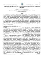
the first administration of CT, irinotecan will not be administered (Fig. 1). This is a peculiar and innovative characteristic of the CIFRA study. ADCC occurs rapidly at the
beginning of the therapy because it involves lympocytes
subpopulatons (i.e. NK) that do not require immunological
priming. Although immunomodulatory effects of irinotecan
are not described, it is reasonable and biologically plausible
that irinotecan-induced lymphopenia could interfere the
ADCC phenomena.
Atropine 0.25 mg sub cutis will be administered for
the prevention of acute cholinergic events before irinotecan infusion. Premedication with antihistamines at
Ottaiano et al. BMC Cancer
(2019) 19:899
Page 5 of 9
Fig. 1 Ideal timeline of CIFRA study
standard doses and dexamethasone 4 mg will be applied
before cetuximab infusion.
Duration of therapy
Administration of folfiri/cetuximab will be allowed until
progression or unacceptable toxicity. In case of unacceptable toxicity related to irinotecan the patients will
continue cetuximab and 5-FU until progression. In case
of unacceptable toxicity related to cetuximab the patient
will continue folfiri until progression. In case of unacceptable toxicity related to 5-FU the patients will continue with cetuximab and irinotecan until progression.
Dose modifications for folfiri
Dose reductions wil be applied in relation to the more severe side effect. Once modified, the dose of the drug will be
the same for the subsequent cycles. A 20% reduction of folfiri will be permitted in case of grade ≥ 3 of haematologic or
non-haematologic toxicities (except for alopecia). Folfiri will
be rechallenged after recovery of toxicity to baseline or
grade 1 or less. At the second appearance of grade ≥ 3 side
effects a dose reduction of 50% will be applied; after a further grade ≥ 3 toxicity or after the first appearance of
grade ≥ 2 cardio-vascular chemotherapy will be permanently discontinued. Chemotherapy will also be interrupted
in case of postponement of the administration for more
than 4 weeks. No prophylactic use of G-CSF o erythropoetin is planned.
Dose modifications for cetuximab
Cetuximab infusion will be delayed in case of grade > 3
cutaneous toxicity for 2 weeks. The investigator will
also consider the administration of topical and/or oral
antibiotic drugs and/or topical corticosteroids [30]. The
treatment will be reintroduced if the cutaneous toxicity
reduces to grade 2 or lower. If grade 3 skin toxicity
reoccurs at a second or third time, cetuximab will be
postponed for a further 2 weeks, but when reintroduced
the dose will be reduced to 200 mg/mq and 150 mg/mq,
respectively. Dose reductions of cetuximab are permanent and it will be permanently discontinued in case of 4
weeks suspension or fourth grade 3 toxicity event occurrence despite appropriate dose adjustments. Allergic
reactions may occur during or after administration of
cetuximab. In case of allergic reaction, the patient will
be treated according to clinical practice. The patient
should also be asked to report immediately to the doctor if any late reaction occurs. In case of grade 1 allergic reaction the cetuximab infusion rate will be reduced
at 50%; it will not be increased subsequently, but rather
reduced at any subsequent administration. In case of a
second allergic or hypersensitive reaction despite the
reduced infusion rate, the infusion will be interrupted
and the patient will continue with folfiri alone. If the
patient experiences an allergic or hypersensitive reaction of grade > 3, cetuximab will be permanently discontinued and chemotherapy will be continued if
considered appropriate by the investigator.
Timing of exams and procedures
Screening phase
The screening phase of the study will begin after signing
of the informed consent and it will consist on the evaluation of the inclusion and exclusion criteria and in the
Ottaiano et al. BMC Cancer
(2019) 19:899
collection of a venous blood sample (10 ml) for the determination of the FcγR polymorphisms. Baseline visit and
exams will be carried out within 21 days before therapy
start (clinical history, clinical examination, PS ECOG, vital
signs) along with the beyond described parameters. Blood
count and clinical biochemistry will be performed at our
local laboratories. The following variables will be evaluated: hemoglobin, blood count with leukocyte formula,
platelets, total bilirurbin, AST, ALT, alkaline phosphatase,
serum creatinine, total proteins, sodium, potassium, calcium, urea, lactic dehydrogenase, creatinine clearance,
CEA and CA19.9. ECG and cardiac ultrasonography with
evaluation of the ventricular ejection fraction will be also
performed. Pregnancy test will be done for all potentially
childbearing women and must be negative during the
screening phase. The test can also be repeated during
treatment if requested by the local Ethics Committee. All
patients, throughout the duration of the study, must use
barrier methods for anti-conception. Total-body Computed Tomography with iv contrast or, if contraindicated,
Magnetic Resonance (MRI) abdomen and chest computed
tomography without iv contrast will be obtained.
Treatment phase
Clinical examination, evaluation of vital signs, blood count
and clinical biochemistry will be performed at day 1 of
each cycle before administration of folfiri/cetuximab
(every 2 weeks). Cardiological evaluation, CEA, CA19.9
and assessment of response to treatment will be performed after 3 months of therapy start and thereafter
every 3 months until progression.
End-of-treatment phase
The suspension of the treatment can occur in case of
toxicity, progression, and/or at the request of the patient. Additional treatment lines or follow-up procedures will be applied at the discretion of the clinician
responsible of the medical treatment. The final visit will
be made immediately after disease progression. The
following data will be collected: ECOG Performance
Status, clinical examination, vital signs, blood count
and clinical biochemistry. The end of study visit should
be done within 30 days of the last administration of folfiri/cetuximab and before starting any second-line CT.
The cutaneous toxicity, if present, will be followed-up
until resolution; if second-line CT will be undertaken
the treatment will not start before its resolution. In any
case, the outcome of adverse events will be recorded
after the the end of study visit. The “patient death
form” must be completed when the patient dies. The
end-of-study form should be completed for patients
who die before the end of study visit and if the patient
withdraws consent, preferably before 28–32 days after
the last folfiri/cetuximab administration.
Page 6 of 9
Response and toxicity assessment
The assessment of response will be perfomed through
total-body computed tomography with iv contrast (if contraindicated: abdomen MRI and chest computed tomography without iv contrast), CEA and CA19.9 after 3
months of folfiri/cetuximab and every 3 months thereafter
until progression. Responses will be classified according to
the RECIST v. 1.1. An independent blinded central review
of radiologic examinations will be performed by a DMC
(Data Monitoring Committee). The independent DMC
will be also responsible of reviewing the PFS data. Toxicity
will be assessed in accordance with the CTCAE, version
4.0. The CTCAE contains 26 categories of adverse events,
organized for pathophysiology, anatomy and etiology.
Each adverse event is graded with a scale ranging from 0
(absence of event or value within the normal range) to 5
(death caused by the adverse event). Patients will be
assessed for the presence of adverse events at each study
visit and will also notify the investigator by telephone in
case of significant adverse events reported between one
visit and the next. For each adverse event, the maximum
grade per patient will be reported. If a patient experiences
a toxic effect of any grade on multiple occasions, the event
will be counted only once.
Translational research
Biomarkers will be evaluated in specimens and correlated
with outcomes and therapy effectiveness. In particular, the
primary tissues, and metastases when available, will be
characterized for the presence of tumor-infiltrating M2
macrophages by immunohistochemistry through the expression of CD163 (ab182422, Abcam), TGF-β (ab92486,
Abcam), Arginase-1 (GTX113131, Genetex), SSP1 Osteopontin antibody (ab218237, Abcam], and PD-L1 (E1L3N®,
XP®). M1 infiltrating macrophages will be detected through
the following antibodies: CD86 (ab53004, Abcam), iNOS
(ab115819, Abcam), IFN-γ (ab218426, Abcam), TNF
(ab1793, Abcam). Natural Killer (NK) and Cytotoxic T
Lymphocites (CTL) will be characterized as follows:
NKP46+ (Clone 195,314, R&D system), granzymeB
(ab134933, Abcam), Foxp3+ (ab20034, Abcam). For FcγR
polymorphisms detection, DNA will be extracted from the
whole blood lymphomonocyte component of each patient
(10 ml of venous blood) by Ficoll-Paque density gradient.
The analysis of the V158F and H131R alleles will involve a
gene amplification phase by PCR reaction with oligonucleotides specific for the regions of interest and a subsequent
PCR products analysis by automatic sequencing (Big Dye
terminators version 3.1 cycle sequencing kit and 3130
Genetic Analyzer; Applied Biosystems) [20, 21]. As an internal quality control, samples with previously known sequenced genotype will be used. These investigations will be
conducted in the laboratories of the “Cellular biology and
Ottaiano et al. BMC Cancer
(2019) 19:899
biotherapies Unit” of the National Cancer Institute of Naples, IRCCS “G. Pascale”.
To study cetuximab-mediated ADCC, peripheral
blood mononuclear cells (PBMC) from patients will be
isolated at diagnosis by Ficoll-Paque Plus gradient
centrifugation (GE Healthcare), then they will be cultured in complete RPMI-1640 medium enriched with
human IL2 (10 ng/mL) for 18 h in order to generate
Lymphokine-Activated Killer (LAK) cells. Target CRC
cells (HT29) will be plated in a 96-well plate at 1 × 10⁴
cells/well. Twenty-four hours later, LAK will be added
at a 10:1 effector:target (E:T) ratio in fresh medium, in
the presence of cetuximab (10 μg/mL), or the rituximab
(anti-CD20, 10 μg/mL), as negative control, or in the
presence of staphylococcal enterotoxin B (SEB) as positive control. Cytotoxicity will be evaluated by sulforhodamine B (SRB) assay [31]. ADCC elicited by not
activated freshly prepared PBMCs will be also evaluated
at E:T cell concentration ratios of 20:1 and 10:1. The
specific cytolysis percentage will be calculated using the
following formula: Cytotoxicity (%) = [1 − (mean test
optical density/mean optical density target)] × 100 [32,
33]. Cetuximab-mediated ADCC is given by cytotoxicitywith cetuximab − cytotoxicitywithout cetuximab. All experiments will be performed in triplicate, and results
expressed as mean values ± standard error. These investigations will be conducted in the laboratories of the
“Functional Genomics” of the National Cancer Institute
of Naples, IRCCS “G. Pascale”.
Discussion
The identification of predictive biomarkers characterizing
the ideal treatment of mCRC patients is subject of intense
investigation. RAS oncogenes in mCRC have not completely
satisfied this unmet need since about 40% of patients with
RAS wt mCRC dose not respond to anti-EGFR treatments.
We previously reported in a series of 74 KRAS wt mCRC
patients, genotypic frequencies of FcγRIIIa and FcγRIIa of
36% VV, 54% VF, 10% FF and 36% HH, 56% HR, 8% RR,
respectively; interestingly, FcγRIIIa but not FcγRIIa polymorphisms were significantly associated with response to
anti-EGFR-based therapy [20]. FcγRIIIa polymorphisms had
also significant prognostic value for PFS [FcγRIIIa: median
PFS 18.2 months in V/V patients (18 patients, 13 events) vs
17.3 months in V/F patients (26 patients, 25 events) vs 9.4
months in F/F patients (5 patients, 5 events); Log Rank test:
p = 0.04] at univariate and multivariate analyses (HR: 2.35;
CI: 1.37–4.01; p = 0.001) with grading and response to firstline chemotherapy. In another study, we described genotipic
frequencies of FcγRIIa and IIIa in 96 consecutive mCRC patients and 148 control subjects and we analyzed the clinical
impact of cetuximab-mediated ADCC in vitro from LAK
patients-derived [21]. There were no statistically significant
differences between the control and study groups in terms
Page 7 of 9
of genotypic frequencies. The incidence of FcγRIIIa V/V, V/
F and F/F were: 28, 47 and 23%, respectively. Interstingly,
the FcγRIIIa V/V and V/F genotypes were associated with
higher cetuximab-mediated ADCC compared with F/F,
median 27% (range: 0–76%) and 20% (0–57%) versus 9%
(0–36%), respectively (p = 0.001). The response rate was
higher in patients with the FcγRIIIa V allele (V/V and V/F
genotypes) compared with the F/F genotype (p = 0.025) as
well as the PFS of patients with FcγRIIIa V/V and V/F was
significantly longer than that of patients with F/F (10.8 vs.
5.1 months respectively, p = 0.05, Log Rank test). The extent
of in vitro cetuximab-mediated ADCC was significantly
correlated with cetuximab clinical response (30% in responders vs 8% in non-responders patients, p = 0.020;
ANOVA test). These observations prompted us to prospectively design the CIFRA trial, which is an investigator initiated phase II study to verify a superior activity of folfiri/
cetuximab of 70% (+ 10% of that previously described) in
mCRC patients selected on a genetic polymorphism of
FcγRIIIa. The risk of a possible unexpected detrimental effect in this selected population is reduced in CIFRA study
by the two-stage design that will stop the enrollment early if
the first condition of acceptable activity is not met.
Notably, the CIFRA study will also prospectively address
whether the ADCC phenomenon contributes to cetuximab
activity. The hypothesis of ADCC as additional or alternative anti-cancer mechanism is supported by two mirror observations: i) a small percent of KRAS mutated mCRC
patients respond to cetuximab [34, 35] and, ii) not all “all
RAS” wt mCRC patients respond to anti-EGFR therapy
[35]. Thus, blockade of signal transduction may not be the
only mechanism of action resulting in the clinical benefit of
cetuximab [36]. In fact, ADCC induced by EGFR-specific
mAbs may inhibit tumor progression in vivo, even in cancers resistant to EGFR signaling inhibition [37]. Immune
mechanisms besides molecular alterations, could contribute
to cetuximab activity as suggested also by indirect data of
Seo and colleagues [38] who reported a significant correlation between EGFR expression and ADCC activity, but
not with the mutational status of RAS and BRAF. To this
regard, CIFRA results could prompt a clinical pilot study to
test the activity of cetuximab-based therapy in RAS mutated FcγRIIIa V/V mCRC patients. Additionally, CIFRA
may suggest a more complex evaluation including FcγRIIIa
polymorphisms and prompt phase III studies in order to
compare panitumumab versus cetuximab with folfiri in
RAS wt/FcγRIIIa V/V mCRC patients.
Furthermore, TM characterization (i.e. NK cells, M1
vs M2 macrophages) and cetuximab-mediated ADCC
will help to understand any relation between specific signaling pathways and treatment response, as recently reported [39, 40]. Although the description of complex
TM biology or dynamics is beyond the scope of this article, herein TM will be evaluated with particular regard
Ottaiano et al. BMC Cancer
(2019) 19:899
to tumor-associated macrophages (TAM), the most represented tumor-infiltrating host cells (may represent up
to 50% of the tumor mass). TAM are classically distinct
in activated (M1) macrophages and alternatively activated (M2) macrophages [41]. M2 TAMs can produce
growth factors and cytokines such as CCL2, CXCL12,
CXCR4, TGFβ, VEGF, PDGF, COX-2 and metalloproteinases that determine immunesuppression and stimulate local tumor growth as well as the metastatic process
[42–44]. NK cells-mediated ADCC is reduced by M2
TAMs through TGFβ release and induction of a
CD27lowCD11bhigh NK phenotype [45]. Conversly, M1
TAMs stimulate anti-tumor CD4+ and CD8+ T cells response and also interact with NK cells that produce
IFN-γ to amplify anti-tumor activity [46, 47]. However,
the role of TAMs during tumor progression and response to anti-cancer therapies is still under investigation. Interestingly, TAMs express both FcγRIIa and IIIA,
but the differential role of M1 vs M2 during administration of therapeutic antibodies remains undetermined.
Data from TAM characterization could help to interpret
CIFRA results as well as provide new insights in TM interactions and cetuximab activity. In selected patients,
cetuximab could be managed as an “immunotherapeutic
drug” and association with NK-activating cytokines
(IFNs, IL-2) [48] could also gain relevance in the attempt
to activate ADCC against antibody-coated cancer cells.
Abbreviations
ADCC: Antibody-Dependent Cell-activated Cytotoxicity; BRAF: B-Rapidly
Accelerated Fibrosarcoma; CCL2: C-C Motif Chemokine Ligand 2; CD: Cluster
of Differentiation; COX-2: Cyclooxygenase-2; CT: ChemoTherapy;
CTCAE: Common Terminology Criteria for Adverse Events; CTL: Cytotoxic T
Lymphocites; CXCL12: C-X-C Motif Chemokine Ligand 12; CXCR4: C-X-C
chemokine receptor type 4; ECOG: Eastern Cooperative Oncology Group;
EGFR: Epidermal Growth Factor Receptor; ESMO: European School of Medical
Oncology; F: Phenylalanine; FA: Folinic Acid; FcγR: Fragment-cc γ Receptor;
FU or 5-FU: 5-FluoroUracile; G-CSF: Granulocyte-Colony Stimulating Factor;
H: Histidine; ICH: International Conference on Harmonization;
IFNs: InterFeroNs; IL-2: InterLeukin-2; IRI: IRInotecan; IV: IntraVenously;
KRAS: Kirsten RAt Sarcoma; LAK: Lymphokine Activated Killer; M1 and
M2: Macrophages subclass 1 and subclass 2; mAb: monoclonal AntiBody;
mCRC: metastatic ColoRectal Cancer; mg: milligram; NCCN: National
Comprehensive Cancer Network; NK: Natural Killer; NRAS: Neuroblastoma Rat
Sarcoma; OS: Overall Survival; PCR: Polymerase Chain Reaction; PDGF: PlateletDerived Growth Factor; PFS: Progression-Free Survival; R: Arginine;
RECIST: Response Evaluation Criteria In Solid Tumors; RR: Response Rate;
SRB: SulfoRhodamine B; TAM: Tumor Associated Macrophages; TGFb: Tumor
Growth Factor b; TIL: Tumor Infiltrating Lymphocites; TM: Tumor
Microenvironment; V: Valine; VEGF: Vascular Endothelial Growth Factor;
wt: wilde type
Acknowledgements
We greatly thank Dr. Manuela Buonanno from the Center for Radiological
Research, Columbia University Irving Medical Center (New York, New York,
United States of America) for critically reading the manuscript.
Authors’ contributions
All Authors have read and approved the manuscript before submission. AO,
SS, NN and GN contributed to study protocol conception and writing. MN,
AMT, CDA, LP contributed to planning the translational research. CR, AC, RC,
LS, AN, ST, AAv, ADS, CP, AP, FI, RP, VA, AAm, AB, UP, MDM, PD contributed
to planning the clinical aspects of the protocol including evaluations of
Page 8 of 9
primary and secondary objectives. MT, PC, GB, GDF contributed to the
statistical design and procedural aspects of the protocol.
Funding
The “Istituto Nazionale Tumori di Napoli - IRCCS G. Pascale”, via M. Semmola,
80131, Naples, Italy, will provide funds for health-insurance, translational
research and publication costs related to the CIFRA study. This funding body
had no role in the design of CIFRA study and will not have any in collection,
execution, analyses or interpretation of data. Furthermore, the funding
source will not have any role in the decision to submit results of the study
for publication.
Availability of data and materials
Not applicable.
Ethics approval and consent to participate
The “Comitato Etico IRCCS Pascale” (President Prof. Francesco Paolo Casavola,
email: , phone+ 39 081 5903397) approved
the CIFRA study and its “consent to participate” form on 12 December, 2018
(document no. 60/18). Written informed consent will be obtained from all
participants to CIFRA study.
Consent for publication
Not applicable.
Competing interests
The authors declare that they have no competing interests.
Author details
Innovative Therapies for Abdominal Metastases Unit, Istituto Nazionale
Tumori di Napoli, IRCCS “G. Pascale”, via M. Semmola, 80131 Naples, Italy.
2
Molecular Immunology and Immunoregulation Unit, Istituto Nazionale
Tumori di Napoli, IRCCS “G. Pascale”, via M. Semmola, 80131 Naples, Italy.
3
Cell Biology and Biotherapy Unit, Istituto Nazionale Tumori di Napoli, IRCCS
“G. Pascale”, via M. Semmola, 80131 Naples, Italy. 4Abdominal Oncology Unit,
Istituto Nazionale Tumori di Napoli, IRCCS “G. Pascale”, via M. Semmola,
80131 Naples, Italy. 5Scientific Directorate, Istituto Nazionale Tumori di Napoli,
IRCCS “G. Pascale”, via M. Semmola, 80131 Naples, Italy. 6Radiology Unit,
Istituto Nazionale Tumori di Napoli, IRCCS “G. Pascale”, via M. Semmola,
80131 Naples, Italy. 7Hepatobiliary Surgical Oncology Unit, Istituto Nazionale
Tumori di Napoli, IRCCS “G. Pascale”, via M. Semmola, 80131 Naples, Italy.
8
Melanoma and Sarcoma Surgery Unit, Istituto Nazionale Tumori di Napoli,
IRCCS “G. Pascale”, via M. Semmola, 80131 Naples, Italy. 9Colorectal Cancer
Surgery Unit, Istituto Nazionale Tumori di Napoli, IRCCS “G. Pascale”, via M.
Semmola, 80131 Naples, Italy. 10Medical Statistics Unit, University of
Campania, Luigi Vanvitelli, Naples, Italy.
1
Received: 19 February 2019 Accepted: 29 August 2019
References
1. Herszényi L, Tulassay Z. Epidemiology of gastrointestinal and liver tumors.
Eur Rev Med Pharmacol Sci. 2010;14:249–58.
2. Altekruse SF, Kosary CL, Krapcho M, Neyman N, Aminou R, Waldron W, et al.
SEER cancer statistics review, 1975–2007. Bethesda, MD: National Cancer
Institute; 2010. (based on November 2009 data submission). Available at
. Accessed 20 Dec 2018
3. Lucas AS, O'Neil BH, Goldberg RM. A decade of advances in cytotoxic
chemotherapy for metastatic colorectal cancer. Clin Colorectal Cancer.
2011;10:238–44.
4. Garrett CR, Eng C. Cetuximab in the treatment of patients with colorectal
cancer. Expert Opin Biol Ther. 2011;11:937–49.
5. Mitchell RA, Luwor RB, Burgess AW. Epidermal growth factor receptor:
structure-function informing the design of anticancer therapeutics. Exp Cell
Res. 2018;371:1–19.
6. Zhao B, Wang L, Qiu H, Zhang M, Sun L, Peng P, et al. Mechanisms of
resistance to anti-EGFR therapy in colorectal cancer. Oncotarget. 2017;8:
3980–4000.
7. Lo Nigro C, Ricci V, Vivenza D, Granetto C, Fabozzi T, Miraglio E, et al.
Prognostic and predictive biomarkers in metastatic colorectal cancer anti-EGFR
therapy. World J Gastroenterol. 2016;22:6944–54.
Ottaiano et al. BMC Cancer
8.
9.
10.
11.
12.
13.
14.
15.
16.
17.
18.
19.
20.
21.
22.
23.
24.
25.
26.
27.
(2019) 19:899
Denkert C, Darb-Esfahani S, Loibl S, Anagnostopoulos I, Jöhrens K. Anticancer immune response mechanisms in neoadjuvant and targeted
therapy. Semin Immunopathol. 2011;33:341–51.
Ochoa MC, Minute L, Rodriguez I, Garasa S, Perez-Ruiz E, Inogés S, et al.
Antibody-dependent cell cytotoxicity: immunotherapy strategies enhancing
effector NK cells. Immunol Cell Biol. 2017;95:347–55.
Mellor JD, Brown MP, Irving HR, Zalcberg JR, Dobrovic A. A critical review of
the role of fc gamma receptor polymorphisms in the response to
monoclonal antibodies in cancer. J Hematol Oncol. 2013;6:1.
Veluchamy JP, Spanholtz J, Tordoir M, Thijssen VL, Heideman DA, Verheul
HM, et al. Combination of NK cells and Cetuximab to enhance anti-tumor
responses in RAS mutant metastatic colorectal Cancer. PLoS One. 2016;11:
e0157830. />Correale P, Marra M, Remondo C, Migali C, Misso G, Arcuri FP, et al.
Cytotoxic drugs up-regulate epidermal growth factor receptor (EGFR)
expression in colon cancer cells and enhance their susceptibility to EGFRtargeted antibody-dependent cell-mediated-cytotoxicity (ADCC). Eur J
Cancer. 2010;46:1703–11.
Ochoa MC, Minute L, López A, Pérez-Ruiz E, Gomar C, Vasquez M, et al.
Enhancement of antibody-dependent cellular cytotoxicity of cetuximab by
a chimeric protein encompassing interleukin-15. Oncoimmunology. 2017;7:
e1393597. />Cartron G, Dacheux L, Salles G, Solal-Celigny P, Bardos P, Colombat P, et al.
Therapeutic activity of humanized anti-CD20 monoclonal antibody and
polymorphism in IgG fc receptor FcγRIIIa gene. Blood. 2002;99:754–8.
Weng WK, Levy R. Two immunoglobulin G fragment C receptor
polymorphisms independently predict response to rituximab in patients
with follicular lymphoma. J Clin Oncol. 2003;21:3940–7.
Musolino A, Naldi N, Bortesi B, Pezzuolo D, Capelletti M, Missale G, et al.
Immunoglobulin G fragment C receptor polymorphisms and clinical efficacy
of trastuzumab-based therapy in patients with HER-2/neu-positive
metastatic breast cancer. J Clin Oncol. 2008;26:1789–96.
Zhang W, Gordon M, Schultheis AM, Yang DY, Nagashima F, Azuma M, et al.
FcγR2A and FcγR3A polymorphisms associated with clinical outcome of
epidermal growth factor receptor–expressing metastatic colorectal cancer
patients treated with single-agent Cetuximab. J Clin Oncol. 2007;25:3712–8.
Bibeau F, Lopez-Crapez E, Di Fiore F, Thezenas S, Ychou M, Blanchard F, et
al. Impact of FcγRIIa-FcγRIIIa polymorphisms and KRAS mutations on the
clinical outcome of patients with metastatic colorectal cancer treated with
Cetuximab plus irinotecan. J Clin Oncol. 2009;27:1122–9.
Pander J, Heusinkveld M, van der Straaten T, Jordanova ES, Baak-Pablo R,
Gelderblom H, et al. Activation of tumor-promoting type 2 macrophages by
EGFR-targeting antibody Cetuximab. Clin Cancer Res. 2011;17:5668–73.
Calemma R, Ottaiano A, Trotta AM, Nasti G, Romano C, Napolitano M, et al.
Fc gamma receptor IIIa polymorphisms in advanced colorectal cancer
patients correlated with response to anti-EGFR antibodies and clinical
outcome. J Transl Med. 2012;10:232.
Trotta AM, Ottaiano A, Romano C, Nasti G, Nappi A, De Divitiis C, et al.
Prospective evaluation of Cetuximab-mediated antibody-dependent cell
cytotoxicity in metastatic colorectal Cancer patients predicts treatment
efficacy. Cancer Immunol Res. 2016;4:366–74.
Van Cutsem E, Köhne CH, Hitre E, Zaluski J, Chang Chien CR, Makhson A, et
al. Cetuximab and chemotherapy as initial treatment for metastatic
colorectal cancer. N Engl J Med. 2009;360:1408–17.
Heinemann V, von Weikersthal LF, Decker T, Kiani A, Vehling-Kaiser U, AlBatran SE, et al. FOLFIRI plus cetuximab versus FOLFIRI plus bevacizumab as
first-line treatment for patients with metastatic colorectal cancer (FIRE-3): a
randomised, open-label, phase 3 trial. Lancet Oncol. 2014;15:1065–75.
Arnold D, Lueza B, Douillard JY, Peeters M, Lenz HJ, Venook A, et al.
Prognostic and predictive value of primary tumour side in patients with RAS
wild-type metastatic colorectal cancer treated with chemotherapy and EGFR
directed antibodies in six randomized trials. Ann Oncol. 2017;28:1713–29.
Holch JW, Ricard I, Stintzing S, Modest DP, Heinemann V. The relevance of
primary tumour location in patients with metastatic colorectal cancer: a
meta-analysis of first-line clinical trials. Eur J Cancer. 2017;70:87–98.
Dienstmann R, Vermeulen L, Guinney J, Kopetz S, Tejpar S, Tabernero J.
Consensus molecular subtypes and the evolution of precision medicine in
colorectal cancer. Nat Rev Cancer. 2017;17:79–92.
Van Cutsem E, Cervantes A, Adam R, Sobrero A, Van Krieken JH, Aderka D,
et al. ESMO consensus guidelines for the management of patients with
metastatic colorectal cancer. Ann Oncol. 2016;27:1386–422.
Page 9 of 9
28. Eisenhauer EA, Therasse P, Bogaerts J, Schwartz LH, Sargent D, Ford R, et al.
New response evaluation criteria in solid tumours: Revised RECIST guideline
(version 1.1). Eur J Cancer. 2009;45:228–47.
29. Simon R. Optimal two-stage designs for phase II clinical trials. Control Clin
Trials. 1989;10:1–10.
30. Lacouture ME, Anadkat M, Jatoi A, Garawin T, Bohac C, Mitchell E.
Dermatologic toxicity occurring during anti-EGFR monoclonal inhibitor
therapy in patients with metastatic colorectal Cancer: a systematic review.
Clin Colorectal Cancer. 2018;17:85–96.
31. Papazisis KT, Geromichalos GD, Dimitriadis KA, Kortsaris AH. Optimization of the
sulforhodamine B colorimetric assay. J Immunol Methods. 1997;208:151–8.
32. Skehan P, Storeng R, Scudiero D, Monks A, McMahon J, Vistica D, et al. New
colorimetric cytotoxicity assay for anticancer-drug screening. J Natl Cancer.
1990;82:1107–12.
33. Studzinski GP, Skehan P. Assays of cell growth and cytotoxicity. In:
Studzinski GP, editor. Cell growth and apoptosis: a practical approach. New
York: Oxford University Press; 1995. p. 169.
34. Tol J, Punt CJ. Monoclonal antibodies in the treatment of metastatic
colorectal cancer: a review. Clin Ther. 2010;32:437–53.
35. Bardelli A, Siena S. Molecular mechanisms of resistance to cetuximab and
panitumumab in colorectal cancer. J Clin Oncol. 2010;28:1254–61.
36. Van Cutsem E, Tejpar S, Vanbeckevoort D, Peeters M, Humblet Y,
Gelderblom H, et al. Intrapatient cetuximab dose escalation in metastatic
colorectal cancer according to the grade of early skin reactions: the
randomized EVEREST study. J Clin Oncol. 2012;30:2861–8.
37. Overdijk MB, Verploegen S, van den Brakel JH, Lammerts van Bueren JJ, Vink
T, van de Winkel JG, et al. Epidermal growth factor receptor (EGFR)
antibody-induced antibody-dependent cellular cytotoxicity plays a
prominent role in inhibiting tumorigenesis, even of tumor cells insensitive
to EGFR signaling inhibition. J Immunol. 2011;187:3383–90.
38. Seo Y, Ishii Y, Ochiai H, Fukuda K, Akimoto S, Hayashida T, et al.
Cetuximab-mediated ADCC activity is correlated with the cell surface
expression level of EGFR but not with the KRAS/BRAF mutational status
in colorectal cancer. Oncol Rep. 2014;31:2115–22.
39. Church SE, Galon J. Regulation of CTL infiltration within the tumor
microenvironment. Adv Exp Med Biol. 2017;1036:33–49.
40. van Pelt GW, Sandberg TP, Morreau H, Gelderblom H, van Krieken JHJM,
Tollenaar RAEM, et al. The tumour-stroma ratio in colon cancer: the
biological role and its prognostic impact. Histopathology. 2018;73:197–206.
41. Zhong X, Chen B, Yang Z. The role of tumor-associated macrophages in
colorectal carcinoma progression. Cell Physiol Biochem. 2018;45:356–65.
42. Franklin RA, Liao W, Sarkar A, Kim MV, Bivona MR, Liu K, et al. The cellular and
molecular origin of tumor-associated macrophages. Science. 2014;344:921–5.
43. Vlaicu P, Mertins P, Mayr T, Widschwendter P, Ataseven B, Hogel B, et al.
Monocytes/macrophages support mammary tumor invasivity by co-secreting
lineage-specific EGFR ligands and a STAT3 activator. BMC Cancer. 2013;13:197.
44. Wyckoff JB, Wang Y, Lin EY, Li JF, Goswami S, Stanley ER, et al. Direct
visualization of macrophage-assisted tumor cell intravasation in mammary
tumors. Cancer Res. 2007;67:2649–56.
45. Krneta T, Gillgrass A, Poznanski S, Chew M, Lee AJ, Kolb M, et al. M2polarized and tumor-associated macrophages alter NK cell phenotype and
function in a contact-dependent manner. J Leukoc Biol. 2017;101:285–95.
46. Luo Y, Zhou H, Krueger J, Kaplan C, Lee SH, Dolman C, et al. Targeting
tumor-associated macrophages as a novel strategy against breast cancer. J
Clin Invest. 2006;116:2132–41.
47. O’Sullivan T, Saddawi-Konefka R, Vermi W, Koebel CM, Arthur C, White JM, et
al. Cancer immunoediting by the innate immune system in the absence of
adaptive immunity. J Exp Med. 2012;209:1869–82.
48. Muntasell A, Ochoa MC, Cordeiro L, Berraondo P. López-Díaz de Cerio a,
Cabo M, et al. targeting NK-cell checkpoints for cancer immunotherapy.
Curr Opin Immunol. 2017;45:73–81.
Publisher’s Note
Springer Nature remains neutral with regard to jurisdictional claims in
published maps and institutional affiliations.