The long-term survival of patients with IIIIVb stage nasopharyngeal carcinoma treated with IMRT with or without Nimotuzumab: A propensity score-matched analysis
Bạn đang xem bản rút gọn của tài liệu. Xem và tải ngay bản đầy đủ của tài liệu tại đây (1.1 MB, 12 trang )
Zhi-Qiang et al. BMC Cancer
(2019) 19:1122
/>
RESEARCH ARTICLE
Open Access
The long-term survival of patients with IIIIVb stage nasopharyngeal carcinoma
treated with IMRT with or without
Nimotuzumab: a propensity score-matched
analysis
Wang Zhi-Qiang1,2†, Mei Qi3†, Li Ji-Bin1,4†, You Rui1,2, Liu You-Ping1,2, Sun Rui1,2, Hu Guang-Yuan3,
Chen Ming-Yuan1,2* and Hua Yi-Jun1,2*
Abstract
Background: To assess the efficacy of Nimotuzumab in combination with first-line chemoradiotherapy treatment in
Chinese patients with primary III-IVb stage nasopharyngeal carcinoma.
Methods: Patients with primary locoregionally advanced nasopharyngeal carcinoma who were treated with
intensity-modulated radiotherapy (IMRT) and concurrent cisplatin-based chemotherapy between January 2008 and
December 2013 at a single institution were retrospectively reviewed. Group A received at least 6 doses of
Nimotuzumab, while Group B did not receive Nimotuzumab. A propensity score matching method was used to
match patients from each group in a 1:3 ratio.
Results: In total, 730 eligible patients were propensity matched, with 184 patients in Group A and 546 patients in
Group B. Significant differences were not observed in the patient and tumor characteristics between Group A and
Group B. At a median follow-up of 74.78 months (range 3.53–117.83 months), locoregional recurrence, distant failure
and death were observed in 10.68, 11.10 and 16.03% of all patients, respectively. The estimated 5-year locoregional
relapse–free survival, distant metastasis–free survival, progression-free survival and overall survival in the Group A
versus Group B were 85.34% versus 89.79% (P = 0.156), 93.09% versus 85.61% (P = 0.012), 79.96% versus 77.99% (P =
0.117) and 88.91% versus 78.30% (P = 0.006), respectively.
Conclusions: This nimotuzumab-containing regimen resulted in improved long-term survival of III-IVb stage NPC
patients and warrants further prospective evaluation.
Keywords: Nasopharyngeal carcinoma, IMRT, Chemotherapy, Nimotuzumab, Prognosis
Background
Nasopharyngeal carcinoma (NPC) is a cancer arising
from the nasopharynx epithelium. Most new cases occur
in Southeast Asia, and it is also endemic in southern
China [1–3]. Due to the large population and high morbidity of nasopharyngeal carcinoma (NPC) in South
* Correspondence: ;
†
Wang Zhi-Qiang, Mei Qi and Li Ji-Bin contributed equally to this work.
1
State Key Laboratory of Oncology in South China, Collaborative Innovation
Center for Cancer Medicine, Guangzhou, China
Full list of author information is available at the end of the article
China [4], the number of NPC patients is considerable,
and nearly 5000 NPC patients are diagnosed at Sun Yatsen University Cancer Centre each year. NPC is distinguished from other types of head and neck cancers by
its unique sensitivity to both radiotherapy and chemotherapy. The current management of loco-regionally advanced NPC is radiotherapy combined with cisplatinbased concurrent chemotherapy. With the development
of modern radiation therapy techniques in recent decades, the treatment outcomes have improved considerably [5]. However, NPC treatment has entered a plateau
© The Author(s). 2019 Open Access This article is distributed under the terms of the Creative Commons Attribution 4.0
International License ( which permits unrestricted use, distribution, and
reproduction in any medium, provided you give appropriate credit to the original author(s) and the source, provide a link to
the Creative Commons license, and indicate if changes were made. The Creative Commons Public Domain Dedication waiver
( applies to the data made available in this article, unless otherwise stated.
Zhi-Qiang et al. BMC Cancer
(2019) 19:1122
period, and new strategies or methods are required to
achieve further improvements.
EGFR is overexpressed in approximately 90% of squamous cell carcinomas of the head and neck [6–8], and
more than 80% of NPC patients overexpress EGFR;
moreover, its expression is associated with unfavorable
T stage and overall survival [9, 10]. With the development of molecular-targeted therapy, EGFR represents a
promising therapeutic target in oncology because of its
correlation with aggressive phenotypes, treatment resistance and poor prognosis. Nimotuzumab is a humanized
anti-EGFR monoclonal antibody that binds to the extracellular domain of EGFR and inhibits EGF binding, and
it is designed to reduce immunoreactivity and enhance
radio sensitivity [11]. Nimotuzumab has demonstrated a
unique clinical safety profile [12], where anti-tumor activity was observed without severe skin, renal, and
gastrointestinal mucosa toxicities commonly associated
with EGFR-targeting antibodies [13]. Previous clinical
studies of nimotuzumab concurrent with radiotherapy in
patients with locally advanced head and neck squamous
cell carcinoma reported that the combination was well
tolerated and may enhance the radio curability of unresectable head and neck neoplasms [14]. In addition, the
side effects from introducing Nimotuzumab to chemoradiotherapy were mild, and this antibody did not affect
the normal execution of radiotherapy [15].
In this study, we aimed to assess the efficacy of nimotuzumab combined with radiotherapy in patients with advanced nasopharyngeal carcinoma. The primary endpoint
was the evaluation of overall survival and progression-free
survival.
Methods
Patients
The Clinical Research Ethics Committee of Sun Yat-sen
University Cancer Center (SYSUCC) approved this
retrospective review. We reviewed the inpatient medical
records of primary nasopharyngeal carcinoma patients
treated with IMRT at SYSUCC between January 2008
and December 2013. A total of 6908 patients were identified, and eligible patients met the following criteria: (i)
III-IVb disease stages; (ii) histologically proven nonmetastatic NPC; (iii) Karnofsky Performance Status (KPS)
≥80; (iv) completion of radical radiotherapy; and (v) no
previous anti-cancer treatment. The exclusion criteria
were as follows: (a) age > 70 years; (b) disease progression
during radiotherapy; (c) pregnancy or lactation; (d) lack
of concurrent chemotherapy; (e) concurrent chemotherapy is not cisplatin-based; (f) received other anti-EGFR
targeting therapy; and (g) previous malignancy or other
concomitant malignant disease. The staging workup included an MRI of the head and neck, a chest radiograph,
a bone scintigraphy, and an ultrasonography of the
Page 2 of 12
abdominal region for all the patients. All the included
patients were restaged according to the Seventh Edition
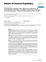
of the American Joint Committee on Cancer (AJCC) staging system. From these criteria, 1274 patients were selected for the matched study (Fig. 1).
We performed an analysis of variance as well as a χ2
test on the patients’ baseline demographics and clinical
characteristics. Variable differences were identified between the two groups, including gender, age, tumor
stage (T stage) and node stage (N stage), clinical stage
and chemotherapy regime, all of which were identified
as prognostic factors for survival outcomes in a previous
study. Using propensity scores to adjust for these 6 factors, we created a well-balanced cohort by matching
each patient who underwent nimotuzumab treatment
with no more than three patients who underwent chemoradiotherapy without nimotuzumab (Table 1). From
this stratification process, we selected a total of 730 patients, including 184 patients in the nimotuzumab arm
and 546 patients in the no nimotuzumab arm (Table 1).
We first conducted case-matched comparisons between
the two arms in terms of efficacy and safety in this wellbalanced cohort of 730. Subsequently, we conducted
univariable and multivariate analyses of the 730 patients.
Treatment
Radiation therapy
All patients received IMRT. The primary nasopharyngeal
gross tumor volume (GTVnx) and the involved cervical
lymph nodes were determined based on MRI/CT and/or
PET-CT imaging, clinical, and endoscopic findings. The
enlarged retropharyngeal nodes together with primary
gross tumor volume (GTV) were outlined as the GTVnx
on the IMRT plans. The clinical tumor volume (CTV)
represents the primary tumor with potential subclinical
disease. The first clinical tumor volume (CTV1) was defined as the GTV plus a 0.5–1.0 cm margin (0.2 to 0.3
margin posteriorly) to encompass the high-risk sites of
microscopic extension and the whole nasopharynx. Clinical target volume 2 (CTV2) was defined as the CTV1
plus a 0.5–1.0 cm margin (0.2 to 0.3 margin posteriorly)
to encompass the low-risk sites of microscopic extension, the level of the lymph node, and the elective neck
area (bilateral levels IIa, IIb, III, and Va are routinely
covered for all N0 patients, whereas ipsilateral levels IV,
Vb, or supraclavicular fossae are also included for N1–3
patients). The prescribed doses were 66–70 Gy to the
planning target volume (PTV) of the primary gross
tumor volume (GTVnx), 60 Gy to PTV1 (PTV of
CTV1), 54 Gy to PTV2 (PTV of CTV2), and 60–66 Gy
to PTVnd of the involved cervical lymph nodes in 28 to
33 fractions. All patients were treated once daily, with
five fractions administered weekly. The doses to critical
structures were within the tolerance range according to
Zhi-Qiang et al. BMC Cancer
(2019) 19:1122
Page 3 of 12
Fig. 1 Study flow diagram
the RTOG 0225 protocol, and efforts were made to meet
the criteria as closely as possible.
Chemotherapy
During the study period, concurrent chemoradiotherapy
(CCRT) ± induction chemotherapy (IC) for stage III to IV
disease was recommended according to our institutional
guidelines. The study-defined concurrent chemoradiotherapy regimen was 80–100 mg/m2 cisplatin on day 1 every
3 weeks for 2–3 cycles or 30 mg/m2 cisplatin weekly. Patients receiving other chemotherapy regimens or who received only one cycle of induction or concurrent
chemotherapy were excluded from this study. The studydefined induction chemotherapy regimens included PF
(n = 161) (80–100 mg/m2 cisplatin on day 1 and 800 mg/
m2 /d fluorouracil civ on days 1–5), TP (n = 176) (75 mg/
m2 docetaxel on day 1 and 75 mg/m2 cisplatin on day 1
or TPF(142) (75 mg/m2 docetaxel on day 1, 75 mg/m2 cisplatin on day 1 and 800 mg/m2 /d fluorouracil civ on days
1–5), and both regimens were repeated every 3 weeks for
2–3 cycles. The reasons for deviating from the institutional guidelines included organ dysfunction suggesting
intolerance to chemotherapy, patient refusal, and the discretion of the doctors in individual cases.
Nimotuzumab delivery
Nimotuzumab was not recommended for NPC patients
by the guideline at that time. Therefore, the use of
Nimotuzumab was determined by the patients’ willingness and the doctors’ experience. Intravenous Nimotuzumab was administered at an initial dose of 200 mg
weekly during the entire radiation period. A total of 184
patients received full doses of Nimotuzumab.
Follow-up
Patient follow-up was measured from the first day of
therapy to the last examination or death. Patients were
examined at least every 3 months during the first 2 years,
with follow-up examinations every 6 months for 3 years
or until death. The last follow-up date was 20 April
2019.
The Common Terminology Criteria for Adverse
Events (version 4.0) was used to evaluate chemotherapyrelated toxic effects, and the Late Radiation Morbidity
Scoring Criteria of the Radiation Therapy Oncology
Group was used to evaluate radiotherapy-related toxic
effects [16]. Acute toxicities were defined as those occurring either during the course of IMRT or within 90 days
of its completion.
Zhi-Qiang et al. BMC Cancer
(2019) 19:1122
Page 4 of 12
Table 1 Baseline characteristics of patients with NPC treated with or without Nimotuzumab
Characteristic
Nimotuzumab Arm
N = 184(%)
No Nimotuzumab Arm
N = 546(%)
Gender
0.704
Female
37(20.11)
117(21.43)
Male
147(79.89)
429(78.57)
Age, Mean (SD)
P value
43.92 (10.53)
44.1 2(10.62)
<44
95(51.63)
253(46.34)
≥ 44
89(48.37)
293(53.66)
WHO pathology
0.822
0.436
I
3(1.6)
11(2.0)
II
8(4.3)
14(2.6)
III
173(94.1)
521(95.4)
T classification
0.966
T1
5(2.72)
17(3.11)
T2
16(8.70)
42(7.69)
T3
107(58.16)
317(58.06)
T4
56(30.42)
170(31.14)
No
19(10.33)
57(10.44)
N1
75(40.76)
230(42.13)
N2
73(39.67)
214(39.19)
N3
17(9.24)
45(8.24)
N classification
0.972
Clinical stage
0.937
III
116(63.04)
346(63.37)
IVa
51(27.72)
155(28.39)
IVb
17(9.24)
45(8.24)
Chemotherapy
0.684
Concurrent
61(33.15)
190(34.80)
Induction + concurrent
123(66.85)
356(65.20)
Statistical analysis
Distant metastasis–free survival (DMFS) and locoregional
relapse–free survival (LRRFS) were calculated from day 1
after completion of treatment to the first distant metastasis and locoregional relapse, respectively. Progression–free
survival (PFS) was calculated from day 1 after completion
of treatment to locoregional relapse, distant relapse or
tumor-related death, whichever occurred first. Overall
survival (OS) was calculated from day 1 after completion
of treatment to the last examination or death.
The clinic-pathologic characteristics of participants are described, and the differences of these characteristics between
the Nimotuzumab group and non-Nimotuzumab group
were compared by the χ2 test for categorical variables and
the t-test for continuous variables. Logistic regression analysis was used to identify confounders between the treatment groups. A propensity score matching method was
used. Propensity scores were calculated based on the
identified potential confounders and other important factors,
such as tumor stage, and then each patient was assigned a
score. Using a caliper width of 0.2, 1:3 matching was performed between patients in the Nimotuzumab group and
non-Nimotuzumab group based on the propensity scores.
LRRFS, DMFS, PFS and OS were calculated using the
Kaplan-Meier method. The differences in LRRFS, DMFS,
PFS and OS between the two groups were tested using
the log-rank test. Multivariate analysis was performed
using the Cox proportional hazards models. All statistical analyses were performed using SPSS 21.0 statistical
software (Chicago, IL, USA). A value of P < 0.05 was
considered statistically significant.
Results
Patient characteristics
Patient characteristics are detailed in Table 1. A total of
6908 consecutive NPC patients who were treated with
Zhi-Qiang et al. BMC Cancer
(2019) 19:1122
IMRT between January 2008 and December 2013 at
SYSUCC were analyzed, and 1274 patients were eligible
for propensity score matching as shown in Fig. 1. Gender, age, T-category, N-category, clinical stage and
chemotherapy regime (IC alone and IC + CCRT) were
used to generate a propensity score model (Fig. 1).
In total, 730 patients were propensity matched in
this study to create two groups: Group A, which received Nimotuzumab, included 184 cases; and Group
B, which did not receive Nimotuzumab, included 546
cases. Among the 730 patients, 154 were female and
576 were male. All 730 patients received cisplatinbased concurrent chemotherapy, and 479 received two
courses of induction chemotherapy. The characteristics of the patients were well balanced between the
propensity-matched groups. The median dose delivered during the initial course of radiation was 70 Gy
(range, 66–80 Gy).
The mean age at the time of reirradiation was 43.92
years (SD = 10.53) for Group A and 44.12 years (SD =
10.62) for Group B. At a median follow-up of 74.78
months (range 3.53–117.83 months), the 1, 3, and 5-year
follow-up rates were 99.6, 96.7 and 90.5%, respectively.
Efficacy and safety
At the median follow-up of 74.78 months (range 3.53–
117.83 months), 117 deaths (16.03%) had occurred. At
the time of the analysis, 68 patients had locoregional
failure (9.32%), 10 showed locoregional failure and distant metastases (1.34%), and 71 developed distant metastases (9.73%).
The 5-year DMFS, LRRFS, PFS and OS rates for Group
A vs. Group B were 93.90% vs. 85.61% (P = 0.012), 85.34%
vs. 89.79% (P = 0.156), 79.96% vs. 77.99% (P = 0.117) and
96.33% vs. 85.97% (P = 0.006), respectively (Table 2).
Page 5 of 12
Significant differences in DMFS and OS were observed between Group A and Group B, although differences in
LRRFS and PFS were not observed. The 5-year DMFS,
LRRFS, PFS and OS according to clinical stage were calculated, and significant differences were only observed in OS
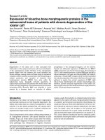
for stage III. The survival curves are shown in Fig. 2.
Table 3 displays the acute toxicities of the 730 patients. Significant differences were not observed in the
hematological toxicities, and significant differences were
not observed between the two groups in terms of hepatoxicity, nephrotoxicity, and gastrointestinal reactions,
including nausea, vomiting, and diarrhea.
Prognosis
The overall survival (OS) of the 730 cases were analyzed by univariate and multivariable cox regression
models, respectively. We included sex, age, T stage, N
stage, clinical stage, nimotuzumab treatment or not
and concurrent chemotherapy (with or without induction chemotherapy) in the model. The results showed
that the T stage, N stage, clinical stage and nimotuzumab or not factors had prognostic significance for
OS (Table 4). The multivariate analysis indicated that
N stage and nimotuzumab treatment were independent prognostic factors for DMFS and OS (Table 5).
Patients with advanced N stage had a poorer prognosis and those who received nimotuzumab had significantly better 5-year OS rates compared with than
those who did not receive nimotuzumab (88.91% versus 78.30%, P<0.01) (Table 2).
Discussion
Even with the administration of cisplatin-based chemoradiotherapy, the treatment outcome for advanced
stages of NPC is still unsatisfactory because of local
Table 2 Five-year survival rates of the 730 NPC patients
All
(N = 730)
Nimotuzumab Arm
(N = 184)
No Nimotuzumab Arm
(N = 546)
DMFS
87.49%
93.09%
85.61%
III
89.81%
94.59%
88.22%
IV
83.43%
90.56%
80.93%
LRRFS
88.60%
85.34%
89.79%
III
90.16%
87.22%
91.21%
IV
85.71%
82.00%
87.17%
PFS
78.47%
79.96%
77.99%
III
78.72%
81.27%
77.88%
IV
63.15%
70.90%
60.52%
80.96%
88.91%
78.30%
III
88.53%
96.33%
85.97%
IV
67.69%
76.24%
64.79%
OS
Chi-square
P value
6.343
0.012
2.012
0.156
2.459
0.117
7.565
0.006
OS overall survival, DMFS distant metastasis–free survival, LRRFS locoregional relapse–free survival, and PFS progression-free survival
Zhi-Qiang et al. BMC Cancer
(2019) 19:1122
Page 6 of 12
Fig. 2 Kaplan-Meier plots of distant metastasis-free survival, locoregional relapse–free survival, progression-free survival, and overall survival of
NPC patients treated with (green lines) or without Nimotuzumab (blue lines) according the clinical stage
recurrence and/or distant metastasis, which represent
the major patterns of disease failure [17]. However,
modern radiation techniques and equipment have enabled the delivery of high doses of radiation to the target tissue while sparing normal organs from risk,
thereby potentially enhancing the therapeutic efficacy
[18]. Previous studies have reported 90% local-regional
control rates for nasopharyngeal carcinoma with the
use of IMRT combined with systematic chemotherapy,
even in patients presenting with advanced locoregional disease [19–21]. Distant metastasis plays an
important role in treatment failure and needs to be
managed properly and urgently. After decades of studies on chemotherapy for NPC, only slight improvements have been achieved in survival and distant
failure; therefore, new treatment strategies must be
developed to address this issue, which has confounded
clinical doctors for a long time.
Zhi-Qiang et al. BMC Cancer
(2019) 19:1122
Page 7 of 12
Table 3 Acute toxicities in the 730 NPC patients
Acute Toxicity
Nimotuzumab Arm
N = 184(%)
No Nimotuzumab Arm
N = 546
Anemia
P value
0.920
G0-G1
137(74.5)
416(76.2)
G2
36(19.6)
98(17.9)
G3
9(4.0)
27(4.9)
G4
2(1.1)
5(0.9)
G0-G1
164(89.1)
464(85.0)
G2
16(8.7)
58(10.6)
G3
4(2.2)
21(3.8)
G4
0(0.0)
3(0.5)
Thrombocytopenia
0.541
Leukopenia
0.845
G0-G1
122(66.3)
370(67.8)
G2
33(17.9)
101(18.5)
G3
27(14.7)
7113.0()
G4
2(1.1)
4(0.7)
G0-G1
139(75.5)
386(70.7)
G2
39(21.2)
118(21.6)
G3
5(2.7)
38(7.0)
G4
1(0.5)
4(0.7)
Neutropenia
0.154
Skin reaction
0.434
G0-G1
126(68.5)
381(69.8)
G2
46(25.0)
117(21.4)
G3
12(6.5)
48(8.8)
Mucositis
0.728
G0-G1
67(36.4)
189(34.6)
G2
73(39.7)
228(41.8)
G3
38(20.7)
102(18.7)
G4
6(3.3)
27(4.9)
G0-G1
62(37.8)
193(35.3)
G2
71(43.3)
214(39.2)
G3
28(17.1)
126(23.1)
G4
3(1.8)
13(2.4)
Nausea
0.394
Vomiting
0.990
G0-G1
138(75.0)
412(75.5)
G2
24(13.0)
68(12.5)
G3
20(10.9)
61(11.2)
G4
2(1.1)
5(0.9)
G0-G1
146(79.3)
437(80.0)
G2
31(16.8)
97(17.8)
G3
7(3.8)
12(2.2)
Diarrhea
0.495
Zhi-Qiang et al. BMC Cancer
(2019) 19:1122
Page 8 of 12
Table 3 Acute toxicities in the 730 NPC patients (Continued)
Acute Toxicity
Nimotuzumab Arm
N = 184(%)
No Nimotuzumab Arm
N = 546
Hepatotoxicity
P value
0.532
G0-G1
137(74.5)
410(75.1)
G2
32(17.4)
80(14.7)
G3
15(8.2)
56(10.3)
Nephrotoxicity
0.770
G0-G1
167(90.8)
505(92.5)
G2
12(6.5)
29(5.3)
G3
5(2.7)
12(2.2)
Weight loss
0.964
G0-G1
14,478.3()
420(76.9)
G2
36(19.6)
112(20.5)
G3
4(2.2)
14(2.6)
With further research of the molecular mechanism of
tumorigenesis and tumor development, molecular targeted therapy in patients with NPC has become a research hotspot. It is known that more than 90% patients
with NPC were positive for the overexpression of EGFR
[6, 7], which is considered an important target in NPC
treatment [22]. Nimotuzumab is a humanized antiEGFR monoclonal antibody, and it is obtained by replacing a murine complementary-determining region
with a human framework. Nimotuzumab has shown
high safety and low toxicity without the severe skin and
mucosa toxicities commonly associated with other
EGFR-targeting antibodies [12, 15]. As reported, compared with other EGFR inhibitors, such as cetuximab,
nimotuzumab shows a greater advantage in terms of less
toxicity [23]. Another advantage of nimotuzumab is that
the affinity constant is quite low, which allows for high
tumor uptake and low normal tissue uptake. Research
has shown that Nimotuzumab demonstrates marked antiproliferative, proapoptotic, and antiangiogenic effects
in tumors that overexpress EGFR [24]. Currently, Nimotuzumab has been approved in several countries for the
treatment of head and neck tumors [25, 26].
The current study retrospectively analyzed the efficacy
of nimotuzumab plus IMRT/CCRT with or without induction chemotherapy in 184 NPC patients. In our
study, encouraging survival rates and distant metastasis
control were attributed to the treatment with nimotuzumab. Our results showed promising clinical outcomes,
with a 5-year DMFS of 93.09%, 5-year LRRFS of 85.34%,
5-year PFS of 79.96%, and 5-year OS of 88.91% observed
in patients who received nimotuzumab and a 5-year
DMFS of 85.61%, 5-year LRRFS of 89.79%, 5-year PFS of
77.99%, and 5-year OS of 78.30% in patients who did
not receive nimotuzumab. The lack of significant difference in the 5-year LRRFS (85.34% vs. 89.79%, P = 0.156)
was reasonable since IMRT provides excellent locoregional control [27]. The current analysis demonstrated that
the addition of nimotuzumab compared with CCRT
alone was associated with a significantly better OS and
DMFS, which presented significantly differences of P =
0.006 and P = 0.012, respectively. Further statistical analyses showed that OS significantly increases in patients
with stage III disease. These data indicated that the increase in survival outcome for NPC patients treated with
nimotuzumab was mainly attributed to the significant
increase in DMFS, which could be related to the greater
ability of nimotuzumab and cisplatin-based chemoradiotherapy to kill tumor cells, especially cisplatin-based
chemotherapy-resistant micrometastases. This improved
tumor killing ability could also partially explain the significant increase in DMFS; however, this is just a postulation, and further investigation is required to explore
the exact mechanism.
Previous studies demonstrated that the main prognostic factors for survival are age, gender, T and N category,
and clinical stage, with the survival rate decreasing as
the T category and N category increased [28]. According
to the multivariate analysis, gender, N stage and nimotuzumab were significant prognostic factors for DMFS; N
stage and nimotuzumab treatment were significant prognostic factors for OS; and node stage was a significant
prognostic factor for PFS. Since only patients with clinical stage III and IV were included in this study and the
local-regional control rate was similar and lacked statistical significance (90.16% vs. 85.71%, P = 0.156), these results can be explained by the use of modern radiation
techniques, which have been proved to improve localregional control. For distant failure, node stage still affects DMFS and OS, and patients with advanced node
stage have a higher likelihood of distant failure, which
leads to overall failure. These results are consistent with
0.450
0.244
−0.213
−0.292
T3
T4
0.325
0.796
With
1
0.230
Yes
0.107
No
Induction chemotherapy
1
0.222
1
Without
Target therapy
IV
III
−0.606
0.278
−1.117
Clinical stage
N3
0.466
0.325
−1.666
−1.960
N1
N2
N0
1
0.486
Node stage
1
0.681
0.223
1
0.372
1
T2
−0.106
−0.943
1.113(0.709–1.747)
2.217(1.174–4.188)
0.546(0.353–0.844)
0.327(0.190–0.564)
0.141(0.074–0.267)
0.189(0.076–0.471)
0.747(0.463–1.205)
0.808(0.335–1.952)
1.975(0.763–5.116)
0.900(0.581–1.393)
0.389(0.188–0.808)
HR(95%CI)
0.642
0.014
0.006
0.000
0.000
0.000
0.232
0.636
0.161
0.636
0.011
P
−0.354
−0.367
−0.405
−0.472
−0.742
−1.554
−0.373
0.261
−0.145
− 0.380
0.189
B
SE
B
T1
Tumor stage
≥ 44
<44
Age
Male
Female
Gender
LRRFS
DMFS
0.270
1
0.260
1
0.244
1
0.380
0.387
0.667
1
0.267
0.408
0.736
1
0.248
1
0.280
1
SE
0.702(0.413–1.193)
0.693(0.416–1.154)
0.667(0.413–1.076)
0.624(0.296–1.315)
0.476(0.223–1.017)
0.211(0.057–0.782)
0.689(0.408–1.162)
1.299(0.583–2.891)
0.865(0.204–3.661)
0.684(0.420–1.111)
1.208(0.697–2.092)
HR(95%CI)
0.191
0.158
0.097
0.215
0.055
0.020
0.162
0.522
0.844
0.125
0.500
P
−0.181
0.292
−0.674
−0.832
−1.216
−1.443
−0.577
−0.126
0.050
− 0.242
−0.270
B
PFS
0.163
1
0.187
1
0.151
1
0.216
0.225
0.342
1
0.162
0.277
0.396
1
0.152
1
0.198
1
SE
Table 4 Prognostic factors associated with overall survival based on univariate cox regression models (N = 730)
0.834(0.607–1.148)
1.340(0.928–1.933)
0.510(0.379–0.685)
0.435(0.285–0.665)
0.296(0.191–0.460)
0.236(0.121–0.462)
0.562(0.409–0.772)
0.882(0.513–1.517)
1.051(0.484–2.285)
0.785(0.582–1.058)
0.764(0.518–1.126)
HR(95%CI)
0.266
0.118
0.000
0.000
0.000
0.000
0.000
0.650
0.900
0.112
0.173
P
−0.279
0.707
−1.166
−1.080
−1.392
−1.568
−1.011
−0.584
0.050
− 0.288
−0.570
B
OS
0.204
1
0.263
1
0.191
1
0.249
0.258
0.406
1
0.200
0.358
0.429
1
0.187
1
0.269
1
SE
0.756(0.507–1.128)
2.028(1.213–3.393)
0.312(0.214–0.453)
0.339(0.209–0.553)
0.249(0.150–0.412)
0.209(0.094–0.463)
0.364 (0.246–0.538)
0.557 (0.276–1.125)
1.052 (0.454–2.438)
0.750(0.519–1.083)
0.566(0.3340.959)
HR(95%CI)
0.170
0.007
0.000
0.000
0.000
0.000
0.000
0.103
0.906
0.125
0.034
P
Zhi-Qiang et al. BMC Cancer
(2019) 19:1122
Page 9 of 12
Zhi-Qiang et al. BMC Cancer
(2019) 19:1122
Page 10 of 12
Table 5 Multivariate analysis of variables correlated with the treatment regimen status and other significant prognostic factors in
730 eligible cases
DMFS
B
LRFS
HR(95%CI)
P
B
PFS
HR (95%CI)
P
B
OS
HR (95%CI)
P
B
HR (95%CI)
P
Gender
Female
1
1
Male
−0.934 0.393 (0.188–
0.823)
0.013
0.247
1
1.280 (0.733–
2.237)
1
0.385 −0.205 0.815 (0.549–
1.210)
0.310
−0.523 0.592 (0.347–
1.011)
0.055
Age
<44
1
1
≥ 44
−0.096 0.909 (0.582–
1.421)
0.675
1
−0.439 0.645 (0.393–
1.058)
1
0.082 −0.256 0.774 (0.571–
1.050)
0.100
−0.252 0.777 (0.534–
1.132)
0.189
Tumor stage
T1
1
T2
0.149
1.161 (0.335–
4.026)
0.814
1
−0.229 0.795 (0.113–
5.603)
0.818 −0.200 0.819 (0.287–
2.339)
1
0.709
1
0.206
1.229 (0.407–
3.714)
0.715
T3
−0.894 0.409 (0.128–
1.305)
0.131
0.109
1.116 (0.246–
5.065)
0.887 −0.510 0.600 (0.256–
1.405)
0.240
−0.546 0.580 (0.215–
1.561)
0.280
T4
−0.474 0.623 (0.248–
1.562)
0.313
−0.329 0.720(0.175–
2.954)
0.648 −0.664 0.515 (0.243–
1.091)
0.083
−0.632 0.531 (0.231–
1.223)
0.137
Node stage
N0
1
1
1
1
N1
−1.979 0.138 (0.046–
0.418)
<
0.001
−1.437 0.238 (0.047–
1.210)
0.083 −1.603 0.201 (0.088–
0.458)
<
0.001
−1.509 0.221 (0.086–
0.567)
0.002
N2
−2.251 0.105 (0.043–
0.260)
<
0.001
−0.594 0.552 (0.160–
1.902)
0.347 −1.329 0.265 (0.138–
0.508)
<
0.001
−1.242 0.289 (0.141–
0.594)
0.001
N3
−1.330 0.264 (0.111–
0.629)
0.003
−0.329 0.720(0.204–
2.542)
0.609 −0.876 0.416 (0.216–
0.804)
0.009
−0.800 0.449 (0.216–
0.934)
0.032
Clinical stage
III
1
IV
0.224
1
1.251 (0.436–
3.593)
0.677
−0.052 0.950 (0.212–
4.254)
1
0.946 0.080
1
1.083 (0.482–
2.434)
0.847
−0.448 0.639 (0.256–
1.598)
0.338
Target therapy
1
1
1
1
Without
With
0.840
2.317 (1.224–
4.384)
0.010
−0.339 0.712 (0.427–
1.189)
0.194 0.353
1.423 (0.985–
2.058)
0.060
0.754
2.125 (1.269–
3.560)
0.004
−0.012 0.988 (0.655–
1.490)
0.955
Induction chemotherapy
No
1
Yes
0.303
1
1.354 (0.849–
2.159)
0.203
−0.209 0.811 (0.471–
1.398)
that of other studies [5, 29]. We must address the significant improvement of overall survival after the administration of a full course of nimotuzumab to NPC
patients in stages III to IV during chemoradiotherapy,
with these patients showing a nearly 10% improvement
in OS (88.91% vs 78.30%, P = 0.006). The results are encouraging and beyond our expectations. The strength of
nimotuzumab combined with radiotherapy for NPC may
be still largely due to the strengthening of the antitumor
effect caused by the increased tumor cell killing ability
1
0.451 0.031
1
1.031 (0.742–
1.434)
0.855
of nimotuzumab and cisplatin-based chemoradiotherapy,
which was mentioned above.
This study presented certain limitations, and the results should be interpreted with caution since this is a
retrospective study. Moreover, the lack of availability of
EGFR expression data is another limitation since a proportion of patients were EGFR negative. Although we
eliminated selection bias, such as gender, age, T and N
stage, and clinical stage, using propensity scores,
whether other confounding factors still exist remains
Zhi-Qiang et al. BMC Cancer
(2019) 19:1122
unclear. In the future, prospective, randomized, welldesigned, and large-sample clinical studies are needed to
evaluate these factors.
Conclusions
In conclusion, our study observed that the administration of nimotuzumab during chemoradiotherapy in stage
III-IV NPC patients showed promising clinical outcomes
compared with the administration of chemoradiotherapy
alone. However, additional studies, especially prospective, well-designed, and large-sample clinical studies, are
needed.
Abbreviations
AJCC: American Joint Committee on Cancer; CCRT: Concurrent
chemoradiotherapy; CT: Computed tomography; CTV: Clinical tumor volume;
DMFS: Distant metastasis–free survival; EGFR: Epidermal growth factor
receptor; GTVnx: The primary nasopharyngeal gross tumor volume;
IC: Induction chemotherapy; IMRT: Intensity-modulated radiotherapy;
KPS: Karnofsky Performance Status; LRRFS: Locoregional relapse–free survival;
MRI: Magnetic Resonance Imaging; NPC: Nasopharyngeal carcinoma;
OS: Overall survival; PET-CT: Positron emission tomography–computed
tomography; PFS: Progression–free survival; PTV: Planning target volume;
RTOG: Radiation Therapy Oncology Group; SYSUCC: Sun Yat-sen University
Cancer Center
Acknowledgments
We gratefully acknowledge the staff at the clinical laboratory, Sun Yat-sen
University Cancer Center for providing support to the research in this study.
We also express our thanks to Xie Si-Yi and Gong Zhi-Da for data collecting.
Authors’ contributions
CMY and HYJ conceived the study. YR, LYP and SR made substantial
contributions to data acquisition, WZQ, MQ and LJB analyzed the data and
performed interpretation of data. WZQ, MQ and LJB involved in drafting the
manuscript. HGY and HYJ edited the manuscript. All authors have read and
approved the final manuscript.
Funding
No funding was received.
Availability of data and materials
The data of this research (RDDA2019001088) is deposited in RDD (http://
www.researchdata.org.cn).
Ethics approval and consent to participate
This study complied with the standards of the Declaration of Helsinki and
current ethical guidelines. It was approved by the Sun Yat-sen University
Cancer Center research ethics committee. All patients provided written informed consent for the collection and publication of their medical information at the first visit to our center, which was filed in their medical records.
Consent for publication
Not applicable.
Competing interests
The authors declare that they have no conflict of interest.
Author details
State Key Laboratory of Oncology in South China, Collaborative Innovation
Center for Cancer Medicine, Guangzhou, China. 2Department of
Nasopharyngeal Carcinoma, Sun Yat-sen University Cancer Center, 651
Dongfeng Road East, Guangzhou 510060, China. 3Department of Oncology,
Tongji Hospital, Tongji Medical College of Huazhong University of Science
and Techology, Wuhan, China. 4Department of Clinical Research, Sun Yat-sen
University Cancer Center, Guangzhou, China.
1
Page 11 of 12
Received: 10 June 2019 Accepted: 13 September 2019
References
1. Wei KR, Zheng RS, Zhang SW, Liang ZH, Ou ZX, Chen WQ. Nasopharyngeal
carcinoma incidence and mortality in China in 2010. Chin J Cancer. 2014;
33(8):381–7.
2. Wei WI, Sham JS. Nasopharyngeal carcinoma. Lancet. 2005;365(9476):
2041–54.
3. Zhang LF, Li YH, Xie SH, Ling W, Chen SH, Liu Q, et al. Incidence trend of
nasopharyngeal carcinoma from 1987 to 2011 in Sihui County, Guangdong
Province, South China: an age-period-cohort analysis. Chin J Cancer. 2015;
34(8):350–7.
4. Wee JT, Ha TC, Loong SL, Qian CN. Is nasopharyngeal cancer really a
"Cantonese cancer"? Chin J Cancer. 2010;29(5):517–26.
5. Lee N, Harris J, Garden AS, Straube W, Glisson B, Xia P, et al. Intensitymodulated radiation therapy with or without chemotherapy for
nasopharyngeal carcinoma: radiation therapy oncology group phase II trial
0225. J Clin Oncol. 2009;27(22):3684–90.
6. Ma BB, Poon TC, To KF, Zee B, Mo FK, Chan CM, et al. Prognostic
significance of tumor angiogenesis, Ki 67, p53 oncoprotein, epidermal
growth factor receptor and HER2 receptor protein expression in
undifferentiated nasopharyngeal carcinoma--a prospective study. Head
Neck. 2003;25(10):864–72.
7. Modjtahedi H, Essapen S. Epidermal growth factor receptor inhibitors in
cancer treatment: advances, challenges and opportunities. Anti-Cancer
Drugs. 2009;20(10):851–5.
8. Zhang ZC, Fu S, Wang F, Wang HY, Zeng YX, Shao JY. Oncogene mutational
profile in nasopharyngeal carcinoma. Onco Targets Ther. 2014;7:457–67.
9. Zhang P, Wu SK, Wang Y, Fan ZX, Li CR, Feng M, et al. p53, MDM2, eIF4E
and EGFR expression in nasopharyngeal carcinoma and their correlation
with clinicopathological characteristics and prognosis: a retrospective study.
Oncol Lett. 2015;9(1):113–8.
10. Campbell NP, Hensing TA, Bhayani MK, Shaikh AY, Brockstein BE.
Targeting pathways mediating resistance to anti-EGFR therapy in
squamous cell carcinoma of the head and neck. Expert Rev Anticancer
Ther. 2016;16(8):847–58.
11. Talavera A, Friemann R, Gomez-Puerta S, Martinez-Fleites C, Garrido G,
Rabasa A, et al. Nimotuzumab, an antitumor antibody that targets the
epidermal growth factor receptor, blocks ligand binding while permitting
the active receptor conformation. Cancer Res. 2009;69(14):5851–9.
12. Si X, Wu S, Wang H, Zhang X, Wang M, Zeng X, et al. Nimotuzumab
combined with chemotherapy as first-line treatment for advanced lung
squamous cell carcinoma. Thorac Cancer. 2018;9(8):1056–61.
13. Garrido G, Tikhomirov IA, Rabasa A, Yang E, Gracia E, Iznaga N, et al. Bivalent
binding by intermediate affinity of nimotuzumab: a contribution to explain
antibody clinical profile. Cancer Biol Ther. 2011;11(4):373–82.
14. Liu ZG, Zhao Y, Tang J, Zhou YJ, Yang WJ, Qiu YF, et al. Nimotuzumab
combined with concurrent chemoradiotherapy in locally advanced
nasopharyngeal carcinoma: a retrospective analysis. Oncotarget. 2016;7(17):
24429–35.
15. You R, Hua YJ, Liu YP, Yang Q, Zhang YN, Li JB, et al. Concurrent
Chemoradiotherapy with or without anti-EGFR-targeted treatment for stage
II-IVb nasopharyngeal carcinoma: retrospective analysis with a large cohort
and long follow-up. Theranostics. 2017;7(8):2314–24.
16. Cox JD, Stetz J, Pajak TF. Toxicity criteria of the radiation therapy oncology
group (RTOG) and the European Organization for Research and Treatment
of Cancer (EORTC). Int J Radiat Oncol Biol Phys. 1995;31(5):1341–6.
17. Ng WT, Lee MC, Hung WM, Choi CW, Lee KC, Chan OS, et al. Clinical
outcomes and patterns of failure after intensity-modulated radiotherapy for
nasopharyngeal carcinoma. Int J Radiat Oncol Biol Phys. 2011;79(2):420–8.
18. Hua YJ, Han F, Lu LX, Mai HQ, Guo X, Hong MH, et al. Long-term treatment
outcome of recurrent nasopharyngeal carcinoma treated with salvage
intensity modulated radiotherapy. Eur J Cancer. 2012;48(18):3422–8.
19. Wang W, Feng M, Fan Z, Li J, Lang J. Clinical outcomes and prognostic factors
of 695 nasopharyngeal carcinoma patients treated with intensity-modulated
radiotherapy. Biomed Res Int. 2014;2014:814948.
20. Setton J, Han J, Kannarunimit D, Wuu YR, Rosenberg SA, DeSelm C,
et al. Long-term patterns of relapse and survival following definitive
intensity-modulated radiotherapy for non-endemic nasopharyngeal
carcinoma. Oral Oncol. 2016;53:67–73.
Zhi-Qiang et al. BMC Cancer
(2019) 19:1122
21. Zhang MX, Li J, Shen GP, Zou X, Xu JJ, Jiang R, et al. Intensity-modulated
radiotherapy prolongs the survival of patients with nasopharyngeal
carcinoma compared with conventional two-dimensional radiotherapy: a
10-year experience with a large cohort and long follow-up. Eur J Cancer.
2015;51(17):2587–95.
22. Zhang JW, Qin T, Hong SD, Zhang J, Fang WF, Zhao YY, et al. Multiple
oncogenic mutations related to targeted therapy in nasopharyngeal
carcinoma. Chin J Cancer. 2015;34(4):177–83.
23. Li HM, Li P, Qian YJ, Wu X, Xie L, Wang F, et al. A retrospective paired study:
efficacy and toxicity of nimotuzumab versus cisplatin concurrent with
radiotherapy in nasopharyngeal carcinoma. BMC Cancer. 2016;16(1):946.
24. Crombet-Ramos T, Rak J, Perez R, Viloria-Petit A. Antiproliferative,
antiangiogenic and proapoptotic activity of h-R3: a humanized anti-EGFR
antibody. Int J Cancer. 2002;101(6):567–75.
25. Crombet T, Osorio M, Cruz T, Roca C, del Castillo R, Mon R, et al. Use of the
humanized anti-epidermal growth factor receptor monoclonal antibody h-R3
in combination with radiotherapy in the treatment of locally advanced head
and neck cancer patients. J Clin Oncol. 2004;22(9):1646–54.
26. Ramakrishnan MS, Eswaraiah A, Crombet T, Piedra P, Saurez G, Iyer H, et al.
Nimotuzumab, a promising therapeutic monoclonal for treatment of
tumors of epithelial origin. MAbs. 2009;1(1):41–8.
27. Mao YP, Tang LL, Chen L, Sun Y, Qi ZY, Zhou GQ, et al. Prognostic factors
and failure patterns in non-metastatic nasopharyngeal carcinoma after
intensity-modulated radiotherapy. Chin J Cancer. 2016;35(1):103.
28. Qu Y, Chen Y, Yu H, Zhao Y, Chen G, Bai L, et al. Survival and prognostic
analysis of primary nasopharyngeal carcinoma in North China. Clin Lab.
2015;61(7):699–708.
29. Yao JJ, Zhang LL, Gao TS, Peng YL, Lawrence WR, Zhang WJ, et al.
Comparing treatment outcomes of concurrent chemoradiotherapy with or
without nimotuzumab in patients with locoregionally advanced
nasopharyngeal carcinoma. Cancer Biol Ther. 2018:1–6.
Publisher’s Note
Springer Nature remains neutral with regard to jurisdictional claims in
published maps and institutional affiliations.
Page 12 of 12