Synergism of wt-p53 and synthetic material in local nano-TAE gene therapy of hepatoma: Comparison of four systems and the possible mechanism
Bạn đang xem bản rút gọn của tài liệu. Xem và tải ngay bản đầy đủ của tài liệu tại đây (6.14 MB, 17 trang )
Li et al. BMC Cancer
(2019) 19:1126
/>
RESEARCH ARTICLE
Open Access
Synergism of wt-p53 and synthetic material
in local nano-TAE gene therapy of
hepatoma: comparison of four systems and
the possible mechanism
Gaopeng Li1 , Wenqin Kang2, Mingliang Jin3, Lidong Zhang4, Jian Zheng5, Kai Jia6, Jinfeng Ma1, Ting Liu1,
Xueyi Dang1, Zhifeng Yan1, Zefeng Gao1*† and Jun Xu6*†
Abstract
Background: TAE-gene therapy for hepatoma, incorporating the tumor-targeted therapeutic efficacy of trans-arterial
embolization, hydroxyapatite nanoparticles (nHAP) and anti-cancer wild-type p53 gene (wt-p53), was presented in our
former studies (Int J Nanomedicine 8:3757-68, 2013, Liver Int 32:998-1007, 2012). However, the incompletely
antitumoral effect entails defined guidelines on searching properer materials for this novel therapy.
Methods: Unmodified nHAP, Ca(2+) modified nHAP, poly-lysine modified nHAP and liposome were separately used to
form U-nanoplex, Ca-nanoplex, Pll-nanoplex, L-nanoplex respectively with wt-p53 expressing plasmid. The four
nanoplexs were then applied in vitro for human normal hepacyte L02 and hepatoma HePG2 cell line, and in vivo for
rabbits with hepatic VX2 tumor by injection of nanoplexs/lipiodol emulsion into the hepatic artery in a tumor target
manner. The distribution, superficial potential, physical structure, morphology and chemical compositions of nanoplexs
were evaluated by TEM, SEM, EDS etc., with the objective of understanding their roles in hepatoma TAE-gene therapy.
Results: In vitro, L-nanoplex managed the highest gene transferring efficiency. Though with the second highest
transfection activity, Pll-nanoplex showed the strongest tumor inhibition activity while maintaining safe to the normal
hepacyte L02. In fact, only Pll-nanoplex can combine both the antitumoral effect to HePG2 and safe procedure to L02
among the four systems above. In vivo, being the only one with successful gene transference to hepatic VX2 tumor,
Pll-nanoplex/lipiodol emulsion can target the tumor more specifically, which may explain its best therapeutic effect
and hepatic biologic response. Further physical characterizations of the four nanoplexs suggested particle size and
proper electronic organic surface may be crucial for nano-TAE gene therapy.
Conclusion: Pll-nanoplex is the most proper system for the combined therapy due to its selectively retention in liver
cancer cells, secondary to its morphological and physico-chemical properties of nanometric particle size, steady
emulsion, proper organic and electronic surface.
Keywords: Nanoparticles, Gene transfer techniques, Hepatocellular carcinoma, Combined therapy, Rabbits, VX2 tumor
* Correspondence: ;
†
Zefeng Gao and Jun Xu contributed equally to this work.
1
Department of General Surgery, Shanxi Cancer Hospital, Shanxi Medical
University, Taiyuan, Shanxi Province, China
6
Department of General Surgery, Shanxi Bethune hospital, Shanxi academy
of medical sciences, Taiyuan, Shanxi Province, China
Full list of author information is available at the end of the article
© The Author(s). 2019 Open Access This article is distributed under the terms of the Creative Commons Attribution 4.0
International License ( which permits unrestricted use, distribution, and
reproduction in any medium, provided you give appropriate credit to the original author(s) and the source, provide a link to
the Creative Commons license, and indicate if changes were made. The Creative Commons Public Domain Dedication waiver
( applies to the data made available in this article, unless otherwise stated.
Li et al. BMC Cancer
(2019) 19:1126
Background
Hepatocellular carcinoma (HCC) is among the most
common and lethal cancers worldwide, especially in
China [3, 4]. To date, the only possible curative treatments are liver resection and transplantation. However,
most cases escape the early detection of small HCCs and
opportunity for radical resection [5]. In addition, the severely impaired hepatic functional reserve, the occurrence of relapse and the shortage of organs also limited
the operations. All the published gene trials on advanced
hepatocellular carcinoma patients have been unsuccessful, due to a lack of understanding of hepatocarcinogenesis and tumor progression [6]. So, most patients with
unresectable HCC have to resort to various nonoperative
strategies [7], among which, combined therapy is the
best solution [8]. Wild-type p53 (wt-p53) is a housekeeping tumour suppressor that is frequently mutated
and disfunctional in more than 50% of HCCs. Former
study [1, 9, 10] successfully combined wt-p53 gene therapy, transcatheter arterial embolization (TAE) and antitumoral nanoparticle for hepatoma by exploiting poly-lysine
modified hydroxyapatite nanoparticles (Pll-nHAP) to
serve as both embolic material and therapeutic target gene
vector at the same time. Unfortunately, ideal transfection
activity and completely tumor eradication were not
achieved and necessitate further improvements. Moreover,
there is no systemic research on identifying the necessary
physico-chemical properties of synthetic material for this
innovative combined therapy. In this study, we compared
the application of Ca(2+) modified nHAP, unmodified
nHAP, liposome and the former-utilized Pll-nHAP system
in TAE-gene therapy both in vitro and in vivo. From that
comparison, we conclude the necessary similarities and
propose basic guidelines for selecting synthetic inorganic
materials in novel strategy of nano-TAE gene therapy.
Methods
Materials
Hydroxyapatite nanoparticles (nHAP), mean radius of 20
nm and zeta potential of − 50.1 mV, were synthesized by
improved precipitation method of Biomaterial Center of
Wuhan University of Technology (Wuhan, China) [11, 12].
nHAP solution (50 mg nHAP/ 1 ml 0.9% NaCl) is first sterilized by high pressure steam sterilization and then emulsificated by ultrasonic processor (H65025T, USA) for 15
mins (0.6~0.8 mA). Human hepatoma HepG2 (Cat.No.:
GDC0024) and normal hepatocyte L02 cell line (Cat.No.:
CL0192) were purchased from China Center for Type Culture Collection (CCTCC), and were maintained in DMEM
medium supplemented with 10% fetal bovine serum (FBS,
Invitrogen, USA.) and kanamycin (100 mg/ml) at 37 °C in
5% CO2 humidified atmosphere. Plasmid DNA (pDNA)
PEGFP-C2 and its wt-p53 containing subclone (PEGFPC2-wt-p53) were prepared and investigated according to
Page 2 of 17
our former studies [1, 9, 10]. New zealand rabbits, female
or male, weighing 2.5-3.5 kg at approximately 17 to 19
weeks of age, were obtained from the laboratory animal
center of Shanxi medical university. VX2 tumor-bearing
rabbits were presented by Zhongnan Hospital of Wuhan
university. All the animal experiments and breeding were
performed under conditions approved by the Ethics
Committee of Shanxi medical university, in compliance
with the NIH guidelines and items for care and use of laboratory animals and in accordance with the Chinese relevant legislation on animal use. The VX2 models were
prepared according to procedures described in the former
reports [1, 9, 10]. All the animals were operated under general anesthesia, intramuscular injection of 0.2 ml per kilo
body weight Sumianxin (Quartermaster University of PLA,
China), by a veterinary anesthetist. The animals for harvesting samples were euthanised by cervical dislocation
after ether anesthesia at the completion of the study. The
animals for observation of survival date were taken care till
the natural death.
Preparations of different nanoplexs and confirmation of
proper charge ratio of nHAP /pDNA
(1): Pll-nHAP and Ca-nHAP were designed and prepared
by using 0.3 ml 0.1% Pll or 0.3 ml 0.1% Cacl2, as reported
in our former work [10]. (2): For preparation of the nanoplexs, different amount (1, 5, 10, 15, 20, 25, 50 μg) of various nHAP (U-nHAP, Ca-nHAP, Pll-nHAP) or 2.5 μl
lipofectamine 2000 (Invitrogen, USA) were mixed and incubated separately with 1 μg pDNA PEGFP-C2 according
to the former reports [10, 13] or commercial protocol. (3):
Cytotoxicity of various nHAP based nanoplex (including
1 μg/ml pDNA PEGFP-C2 and 1, 5, 10, 15, 20, 25, 50 μg/
ml nHAP, Ca-nHAP or Pll-nHAP separately) for HepG2
and L02 were evaluated by MTT to exploit and confirm a
proper charge ratio of nHAP /pDNA with maximal
HepG2 cytotoxicity and minimal L02 cytotoxicity. The incubation time of the nanoplexs for MTT is 72 h. All the
following tests in this study utilized nHAP nanoplexs with
that proper charge ratio (w/w nanoparticles: pDNA
PEGFP-C2-wt-p53 15:1). Comparative evaluation of the
four nanoplexs was carried out through investigating the
cell viability, transfections efficiency, necrosis and apoptosis of HepG2 and L02 by MTT, fluorescence microscope
(FM) and flowcytometry respectively. pDNA with normal
saline solution served as controls. The experiment details
are according to our former reports [1, 9, 10]. (4): The
nHAP/lipiodol and nanoplex/lipiodol W/O emulsions
were prepared by emulsionizing 1 ml lipiodol and 1 ml
nanoplexs (containing 3.75 mg various nHAP and 250 μg
of pEGFPC2-wt-p53 pDNA), according to the pumping
method in our former report, followed by storage at room
temperature prior to the surgical procedure [1, 9, 10]..
Li et al. BMC Cancer
(2019) 19:1126
Specific gene delivery and retention of nanoplex/lipiodol
emulsion to VX2 tumor in vivo
The surgical procedures were taken by selective
catheterization to the left hepatic artery of VX2 tumorbearing rabbits, followed by trans-arterial injection 2 ml
of random one emulsion per kg body weight: pDNA/
lipiodol (A, 13 animals), L-nanoplex/lipiodol (B, 10 animals), U-nanoplex/lipiodol (C, 10 animals), Cananoplex/lipiodol (D, 10 animals) and Pll-nanoplex/
lipiodol (E, 13 animals). Seventy two hs post-injection,
all the animals were anesthetized, scanned by spiral
computed tomography (CT, GE Prospeed, USA) in the
supine position for observation of polyplex emulsion retention in liver and then for harvesting tumors and liver
samples. All the samples were then divided into four
parts: ① One part were fixed in 10% neutral buffered
formalin (0.1 M phosphate buffered saline) and embedded in paraffin for immunohistochemistry and histomorphometric evaluation. ② One part from each sample
was fixed in methylmethacrylate and then analyzed by
transmission electron microscope (TEM) and scanning
electron microscope (SEM) for evaluating Cell uptake of
nHAP and nanoplex. Chemical elemental mapping and
energy-dispersive spectroscopy (EDS) were subsequently
performed, using high-resolution SEM (Bruker Nano
GmbH Berlin, Germany) equipped to EDS analyzer and
operated at 20 keV in the Electronic Microscopy Laboratory of Chinese Academy of Sciences Coal Chemistry. ③
One part was analyzed by western blotting for the investigation of EGFP-wt-p53 fusion protein according to reference [1, 9, 10]. ④ One part was digested by trypsin
method for parenchyma cells, whose green fluorescent
fusion protein were first observed under fluorescence
microscope and then analyzed by flowcytometry for
transfection efficiency (TE) and mean fluorescence intensity (MFI).
Therapeutic effects of nanoplex/lipiodol emulsion
mediated combined therapy in vivo
The operations were taken by selective catheterization of
the left hepatic artery and trans-arterial injection 2 ml of
different emulsion per kg body weight to former described
rabbits VX2 models:pDNA/lipiodol (A, 16 animals), Lnanoplex/lipiodol (B, 20 animals), U-nanoplex/lipiodol (C,
10 animals), Ca-nanoplex/lipiodol (D, 10 animals), Pllnanoplex/lipiodol (E, 30 animals). For all the animals,
blood hepatic biochemical levels of total biliflavin (TBL),
aspartate aminotransferase (AST) and alanine aminotransferase (ALT) was investigated 1 day before and 1, 3, 5, 7
days after operation. The longest (L) and shortest (S) of
tumor diameter was measured by spiral computed tomography (CT, GE Prospeed, USA) on dopy rabbits of each
group in the supine position 1day pre-operation, 1 week
and 2 weeks post-operation. The volume (V) was
Page 3 of 17
calculated according to the eq. V = L × S2/2. The tumor
growth rate (TGW) was defined as (postoperative volume/
preoperative volume) × 100%. All survival time of the
animals were daily documented.
Physical characterizations of nanoparticles and nanoplexs
(1): The size and polydispersity of the nanoplexs were
evaluated by TEM (Osaka, Japan). (2): The zeta-potential
was measured by zeta-potential analyzer (BDL-B, Shanghai) at 25 °C after diluting the dispersion to an appropriate volume with water. (3): For the DNA combination
assay, 10 μl of each polyplex solutions with varying ratios
of pDNA/ nanoplex mentioned above were analyzed by
1.0% agarose gel electrophoresis in Tris-Borate-EDTA
buffer and visualized by SYBR Green I dye according to
the protocol (Invitrogen, Carlsbad, CA, USA). (4): For
the pDNA protection assay, 10 μl of each polyplex solution was first incubated with isovolumic rabbit serum at
37 °C for 12 h followed by addition of isovolumic alkaline
lysis solution (0.2 N NaOH, 1% SDS). After gentle reversal and 3 min incubation at 4 °C, 7.5 μl acid solution (5
M AcO−/AcOH, pH 4.8) was added and incubated at
4 °C for 10 mins. After centrifugation at 5000 g for 10
mins at 4 °C, the supernatant was mixed with 0.6 volume
of 100% dimethylcarbinol for 10 mins at − 20 °C. Following centrifugation same to the above, the pellet was resuspended in isovolumic TE buffer (10 mM Tris-Cl, 1
mM EDTA, pH 8.0). Eventually, the pDNA was purified
by HiSpeed Plasmid Mini Kit (Qiagen, German) and an
aliquot was analyzed by agarose gel electrophoresis.
Statistical analysis
All data were expressed as Mean ± SD. Means between
multi-groups were compared using one-way ANOVA
and Fisher-LSD multiple comparison test. Survival analysis was estimated by the Kaplan-Meier survival
method, with the statistical significance of survival distributions evaluated by log-rank tests. The event used as
an end point was death. A p value of 0.05 or less was
considered significant. Statistical analysis was performed
using SPSS 12.0.
Results
Optimal dosage for safe procedure and antitumoral effect
of nHAP based nanoplexs in vitro
In general, cell viability of both cell lines decreased with
increased concentration of nanoplexs. Slight L02 normal
liver cell viability was decreased, whereas much more
HepG2 tumor cell viability was decreased when both
treated by same concentration of Pll-nHAP-PEGFP-C2
(Pll-nanoplex). The contrast were most obvious when
Pll-nanoplex concentration is 15 μg/ml, striking a balance between safe transfection (about 4% reduction of
L02 cell viability) and most antitumoral effect (about
Li et al. BMC Cancer
(2019) 19:1126
Page 4 of 17
30% reduction of HepG2 cell viability). For Ca-nHAPPEGFP-C2 (Ca-nanoplex), the results were just the opposite, showing much more cytotoxicity for L02 than
HepG2, especially at the concentration of 15 μg/ml. For
unmodified nHAP-PEGFP-C2 (U-nanoplex), cell viability
of both HepG2 and L02 were same decreased. The obvious conflicting cell viability of HepG2 versus L02 in
15 μg/ml of Ca-nanoplex and Pll-nanoplex makes us
choose 15 μg/ml concentration of nanoplex for the following test (Fig. 1).
but in the order of Pll-nanoplex, Ca-nanoplex >Lnanoplex>U-nanoplex, with no statistical significance between Ca-nanoplex and Pll-nanoplex. In all, Pll-nanoplex
showed the most L02 cell viability and HepG2 tumoricidal
acivity, whereas the U-nanoplex showed the least L02 cell
viability and HepG2 tumoricidal effect (Fig. 2). Thus, Pllnanoplex is the best system in vitro, taking into account
safe process and antitumoral activity.
Pll-nanoplex shows safest procedure and most effective
tumoricidal activity in vitro
HepG2 cells in group NS (normal saline+ − PEGFP-C2, A)
undergo unsuccessful gene transfection in the absence of
transfection reagent (liposome) or nHAP carrier particles.
In contrast, obvious green fluorescence of transfectedpositive cells can be observed by fluorescence microscope
in all the four nanoplex groups, increased with extension
of time (72 hs > 36 hs > 12 hs) and was in the order of L-
When 15 μg/ml of three nHAP based nanoplexs and liposome were compared, cell viability of HepG2 was decreased by all the four polyplexs, in the order of Cananoplex> L-nanoplex >Pll-nanoplex> U-nanoplex. Cell
viability of L02 was also decreased by all the four polyplexs
Pll-nanoplex mediated best therapeutic effect and nHAP
based gene delivery in vitro
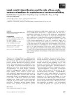
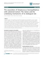
Fig. 1 Cell viability of hepatoma HepG2 and hepatocytes L02 cell line in different concentration of three nHAP based nanoplexs. I: Comparison
among different nanoplexs at same concentration. II: Comparison among various concentration of same nanoplex. Note for graphic II, §☆★○
represent significant difference from NS (control group), 1 μg/ml, 5 μg/ml and 10 μg/ml respectively as calculated with one-way analysis of
variance and Fisher-LSD multiple comparison test
Li et al. BMC Cancer
(2019) 19:1126
Page 5 of 17
Fig. 2 Viability comparison of among cells treated by 15 μg/ml of three nHAP based nanoplexs and L-nanoplex. Note:*△▲represent significant
difference from Pll-nanoplex, Ca-nanoplex and Un-nanoplex with one-way analysis of variance and Fisher-LSD multiple comparison test
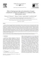
nanoplex(E) > Pll-nanoplex (D) > Ca-nanoplex (C) > Unanoplex (B) at same Observation time points (Fig. 3I).
Transfection efficiency (TE) and mean fluorescence intensity (MFI) was then analyzed by flowcytometry for HepG2
cells. Both TE and MFI of all groups increased in parallel
with time (72hs > 36hs > 12hs) and increased in the order
of E > D > C > B > A for different polyplexs at same Observation time points. However, group D and E showed statistically significant higher TE, MFI, apoptosis and necrosis
rates than other groups. The liposome showed the highest
TE and MFI, whereas Pll-nanoplex induced the most apoptosis and necrosis of HepG2 cell at 36 and 72 hs, respectively, significantly compared to the other three nanoplexs
(P<0.05). As for apoptosis and necrosis analysis, PEGFPC2-wt-p53 is used instead of PEGFP-C2 (Table 1). For the
target gene expression, the expression of EGFP-wt-p53 fusion protein only be detected by in L-nanoplex(E) and Pllnanoplex (D) group at 72 hs and 36 hs (Fig. 3 II).
Only Pll-nanoplex/lipiodol emulsion selectively targeted
and successfully transfer gene to VX2 tumor
The successful transfer of wt-p53 into HepG2 cell line
in vitro could not recapitulate all the necessary process
that happen in HCC in vivo. We therefore sought to address this concern by applying nanoplexs/lipiodol in
rabbit VX2 hepatic cancer model. For target gene expression, western blot showed that the expression of
EGFP-wt-p53 fusion protein only be detected by in
tumor cells of Pll-nanoplex/lipiodol group, whose obvious green fluorescent of also be observed from fluorescent microscope (Fig. 4). Subsequent flowcytometry
showed that TE and MFI of tumor cells in Pll-nanoplex/
lipiodol group were significantly higher than other
groups (Table 2). Transverse CT scan (Fig. 4) revealed
that the specific retention of nanoplex/lipiodol emulsions in implanted VX2 tumor 72 hs after the transarterial delivery, increased with decreased diffuse in liver and
was in the order Pll-nanoplexPll-nanoplex/lipiodol
(D)U-nanoplex/lipiodol (B) > lipiodol (A), liposome-wtp53/lipiodol (E), Ca-nanoplex/lipiodol (C). In fact, group
A, E, C showed no selective retention in tumor (Fig. 4).
For the nanoparticle distribution, TEM, EDS and subsequent elemental mapping all showed that the Pll-nHAP
can only be observed in the cytoplasm of tumor cells but
liver cells, whereas the Ca-nHAP can only be observed
in the cytoplasm of the liver cells but tumor cells, and
the unmodified nHAP can be observed in both tumor
and liver cells (Fig. 5, Fig. 6).
Pll-nanoplex/lipiodol emulsion mediated the most
effective procedure safely in vivo
Overall tumor volumes
As shown in Table 3: There were no significant difference among all groups in preoperative overall tumor
Li et al. BMC Cancer
(2019) 19:1126
Page 6 of 17
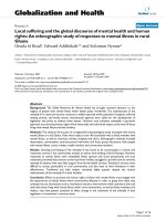
Fig. 3 Obvious green fluorescence of transfected-positive HepG2 cells observed by fluorescence microscope (FM). II:Expression of EGFP-p53
protein transfected-positive HepG2 cells observed by western blot. Note: NS (normal saline+ − PEGFP-C2, A), L-nanoplex(E), Pll-nanoplex (D), Cananoplex (C), U-nanoplex (B)
Table 1 Transfection efficiency (TE), mean fluorescence intensity (MFI), apoptosis rate (AR) and necrosis rates (NR) of HepG2 cells
analyzed by flowcytometry in vitro: pDNA (A),U-nanoplex (B), Ca-nanoplex (C), Pll--nanoplex (D), L-nanoplex (E). All the data were
calculated with one-way analysis of variance and Fisher-LSD multiple comparison tests
TE (%)
MFI
12hs
NR (%)
Group C
Group D
Group E
0
0.1 ± 0.05
0.1 ± 0.10
0.1 ± 0.08
0.7 ± 0.10a
0
0.1 ± 0.06
0.3 ± 0.08
2.1 ± 0.26
20.1 ± 1.53a,b,c,d
72hs
0
0.2 ± 0.02
0.4 ± 0.14
6.3 ± 0.33a,b,c
16.8 ± 1.48a,b,c,d
12hs
86.4 ± 7.22
88.0 ± 5.61
89.5 ± 2.70
96.5 ± 16.00
90.3 ± 2.80
86.3 ± 5.59
93.3 ± 3.77
a,b,c
93.8 ± 3.56
a,b,c
189.9 ± 10.03a,b,c,d
a,b,c
106.7 ± 10.49
72hs
85.4 ± 2.68
97.2 ± 4.62
95.3 ± 3.53
135.4 ± 17.10
143.2 ± 17.66a,b,c,d
36hs
0.2 ± 0.08
5.0 ± 1.47a
0.4 ± 0.06b
6.5 ± 0.71a,b,c
2.0 ± 0.57a,b,c,d
72hs
1.7 ± 0.58
2.5 ± 0.75
1.85 ± 0.28
36.0 ± 1.70
24.6 ± 1.93a,b,c,d
36hs
0.8 ± 0.17
1.7 ± 0.48
1.0 ± 0.06
6.8 ± 0.64a,b,c
9.8 ± 3.38a,b,c,d
72hs
a,b,c,d
Group B
36hs
36hs
AR (%)
Group A
2.1 ± 0.41
3.2 ± 0.89
represent significant difference from group A, B, C, D respectively
2.6 ± 0.41
a,b,c
a,b,c
15.3 ± 4.08
18.0 ± 10.92a,b,c,d
Li et al. BMC Cancer
(2019) 19:1126
Page 7 of 17
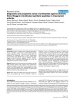
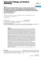
Fig. 4 VX2 tumor can be shown clearly by CT on the left lobe of liver (T, area showed by white cross) before emulsion injection. After in vivo
intra-arterial injection of PEGFP-C2-wt-P53/lipiodol (A), L-nanoplex/lipiodol (E), U-nanoplex/lipiodol (B), Ca-nanoplexCa-nanoplex/lipiodol (C), Pllnanoplex/lipiodol (D), nanoplex emulsion in group D displayed significantly stronger and more selectively deposits in tumor area (D, area showed
by black cross), compared to the slight but selective deposits in group B (B, area showed by black cross), whereas emulsions in group A, C, E
produced no tumor-selective retention potency but diffuse distribution in liver. In contrast to group A, B, C and E, EGFP-wt-P53 expression was
observed by fluorescence microscope (FM) for green fluorescence (the arrow) and by western blot for a ∼ 72 kDa molecular weight band only in
tumor of group D
Table 2 Flowcytometry was utilized to measure and normalize transfection efficiency (TE) and mean fluorescence intensity (MFI) of
harvested tumor cells across different groups in vivo: pDNA/lipiodol (A), L-nanoplex/lipiodol (E), U-nanoplex/lipiodol (B), Cananoplex/lipiodol (C), Pll-nanoplex/lipiodol (D)
Group A
Group E
Group B
Group C
Group D
TE (%)
0.1 ± 0.06
0.2 ± 0.06
0.2 ± 0.07
0.2 ± 0.07
4.1 ± 0.64a,b,c,d
MFI
95.6 ± 4.71
106.5 ± 11.15
05.3 ± 9.27
100.2 ± 12.39
124.4 ± 17.23a,c,d
a,b,c,d
represent significant difference from group A, E, B, C respectively (P < 0.05) . The almost 0% transfected cells in group A exhibit strong autofluorescence,
which attributes to the high background fluorescence. However, group E have more MFI due to the enormous green fluorescent of EGFP-wt-P53 fusion protein in
its 4% pEGFPC2-wt-P53 positive transfected cells. All the data were expressed as mean ± SD and calculated with one-way analysis of variance and Fisher-LSD
multiple comparison tests
Li et al. BMC Cancer
(2019) 19:1126
Page 8 of 17
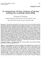
Fig. 5 (See legend on next page.)
volume (PA VS E = 0.282, PA VS B = .054, PA VS C = .344,
PA VSD = .081, PE VS B = .274, PE VS C = .958, PE VS B =
0.526, PB VS C = 0.367, PB VS D = 0.508, PC VSB = 0.656).
One week after trans-arterial administration of different
nanoplex/lipiodol emulsions, significant smaller tumor
volume were observed in group E than other groups (PA
VS E
= 0.598, PA VS B = .057, PA VS C = .834, PA VS
B
= .000, PE VS B = .125, PE VSC = .812, PE VS B < 0.001, PB
Li et al. BMC Cancer
(2019) 19:1126
Page 9 of 17
(See figure on previous page.)
Fig. 5 a Observation of nHAP presence (small black spots showed by arrows) under transmission electron microscopy (TEM) with magnification
of 25,000 times after in vivo intra-arterial injection of polyplex/lipiodol emulsion to VX2 tumor-bearing Rabbits: no nHAP deposit in VX2 tumor cell
(TN) and normal liver cell (LN) of PEGFP-C2-wt-P53/lipiodol group. nHAP deposit in both VX2 tumor cell (TC) and normal liver cell (LC) of Unanoplex/lipiodol group. nHAP deposit in cytoplasm of normal liver cell (LD) but VX2 tumor cell (TD) of Ca-nanoplexCa-nanoplex/lipiodol group.
nHAP can selectively deposit in cytoplasm of VX2 tumor cell (TE) but normal liver cell (LE) of Pll-nanoplex/lipiodol group. b Semi-qualitative
energy dispersive spectroscopy (EDS) spectra of all the tissues above in Fig. 5a were investigated under scanning electron microscopy (SEM): As
presented, their spectra have been overlapped except in the region of 2.010 and 3.692 keV which represent the calcium and phosphorus element
respectively. The peak area of calcium and phosphorus element can be seen in the samples of TC, LC, LD, TE but LN, TN, TD, LE. The main
components of nHAp were calcium and phosphorus in the molar ratio Ca/P around of 2.0, which is similar to the estimated Ca/P molar ratio of
TC, LC, LD, TE . In contrast, the Ca/P molar ratios of LN, TN, TD, LE had similar consequences around 0.6. The EDS analysis further confirm
presence of nHAPs shown in Fig. 5a. Therefore, the existence of nHAP was confirmed in the samples of TC, LC, LD, TE but LN, TN, TD, LE
= 0.125, PB VS D = 0.009, PC VSB < 0.001). Two weeks
after operation, trans-arterial administration of B and D
led to significant delay of tumor growth than group A,
E, C (PA VS E = 0.797, PA VS B = .000, PA VS C = .894, PA
VS D
= .000, PE VS B = .000, PE VS C = .934, PE VS D < 0.001,
B VS C
P
< 0.001, PC VS D < 0.001). In addition, no smaller
tumor volume was noted in Group E than group C 2
weeks after operation(PB VS D865).
VS C
Tumor growth rate (TGW)
For all groups, TGW of all groups increased with the extension of time (2 weeks> 1 week). However, 1 week TGW
of only group D is statistically significant more than other
groups. Two weeks TGW of group B and D were statistically significant more than other groups. Group D has the
least 2 weeks TGW. The overall tumor growth changes
revealed that Pll-nanoplex/lipiodol emulsion can inhibit
Fig. 6 Furthermore, elemental mapping examination has shown the abundant presence of element Calcuim (Ca) and phosphorus (P) in TC, LC,
LD, TE (with the order of TE > LC > TC > LD), while these observations were not observed in LN, TN, TD, LE. Element Oxygenium (O), Carbon (C),
Sulfur (S), Nitrogen (N) present in all tissues show no obvious difference. F represent the fusion image of all element above in tissue
Li et al. BMC Cancer
(2019) 19:1126
Page 10 of 17
Table 3 Preoperational and postoperational VX2 tumor volume
(mm3, mean ± SD) of different groups: pDNA/lipiodol (A), Lnanoplex/lipiodol (E), U-nanoplex/lipiodol (B), Ca-nanoplex/
lipiodol (C), Pll-nanoplex (D). All the data were calculated with
one-way analysis of variance and Fisher-LSD multiple
comparison tests
Groups
preopertion
1 w postopertion
2 w postopertion
A
1257.8 ± 259.49
1937.7 ± 691.15
3873.2 ± 1632.08
E
1169.9 ± 264.69
1860.2 ± 520.80
3789.2 ± 991.56
B
1066.6 ± 220.95
1598.3 ± 323.04
2010.6 ± 546.49a,b
C
1164.9 ± 258.87
1900.6 ± 375.93
3820.6 ± 1059.55c
D
1125.4 ± 216.84
a,b,c,d
1168.6 ± 177.51
a,b,d
1950.1 ± 417.13
a,b,c,d
represent significant difference from group A, E, B, C
respectively (P < 0.05)
the one and two-week significantly more than the others
(Fig. 7). Group E and C inhibited the least tumor growth
than the remaining 3 groups in vivo.
Hepatic function investigation
There is no significant difference in all groups for the
plasma levels of TBL, AST and ALT before operation.
One day postoperation: group E exhibit enhanced ALT
and TBL compared to other groups. Group E and B exhibit enhanced AST compared to other groups. Three
days postoperation: group E exhibit enhanced ALT and
AST compared to other groups. Group E and B exhibit enhanced AST compared to other groups. There is no significant difference in all groups for the plasma levels of
TBL. Five days postoperation: group E exhibit enhanced
ALT than other groups and group D exhibit lower ALT
than group A. Group A exhibit less AST compared to all
other nanoplex groups. There is no significant difference
in all groups for the plasma levels of TBL. Seven days postoperation: group B exhibit enhanced TBL, AST and ALT
than all other groups. In all, contrast to the severe hepatic
function damage of liposome/lipiodol, all the nHAP based
emulsion enhanced the plasma levels of liver markers transiently but all recovered within 1 week post operation, except the slightly increased Tbil of Ca-nanoplex/lipiodol
group (Fig. 6). So nHAP/lipiodol based emulsion is same
safe for long term hepatic function (Fig. 7).
Survival benefit
Log-rank test for Kaplan-Meier curves denied the null
hypothesis “all survival curves are the same”. Further
pairwise comparison show that, compared to group A,
significant longer survival time can be observed in group
B (p = 0.002) and D (p < 0.001) while significant shorter
survival time can be observed in group E (p < 0.001).
There is no significant difference for the survival time
between the Group A and C (p = 0.591). Group D can
significantly enhance the survival benefit than Group B
(p < 0.001). Group D enhance the most survival benefit
(Fig. 8a). The survival time (mean ± SD) for group A, E,
B, C, D are 39.7 ± 4.69, 24.1 ± 6.61, 47.4 ± 9.20, 37.8 ±
7.60 and 60.4 ± 7.99 days, respectively (Fig. 8b).
In all, Pll-nanoplex/lipiodol supplied to the best therapeutic effect without severe influence of hepatic function, whereas liposome/ lipiodol emulsion resulted in
the least survival benefit with most severe influence of
hepatic function despite of its good inhibition of tumor
growth in 2 weeks (Fig. 7).
Surface modified nHAP with pll became cationic and
much smaller
I: As for the zeta-potential, both lipsome and Pll modification can turn very negatively charged nHAP to slightly
cationic nanoplex (Fig. 9I). In all, Only Pll-nHAP can
form cationic nanometeric nanoplex with pDNA. II: Unmodified nHAP (A) and unmodified nHAP-PEGFP-C2wt-p53 complex (E) can easily congregated into large
particles of 251 ± 53.6 nm and 282 ± 65.9 nm in diameter
respectively. Ca(2+) modified nHAP (B) and Ca-nHAPPEGFP-C2-wt-p53 complex (F) crystallized to much larger particles of 851 ± 651.2 nm and 883 ± 658.7 nm in
diameter respectively, even precipitate with very slight
water solubility. Pll modified nHAP (C) disperse with
small particles of 15 ± 3.2 nm but easily congregated,
whereas Pll-nHAP-PEGFP-C2-wt-p53 complex (G) scattered and keep even small particles of 97 ± 13.2 nm in
steady solution. Lipsome (D) and lipsome-PEGFP-C2wt-p53 (H) complex scattered and keep big particles of
555 ± 63.2 nm and 658 ± 71.8 nm respectively. So, TEM
results showed only the Pll-nHAP–pDNA nanoplex can
keep the diameter below 100 nm when any of the others
either can’t form real nanoplex or the one smaller than
500 nm (Fig. 9 II).
Only Pll-nHAP can combine and protect the most pDNA
Gel retardation experiment (Fig. 10) show that, contrary
to U-nanoplex’s disability of pDNA absorption and protection, the positive charged Pll-nanoplex (Pll-nHAP
/pDNA mass ratio more than 15), Ca-nanoplex (CanHAP/pDNA mass ratios more than 25), liposome/
pDNA complex exhibited strong potency of pDNA absorption and protection from the destruction of nucleinase in rabbit serum. Pll-nanoplex can absorb and
protect more pDNA than Ca-nanoplex when same
nHAP was used, which may explain its stronger capability of pDNA transfection efficiency.
No significant differnece for water-in-oil percentage [W/
O], droplet sizes and viscosity of different emulsion
As shown in Table 4, there is no significant difference
for the mean percentage of water-in-oil [W/O], droplet
sizes and viscosity for different emulsion: pDNA/lipiodol
Li et al. BMC Cancer
(2019) 19:1126
Page 11 of 17
Fig. 7 Tumor growth rate (TGW), plasma levels of total biliflavin (TBL), aspartate aminotransferase (AST) and alanine aminotransferase (ALT) in
different groups: PEGFP-C2-wt-P53/lipiodol (A), L-nanoplex/lipiodol (E), U-nanoplex/lipiodol (B), Ca-nanoplex/lipiodol (C), Pll-nanoplex/lipiodol (D).
*△▲☆ represent significant difference from group A,E,B,C respectively as calculated with one-way analysis of variance and Fisher-LSD multiple
comparison test
(A), L-nanoplex/lipiodol (E), U-nanoplex/lipiodol (B),
Ca-nanoplex/lipiodol (C), Pll-nanoplex (D).
Discussion
Our former reports [1, 9, 10, 14] successfully innovated
TAE-gene therapy for hepatocellular carcinoma (HCC)
through application of Pll-nanoplex. This study focus on
comparing and investigating the crucial physicochemical characterizations of four nanoplexs that give
better therapeutic effect and more safety for nano-TAE
gene therapy. The purpose of this new therapy is to
combine the antitumoral effect of nanoparticle, target
gene therapy and transarterial embolization (TAE)
through application of one system. So, all that three requirements must be satisfied when searching the proper
systems for HCC treatment.
First, the nanoplex must have specific anti-tumor activity. Among various non-viral gene carriers, liposome
remain most efficient and prevalent to date. However,
general serious toxicity to the cell membrane [15, 16]
makes it hard to have specific antitumoral effect. HAP,
with molecular formula Ca10(PO4)6(OH)2, is the essential component of human enamel [17–19] and its nanoparticle (nHAP, 0.1-100 nm in diameter) proved to have
good tissue compatibility both in vitro and vivo [20–23].
However, that safety is only observed in bone tissue and
nonparenchymal cell. In the present study, the unmodified nHAP showed comparable cytotoxicity both to
HepG2 and L02 cells, mostly due to its surface properties as well as high negative zeta-potential, whose inner
expulsion also induce nHAP precipitation and congregation [18, 21, 24]. As surface coating is a primary determinant of cytotoxicity, nHAP was surface-modified by
Li et al. BMC Cancer
(2019) 19:1126
Page 12 of 17
Fig. 8 Overall survival curves (a) and survival time (b) of animals from different groups. *△▲☆ represent significant difference from group A,E,B,C
respectively as calculated with one-way analysis of variance and Fisher-LSD multiple comparison test
utilizing Ca(2+) and Pll, representing popular strategies of
inorganic and organic respectively. For Ca-nHAP nanoplex, the particles precipitate to microparticles right after
the Ca(2+) addition and the big particles definitely cover
up the cell membrane and may influence the normal
substance exchange, the main reason for its nonspecific
cytotoxicity. As expected, Pll-nanoplex obviously inhibited the proliferation of hepatoma cells whereas proliferation of normal hepatocyte was relatively slightly
affected, which coincide with the report about TIO2 (titanium oxide) nanoparticles [25]. Contrary to liposome,
U-nHAP and Ca-nHAP nanoplex, we attribute the
Li et al. BMC Cancer
(2019) 19:1126
Page 13 of 17
Fig. 9 Zeta-potential and sizecomparation of various nanoplexs under zeta-potential analyzer (I) and transmission electron microscopy (TEM) with
magnification × 25,000 (II) respectively
privileges of Pll-nanoplex to its nanomentric diameter
and slightly positive organic surface, which have stronger
affinity for cell membranes to accomplish the endocytosis process. As organic molecule with strong affinity for
cell membrane, Pll incorporation reduce nHAP diameter
and cationize its surface, which in turn favor the interaction of nHAP to cell membrane and the following
phagocytosis by tumor under physical conditions. In
addition, the different phagocytosis capability of cancer
and normal cell may also account for that phenomenon.
After phagocytosis, the nanoparticle can distribute in
cytosolic organelles and elevate its Ca(2+) concentration
and in turn induce tumor apoptosis by Ca(2+)-dependent
endonuclease activation [26–28]. Take together, specific
antitumoral effect may be better achieved by particles
with organic surface, proper size (about 100 nm) and
positive superficial zeta-potential (about +10mv) to favor
the swallow of tumor cell but normal cell. In this way,
we can turn cytotoxicity of nanoparticles to specific antitumoral effect [29, 30].
Second, effective gene transfer need an ideal vector to
deliver naked pDNA into cells. pDNA condensation is the
first step for the vector mediated gene delivery [31]. The
features of large surface and high surface energy of nHAP
hold strong DNA binding potency. The unmodified
nHAP, however, with very negative zeta-potential value,
may repel pDNA of same negative potential and thus
inhibited the formation of nHAP-pDNA nanoplex, accounting for the subsequent gene delivery failure. So, the
nanoplex need cationic surface to bind pDNA of negative
potential by the law of opposite charges attract. For that
reason, liposome, Pll-nHAP and Ca-nHAP successfully
compacted the pDNA and formed nanoplexs in this study.
After that, synthetic material employed for gene delivery
should be or become cationic for a higher affinity for the
negatively charged cytoplasm membrane followed by
endocytosis [32–34]. Obviously, all the three above satisfy
this requirement. Ca(2+) have been demonstrated to be the
most potential surface improver for nHAP [35]. However,
the cationic improvement for nHAP was too poor to keep
positive potential of Ca-nanoplex at same concentration
(Ca-nHAP/pDNA mass ratio less than 20). In addition,
Ca(2+) modification promoted congregation and fusion of
nHAP, which in turn decreases their surface area, porosity
Li et al. BMC Cancer
(2019) 19:1126
Page 14 of 17
Fig. 10 PDNA combination (A, B) and protection (C, D) effects of different nanoplex: 0, 1, 5, 10, 15, 20, 25, 50 represent unmodified nHAP /PDNA
mass ratio. a, b, c, d, e, f, g represent Pll-nHAP /PDNA mass ratios of 1, 5, 10, 15, 20, 25, 50 respectively. I, II, III, IV, V, VI, VII represent Ca (2+)-nHAP
/PDNA mass ratios of 1, 5, 10, 15, 20, 25, 50 respectively. L and N represent liposome/PDNA complex and nude PDNA respectively. P represent
PDNA without enzymes
and results in particle bigger, less stable in emulsion and
reduced absorption to pDNA. Moreover, microparticles of
Ca-nHAP is too big to be swallowed by the cells, let alone
the following gene transfer. The reason may be that bivalent cations, such as Mg(2+), Ca(2+) and Zn(2+) atoms
[36–38] may bond to PO4(3−) ionic group of nHAP as tricalcium phosphate (TCP, Ca3(PO4)2), which in turn
changes the microstructure of nHAP, reduces its crystallinity of structure, increase its particle size, as well as promoting its congregation and precipitation. So, Ca(2+)
modification is not suitable for nHAP gene therapy. The
liposome can be swallowed by the cells in vitro but its
diameter (about 500 nm) is also too big to penetrate the
barrier between blood and tumor cells during the processes before when endocytosis can possibly occurs
in vivo [24, 39, 40]. Similar transfection failure of particles
bigger than 250 nm were obtained by synergism of PEI
and liposome [41] and this diameter is proved to selectively target Kupffer cells but the tumor parenchyma cell
[42], indicating that similar system can’t mediated effective
gene therapy to HCCs. In the present study, Pll of organic
polymer, known for pDNA loading and protection, was
also used for the nHAP modification. As expected, the
cationic nHAP-Pll-nanoplex successfully absorbed and
condensed the pDNA into polyplex below 100 nm. Similar
to reports of other cell lines in vitro [43], nHAP mediated
transfection efficiency to HepG2 was much lower than
that of commercial liposome products such as lipofectamine 2000 in this study. However, only Pll-nanoplex can
successfully transfer pDNA to rabbit VX2 tumor in vivo
due to its small enough diameter(< 100 nm) and cationic,
organic polyer surface, which is easier for cell to adhere.
Li et al. BMC Cancer
(2019) 19:1126
Page 15 of 17
Table 4 Mean percentage of water-in-oil [W/O], droplet sizes
and viscosity for different emulsion: pDNA/lipiodol (A), Lnanoplex/lipiodol (E), U-nanoplex/lipiodol (B), Ca-nanoplex/
lipiodol (C), Pll-nanoplex (D)
Groups
W/O (%)
Droplet Size (μm)
Viscosity (cP)
A
65.5 ± 3.23
30.5 ± 3.08
141.6 ± 1.36
E
67.9 ± 4.69
30.2 ± 2.89
138.2 ± 1.58
B
66.6 ± 2.91
28.3 ± 3.08
140.6 ± 2.43
C
64.8 ± 2.82
30.6 ± 3.09
139.6 ± 3.05
D
65.4 ± 2.32
29.6 ± 3.01
139.1 ± 2.91
Similar to that presented here, Zauner [44] observed internalization of only few particles of polystyrene microsphere of > 100 nm in Hepa and HepG2 cell line. So, to
exploit the potential of the complex mediated gene delivery for HCC in vivo, we suggest pDNA entrapment into a
cationic nanometric nanoplex with organic surface (about
100 nm) as the prerequisite criteria.
Third, the specific deposition and retention of the
complexes in HCC is also necessary due to the reason
that all the antitumoral factors, including gene therapy,
TAE and nanoparticle, need long enough time to be
fully exploited in the local tumor site. Lipiodol can
selectively stagnate in HCCs as different time required
for its removal from normal capillaries and tumor neovasculature [45]. Trans-arterial injection of nHAP/lipiodol emulsion successfully achieve tumor embolism,
target retention of nHAP in tumor and subsequent
inhibition of tumor growth. In this study, the specific
deposition of lipiodol and nHAP only in the tumor site
was observed in Pll-nanoplex by CT images and TEM.
Subsequent energy-dispersive spectroscopy (EDS) confirmed specific existence of Pll-nHAPs in VX2 tumor
site. The liposome couldn’t absorb the lipiodol and
develop a integral liposome-based composite, maybe
due to its nonporous fat-soluble surface. That diffused
localization of lipiodol and liposome do no help to the
specific stagnation of liposome-pDNA in tumor. The
Ca-nanoplex can indeed absorb the lipiodol and integrated into one component, but the micrometer particle can easily block the big vessel and make the
lipiodol contraflow to nearly the whole liver, as illustrated in Fig. 9. The unmodified nHAP/lipiodol was
observed in the tumor target of CT images. However,
TEM and EDS result show that more nHAP distribute
in the liver cells than in the tumor cells, suggesting that
the lipiodol in fact may be eliminated by liver but stagnate in the tumor. Energy-dispersive X-ray spectroscopy
(EDS) is an analytical technique used for determining the
presence of chemical elements in a sample and their relative abundance. Its characterization capabilities are due to
the unique atomic structure of each element that can
generate a unique set of peaks on its electromagnetic
emission spectrum after excited by the incoming beam of
X-ray. Electron beam excitation and detection is processed
under scanning electron microscopes (SEM) and transmission electron microscopes (TEM). However, of particular note, elemental mapping and EDS wasn’t be
performed with TEM in this study due to its high working
temperature environment (about 200 °C) operated at 200
keV, which obviously may burn the tumor tissue. With regard to the subcellular distribution, nHAP can distribute
in the cytoplasm in the present study, similar to Radoslav’s
result of rat pheochromocytoma PC12 cells [28].
Fourth, it’s well admitted that the advantages of waterin-oil [W/O] emulsion to oil-in-water [O/W]) emulsion
in embolic effect and longer tumor retention of conventional trans-arterial chemoembolization for hepatocellular carcinoma, due to its higher viscosity, drug carriage
capacity; and a longer drug release time [46]. All the
four emulsions here has similar W/O percentage, droplet
sizes and viscosity. So, there was no significant difference
in the impact of each gene vector system on that three
emulsion characteristics, which then may influence the
tumor uptake and locoregional drug delivery.
From comparisons above, it is easy to understand that
the crystallographical and chemical characteristics of Pllnanoplex, which may satisfy all the nanometric features
called for the combination of TAE-gene therapy, result in
best cyto-tissue compatibility, safe procedure and excellent therapeutic efficiency in vitro and in vivo. Moreover,
the application of lipiodol to nanoplex dramatically improved stability of nHAP emulsion, its stagnation in tumor
target and favor its uptake by tumor cell. Indeed, thoroughly achievements and ideal transfection efficiency were
not observed by using all the four systems in this study
and the most frequently used polyethylenimine (PEI) in
references [33, 34, 41, 47]. However, through comparing
the four systems, a systemic requirement for nanometric
features of materials in this new therapy is proposed and
these guidelines may benefit the screening and identification for future systems. Many studies report the combination of Dosper liposome plus PEI 700 or 2000 as effective
transfection synergism [33, 34, 41]. Recently, we has also
managed to utilize branched PEI modified hydroxyapatite
nanoparticles to transfer siRNA transfection of hepatoma
cells in vitro [14]. Whether this combination can be applied in additional TAE-gene therapy in vivo is our future
interest.
Conclusion
We systematically apply and compare the usage of four
different systems in vitro and in vivo. Though no better
treatments is found than the former study [10], it is important to note that Pll-nHAP differs from unmodified
nHAP, Ca-nHAP in several ways i.e., proper positive organic surface and smaller nano-sized diameter. Though
Li et al. BMC Cancer
(2019) 19:1126
the preliminary investigations in this study for the choice
of synthetic material in hepatoma nano-TAE gene therapy is not adequate to draft defined guidelines concerning this issue, the practical experiences and mechanisms
concluded could potentially be exploited to spur higher
grade of evidence, particularly in vivo studies for TAEgene therapy to HCC.
Abbreviations
Ca-nanoplex: Ca-nHAP-PEGFP-C2; HCC: Hepatocellular carcinoma; MFI: Mean
fluorescence intensity; pDNA: Plasmid DNA; PEI: Polyethylenimine; Pllnanoplex: Pll-nHAP-PEGFP-C2; Pll-nHAP: Hydroxyapatite nanoparticles;
SEM: Scanning electron microscopes; TAE: Transcatheter arterial embolization;
TE: Transfection efficiency; TEM: Transmission electron microscopes;
TGW: Tumor growth rate; U-nanoplex: Unmodified nHAP-PEGFP-C2; wtp53: Wild-type p53
Acknowledgements
Not applicable.
Authors’ contributions
GL carried out the molecular genetic studies, participated in the sequence
alignment and drafted the manuscript. WK, MJ, LZ carried out the western
blot and immunoassays. JZ, KJ, JM participated in the animal research., TL,
XD participated in the design of the study and performed the statistical
analysis. ZY, ZG, JX conceived of the study, and participated in its design and
coordination and helped to draft the manuscript. All authors read and
approved the final manuscript.
Funding
This work was supported by grants from the doctor project of Shanxi Cancer
Hospital, China (2017A06), National Natural Science Foundation of China for
Young Scholars (Grant No: 81201810), science and research fund of Shanxi
Health and Family Planning Commission (Grant No: 201601063), The Key
research Project of Shanxi Province, China (socail development:
201703D321010-1), Natural Science Foundation of Guangdong Province,
China (2015A030313057). The funders had no role in the design of the study
and collection, analysis, and interpretation of data and in writing the
manuscript.
Availability of data and materials
The datasets generated during and/or analysed during the current study are
available from the corresponding author on reasonable request.
Ethics approval and consent to participate
All the animal experiments and breeding were performed under conditions
approved by the Ethics Committee of Shanxi medical university, in
compliance with the NIH Guidelines and items for Care and Use of
Laboratory Animals and in accordance with the Chinese relevant legislation
on animal use.
Consent for publication
Not applicable.
Competing interests
The authors declare that they have no competing interests.
Author details
1
Department of General Surgery, Shanxi Cancer Hospital, Shanxi Medical
University, Taiyuan, Shanxi Province, China. 2Department of Critical Care
Medicine, First Hospital of Shanxi Medical University, Taiyuan, Shanxi
Province, China. 3Department of Anesthesia, Taiyuan Central Hospital,
Taiyuan, Shanxi Province, China. 4Department of General Surgery, Qingxu
People’s hospital, Taiyuan, Shanxi Province, China. 5Department of General
Surgery, Shanxi Cancer Hospital, Shanxi Medical University, Taiyuan, Shanxi
Province, China. 6Department of General Surgery, Shanxi Bethune hospital,
Shanxi academy of medical sciences, Taiyuan, Shanxi Province, China.
Page 16 of 17
Received: 20 April 2019 Accepted: 13 September 2019
References
1. Dong S, Tang Q, Long M, Guan J, Ye L, Li G. The cooperative effect of p53
and Rb in local nanotherapy in a rabbit VX2 model of hepatocellular
carcinoma. Int J Nanomedicine. 2013;8:3757–68.
2. Li G, Ye L, Pan J, et al. Antitumoural hydroxyapatite nanoparticles-mediated
hepatoma-targeted trans-arterial embolization gene therapy: in vitro and
in vivo studies. Liver Int. 2012;32:998–1007.
3. Kulik L, El-Serag HB. Epidemiology and Management of Hepatocellular
Carcinoma. Gastroenterology. 2019;156:477–91.e1.
4. Kim NG, Nguyen PP, Dang H, Kumari R, Garcia G, Esquivel CO, et al.
Temporal trends in disease presentation and survival of patients with
hepatocellular carcinoma: a real-world experience from 1998 to 2015.
Cancer. 2018;124:2588–98.
5. Yang A, Ju W, Yuan X, Han M, Wang X, Guo Z, et al. Comparison between
liver resection and liver transplantation on outcomes in patients with
solitary hepatocellular carcinoma meeting UNOS criteria: a population-based
study of the SEER database. Oncotarget. 2017;8:97428–38.
6. Facciorusso A, Licinio R, Carr BI, Di LA, Barone M. MEK 1/2 inhibitors in the
treatment of hepatocellular carcinoma. Expert Rev Gastroenterol Hepatol.
2015;9:993–1003.
7. Lo CM, Ngan H, Tso WK, et al. Randomized controlled trial of transarterial
lipiodol chemoembolization for unresectable hepatocellular carcinoma.
Hepatology. 2002;35(5).
8. Rognoni C, Ciani O, Sommariva S, Facciorusso A, Tarricone R, Bhoori S, et al.
Trans-arterial radioembolization in intermediate-advanced hepatocellular
carcinoma: systematic review and meta-analyses. Oncotarget. 2016;7:72343–55.
9. Li G, Dong S, Qu J, et al. Synergism of hydroxyapatite nanoparticles and
recombinant mutant human tumour necrosis factor-alpha in chemotherapy
of multidrug-resistant hepatocellular carcinoma. Liver Int. 2010;30(4):585–92.
10. Li G, Ye L, Pan J, et al. Antitumoural hydroxyapatite nanoparticles-mediated
hepatoma-targeted trans-arterial embolization gene therapy: in vitro and
in vivo studies. Liver Int. 2012;32(6):998–1007.
11. Han Y, Li S, Wang X, Bauer I, Yin M. Sonochemical preparation of
hydroxyapatite nanoparticles stabilized by glycosaminoglycans. Ultrason
Sonochem. 2007;14(3).
12. Bauer IW, Li SP, Han YC, Yuan L, Yin MZ. Internalization of hydroxyapatite
nanoparticles in liver cancer cells. J Mater Sci Mater Med. 2008;19(3).
13. Li G, Chen X, Wang Q, Xu Z, Zhang W, Ye L. The roles of four multi-drug
resistance proteins in hepatocellular carcinoma multidrug resistance. J
Huazhong Univ Sci Technolog Med Sci. 2007;27(2):173–5.
14. Xu XL, Yang HY, Ou B, et al. Hydroxyapatite nanoparticles modified by
branched polyethylenimine are effective non-viral vectors for siRNA
transfection of hepatoma cells in vitro. Int J Oncol. 2015;46(5):2138–42.
15. Bose RJ, Arai Y, Ahn JC, Park H, Lee SH. Influence of cationic lipid
concentration on properties of lipid-polymer hybrid nanospheres for gene
delivery. Int J Nanomedicine. 2015;10:5367–82.
16. Fortier C, Durocher Y, De Crescenzo G. Surface modification of nonviral
nanocarriers for enhanced gene delivery. Nanomedicine (Lond). 2014;9:135–51.
17. Leroy C, Aussenac F, Bonhomme-Coury L, Osaka A, Hayakawa S, Babonneau
F, et al. Hydroxyapatites: key structural questions and answers from dynamic
nuclear polarization. Anal Chem. 2017;89:10201–7.
18. Gelli R, Del BS, Tempesti P, Bonini M, Ridi F, Baglioni P. Enhanced formation
of hydroxyapatites in gelatin/imogolite macroporous hydrogels. J Colloid
Interface Sci. 2018;511:145–54.
19. John Ł, Janeta M, Szafert S. Designing of macroporous magnetic bioscaffold
based on functionalized methacrylate network covered by hydroxyapatites
and doped with nano-MgFe2O4 for potential cancer hyperthermia therapy.
Korean J Couns Psychother. 2017;78:901–11.
20. Iannotti V, Adamiano A, Ausanio G, Lanotte L, Aquilanti G, JMD C, et al.
Fe-doping-induced magnetism in Nano-hydroxyapatites. Inorg Chem.
2017;56:4447–59.
21. Petit S, Gode T, Thomas C, Dzwigaj S, Millot Y, Brouri D, et al. Incorporation
of vanadium into the framework of hydroxyapatites: importance of the
vanadium content and pH conditions during the precipitation step. Phys
Chem Chem Phys. 2017;19:9630–40.
22. Kolmas J, Piotrowska U, Kuras M, Kurek E. Effect of carbonate substitution on
physicochemical and biological properties of silver containing
hydroxyapatites. Korean J Couns Psychother. 2017;74:124–30.
Li et al. BMC Cancer
(2019) 19:1126
23. Lambert F, Bacevic M, Layrolle P, Schüpbach P, Drion P, Rompen E. Impact
of biomaterial microtopography on bone regeneration: comparison of three
hydroxyapatites. Clin Oral Implants Res. 2017;28:e201–201e207.
24. Gril B, Paranjape AN, Woditschka S, Hua E, Dolan EL, Hanson J, et al.
Reactive astrocytic S1P3 signaling modulates the blood-tumor barrier in
brain metastases. Nat Commun. 2018;9:2705.
25. Xian-ying C. Selective anti hepatoma with IIO2 nanop. Wuhan Univ Technol.
2003;18(1).
26. Bellomo G, Perotti M, Taddei F, et al. Tumor necrosis factor alpha induces
apoptosis in mammary adenocarcinoma cells by an increase in intranuclear
free Ca2+ concentration and DNA fragmentation. Cancer Res. 1992;52(5).
27. Baumann S, Fas SC, Giaisi M, et al. Wogonin preferentially kills malignant
lymphocytes and suppresses T-cell tumor growth by inducing PLCgamma1and Ca2+−dependent apoptosis. Blood. 2008;111(4).
28. Savic R, Luo L, Eisenberg A, Maysinger D. Micellar nanocontainers distribute
to defined cytoplasmic organelles. Science. 2003;300(5619).
29. Colvin VL. The potential environmental impact of engineered nanomaterials.
Nat Biotechnol. 2003;21(10).
30. Roco MC. Environmentally responsible development of nanotechnology.
Environ Sci Technol. 2005;39(5).
31. Fink TL, Klepcyk PJ, Oette SM, et al. Plasmid size up to 20 kbp does not limit
effective in vivo lung gene transfer using compacted DNA nanoparticles.
Gene Ther. 2006;13(13).
32. Guo Y, Wu Z, Shen S, Guo R, Wang J, Wang W, et al. Nanomedicines reveal
how PBOV1 promotes hepatocellular carcinoma for effective gene therapy.
Nat Commun. 2018;9:3430.
33. Meneksedag-Erol D, KC RB, Tang T, Uludağ H. A delicate balance when
substituting a small Hydrophobe onto low molecular weight
Polyethylenimine to improve its nucleic acid delivery efficiency. ACS Appl
Mater Interfaces. 2015;7:24822–32.
34. Choi JW, Nam JP, Nam K, Lee YS, Yun CO, Kim SW. Oncolytic adenovirus
coated with multidegradable bioreducible Core-cross-linked
Polyethylenimine for cancer gene therapy. Biomacromolecules. 2015;16:
2132–43.
35. Mozafari MR, Omri A. Importance of divalent cations in nanolipoplex gene
delivery. J Pharm Sci. 2007;96(8).
36. Sun J, Zheng X, Li H, et al. Monodisperse selenium-substituted
hydroxyapatite: controllable synthesis and biocompatibility. Korean J Couns
Psychother. 2017;73:596–602.
37. Suruagy AA, Alves AT, Sartoretto SC, Calasans-Maia JA, Granjeiro JM,
Calasans-Maia MD. Physico-chemical and Histomorphometric evaluation of
zinc-containing hydroxyapatite in rabbits Calvaria. Braz Dent J. 2016;27(6):
717–26.
38. de Val JEMS, Calvo-Guirado JL, Gómez-Moreno G, Pérez-Albacete MC,
Mazón P, De Aza PN. Influence of hydroxyapatite granule size, porosity, and
crystallinity on tissue reaction in vivo. Part a: synthesis, characterization of
the materials, and SEM analysis. Clin Oral Implants Res. 2016;27(11):1331–8.
39. Arvanitis CD, Askoxylakis V, Guo Y, Datta M, Kloepper J, Ferraro GB, et al.
Mechanisms of enhanced drug delivery in brain metastases with focused
ultrasound-induced blood-tumor barrier disruption. Proc Natl Acad Sci U S
A. 2018;115:E8717–8717E8726.
40. Zhou W, Chen C, Shi Y, Wu Q, Gimple RC, Fang X, et al. Targeting glioma
stem cell-derived Pericytes disrupts the blood-tumor barrier and improves
chemotherapeutic efficacy. Cell Stem Cell. 2017;21:591–603.e4.
41. Lampela P, Elomaa M, Ruponen M, Urtti A, Mannisto PT, Raasmaja A.
Different synergistic roles of small polyethylenimine and Dosper in gene
delivery. J Control Release. 2003;88(1).
42. Popielarski SR, Hu-Lieskovan S, French SW, Triche TJ, Davis ME. A
nanoparticle-based model delivery system to guide the rational design of
gene delivery to the liver. 2. In vitro and in vivo uptake results. Bioconjug
Chem. 2005;16(5).
43. Frayssinet P, Rouquet N, Mathon D. Bone cell transfection in tissue culture
using hydroxyapatite microparticles. J Biomed Mater Res A. 2006;79(2).
44. Zauner W, Farrow NA, Haines AM. In vitro uptake of polystyrene
microspheres: effect of particle size, cell line and cell density. J Control
Release. 2001;71(1):39–51.
45. Vogl TJ, Lahrsow M, Albrecht MH, Hammerstingl R, Thompson ZM, GruberRouh T. Survival of patients with non-resectable, chemotherapy-resistant
colorectal cancer liver metastases undergoing conventional lipiodol-based
transarterial chemoembolization (cTACE) palliatively versus neoadjuvantly
prior to percutaneous thermal ablation. Eur J Radiol. 2018;102:138–45.
Page 17 of 17
46. Masada T, Tanaka T, Nishiofuku H, et al. Techniques to form a suitable
Lipiodol-Epirubicin emulsion by using 3-way stopcock methods in
Transarterial chemoembolization for liver tumor. J Vasc Interv Radiol. 2017;
28(10):1461–6.
47. Wang W, Balk M, Deng Z, Wischke C, Gossen M, Behl M, et al. Engineering
biodegradable micelles of polyethylenimine-based amphiphilic block
copolymers for efficient DNA and siRNA delivery. J Control Release. 2016;
242:71–9.
Publisher’s Note
Springer Nature remains neutral with regard to jurisdictional claims in
published maps and institutional affiliations.