The diversity between curatively resected pancreatic head and body-tail cancers based on the 8th edition of AJCC staging system: A multicenter cohort study
Bạn đang xem bản rút gọn của tài liệu. Xem và tải ngay bản đầy đủ của tài liệu tại đây (996.87 KB, 11 trang )
Sheng et al. BMC Cancer
(2019) 19:981
/>
RESEARCH ARTICLE
Open Access
The diversity between curatively resected
pancreatic head and body-tail cancers
based on the 8th edition of AJCC staging
system: a multicenter cohort study
Weiwei Sheng1, Ming Dong1*, Guosen Wang1, Xiaoyang Shi1, Wei Gao1, Kewei Wang1, He Song1, Gang Shi2 and
Xiaodong Tan3
Abstract
Background: To our knowledge, there are no studies to systematically compare the detailed clinical significance
between curatively resected pancreatic head (ph) and body-tail (pbt) ductal adenocarcinoma based on the new 8th
edition of AJCC staging system (8th AJCC stage) that was just applied in clinical practice in 2018.
Methods: Three hundred fifty-one patients with curatively resected pancreatic adenocarcinoma (PC) from three
center hospitals were entered into this multicenter cohort study.
Results: Increasing tumor size (P < 0.001), T stage (T1 + T2 vs T3 + T4, P = 0.003), frequent postoperative liver
metastasis (PLM) (P = 0.002) and 8th AJCC stage (IA to VI, P < 0.001; I + II vs III + IV, P = 0.002) were closely associated
with the progression of pbt cancers compared with that in ph cancer patients. Moreover, tumor size≥3 cm (P =
0.012), 8th AJCC stage (III + IV) (P = 0.025) and PLM (P = 0.010) were identified as independent risk factors in pbt
cancers in logistic analysis. Patients with pbt cancers had a significantly worse overall survival compared with ph
cancer patients (P = 0.003). Moreover, pbt was an independent unfavorable factor in multivariate analysis (P = 0.011).
In addition to lymph nodes metastasis, 8th AJCC stage, vascular invasion and PLM, increasing tumor size and
advanced T stage were also closely associated with the poor prognosis in 131 cases of pbt cancer patients
compared with Ph cancer patients.
Conclusion: Pbt, as an independent unfavorable factor for the prognosis of PC patients, are much more aggressive
than that in ph cancers according to 8th AJCC staging system. 8th AJCC staging system are more comprehensive
and sensitive to reflect the malignant biology of pbt cancers.
Keywords: Pancreatic head and body-tail cancers, Clinical significance, Prognosis, 7th and 8th edition of AJCC
staging system
Background
From 2000 to 2011, pancreatic adenocarcinoma (PC)
takes up the second upward trend of age-standardized
mortality rates in the Chinese male population [1].
Meanwhile, it is the fourth most common cause of cancer death in the United States and Japan [2, 3]. Despite
advances in multimodality treatment, long-term survival
* Correspondence:
1
Department of gastrointestinal surgery, the First Hospital, China Medical
University, Shenyang 110001, China
Full list of author information is available at the end of the article
hasn’t shown improvement over the past several decades
[4]. The poor prognosis of PC is mainly due to the late
diagnosis and advanced progression, most patients with
PC are diagnosed at stages III and IV [5]. Even following
curative resection, the reported 5-year survival rate remains low (7–24%) [6]. Accurate evaluation of tumor
stage is a prerequisite for further treatment and prognostic prediction. The AJCC TNM staging system has
been widely applied worldwide as the most authorized
tool for tumor staging assessment. AJCC released the
© The Author(s). 2019 Open Access This article is distributed under the terms of the Creative Commons Attribution 4.0
International License ( which permits unrestricted use, distribution, and
reproduction in any medium, provided you give appropriate credit to the original author(s) and the source, provide a link to
the Creative Commons license, and indicate if changes were made. The Creative Commons Public Domain Dedication waiver
( applies to the data made available in this article, unless otherwise stated.
Sheng et al. BMC Cancer
(2019) 19:981
8th edition (8th AJCC stage), which incorporated significant changes in the T and N classification of PC [7].
Most studies in terms of PC focuse on the head of the
pancreas (ph), whereas rare data is regarding pancreatic
tail and body (pbt) cancers. Previous studies investigate
the incidence rate and survival time between ph and pbt
ductal cancers [8]. However, the results remain controversial and the relationship between tumor location and
clinical characters is rarely reported. Meanwhile, to
the best of our knowledge, there is no studies to systematically compare the clinical significance between
curatively resected ph and pbt cancers based on the
new8th AJCC stage [8]. Based on the new 8th AJCC
stage, we find new clinical difference between curatively resected ph and pbt cancers, which provides a
new clinical sight in revealing the malignant biology
of PC, especially in pbt cancer.
Methods
Patients
This research protocol was approved by the ethical committee of the institutional review board of China Medical
University and a consent form was signed by each participating patient. All patients enrolled from the First hospital
of China Medical University, Shengjing hospital of China
Medical University and Cancer hospital of China Medical
University were histologically proven to be pancreatic
ductal adenocarcinomas. Contrast computed tomography
(CT)/positron emission tomography (PET), contrast nuclear magnetic resonance (MRI) and surgical exploration
were used to ensure whether all PC patients meet our resection criteria as Sugiura et al. previously reported [9], including: a) no distant metastasis, b) tumor extension to
Page 2 of 11
the superior mesenteric artery or celiac trunk was less
than 90°and can be completely resected and constructed.
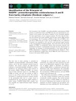
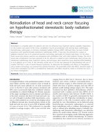
The detailed enrollment procedure was shown in Fig. 1.
Based on above criteria, 351 cases of consecutive PC patients underwent pancreatoduodenectomy (PD) or distal
pancreatectomy (PDP) were finally entered into this study
between 2008 and 2016. In order to achieve R0 resection,
cancer resection margins were at least 1 mm as cut-off.
Meanwhile, some cases with peripancreatic invasion
underwent corresponding organ resection, such as spleen,
left adrenal gland, gastrointestine (partial stomach, duodenum, intestine or colon), artery (hepatic, superior mesenteric and celiac artery) and vein (portal or superior and
inferior mesenteric vein). 6 PC patients were detected a
single liver metastasis (preoperative CT examination is
not detected) in surgery, we additionally executed partial
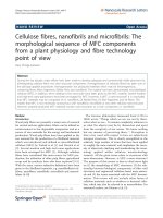
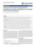
hepatectomy. A dedicated table for patients’ characteristics was summarized in Table 1. Four classic samples from
consecutive PC patients underwent radical PD and PDP
showed in Fig. 2.
Follow-up
All patients were followed up by the operating surgeons.
As described previously [6], postoperative patients were
performed routinely laboratory examinations, including
tumor markers, liver function, US, abdominal CT/PET
or contrast MRI every 3–6 months. For postoperative
liver metastasis (PLM), if the liver metastasis showed no
definite evidence of other metastasis or recurrence
elsewhere, we characterized the newly developed
hepatic lesion as PLM [10]. Patient follow-up examinations was performed each 3 months for the first 2
postoperative years, every 6 months for > 2 years, and
yearly thereafter. One hundred twenty-five cases of ph
cancer patients (125/220, 56.8%) and 73 cases of pbt
cancer patients (73/131, 55.7%) accepted postoperative
gemcitabine-based chemotherapy, no difference was
shown in two groups with or without chemotherapy
treatment.
Statistical analysis
Fig. 1 Study flow chart. Undergoing strict selection, 351 cases of PC
patients were finally entered into this study from three multiple
centers. PC: pancreatic adenocarcinoma; Ph: pancreatic head; Pbt:
pancreatic body-tail
Statistical analysis was performed using SPSS software
19.0. The differences between curatively resected ph and
pbt cancers was analyzed using a Chi-Squared test. A logistic regression analysis was performed to determine
the pathologic impact findings that were significant with
regard to differences in the univariate analysis. The
Kaplan-Meier method was used to estimate PC
patients’ survival, and differences were analyzed by
the log-rank test. The variables that were significant
by the univariate analysis were subjected to a multivariate Cox proportional hazards regression analysis
Sheng et al. BMC Cancer
(2019) 19:981
Page 3 of 11
Table 1 The clinical data in 351 cases of PC patients with curatively surgical resection
Parameters
No. of patients
Parameters
No. of patients
Cases
351
Cases
351
Age (years)
Perineural invasion
≤ 65
240
Absent
278
> 65
111
Present
73
Gender
Vascular permeation
Male
210
Female
141
Tumor size (cm)
Absent
266
Present
85
Pre-therapeutic CA19–9 level
<3
108
Mean ± SD
≥3
243
PLM
Absent
227
Ph
220
Present
124
Pbt
131
Tumor location
Differentiation
355 ± 283
7th AJCC stagea
IA
19
Well
140
IB
140
Moderate
118
IIA
76
Poor
93
IIB
104
IV
6
8th T stage
T1
35
T2
167
PD alone
T3
143
PD + gastrointestine
8
T4
6
PD + portal or superior
mesenteric vein
11
PD+ hepatic or superior
mesenteric artery
3
7th T stagea
Surgical procedures
196
T1
27
PD + liver
2
T2
215
PDP alone
97
T3
103
PDP + gastrointestine
12
PDP + portal vein or inferior mesenteric vein
6
Lymph nodes metastasis
N0
244
N1
91
PDP + liver+ left adrenal
1
N2
16
PDP + liver
3
PDP + left adrenal
4
IA
24
PDP+ celiac artery
3
IB
119
Postoperative chemotherapy
IIA
93
Ph cancers
125/220
IIB
86
Pbt cancers
73/131
III
23
IV
6
8th AJCC stage
PDP + gastrointestine
+left adrenal
5
N1 Lymph nodes metastasis 1–3; N2 Lymph nodes metastasis> 3; PLM postoperative live metastasis; PD Pancreatoduodenectomy; PDP Distal pancreatectomy7th
and 8th AJCC stage 7th and 8th edition of AJCC staging system in PC; Ph Pancreatic head; Pbt Pancreatic body-tail. a 6 cases of T4 stage in 8th AJCC stage (III)
were exclude in 7th AJCC stage
Sheng et al. BMC Cancer
(2019) 19:981
Page 4 of 11
Fig. 2 Four classic samples from consecutive PC patients underwent PD or PDP. a, c Under PD treatment, two ph tumor samples was shown as
arrows suggested. b, d Under PDP treatment, two pbt tumor samples was shown as arrows suggested. PD: Pancreatoduodenectomy; PDP: distal
pancreatectomy. Ph: pancreatic head; Pbt: pancreatic body-tail
in a stepwise manner. A value of P < 0.05 was considered
to be statistically significant.
IA, IB, IIA, IIB and IV was 5.5, 40.5, 22, 30.1 and 1.7% in
7th AJCC stage (III stage that was defined as “unresectable” PC were excluded).
Results
Comparison of the 7th and 8th editions of the TNM
staging system for patients
The detailed information of 7th and 8th AJCC stage in
PC was summarized in Additional file 1: Table S1 and
Additional file 2: Table S2. Briefly, in the 8th edition,
stages T1-T3 are redefined according to tumor size.
When the tumor invades the celiac axis, hepatic artery
and/or superior mesenteric artery, it is defined as T4,
and the classification as “unresectable” was removed. Because all the patients enrolled in this study accept the
curative resection, 6 PC patients with T4 stage (III stage)
based on 8th AJCC stage were exclude in 7th AJCC system (Table 1). In addition, the N classification was further subdivided according to the number of positive
lymph nodes as N0, N1 and N2. T1–3N2M0 was defined
as stage III in 8th AJCC stage, while it was defined as
stage IIB in 7th AJCC stage. In current study, 14.9% (16/
107) of these patients had metastasis more than 3 lymph
nodes (pN2) (Table 1). The ratio of stage IA, IB, IIA,
IIB, III and IV of 8th AJCC stage was 6.8, 33.9, 26.4,
24.3, 6.5 and 1.7%, respectively, while the ratio of stage
Different clinical significance between ph and pbt cancers
in 351 cases PC patients with curative resection
Chi-Squared test in Table 2 showed that tumor size, T
stage, 8th AJCC stage and PLM were significantly different between ph and pbt cancers. Increasing tumor size
≥3 cm (ph 60.7% vs 81.6%; P < 0.001), frequent PLM (ph
29% vs pbt 45.8%, P = 0.002) and advanced T (T3 + T4,
ph 36.3% vs pbt 52.7%, P = 0.003) and 8th AJCC stage
(IA to VI, P < 0.001; III + IV, ph 5.2% vs pbt 14.5%, P =
0.002) were closely associated with the progression of
pbt cancers compared with that in ph cancers. However,
age, gender, tumor differentiation, lymph nodes metastasis, CA199 level and perineural and vascular invasion
showed no difference (P > 0.05). A multivariate analysis
(logistic regression analysis) identified tumor size≥3 cm
(P = 0.012), 8th AJCC stage (III + IV) (P = 0.025) and
PLM (P = 0.010) as independent risk factors in pbt cancers (Table 2). It was worthy noted that T and TNM
stage based on 7th AJCC stage system showed no significant difference in both cohorts, which implying a close
(2019) 19:981
Sheng et al. BMC Cancer
Page 5 of 11
Table 2 Clinical significance between ph and pbt cancers in 351 cases PC patients with curatively resection
Parameters
Cases
No. of
patients
Chi square
P
Head Body-tail
Multivariate
analysis
Odds ratio
(95% CI)
P
2.133(1.180–3.856)
0.012
1.344(0.805–2.243)
0.258
2.520(1.121–5.665)
0.025
351
Age (years)
≤ 65
240
157
83
> 65
111
63
48
Male
210
131
79
Female
141
89
52
0.125
Gender
0.911
Tumor size (cm)
<2
35
28
7
≥2
316
192
124
<3
108
84
24
≥3
243
136
107
Well
140
86
54
Moderate
118
73
45
poor
93
61
32
N0
244
155
89
N1
91
59
32
N2
16
6
10
T1 + T2
242
159
83
T3
103
58
45
IA
19
13
6
IB
140
86
54
IIA
76
45
31
IIB
104
71
33
IV
6
2
4
T1 + T2
202
140
62
T3 + T4
149
80
69
0.027
Tumor size (cm)
0.000
Differentiation
0.793
Lymph nodes metastasis
0.101
7th T stagea
7th AJCC stageb
0.114
0.360
8th T stage
8th AJCC stage
0.003
0.000
IA
24
18
6
IB
119
84
35
IIA
93
42
51
IIB
86
66
20
III
23
8
15
IV
6
2
4
321
209
112
8th AJCC stage
I + II
0.002
(2019) 19:981
Sheng et al. BMC Cancer
Page 6 of 11
Table 2 Clinical significance between ph and pbt cancers in 351 cases PC patients with curatively resection (Continued)
Parameters
III + IV
No. of
patients
Chi square
P
Head Body-tail
30
11
19
Absent
278
180
98
Present
73
40
33
Absent
266
172
94
Present
85
48
37
/
389.6 ± 255.7
324.2 ± 283.3
0.429
Absent
227
156
71
0.002
Present
124
64
60
Multivariate
analysis
Odds ratio
(95% CI)
P
1.854(1.160–2.963)
0.010
Perineural invasion
0.118
Vascular permeation
0.174
Pre-therapeutic CA19–9 level
Mean ± SD
PLM
N1 Lymph nodes metastasis 1–3; N2 Lymph nodes metastasis> 3; PLM postoperative live metastasis; 7th and 8th AJCC stage 7th and 8th edition of AJCC staging
system in PC; Ph Pancreatic head; Pbt Pancreatic body-tail. a, b 6 cases of T4 stage in 8th TNM stage (III) were exclude
Table 3 Univariate and multivariate analysis for prognostic factors in 351 cases of PC patients with curatively surgical resection
Parameters
median survival
(days)
Univariate analysis
P (log rank)
Multivariate analysis
hazard ratio
(95% CI)
Age (< 65/ ≥65 years)
432/421
0.127
─
Gender (male/female)
421/472
0.366
─
Tumor location
(ph/pbt)
488/340
0.003
1.405(1.082–1.826)
Tumor size (< 2/ ≥2 cm)
472/421
0.371
─
Tumor size (< 3/ ≥3 cm)
472/418
0.096
─
Well/Moderate/poor
Differentiation
452/420/382
0.071
─
T stage
(T1 + T2/ T3 + T4)
472/381
0.068
─
Lymph nodes metastasis
8th (N0/N1/N2)
480/330/284
0.001
1.451(1.123–1.874)
Lymph nodes metastasis
7th (N0/N1 + N2)a
472/399
0.090
─
8th AJCC stage
(I + II /III + VI)
468/284
0.007
1.442(1.085–1.915)
Perineural invasion (absent/present)
454/400
0.179
─
Vascular permeation
(absent/present)
480/330
0.004
1.401(0.905–2.168)
CA19–9 level
(< 37 U/ml/ ≥37 U/ml)
565/395
0.104
─
PLM
(absent/present)
499/330
0.000
1.594(1.224–2.076)
468/172
0.012
Not included
7th AJCC stage
P
0.011
0.004
0.012
0.131
0.001
N1 Lymph nodes metastasis 1–3; N2 Lymph nodes metastasis> 3; 7th and 8th AJCC stage 7th and 8th edition of AJCC staging system in PC; Ph Pancreatic head;
Pbt Pancreatic body-tail. a In 7th AJCC stage, N1 and N2 combined together
Sheng et al. BMC Cancer
(2019) 19:981
relationship of pbt cancers with the advanced clinal
stage based on 8th AJCC stage.
Prognostic factors of PC patients who underwent curative
pancreatectomy
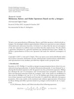
Patients with pbt cancers had a significantly worse overall survival compared with ph cancer patients (P = 0.003)
in univariate analysis (Table 3) (Fig. 3a). Meanwhile,
lymph nodes metastasis (P = 0.001), 8th AJCC stage (P =
0.007), vascular permeation (P = 0.004) and PLM (P <
0.001) were also associated with PC patients’ poor prognosis. In multivariate model, tumor location (P = 0.011),
lymph nodes metastasis (P = 0.004), 8th AJCC stage (P =
0.012) and PLM (P = 0.001) were independent unfavorable prognostic indicators in PC (Table 3). 7th AJCC
stage was also associated with the poor prognosis of PC
patients (P = 0.012). Interestingly, previous studies show
that pbt cancer patients have a better prognosis than ph
cancer patients in early 7th AJCC I and II stage [11]. In
current study, pbt cancer patients showed worse prognosis in both 8th AJCC I-III stage and I-II stage compared
with ph cancer patients (Fig. 3b, c). Only in 8th AJCC I
stage, the median survival days of pbt cancer patients
was longer than that in ph cancer patients, but no statistic difference (data not shown). In addition, lymph node
metastasis (N0/N1) in 7th AJCC stage failed to stratify
patients by survival, whereas lymph node metastasis
(N0/N1/N2) based on 8th AJCC stage was an
independent unfavorable prognostic indicator in our
current study. It indicated that lymph node metastasis
in 8th AJCC stage is more comprehensive to reflect
the malignant progression and poor prognosis of PC
patients.
Page 7 of 11
Different prognostic indicators in ph and pbt cancer
patients with curative surgical resection
Lymph node metastasis, 8th AJCC stage, vascular invasion and PLM were associated with the poor prognosis
in 220 cases of Ph cancer patients (Table 4). In 131 cases
of pbt cancer patients, in addition to above characters,
tumor size and T stage were identified as the poor prognostic indicators (Table 4). More clinical factors based
on 8th AJCC stage were the prognostic indicators in pbt
cancer compared with the ph cancer.
Discussion
8th AJCC stage demonstrates a more equal distribution
among stages and increases prognostic accuracy compared with 7th AJCC stage. In an international multicenter cohort study including 1525 consecutive patients, the
new T stage does not demonstrate significant correlation
with survival on univariate or multivariate analysis,
whereas the new N stage showed accurate discrimination
of survival [12]. These results were consistent with our
current study. However, the superiority of the 8th edition
in evaluating the relationship between tumor location and
clinical characters has not been investigated in PC patients, to our knowledge. Based on the new 8th AJCC
stage, we found new diversity between ph and pbt cancers
from a multicenter cohort study.
In anatomy, cell composition, blood supply,
lymphatic and venous backflow,
and innervations are significantly different between ph
and pbt cancers [13]. In clinic, tumors at different locations (ph vs pbt) display different clinical presentation,
treatment efficiency (surgery and chemoradiotherapy) and prognosis [14]. The incidence rate for ph cancer has remained at 5.6% per 100,000, whereas the rate
Fig. 3 The prognosis between ph and pbt cancers with different clinical stage of 8th AJCC. a. The prognosis between ph and pbt cancers in 8th
AJCC stage I to III. b. The prognosis between ph and pbt cancers in 8th AJCC stage I to III. c. The prognosis between ph and pbt cancers in 8th
AJCC stage I to II
Sheng et al. BMC Cancer
(2019) 19:981
Page 8 of 11
Table 4 Difference of prognostic factors in Ph and Ptb cancer patients with curatively surgical resection
Tumor location
Parameters
Median survival
(days)
Univariate analysis
P (log rank)
220
Ph cancers
Age (< 65/ ≥65 years)
499/480
0.131
Gender (male/female)
454/615
0.335
Tumor size (< 3/ ≥3 cm)
555/488
0.358
Well/Moderate/poor
Differentiation
615/555/411
0.155
T stage
(T1 + T2/T3)
488/454
0.105
Lymph nodes metastasis (N0/N1/N2)
565/418/185
0.004
8th AJCC stage
(I + II /III + IV)
360/273
0.017
Perineural invasion (absent/present)
880/545
0.298
Vascular permeation
(absent/present)
565/350
0.005
CA19–9 level
(< 37 U/ml/ ≥37 U/ml)
666/450
0.171
PLM
131
Pbt cancers
absent/present
586/365
0.039
Age (< 65/ ≥65 years)
381/300
0.111
Gender (male/female)
395/340
0.439
Tumor size (< 3/ ≥3 cm)
530/320
0.023
Well/Moderate/poor
Differentiation
400/280/273
0.070
T stage
(T1 + T2/ T3 + T4)
418/320
0.016
Lymph nodes metastasis (N0/N1/N2)
360/259/234
0.007
8th AJCC stage
(I + II /III + IV)
499/273
0.001
Perineural invasion (absent/present)
360/333
0.104
Vascular permeation
(absent/present)
400/265
0.009
CA19–9 level
(< 37 U/ml/ ≥37 U/ml)
468/331
0.099
PLM
432/275
0.001
absent/present
N1 Lymph nodes metastasis 1–3; N2 Lymph nodes metastasis> 3; 8th AJCC stage 8th edition of AJCC staging system in PC; Ph Pancreatic head; Pbt
Pancreatic body-tail
for pbt cancers has increased by 46% between 1973 and
2002 in the SEER database [7]. Though both ph and pbt
cancers had a higher proportion diagnosis in the distant
stages (a neoplasm that has spread to parts of the body
remotes from the primary tumor or to distant lymph
nodes), patients with ph cancer were more likely to have
localized and regional diseases (12.9 and 32.2%, respectively) as compared with pbt cancers (6.6 and 13.9%, respectively) [7]. According to 7th AJCC stage, there was
no significant difference in TNM stage between resected
ph and pbt cancers [15]. However, in current study, we
find new clinical difference between curatively resected
ph and pbt cancers bases on 8th AJCC stage, which
hasn’t been reported previously to our knowledge.
The alteration of the definitions of T and N is the
main changes in 8th AJCC stage compared with the 7th
AJCC stage [16]. Just shown in Additional file 1: Table
S1 and Additional file 2: Table S2, extra-pancreatic invasion can be difficult to predict accurately before surgery
and may be inconsistently assessed by pathologists [17].
T3 tumors are now defined as those ≥4 cm, while nodal
involvement has been improved from a binary system to
one based on extent of nodal involvement. In current
study, increasing tumor size and advanced T stage and
Sheng et al. BMC Cancer
(2019) 19:981
8th AJCC stage were closely associated with the progression of pbt cancers compared with ph cancers. Only one
study shows tumor size but not T and clinical stage in
7th AJCC stage exhibits difference in resected ph (56
cases) and pbt (24 cases) cancers [15], which is consistent with our study. Based on the alteration of T and N
status in 8th AJCC stage, T1–3 stage was likely a stratified analysis of tumor size. Meanwhile, new 8th AJCC
stage mainly increased III stage (16 vs 0) but decreased
IIB stage (86 vs 104) in PC patients compared with 7th
AJCC stage in our study, which is the critical reason for
the discrimination in above results just as Omar AbdelRahman suggested [18]. We additionally found PLM was
more frequent in pbt cancers, which is consistent with
the study by Maria Chiara Ambrosetti et al. [19]. However, Nakata B et al. show that the recurrence of peritoneum, liver, lung and bone showed no difference in
tumor location [15]. Among 707 unresectable PC patients with stage III, 30.1% developed PLM. However, no
risk factors were identified among these patients [20].
The inconsistence might be due to the different sample
size and diversity in national population included in different studies.
Currently, prognostic difference between ph and pbt
cancer patients remain controversial. Data from SEER database (1988–2004) including 33,752 PC patients
presents a significant lower median survival (4 months vs 6
months)inpatientswithpbtcancercomparedwiththosewith
ph cancer [21]. However, data from the national PC registry of Japan showed a significant lower 5-year survival rate (10.7% vs 13.8%) for patients with ph cancers (n =
5788) than those with pbt cancers (n = 1629) [22]. Both
unresectable and resectable PC patients are enrolled in
above studies. In our current study, we only enrolled curatively resected PC patients from three multiple centers.
Our study showed that pbt cancer patients had a worse
survival compared with ph cancers and was an independent unfavorable prognostic factor. However, a Japanese
study enrolling. Eighty consecutive patients with resectable PC presents similar overall survival and recurrence
rates after a curative resection between ph (n = 56) and
pbt (n = 24) cancers [15]. Wentz SC et al. also show no relationship of tumor location (151 ph vs 18 pbt) with
resected PC patients [23]. Interestingly, in 43,946 PC patients from SEER registry database, higher survival rates is
shown in ph cancer compared with pbt cancer in several
variables (age, sex, race, geography, and time). But the 3year survival rate for local-stage (neoplasm confined to
the organ of origin) pbt cancer is 20.0% compared with
9% for local-stage in ph cancer [7]. In 32 PC patients with
7th AJCC stage II, both overall and tumor-free survival
were significantly higher in the patients with pbt cancer
compared with those with ph cancers [11]. Our study
showed that the survival time of pbt cancer patients was
Page 9 of 11
longer than that in ph cancer patients only in 8th AJCC I
stage but no statistic difference. Indeed, some small metastases (liver metastasis) known as “micrometastases”
from PC may be overlooked even with advanced imaging
and surgical exploration [24]. In our study, 6 PC patients
had a simultaneous single liver metastasis resection that
was not detected in preoperative examination. 4 of 6 patients were evaluated in early stage (less than IIA) if we
neglected the small single liver metastasis. Generally, pbt
cancers were associated with much more advanced stage
and worse prognosis in PC patients.
Finally, compared with ph cancers, we first showed
tumor size and T stage were not only independent risk
factors in the development of pbt cancers, but also poor
prognostic indicators based on 8th AJCC stage. Taken
together, 8th AJCC stage are more comprehensive to
reflect the poor prognosis of pbt cancer patients.
Limitations
Generally, one limitation in this study is that we don’t
have a systematical standardization in surgical procedure
and postoperative pathological examination throughout
3 centers, resulting in unstablebilty in lymph node yield,
tumor size, and margin status [25, 26]. In addition, the
sample size is still small in our current study. That is the
reason that some important clinical characters, such as
tumor differentiation (P = 0.071), only get bordering statistic association with PC patients’ survival. Finally, our
study enrolls a few patients with extended R0 resection
(combining with surrounding organ resection) in both
cohorts that is recommended according to NCCN guidelines but might bring some confounder in current study.
Two relatively larger studies show favorable results following hepatic metastasis resection for PC in a highly selected cohort of patients [27, 28]. That is one reason
that we enroll 6 cases with synchronous hepatectomy for
the single liver metastasis that was not found by preoperative enhanced CT. Because only 2 and 4 cases of
synchronous hepatectomy are included in ph and pbt
cohorts, respectively, it has little effect in our statistic results even though we deleted these 6 cases.
Conclusion
Based on the 8th AJCC staging system, tumor size, T
stage, AJCC stage and PLM are independent risk factors
in the development of pbt cancers compared with ph
cancers. Pbt, as an independent unfavorable factor for
the prognosis of PC patients, are much more aggressive
than that in ph cancers according to 8th AJCC staging
system. 8th AJCC staging system are more comprehensive and sensitive to reflect the malignant biology of pbt
cancers compared with ph cancers.
Sheng et al. BMC Cancer
(2019) 19:981
Page 10 of 11
Supplementary information
4.
Supplementary information accompanies this paper at />1186/s12885-019-6178-z.
5.
Additional file 1: Table S1. 8th AJCC stage for PC. The details of TNM
Stage in 8th edition of American Joint Committee on Cancer according
to primary tumor, regional lymph node and Distant metastasis.
6.
Additional file 2: Table S2. 7th AJCC stage for PC. 7th AJCC stage for
PC. The details of TNM Stage in 7th edition of American Joint Committee
on Cancer according to primary tumor, regional lymph node and Distant
metastasis.
7.
Abbreviations
7th AJCC stage: 7th edition of AJCC staging system; 8th AJCC stage: 8th
edition of AJCC staging system; Pbt: Pancreatic body-tail; PC: Pancreatic
adenocarcinoma; PD: Pancreatoduodenectomy; PDP: Distal pancreatectomy;
Ph: Pancreatic head; PLM: Postoperative liver metastasis
Acknowledgements
We thank for the clinical surgeons from the First hospital of China Medical
University, Shengjing hospital of China Medical University and Cancer
hospital of China Medical University for the clinical data collection.
Authors’ contributions
WS and MD contributed the study design and concept. Data acquisition was
performed by WS, GW, GS and XT. XS and WG performed the statistical
analysis. MD, GS and XT contributed to the data analysis and interpretation.
HS and KW were performed for administrative, technical and material
support. All of the authors read and approved the final manuscript.
Funding
This work was supported by the Chinese National.
Science Foundation for youth scholar (No.81401941 to WS) and by the Chinese.
National Science Foundation (No. 81672835 to MD). The funding bodies had no role.
in the study design, data collection, analysis and interpretation, or in writing.
the manuscript.
Availability of data and materials
The datasets used and/or analyzed during the current study are available.
from the corresponding author on reasonable request.
Ethics approval and consent to participate
This research protocol was approved by the ethical committee of the
institutional review board of China Medical University and a consent form
was signed by each participating patient.
8.
9.
10.
11.
12.
13.
14.
15.
16.
17.
18.
Consent for publication
Not applicable.
19.
Competing interests
The authors declare no conflict of interest.
20.
Author details
Department of gastrointestinal surgery, the First Hospital, China Medical
University, Shenyang 110001, China. 2Department of general surgery, Cancer
hospital of China Medical University, Shenyang 110042, China. 3Department
of thyroid and pancreatic surgery, Shengjing Hospital of China Medical
University, Shenyang 110004, China.
21.
1
22.
23.
Received: 31 May 2019 Accepted: 20 September 2019
References
1. Chen W, Zheng R, Baade PD, Zhang S, Zeng H, Bray F, et al. Cancer statistics
in China, 2015. CA Cancer J Clin. 2016;66:115–32.
2. Siegel RL, Miller KD, Jemal A. Cancer statistics. CA Cancer J Clin. 2016;66(1):
7–30.
3. Kanno A, Masamune A, Hanada K, Maguchi H, Shimizu Y, et al. Multicenter
study of early pancreatic cancer in Japan. Pancreatology. 2017;17:1–7.
24.
25.
Ryan DP, Hong TS, Bardeesy N. Pancreatic adenocarcinoma. N Engl J Med.
2014;371:1039–49.
UICC. TNM classification of malignant tumors. 8th ed. Hoboken: WileyBlackwell; 2017.
Sheng W, Dong M, Zhou J. Yuji li, Fanmin Kong, Yulin tian. Tumor size and
clinical stage are independent risk predictors for the high occurrence and
poor prognosis of postoperative liver metastasis in patients with radically
resectable pancreatic cancer. Int J Clin Exp Pathol. 2016;9(2):854–65.
Shi S, Hua J, Liang C, Meng Q, Liang D, Xu J, et al. Proposed modification of
the 8th edition of the AJCC staging system for pancreatic ductal
adenocarcinoma. Ann Surg. 2019;269(5):944–50.
Lau MK, Davila JA, Shaib YH. Incidence and survival of pancreatic head and
body and tail cancers: a population-based study in the United States.
Pancreas. 2010;39(4):458–62.
Sugiura T, Uesaka K, Mihara K, Sasaki K, Kanemoto H, Mizuno T, et al. Margin
status, recurrence pattern, and prognosis after resection of pancreatic
cancer. Surgery. 2013;154(5):1078–86.
Park JB, Kim YH, Kim J, et al. Radiofrequency ablation of liver metastasis in
patients with locally controlled pancreatic ductal adenocarcinoma. J Vasc
Interv Radiol. 2012;23(5):635–41.
Ling Q, Xu X, Ye P, Xie H, Gao F, Hu Q, et al. The prognostic relevance of
primary tumor location in patients undergoing resection for pancreatic
ductal adenocarcinoma. Oncotarget. 2017;8(9):15159–67.
van Roessel S, Kasumova GG, Verheij J, Najarian RM, Maggino L, de Pastena
M, et al. International validation of the eighth edition of the American joint
committee on Cancer (AJCC) TNM staging system in patients with resected
pancreatic Cancer. JAMA Surg. 2018;153(12):e183617.
Ling Q, Xu X, Zheng SS, Kalthoff H. The diversity between pancreatic head
and body/tail cancers: clinical parameters and in vitro models. Hepatobiliary
Pancreat Dis Int. 2013;12(5):480–7.
Kikuyama M, Kamisawa T, Kuruma S, Chiba K, Kawaguchi S, Terada S, et al.
Early Diagnosis to Improve the Poor Prognosis of Pancreatic Cancer.
Cancers (Basel). 2018;10(2):E48.
Nakata B, Yamada N, Amano R, Tendo M, Inoue M, Sakurai K, et al.
Comparison of clinicopathological characteristics of curatively resected
pancreatic head and body/tail ductal cancers. J Exp Clin Cancer Res. 2007;
26(4):459–66.
Kamarajah SK, Burns WR, Frankel TL, Cho CS, Nathan H. Validation of the
American joint commission on Cancer (AJCC) 8th edition staging system for
patients with pancreatic adenocarcinoma: a surveillance, epidemiology and
end results (SEER). Ann Surg Oncol. 2017;24(7):2023–30.
Adsay NV, Bagci P, Tajiri T, Oliva I, Ohike N, Balci S, et al. Pathologic staging
of pancreatic, ampullary, biliary, and gallbladder cancers: pitfalls and
practical limitations of the current AJCC/UICC TNM staging system and
opportunities for improvement. Semin Diagn Pathol. 2012;29(3):127–41.
Abdel-Rahman O. Evaluation of the 8th AJCC staging system for
pathologically versus clinically staged pancreatic adenocarcinoma: a time to
revisit a dogma? Hepatobiliary Pancreat Dis Int. 2018;17(1):64–9.
Ambrosetti MC, Zamboni GA, Mucelli RP. Distribution of liver metastases
based on the site of primary pancreatic carcinoma. Eur Radiol. 2016;26(2):
306–10.
D S, W L, GY B, YH F, SX H, MZ Q, et al. Risk factors of liver metastasis from
advanced pancreatic adenocarcinoma: a large multicenter cohort study.
World J Surg Oncol. 2017;15(1):120.
Artinyan A, Soriano PA, Prendergast C, Low T, Ellenhorn JD, Kim J. The
anatomic location of pancreatic cancer is a prognostic factor for survival.
HPB (Oxford). 2008;10(5):371–6.
Matsuno S, Egawa S, Fukuyama S, Motoi F, Sunamura M, Isaji S, et al.
Pancreatic Cancer registry in Japan: 20 years of experience. Pancreas. 2004;
28(3):219–30.
Wentz SC, Zhao ZG, Shyr Y, Shi CJ, Merchant NB, Washington K, et al.
Lymph node ratio and preoperative CA 19-9 levels predict overall survival
and recurrence-free survival in patients with resected pancreatic
adenocarcinoma. World J Gastrointest Oncol. 2012;4(10):207–15.
Hatwell C, Zappa M, Wagner M, Michoux N, Paradis V, Vilgrain V, Maggiori L,
Panis Y. Detection of liver micrometastases from colorectal origin by
perfusion CT in a rat model. Hepatobiliary Pancreat Dis Int.
2014;13(3):301–8.
Chandrasegaram MD, Goldstein D, Simes J, et al. Meta-analysis of radical
resection rates and margin assessment in pancreatic cancer. Br J Surg. 2015;
102(12):1459–72.
Sheng et al. BMC Cancer
(2019) 19:981
26. Soer E, Brosens L, van de Vijver M, et al. Dilemmas for the pathologist in the
oncologic assessment of pancreatoduodenectomy specimens: an overview
of different grossing approaches and the relevance of the histopathological
characteristics in the oncologic assessment of pancreatoduodenectomy
specimens. Virchows Arch. 2018;4:533–43.
27. Shrikhande SV, Kleeff J, Reiser C, et al. Pancreatic resection for M1 pancreatic
ductal adenocarcinoma. Ann Surg Oncol. 2007;14:118–27.
28. Yamada H, Hirano S, Tanaka E, et al. Surgical treatment of liver metastases
from pancreatic cancer. HPB (Oxford). 2006;8:85–8.
Publisher’s Note
Springer Nature remains neutral with regard to jurisdictional claims in
published maps and institutional affiliations.
Page 11 of 11