Overexpression of iASPP is required for autophagy in response to oxidative stress in choriocarcinoma
Bạn đang xem bản rút gọn của tài liệu. Xem và tải ngay bản đầy đủ của tài liệu tại đây (2.66 MB, 13 trang )
Chan et al. BMC Cancer
(2019) 19:953
/>
RESEARCH ARTICLE
Open Access
Overexpression of iASPP is required for
autophagy in response to oxidative stress
in choriocarcinoma
Ka-Kui Chan1* , Esther Shuk-Ying Wong1, Ivy Tsz-Lo Wong1, Claire Ling-Yang Cheung1, Oscar Gee-Wan Wong1,
Hextan Yuen-Sheung Ngan2 and Annie Nga-Yin Cheung1,3*
Abstract
Background: Gestational trophoblastic disease (GTD) is a heterogeneous group of diseases developed from
trophoblasts. ASPP (Ankyrin-repeat, SH3-domain and proline-rich region containing protein) family proteins, ASPP1
and ASPP2, have been reported to be dysregulated in GTD. They modulate p53 activities and are responsible for
multiple cellular processes. Nevertheless, the functional role of the ASPP family inhibitory member, iASPP, is not well
characterized in GTD.
Methods: To study the functional role of iASPP in GTD, trophoblastic tissues from normal placentas, hydatidiform
mole (HM) and choriocarcinoma were used for immunohistochemistry, whereas siRNAs were used to manipulate
iASPP expression in choriocarcinoma cell lines and study the subsequent molecular changes.
Results: We demonstrated that iASPP was overexpressed in both HM and choriocarcinoma when compared to
normal placenta. Progressive increase in iASPP expression from HM to choriocarcinoma suggests that iASPP may be
related to the development of trophoblastic malignancy. High iASPP expression in HM was also significantly
associated with a high expression of autophagy-related protein LC3. Interestingly, iASPP silencing retarded the
growth of choriocarcinoma through senescence instead of induction of apoptosis. LC3 expression decreased once
iASPP was knocked down, suggesting a downregulation on autophagy. This may be due to iASPP downregulation
rendered decrease in Atg5 expression and concomitantly hindered autophagy in choriocarcinoma cells. Autophagy
inhibition per se had no effect on the growth of choriocarcinoma cells but increased the susceptibility of
choriocarcinoma cells to oxidative stress, implying a protective role of iASPP against oxidative stress through
autophagy in choriocarcinoma.
Conclusions: iASPP regulates growth and the cellular responses towards oxidative stress in choriocarcinoma cells.
Its overexpression is advantageous to the pathogenesis of GTD. (266 words).
Background
Gestational trophoblastic disease (GTD) comprises a heterogeneous group of diseases arisen from the placental trophoblasts [1]. Hydatidiform mole (HM) is the most
common form of GTD which may progress to persistent
trophoblastic disease or even choriocarcinoma, a frankly
malignant neoplasm and chemotherapy may be needed [2].
HM can be subclassified into partial and complete HM depending on the genetic and histopathological features. The
* Correspondence: ;
1
Department of Pathology, Queen Mary Hospital, University of Hong Kong,
Hong Kong SAR, China
Full list of author information is available at the end of the article
molecular mechanism contributing to the malignant progression remains unclear. ASPP family is a group of evolutionary conserved serine-threonine kinases with three
members, ASPP1, ASPP2 and iASPP, identified so far [3].
All these proteins share homology in their C-termini which
are composed of ankyrin repeats, a SH3 domain and a
proline-rich region. ASPP family proteins play various roles
in cellular processes through affecting p53 and related proteins p63 and p73 [4]. Both ASPP1 and ASPP2 positively
regulate p53-mediated activities, whereas iASPP is inhibitory on p53 functions [5]. Thus, a coordinated expression
between ASPP members may be crucial for the prevention
of GTD pathogenesis. We have previously demonstrated
© The Author(s). 2019 Open Access This article is distributed under the terms of the Creative Commons Attribution 4.0
International License ( which permits unrestricted use, distribution, and
reproduction in any medium, provided you give appropriate credit to the original author(s) and the source, provide a link to
the Creative Commons license, and indicate if changes were made. The Creative Commons Public Domain Dedication waiver
( applies to the data made available in this article, unless otherwise stated.
Chan et al. BMC Cancer
(2019) 19:953
the implication of downregulation of ASPP1 and ASPP2 in
GTD [5, 6]. Ectopic overexpression of these two genes triggered apoptosis in choriocarcinoma cells, whereas ASPP2
was also involved in the control of the migration potential
in choriocarcinoma cells, suggesting that ASPP1/2 played a
tumor suppressive role in multiple cellular functions in
GTD. On the contrary, iASPP was shown to be overexpressed in various cancers and possessed anti-apoptotic
functions which rendered chemoresistance [7]. Nevertheless, the oncogenic as well as other cellular effects of iASPP
have yet been clearly characterized in GTD.
Autophagy refers to a process of lysosomal degradation
to maintain the cellular homeostasis [8]. It is a multi-step
process which is tightly regulated by numerous molecules
involved at different stages. Autophagy starts from vesicle
initiation by Beclin1 and VPS34, then the vesicle elongates
with the coupling of Atg5 and other Atg members. Light
chain (LC)3, on the other hand, is necessary for the formation of autophagosome and thus is a good indicator for autophagic activity. Fusion of autophagosome with lysosome
triggers the degradation processes. Autophagy plays contradictory roles during carcinogenesis. It was thought to be a
barrier for cancer initiation in breast cancer [9] but can also
promote progression and chemoresistance in cancers of
breast and ovary [10, 11]. The effect of iASPP on autophagy
has also been investigated recently such as in regulating
keratinocyte differentiation [12] but the possible interaction
between iASPP and autophagy in the context of trophoblastic disease has yet been characterized. In this study, we
have shown that LC3 expression was upregulated in choriocarcinoma cells when compared to normal trophoblastic
cells and exhibited a close association with iASPP expression in GTD. Knockdown of iASPP decreased LC3 expression in choriocarcinoma cells. On the other hand, the
ability of trophoblasts to handle the oxidative stress in pregnancy is crucial to the well-being of placenta and fetus. Autophagy is known to be an essential process induced by
oxidative stress [13]. Herein, we have demonstrated that
iASPP level is important for choriocarcinoma cell survival
under hydrogen peroxide treatment, indicating that a functional role of iASPP on autophagy may help to deal with
the oxidative stress in placenta.
Methods
Clinical samples and cell lines
A total of 91 normal trophoblastic tissues and GTD specimens including 10 first trimester placentas, 11 term placentas, 63 HM and 7 choriocarcinoma were used in this
cohort. The patients’ age and the gestational age of HM
cases ranged from 17 to 51 years and 5 to 37 weeks, respectively (gestational age of 36 cases cannot be ascertained). Follow up results were available in 39 HM with 26
cases regressed and 13 cases developed persistent trophoblastic disease requiring chemotherapy. They were retrieved
Page 2 of 13
from the archives of Department of Pathology, Queen Mary
Hospital, Hong Kong and their corresponding clinical
follow-up data were obtained. Ethical approval has been obtained from Institutional Review Board, University of Hong
Kong/Hospital Authority Hong Kong West Cluster (UW
13–264) waiving need for consent.
For in vitro studies, choriocarcinoma cell lines, BeWo
[American Type Culture Collection (ATCC), Manassas,
VA] which was cultured in Ham’s 12 K (Kaighn’s)
medium (ThermoFisher Scientific, Waltham, MA), JEG3 and JAR cells (ATCC), were cultured in minimum essential Eagle’s medium (Caisson Labs, Smithfield, UT).
HTR8/SVneo, a transformed first trimester trophoblast
cell line (kindly provided by Prof. Peeyush K. Lala) [14],
was cultured in RPMI 1640 medium (ThermoFisher Scientific). All media were supplemented with 10% fetal bovine serum, 100 U/ml penicillin, and 100 μg/ml
streptomycin (ThermoFisher Scientific). All cell lines
were cultured in a humidified incubator at 37 °C supplemented with 5% CO2.
Transfection
For siRNA transfection, siRNA negative control and siiaspp
(clone ID: s21296, s195072 and 4,390,846, ThermoFisher
Scientific) at the concentration of 20 nM were used to transfect cells with siLentFect Lipid Reagent (Bio-Rad, Hercules,
CA) for 24 h. The cells were then replenished with fresh,
complete medium and incubated for further 48 h. For transfection of EGFP-C2-LC3 plasmid, a gift from Dr. James
Murray (Trinity College Dublin, Dublin), Lipofectamine®
2000 (ThermoFisher Scientific) was used instead. The GFP
signal was captured by fluorescence microscopy.
Immunohistochemistry
Paraffin sections of 5 μm thick were cut and deparaffinized. Antigen retrieval was done by heating in Tris buffer
(pH 8.0) for 10 min using a pressure cooker. Mouse
monoclonal anti- iASPP antibody (Clone LXO49.3;
Sigma-Aldrich, St Louis, MO) and rabbit polyclonal antiLC3 antibody (Proteintech, Rosemont, IL) were both applied in 1:100 dilutions accordingly. The sections were
incubated with antibodies at 4 °C overnight. REAL™ EnVision™ Detection System (Dako, Cambridge, UK) and DAB
(3,30-diaminobenzidine tetrahydrochloride) was used to
develop the signal followed by counter-staining with
hematoxylin. Ovarian cancer samples with known iASPP
expression status and reagent blank without primary antibody were used as positive and negative controls, respectively. Each immunostained slide per case was scanned at
20X magnification by Aperio CS2 system (Leica, Nussloch, Germany) and 4–6 regions per section were annotated for scoring with the system software ImageScope
using positive pixel count v9 algorithm and generated
scores as continuous values.
Chan et al. BMC Cancer
(2019) 19:953
Western blot
Total protein lysate was extracted with RIPA lysis buffer [50
mM Tris–HCl (pH 8.0), 150 mM NaCl, 1% (v/v) NP-40,
0.5% (w/v) deoxycholate, and 0.1% (w/v) sodium dodecyl
sulfate (SDS)], supplemented with 2 mM phenylmethylsulfonyl fluoride, 1 mM sodium orthovanadate and 0.1 μM sodium okadate. Twenty μg of each sample was added and
resolved by sodium dodecyl sulfate–polyacrylamide gel electrophoresis. Proteins were then transferred to polyvinylidene
difluoride membrane. The membrane was blocked with 5%
non-fat milk for 1 h and probed with corresponding primary
antibodies at 4 °C overnight. The signal was developed with
WesternBrightTM ECL (Advansta Inc., Menlo Park, CA).
Rabbit polyclonal anti-LC3, p21WAF1/Cip1 and Atg5 antibodies were purchased from Cell Signaling Technology
(Danvers, MA), while mouse monoclonal anti-iASPP and βactin antibodies were purchased from Sigma-Aldrich. Antiα-tubulin mouse monoclonal antibody was purchased from
Santa Cruz Biotechnology (Dallas, TX).
Quantitative reverse transcription PCR (qRT-PCR)
TRIzol reagent (ThermoFisher Scientific) was used to
extract the RNA according to the manufacturer’s instruction. One μg RNA was used to synthesize cDNA
with OligoDT by SuperScript™ III system (Invitrogen,
Carlsbad, CA). cDNA was mixed with 2 × HotStart SYBR
Green qPCR Master Mix (ExCell Bio) and 0.5 μM forward and reverse primers. The PCR reaction was 15 s at
95 °C and 45 s at 60 °C for 40 cycles in a 7900HT Fast
Real-Time PCR System (Applied Biosystems). The ΔΔCt
method was used to determine the relative mRNA expression. The sequences of the primers are: p21 forward 5′
GCAGACCAGCATGACAGATTTC 3′, reverse 5’GGATTAGGGCTTCCTCTTGGA; GAPDH forward 5′ CGACAGTCAGCCGCATCTT 3′, reverse 5′ CCCCATGGTG
TCTGAGCG 3′.
MTT, clonogenic, trypan blue exclusion and BrdU
incorporation assays
Choriocarcinoma cell lines were seeded in 96-well plate at a
density of 6000 cells/well. Chloroquine (Sigma-Aldrich) was
reconstituted in sterile water and diluted to corresponding
concentrations with medium. After treatments as indicated,
10 μl MTT solution at a concentration of 5 mg/ml was
added to 100 μl medium per well and incubated for 2 h at
37 °C. The formazan formed was dissolved with 100 μl
DMSO and the absorbance at 570 nm was determined using
Microplate Reader Infinite® 200 (Tecan, Männedorf,
Switzerland). For 2D clonogenic assay, 800 cells were seeded
per well in 6-well plate after transfection and were allowed
to grow for 14 days. Cells were then fixed and stained in
Giemsa solution (Merck, Darmstadt, Germany) containing
50% methanol for 30 min. After washing with tap water several times, colonies of at least 50 cells were counted [15].
Page 3 of 13
Trypan blue exclusion assay was performed by staining cells
with 0.4% trypan blue and counting cells with
hemocytometer. BrdU incorporation was carried out using
BrdU cell Proliferation assay kit (Cell Signaling Technology).
Senescence detection
After cells recovered from transfection with siRNAs for
72 h, the presence of β-glalactosidase was detected by
using Senescence (SA) β-Galactosidase staining kit (Cell
Signaling Technology) as the manual instructed. The cell
images (200X) were captured by an Inverted Microscope
(Nikon Eclipse TS100). The percentage of SA-βGalactosidase positive cells was assessed.
Flow cytometry and TUNEL assay for apoptosis detection
Propidium iodide staining was performed. Cell pellets
were collected and washed with phosphate-buffered saline (PBS), fixed with ice-cold 70% ethanol overnight,
and re-suspended in PBS containing 200 μg/ml RNaseA
(Thermo Fisher Scientific) and 20 μg/ml propidium iodide (Sigma-Aldrich). The samples were examined using a
FACS Calibur flow cytometer (BD Bioscience, San Jose,
CA). Aliquots of cells in different phases of the cell cycle
were analysed with FlowJo v10. TUNEL assay was performed using In Situ Cell Death Detection Kit, Fluorescein (Sigma-Aldrich). Cells were collected and stained
according to the manufacturer instruction.
Statistical analysis
Immunohistochemical scores for the normal and GTD
groups were compared with Mann-Whitney test, using
SPSS version 24.0 for Windows (SPSS Inc., Chicago, IL,
USA). The data generated in MTT, clonogenic and BrdU
incorporation assays was examined by Student’s t-test.
Spearman’s test was carried out for correlation analysis
between two variables, iASPP and LC3 scores in immunohistochemical studies. Three independent experiments
were performed unless specified. All data are expressed
as mean ± standard error of mean (S.E.M.). A P-value
less than 0.05 was considered statistically significant.
Results
Overexpression of iASPP in GTD
We have previously demonstrated that HM or choriocarcinoma had lower ASPP1 and ASPP2 expression than normal
placentas [5, 6]. Here, we evaluated the endogenous iASPP
level in GTD samples. The iASPP protein expression was
predominantly found at the cytoplasm. In contrast to ASPP1
and ASPP2, HM expressed significantly higher (P < 0.001)
iASPP than first trimester and term placentas (Fig. 1a&b).
There was, however, no statistically significant difference in
iASPP immunoscores between HM that spontaneously
regressed (n = 26) and those developed persistent trophoblastic disease requiring chemotherapy (n = 13) (P = 0.231).
Chan et al. BMC Cancer
(2019) 19:953
Page 4 of 13
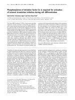
Fig. 1 Overexpression of iASPP in GTD. a Photomicrographs showing higher iASPP expression level in hydatidiform moles (HM) than 1st trimester
and term placenta as assessed by immunohistochemistry. Scale bar, 100 μm. b Statistically, higher iASPP level was demonstrated in HM than
normal placenta (1st trimester and term, *P = 0.017). c Choriocarcinoma cell lines (BeWo, JEG-3 and JAR) showed higher iASPP expression than
normal trophoblast cell line, HTR8/SV neo (HTR). Total forms of iASPP (both phospho and unphospho- forms) were detected and their relative
intensities normalized with actin were measured by ImageJ and depicted as numbers on the top
The mean iASPP expression of those progressive cases was
0.91 which was slightly higher than that of regressed cases
(0.89). Choriocarcinoma had the highest iASPP score among
all sample types but statistical significance cannot be reached
when compared to normal placenta or HM (Fig. 1b). This
may be due to the diverse status of chemotherapy among
those choriocarcinoma cases. Three choriocarcinoma cell
lines, BeWo, JEG-3 and JAR, were also used to compare
iASPP expression to that in a normal trophoblast cell line,
HTR8/SVneo. Consistently, a higher iASPP expression was
found in all choriocarcinoma cell lines compared with
HTR8/SVneo cells (Fig. 1c).
Functional importance of iASPP on the growth of
choriocarcinoma cells
Two independent siRNAs (siiaspp#1& siiaspp#2) were used
to knock down the iASPP expression in choriocarcinoma
cell lines JEG-3 and JAR. Silencing iASPP in JEG-3 and JAR
cells decreased their growth as less viable cells and colonies
were illustrated in MTT and clonogenic assays, respectively
(Fig. 2a&b). The effects were likely exerted by inhibition on
cell proliferation rather than apoptosis induction. Less
BrdU incorporation was observed in choriocarcinoma cells
after iASPP knockdown, suggesting a decrease in DNA synthesis upon iASPP downregulation (Fig. 2c). No increase in
cleaved caspase 3 protein expression could be detected after
iASPP knockdown as well (Fig. 2d). Decrease in cell viability after iASPP knockdown was corroborated by trypan
blue exclusion assay (Fig. 2e). On the contrary, no remarkable increase in DNA breaks and fragmentation were observed in iASPP knockdown cells as detected by TUNEL
and PI staining assays, respectively (Fig. 2f &g). More importantly, senescence was induced after iASPP silencing.
More cells with iASPP downregulation were stained with
Chan et al. BMC Cancer
(2019) 19:953
Fig. 2 (See legend on next page.)
Page 5 of 13
Chan et al. BMC Cancer
(2019) 19:953
Page 6 of 13
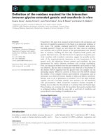
(See figure on previous page.)
Fig. 2 Knockdown of iASPP affected the growth of choriocarcinoma cells. a Choriocarcinoma cells with iASPP knockdown by using two siRNA
(siiaspp#1 & siiaspp#2) grew slower than those transfected with scramble control (si) as indicated in MTT assay. (For JEG-3, si vs siiaspp#1, P =
0.001; si vs siiaspp#2, P = 0.002. For JAR, both si vs siiaspp, P < 0.001). b Both JEG-3 and JAR cells with iASPP silencing formed less colonies than
the scramble control. (For JEG-3, si vs siiaspp#1, P = 0.03; si vs siiaspp#2, P = 0.19]; For JAR, si vs siiaspp#1, P = 0.007, si vs siiaspp#2, P = 0.01). c The
incorporation of BrdU was less in choriocarcinoma cells with iASPP knockdown than the scramble control. (For JEG-3, si vs siiaspp#1, P = 0.03; si vs
siiaspp#2, P = 0.008; For JAR, si vs siiaspp#1, P = 0.01; si vs siiaspp#2, P = 0.004 (d) No apparent increase in cleaved caspase 3 (cl. casp3) was seen
after iASPP was downregulated. e Trypan blue exclusion assay was used to assess the number of viable cells (left panel) and dead cells (right
panel) under different transfection conditions. f TUNEL assay was used to measure the presence of DNA breaks. Cisplatin (CDDP, 10 μM for 24 h)
treated cells were used as positive controls. g Histograms showing different cell cycle phases of choriocarcinoma cells with or without
iASPP knockdown
SA-β-Gal than scramble control (Fig. 3a&b). Higher mRNA
and protein expression of p21WAF1/Cip1, a CDK inhibitor
which is p53 dependent, was expressed in cells with iASPP
knockdown, corroborating the induction of senescence
(Fig. 3c&d). All these evidence suggest that iASPP affects
the growth of choriocarcinoma cells.
Functional relationship between iASPP and autophagy in
GTD
We have also evaluated the effect of iASPP on autophagy.
Endogenous level of LC3 is closely associated with the autophagic activity. In general, HM samples expressed significantly higher (P = 0.043) LC3 level than normal
placenta (Fig. 4a). Processing of LC3 during autophagy is a
good readout for autophagic activity [16]. LC3 is firstly
cleaved into cytosolic LC3-I which is then lipidated to
form LC3-II on the membrane of autophagosome during
an autophagic flux. Thus, an increase in LC3-II to LC3-I
expression ratio indicates a more active autophagy. Consistent with the immunohistochemistry results, the overall
expression of LC3 (I & II) was higher in all choriocarcinoma cells BeWo, JEG-3 and JAR while they also attained
higher LC3-II to LC3-I ratio than that in HTR8/SVneo
cells (Fig. 4b). All together suggests that autophagy may
be more active in choriocarcinoma cells. Moreover, iASPP
expression was significantly associated with LC3 expression in HM tissues, as assessed immunohistochemically
(Pearson correlation = 0.419, P = 0.001).
Choriocarcinoma cells with iASPP downregulation
presented less LC3-II expression than scramble control
with the absence of LC3-I in all samples (Fig. 4c).
Autophagosome formation can be illustrated by the
presence of LC3 puncta and act as an indication of active autophagy. Bafilomycin A1, a lysosomal inhibitor,
was added and resulted in the formation of GFP-LC3
puncta. Less puncta was observed in choriocarcinoma
cells with iASPP silencing by fluorescence microscopy
(Fig. 4d). All these evidence suggest an obstruction on
autophagy upon iASPP downregulation. Such regulation
on autophagy may be mediated by Atg5 which is responsible for autophagosomal membrane formation, and its
downregulation has been shown to affect the autophagy
[17]. Indeed, we here observed that iASPP downregulation
reduced Atg5 expression in both JEG-3 and JAR cells (Fig.
4c) that may lead to suppression on autophagic function.
Silencing of iASPP or autophagy inhibition sensitized
choriocarcinoma cell towards oxidative stress
Chloroquine, a clinically used lysosomal inhibitor, was
also effective in blocking autophagy. In the context of
choriocarcinoma, chloroquine did not show strong impact on the cell viability during a 24 h incubation period
unless a high concentration (40 μM) was used (Fig. 5a).
Autophagy is usually induced under oxidative stress, the
consequence of which can be protective or detrimental
depending on the cell context [18]. Hydrogen peroxide
is a strong oxidizing agent that can induce apoptosis
[19]. By using a sub lethal dose of chloroquine (20 μM),
addition of hydrogen peroxide (H2O2) along with
chloroquine resulted in less viable cells when compared
to treatment with H2O2 alone especially at lower dose
(Fig. 5b), suggesting that autophagy inhibition sensitizes
choriocarcinoma cells to oxidative stress. Chloroquine
blocks autophagic flux and leads to accumulation of
LC3-II. We found that H2O2 slightly increased LC3-II in
JEG-3 cells, whereas it resulted in the highest LC3-II expression when chloroquine was added simultaneously
(Fig. 5d). Similarly, choriocarcinoma cells with iASPP
knockdown were more sensitive to H2O2 inhibition with
more reduction in cell viability than the scramble control under a wider range of concentration (Fig. 5c). The
decrease in viable cell was unlikely due to an induction
of apoptosis as the levels of cleaved caspase 3 were comparable among different treatment groups (Fig. 6a). Instead, we found that the cell proliferation was affected as
cells with silencing iASPP accumulated more in G2/M
phase once treated with H2O2 when compared to scramble controls (Fig. 6b).
Discussion
iASPP is a discrete member of the ASPP family with respect to its functions on p53 and p63 activation. Structurally, iASPP lacks the α–helical domain which is
present in other members ASPP1/2 [4]. Functionally,
iASPP is considered anti-apoptotic and oncogenic,
whereas ASPP1/2 is pro-apoptotic and tumor
Chan et al. BMC Cancer
(2019) 19:953
Fig. 3 (See legend on next page.)
Page 7 of 13
Chan et al. BMC Cancer
(2019) 19:953
Page 8 of 13
(See figure on previous page.)
Fig. 3 Knockdown of iASPP induced senescence in choriocarcinoma cells. a The presence of senescent cells was detected by SA-β-Galactosidase
staining (blue color, indicated with arrows) in both JEG-3 and JAR cells with iASPP knockdown (200X magnification). b The percentage of SA-βGalactosidase positive cells was measured and compared. Statistically significant increase in senescence was found in JEG-3, si vs siiaspp#1 (P =
0.04), and JAR, si vs siiaspp#2 (P = 0.005), respectively. c Increase in p21WAF1/Cip1 mRNA levels in JEG-3 and JAR cells with iASPP knockdown as
measured by qRT-PCR. d At protein level, knockdown of iASPP also induced the expression of p21WAF1/Cip1 in choriocarcinoma cells
suppressive. Thus, dysregulation on ASPP expression
may be common but different among diseases [7, 20].
Our previous studies have also demonstrated the downregulation of ASPP1/2 in GTD [5, 6]. Here, we showed
higher expression of iASPP in HM and choriocarcinoma
when they were compared to normal placenta although
there was no significant correlation between iASPP expression level and the progression or regression of HM,
suggesting that iASPP may not be a good predictive
marker for HM progression. Altogether, it seems that an
imbalanced expression between iASPP (upregulated) and
ASPP1/2 (downregulated) is important for the pathogenesis of GTD.
The primary role of iASPP in apoptosis has been well
characterized in cancer cells. In recent years, more alternative functions of iASPP have emerged. Here we show that
iASPP also plays a role in cellular senescence. ASPP family
members have been reported to participate in senescence
through mediating the activities of different p53 family
members [21, 22]. In the context of choriocarcinoma, we
also showed that iASPP deficiency triggered senescence
through the induction of p21WAF1/Cip1 expression to
suppress cell growth but not through its well established
anti-apoptotic effect. A high iASPP level may prevent p53
to induce senescence through the transcription of
p21WAF1/Cip1. Direct binding of p53 to the promoter region
of p21WAF1/Cip1 and activation of its transcription has been
demonstrated [23]. In a more recent study, overexpression
of antiproliferative gene, TIS21, though inhibited p53iASPP interaction, shifted p53-induced senescence to
apoptosis through posttranslational modification of p53
[24], suggesting that additional mediators are involved in
determination of p53-induced senescence or apoptosis. In
contrast, another group showed iASPP silencing reduced
p21WAF1/Cip1 expression in keratinocytes and promoted
terminal differentiation through an iASPP-p63 feedback
loop mechanism [21]. Such discrepancy indicates that depending on the cell context and mediators iASPP interacts,
different cellular responses may result.
iASPP has also been illustrated to regulate autophagy in
keratinocytes [12]. On the contrary to the inhibitory effect
in keratinocytes, iASPP may be necessary for maintaining
an active autophagy in choriocarcinoma cells via regulating
the Atg5 expression. The positive correlation between
iASPP and LC3 expressions in clinical samples further suggested a possible link between iASPP and autophagy in
GTD. Autophagy is important for cellular homeostasis and
its dysregulation has been found in various diseases [25].
Autophagy was firstly linked to tumorigenesis when monoallelic deletion of Beclin1, a modulatory gene on autophagy, was found in breast and ovarian cancers [26]. In
contrast, studies have also demonstrated that autophagy inhibition enhanced cytotoxic effects of chemotherapy but
promoted proliferation in certain cellular context [27, 28],
suggesting that autophagy may play a role in cancer survival under stress. Autophagy provides not only the nutrients and energy but also the cellular restructuring in
response to metabolic stress. Such paradox on autophagy
effect is mainly because autophagy participates in processes
promoting cell death and cell survival [29, 30], indicating
that a tight regulation on autophagy is crucial. Based on a
higher LC3-II to LC3-I expression ratio and LC3 level
found in choriocarcinoma cells and HM respectively, it is
likely that an upregulated autophagy may exhibit prosurvival effect for GTD. Active autophagy is proven to be
necessary for the progression in other cancer types [31].
Our evidence showing autophagy promoting effect of
iASPP in choriocarcinoma was different from studies on
keratinocytes where iASPP was shown to be an autophagy
inhibitor in keratinocytes [12]. Such discrepancy may be
mainly due to the differences in the nature of cells. Choriocarcinoma cells have a high basal autophagy activity as we
noticed choriocarcinoma cell lines expressing higher LC3-II
level than normal trophoblastic cell. We have shown that
iASPP knockdown suppressed expression of Atg5 and subsequent GFP-LC3 puncta formation. Atg5 is responsible for
autophagosome formation [32]. Overexpression of Atg5
has been shown to activate autophagy, whereas knockdown
of Atg5 resulted in autophagic downregulation. A recent
study also reported that knockdown of iASPP downregulated autophagy in lung cancer cells through interfering the
autophagosome formation [33].
Interestingly, we also demonstrated that either iASPP silencing or autophagy inhibition sensitized choriocarcinoma
cells towards oxidative stress induced by hydrogen peroxide.
Crosstalk between autophagy and oxidative stress signal has
been reported [13]. Generation of hydrogen peroxide activates AMPK and triggers the initiation of autophagy [34]
which is found to be cytoprotective for cells in response to
oxidative stress [35]. Blockage of autophagy with chloroquine may prevent protecting choriocarcinoma cells from
the oxidative stress induced by hydrogen peroxide. This provides a novel therapeutic approach against choriocaricoma.
Silencing iASPP may also render choriocarcinoma cells
Chan et al. BMC Cancer
(2019) 19:953
Page 9 of 13
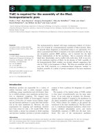
Fig. 4 Effect of iASPP on autophagy. a Higher expression of LC3 was demonstrated in HM than normal placentas by immunohistochemistry (normal vs HM
*; P = 0.043). Scale bar, 100 μm. b Choriocarcinoma cell lines, BeWo (2), JEG-3 (3) and JAR (4), expressed more LC3-II than normal trophoblast cell line HTR8/
SVneo (1). The LC3 bands were quantified by using ImageJ. The LC3-II to LC3-I ratio on each cell line was analyzed and listed. c Knockdown of iASPP in
choriocarcinoma cells decreased Atg5 and LC3-II expressions. d More GFP-LC3 puncta (arrows) were observed in choriocarcinoma cells treated with scramble
control than those cells with iASPP knockdown. All the cells were treated with Bafilomycin A1 (20 nM) for 6 h before captured under microscope (200X)
Chan et al. BMC Cancer
(2019) 19:953
Fig. 5 (See legend on next page.)
Page 10 of 13
Chan et al. BMC Cancer
(2019) 19:953
Page 11 of 13
(See figure on previous page.)
Fig. 5 Autophagy inhibition sensitized choriocarcinoma cells in response to hydrogen peroxide. a Chloroquine inhibition on autophagy per se
had no effect on the viability of choriocarcinoma cells as measured by MTT assay in the first 24 h. b Addition of chloroquine (20 μM) enhanced
the toxic effect of hydrogen peroxide at low dosage after 24 h (*P < 0.001; #P = 0.02). c Choriocarcinoma cells with iASPP knockdown (siiaspp#1/2)
were more sensitive to hydrogen peroxide than scramble control (si). d Change of LC3 expression after addition of hydrogen peroxide (H2, 0.3
mM) with or without chloroquine (C, 20 μM). (** P < 0.05)
Fig. 6 Increased proportion of cells at G2/M in choriocarcinoma cells with iASPP silencing when treated with hydrogen peroxide. a No obvious
increase in cleaved caspase 3 between scramble control and iASPP knockdown cells. b The G2/M population in cells with iASPP silencing was
higher than the scramble control when they were all treated with hydrogen peroxide. However, no statistically significant difference was
achieved. Representative results of two independent experiments were shown
Chan et al. BMC Cancer
(2019) 19:953
more susceptible to hydrogen peroxide through regulating
autophagy. Further investigation is needed to delineate the
underlying mechanisms by identifying the common mediators affected by iASPP knockdown and chloroquine in future studies. However, we should not exclude other factors
such as effects of cell cycle alteration. A recent study has
shown that iASPP regulates the recruitment of CEP55 to
the midbody and concomitantly controls cytokinesis [36]. In
addition, oxidative stress can induce mitotic arrest [37], suggesting that cell cycle may be potently deregulated when
hydrogen peroxide is applied to cells with iASPP deficiency.
Our findings on cell cycle analysis also show an apparent increase in the proportion of cells at G2/M phase under
hydrogen peroxide treatment in iASPP knockdown cells,
despite statistical significance was not achieved. On the
other hand, iASPP has recently reported as an antioxidative
factor to participate in regulating the reactive oxygen species
homeostasis [38], again supporting that iASPP may also play
a role in regulation of oxidative stress in GTD.
Conclusions
iASPP may be a potential therapeutic target for choriocarcinoma as iASPP silencing not only inhibits cell growth
but also renders higher susceptibility to oxidative stress.
Supplementary information
Supplementary information accompanies this paper at />1186/s12885-019-6206-z.
Additional file 1. A list of average immunoscores for patients and their
individual diagnosis.
Additional file 2. The raw western blots for iASPP shown in the
manuscript.
Abbreviations
ASPP: Ankyrin-repeat, SH3-domain and proline-rich region containing protein;
BrdU: 5-bromo-2′-deoxyuridine; GTD: gestational trophoblastic disease;
HM: hydatidiform mole; LC3: Light chain 3; MTT: 3-(4, 5-dimethylthiazolyl-2)-2,
5-diphenyltetrazolium bromide
Acknowledgements
We thank Prof. Peeyush K. Lala for the HTR8/SVneo trophoblast cell line.
Authors’ contributions
KKC and ANYC designed the study and wrote the manuscript. KKC and
ESYW carried out the immunohistochemistry and statistical analyses. KKC,
ITLW, CLYC and OGWW carried out and interpreted western blots and flow
cytometry. HYSN and ANYC also helped to collect and analyze clinical
samples. All authors have read and approved the final manuscript.
Funding
This study was supported by Health and Medical Research Fund (#01121336),
Hong Kong Special Administrative Region. Other than providing financial
support and owning the intellectual rights of this study, the funding body
was not involved in the design of the study and collection, analysis, and
interpretation of data and writing the manuscript.
Availability of data and materials
A table listing cases for immunohistochemical evaluation is provided as
Additional files 1 and 2.
Page 12 of 13
Ethics approval and consent to participate
Ethical approval has been obtained from Institutional Review Board,
University of Hong Kong/Hospital Authority Hong Kong West Cluster (UW
13–264) waiving need for consent.
Consent for publication
Not applicable
Competing interests
The authors declare that they have no competing interests.
Author details
Department of Pathology, Queen Mary Hospital, University of Hong Kong,
Hong Kong SAR, China. 2Department of Obstetrics and Gynaecology, Queen
Mary Hospital, University of Hong Kong, Hong Kong SAR, China.
3
Department of Pathology, University of Hong Kong-Shenzhen Hospital,
Shenzhen, China.
1
Received: 13 January 2019 Accepted: 25 September 2019
References
1. Cheung AN. Gestational trophoblastic disease. In: Robboy S, Mutter G, Prat J,
Bentley RC, Russell P, Anderson MC, editors. Robboy's pathology of the female
reproductive tract. China: Elsevier Churchill Livingstone; 2009. p. 881–907.
2. Seckl MJ, Sebire NJ, Berkowitz RS. Gestational trophoblastic disease. Lancet.
2010;376(9742):717–29.
3. Trigiante G, Lu X. ASPP [corrected] and cancer. Nat Rev Cancer. 2006;6(3):
217–26.
4. Sullivan A, Lu X. ASPP: a new family of oncogenes and tumour suppressor
genes. Br J Cancer. 2007;96(2):196–200.
5. Mak VC, Lee L, Siu MK, Wong OG, Lu X, Ngan HY, et al. Downregulation of
ASPP1 in gestational trophoblastic disease: correlation with
hypermethylation, apoptotic activity and clinical outcome. Mod Pathol.
2011;24(4):522–32.
6. Mak VC, Lee L, Siu MK, Wong OG, Lu X, Ngan HY, et al. Downregulation of
ASPP2 in choriocarcinoma contributes to increased migratory potential
through Src signaling pathway activation. Carcinogenesis. 2013;34(9):2170–7.
7. Jiang L, Siu MK, Wong OG, Tam KF, Lu X, Lam EW, et al. iASPP and
chemoresistance in ovarian cancers: effects on paclitaxel-mediated mitotic
catastrophe. Clin Cancer Res. 2011;17(21):6924–33.
8. Edinger AL, Thompson CB. Defective autophagy leads to cancer. Cancer
Cell. 2003;4(6):422–4.
9. Qu X, Yu J, Bhagat G, Furuya N, Hibshoosh H, Troxel A, et al. Promotion of
tumorigenesis by heterozygous disruption of the beclin 1 autophagy gene.
J Clin Invest. 2003;112(12):1809–20.
10. Chittaranjan S, Bortnik S, Dragowska WH, Xu J, Abeysundara N, Leung A,
et al. Autophagy inhibition augments the anticancer effects of epirubicin
treatment in anthracycline-sensitive and -resistant triple-negative breast
cancer. Clin Cancer Res. 2014;20(12):3159–73.
11. Chan KK, Wong OG, Wong ES, Chan KK, Ip PP, Tse KY, et al. Impact of iASPP
on chemoresistance through PLK1 and autophagy in ovarian clear cell
carcinoma. Int J Cancer. 2018;143(6):1456–69.
12. Chikh A, Sanzà P, Raimondi C, Akinduro O, Warnes G, Chiorino G, et al.
iASPP is a novel autophagy inhibitor in keratinocytes. J Cell Sci. 2014;
127(14):3079–93.
13. Filomeni G, De Zio D, Cecconi F. Oxidative stress and autophagy: the clash
between damage and metabolic needs. Cell Death Differ. 2015;22(3):377–88.
14. Graham CH, Hawley TS, Hawley RG, MacDougall JR, Kerbel RS, Khoo N, et al.
Establishment and characterization of first trimester human trophoblast cells
with extended lifespan. Exp Cell Res. 1993;206(2):204–11.
15. Franken NA, Rodermond HM, Stap J, Haveman J, van Bree C. Clonogenic
assay of cells in vitro. Nat Protoc. 2006;1(5):2315–9.
16. Tanida I, Waguri S. Measurement of autophagy in cells and tissues. Methods
Mol Biol. 2010;648:193–214.
17. Xiong X, Wu M, Zhang H, Li J, Lu B, Guo Y, et al. Atg5 siRNA inhibits
autophagy and enhances norcantharidin-induced apoptosis in
hepatocellular carcinoma. Int J Oncol. 2015;47(4):1321–8.
18. Navarro-Yepes J, Burns M, Anandhan A, Khalimonchuk O, del Razo LM,
Quintanilla-Vega B, et al. Oxidative stress, redox signaling, and autophagy:
cell death versus survival. Antioxid Redox Signal. 2014;21(1):66–85.
Chan et al. BMC Cancer
(2019) 19:953
19. Singh M, Sharma H, Singh N. Hydrogen peroxide induces apoptosis in HeLa
cells through mitochondrial pathway. Mitochondrion. 2007;7(6):367–73.
20. Song B, Bian Q, Zhang YJ, Shao CH, Li G, Liu AA, et al. Downregulation of
ASPP2 in pancreatic cancer cells contributes to increased resistance to
gemcitabine through autophagy activation. Mol Cancer. 2015;14:177.
21. Chikh A, Matin RN, Senatore V, Hufbauer M, Lavery D, Raimondi C, et al.
iASPP/p63 autoregulatory feedback loop is required for the homeostasis of
stratified epithelia. EMBO J. 2011;30(20):4261–73.
22. Wang Y, Godin-Heymann N, Dan Wang X, Bergamaschi D, Llanos S, Lu X.
ASPP1 and ASPP2 bind active RAS, potentiate RAS signalling and enhance
p53 activity in cancer cells. Cell Death Differ. 2013;20(4):525–34.
23. Laptenko O, Beckerman R, Freulich E, Prives C. p53 binding to nucleosomes
within the p21 promoter in vivo leads to nucleosome loss and
transcriptional activation. Proc Natl Acad Sci U S A. 2011;108(26):10385–90.
24. Choi OR, Ryu MS, Lim IK. Shifting p53-induced senescence to cell death by
TIS21(/BTG2/Pc3) gene through posttranslational modification of p53
protein. Cell Signal. 2016;28(9):1172–85.
25. Levine B, Kroemer G. Autophagy in the pathogenesis of disease. Cell. 2008;
132(1):27–42.
26. Liang XH, Jackson S, Seaman M, Brown K, Kempkes B, Hibshoosh H, et al.
Induction of autophagy and inhibition of tumorigenesis by beclin 1. Nature.
1999;402(6762):672–6.
27. Thorburn J, Staskiewicz L, Goodall ML, Dimberg L, Frankel AE, Ford HL, et al.
Non-cell-autonomous effects of autophagy inhibition in tumor cells
promote growth of drug-resistant cells. Mol Pharmacol. 2017;91(1):58–64.
28. Yang W, Hosford SR, Traphagen NA, Shee K, Demidenko E, Liu S, et al.
Autophagy promotes escape from phosphatidylinositol 3-kinase inhibition
in estrogen receptor-positive breast cancer. FASEB J. 2018;32(3):1222–35.
29. Fitzwalter BE, Thorburn A. Recent insights into cell death and autophagy.
FEBS J. 2015;282(22):4279–88.
30. Kroemer G, Levine B. Autophagic cell death: the story of a misnomer. Nat
Rev Mol Cell Biol. 2008;9(12):1004–10.
31. Yang A, Kimmelman AC. Inhibition of autophagy attenuates pancreatic cancer
growth independent of TP53/TRP53 status. Autophagy. 2014;10(9):1683–4.
32. Sheng Y, Song Y, Li Z, Wang Y, Lin H, Cheng H, et al. RAB37 interacts
directly with ATG5 and promotes autophagosome formation via regulating
ATG5-12-16 complex assembly. Cell Death Differ. 2018;25(5):918–34.
33. Xue Y, Han H, Wu L, Pan B, Dong B, Yin CC, et al. iASPP facilitates tumor
growth by promoting mTOR-dependent autophagy in human non-smallcell lung cancer. Cell Death Dis. 2017;8(10):e3150.
34. Zmijewski JW, Banerjee S, Bae H, Friggeri A, Lazarowski ER, Abraham E.
Exposure to hydrogen peroxide induces oxidation and activation of AMPactivated protein kinase. J Biol Chem. 2010;285(43):33154–64.
35. Dutta D, Xu J, Kim JS, Dunn WA Jr, Leeuwenburgh C. Upregulated
autophagy protects cardiomyocytes from oxidative stress-induced toxicity.
Autophagy. 2013;9(3):328–44.
36. Gao K, Zhang Y, Shi Q, Zhang J, Zhang L, Sun H, et al. iASPP-PP1 complex is
required for cytokinetic abscission by controlling CEP55 dephosphorylation.
Cell Death Dis. 2018;9(5):528.
37. Wang GF, Dong Q, Bai Y, Yuan J, Xu Q, Cao C, et al. Oxidative stress induces
mitotic arrest by inhibiting Aurora A-involved mitotic spindle formation.
Free Radic Biol Med. 2017;103:177–87.
38. Ge W, Zhao K, Wang X, Li H, Yu M, He M, et al. iASPP Is an Antioxidative
Factor and Drives Cancer Growth and Drug Resistance by Competing with
Nrf2 for Keap1 Binding. Cancer Cell. 2017;32(5):561–573.e6.
Publisher’s Note
Springer Nature remains neutral with regard to jurisdictional claims in
published maps and institutional affiliations.
Page 13 of 13