The prognostic impact of GSTM1/GSTP1 genetic variants in bladder Cancer
Bạn đang xem bản rút gọn của tài liệu. Xem và tải ngay bản đầy đủ của tài liệu tại đây (1.14 MB, 11 trang )
Albarakati et al. BMC Cancer
(2019) 19:991
/>
RESEARCH ARTICLE
Open Access
The prognostic impact of GSTM1/GSTP1
genetic variants in bladder Cancer
Nada Albarakati1, Dareen Khayyat2, Asharf Dallol3, Jaudah Al-Maghrabi4,5 and Taoufik Nedjadi1*
Abstract
Background: The glutathione S-transferases (GSTs) are a superfamily of phase II detoxifying enzymes that
inactivates a wide variety of potential carcinogens through glutathione conjugation. Polymorphic changes in the
GST genes have been reported to be associated with increased susceptibility to cancer development and anticancer
drug resistance. In this study, we investigated the association between genetic variants in GSTM1 and GSTP1 and
patients’ clinicopathological parameters. The prognostic values of such associations were evaluated among bladder
cancer patients.
Methods: Genotyping of GSTM1 and GSTP1 in bladder cancer patients was assessed using polymerase chain
reaction followed by DNA sequencing. Overall survival was estimated using the Kaplan-Meier method and multiple
logistic regression and correlation analysis were performed.
Results: The GSTM1 null genotype was significantly associated with poor overall survival compared with the wild-type
GSTM1 genotype. There was a trend towards better overall survival in patients with wild-type GSTP1 allele (AA)
compared with GSTP1 (AG/GG) genotype. Interestingly, Kaplan-meier survival curve for GSTM1 null patients adjusted for
sub-cohort with amplified HER2 gene showed poor survival compared with the GSTM1 null/ non-amplified HER2 gene.
Also the same population when adjusted with HER2 protein expression, data showed poor survival for patients
harboring GSTM1 null/high HER2 protein expression compared with low protein expression.
Conclusion: This study focuses on the impact of GSTM1 null genotype on bladder cancer patients’ outcome. Further
investigations are required to delineate the underlying mechanisms of combined GSTM−/− and HER2 status in bladder
cancer.
Keywords: Bladder cancer, GSTM1, GSTP1, HER2, Polymorphism, Prognosis
Background
Bladder cancer is the 9th most common cancer and a
leading cause of cancer-related death worldwide. It has
been estimated that around 550,000 new bladder cancer
cases and 199,922 deaths occurred in the year 2018 worldwide and these numbers are expected to double in the upcoming years [1]. The disease is highly recurring and do
frequently progress to a muscle invasive phenotype which
necessitate a vigilant and continuous monitoring protocol
[2]. Despite advances in diagnostic and treatment modalities, bladder cancer remains source of co-morbidity and
continues to pose challenges for clinicians given that
* Correspondence:
1
King Abdullah International Medical Research Center, King Saud bin
Abdulaziz University for Health Sciences, Ministry of the National Guard Health Affairs, Jeddah, Kingdom of Saudi Arabia
Full list of author information is available at the end of the article
patients’ outcome being solely dependent on the grading
and staging system [3]. Therefore, a deeper understanding
of the bladder cancer pathogenesis and associated mechanisms will undoubtedly improve patients’ outcome via
prevention of disease progression and recurrence.
It is well documented that occupational exposure to
chemical carcinogens including aromatic amines and
polycyclic aromatic hydrocarbons is associated with
the risk of bladder cancer development [2, 4]. Kellen
et al. reported an increased risk of developing bladder
cancer associated with cumulative exposure to aromatic amines, but not to PAHs and diesel [5]. In an
independent study, Ferrís et al. concluded that
bladder cancer is a result of the interaction between
constitutional and environmental risk factors including aromatic amines and polycyclic aromatic
© The Author(s). 2019 Open Access This article is distributed under the terms of the Creative Commons Attribution 4.0
International License ( which permits unrestricted use, distribution, and
reproduction in any medium, provided you give appropriate credit to the original author(s) and the source, provide a link to
the Creative Commons license, and indicate if changes were made. The Creative Commons Public Domain Dedication waiver
( applies to the data made available in this article, unless otherwise stated.
Albarakati et al. BMC Cancer
(2019) 19:991
hydrocarbons [6]. The involvement of environmental
factors such as cigarette smoking in bladder carcinogenesis has been extensively investigated [7, 8]. Recent evidence supports the dynamic interplay between
environmental factors and other co-factors, including
genetic predisposition, in the pathogenesis of bladder
cancer [9].
Protecting against carcinogen-induced and chemotherapyinduced oxidative stress involves a series of event
characterized by the activation of phase-II cellular detoxifying enzymes; Glutathione S-transferases (GSTs) or Nacetyltransferases (NATs) [10]. GSTs enzymes superfamily
consist of at least 16 genes located on more than 7 chromosomes [11]. Although they are structurally different with distinct evolutionary origins, all GSTs isoenzymes are
functionally similar in protection against electrophiles and
oxidative stressors. The cytosolic sub-family of GST is found
to be active in a homo- or heterodimeric state and is subdivided into eight classes designated as follow: GST alpha
(α), mu (μ), kappa (κ), omega (ω), pi (π), sigma (σ), theta (θ),
and zeta (ζ) [12]. GSTs play a critical protective anticancer
role through glutathione conjugation with a range of potentially cytotoxic exogenous or endogenous molecules making
them less toxic. Allelic polymorphisms in these genes elicit
changes in enzyme activities leading to biotransformation
and play important role in the development and progression
of different cancers, such as lung, colorectal, leukemia,
breast and bladder cancers. Furthermore, Sau et al. showed
the contribution of GSTs overexpression in resistance
against several anti-cancer drugs [13].
GSTM1 gene is located on chromosome 1p13.3 and
the most common polymorphic variant of GSTM1
gene is the homozygous deletion (GSTM1 null genotype) characterized by abolished enzyme activity [14].
Many studies have investigated the relationship between the genetic polymorphism of GSTM1 and the
risk of cancer, but the association remains controversial among different populations. Previous epidemiological studies showed an association between the
homozygous deletion of GSTM1 and increased risk of
lung, colorectal and head and neck cancers [15–17].
However other studies failed to establish the association between GSTM1 null and the risk of several
types of cancers [18–21].
GSTP1 is encoded by a single gene located on
chromosome 11 [22]. The common functional GSTP1
polymorphism at codon 105 is an A to G substitution
resulting in an amino acid switch from isoleucine to
valine (Ile105Val) and lowering the catalytic activity of
GSTP1enzyme [23]. The decreased detoxification capacity of the GSTP1 enzyme resulted in differences in
chemotherapeutic responses. The increased expression
of the GSTP1 Val105 genotype was shown to be associated with a variety of tumors, such as ovarian, breast,
Page 2 of 11
colon, lymphoma, and pancreas [24]. The hypothesis
that GSTP1 variants modulate the risk of urinary bladder cancer has also been investigated [24, 25]. However, inconclusive results have been reported on the
association between GSTP1 gene polymorphisms and
the risk of bladder cancer: while a number of studies
identified an obvious association between GSTP1
polymorphisms Ile105Val and bladder carcinoma risk
[26–28], other studies illustrated that there are no association between GSTP1 Ile105Val polymorphism and
bladder cancer [29, 30].
HER2 is a trans-membrane glycoprotein receptor
tyrosine kinase of the epidermal growth factor receptor family EGFR/ErbB. It plays an important role in
the development and progression of many tumor types
including breast, gastric and bladder cancers [31].
Recent sequencing efforts to uncover the complex
genomic landscape of bladder cancer identified six distinct molecular subtypes. HER2-like is one of the main
subtypes characterise by higher ERBB2 amplification
and signalling [32]. HER2 is considered one of the
most important prognostic biomarkers that play an
important role in the patho-physiology of bladder cancers and a potential therapeutic target in bladder cancer [31, 33, 34]. Also, interactions between GST gene
family and other genes including HER2 may be involved in cancer susceptibility and clinical management of cancer patients. In the present study, we aim
to investigate the prognostic value of GSTM1 and
GSTP1 genetic polymorphisms in patients with bladder cancer and evaluate their association with patients’
clinicopathological parameters. We also attempted to
evaluate the clinical significance of HER2 status in
cases confirmed to have GSTM1/ GSTP1 variants with
bladder cancer prognosis.
Methods
Patients and sample collection
Formalin-fixed paraffin-embedded (FFPE) tissue samples were obtained from histologically confirmed bladder cancer patients who underwent bladder resection
between 2005 and 2012 at King Abdulaziz University
Hospital (KAUH), Jeddah, Saudi Arabia. The study
group consists of 93 patients; only specimens containing more than 80% cellular composition were used in
the analysis. All patients have not been subjected to
any chemotherapy or radiotherapy prior to sample
collection. Clinical and pathological data including
age, gender, tumor grade, tumor stage, lymph node,
vascular invasion, metastasis, and survival were gathered from patients’ medical records and summarized
in Table 1. This study was ethically approved by the
institutional research ethics committee, faculty of
medicine, King Abdulaziz University (ref. N. 149–14).
Albarakati et al. BMC Cancer
(2019) 19:991
Page 3 of 11
Table 1 The clinicopathological characteristics of 93 patients
with bladder cancer
The clinicopathological characteristics
Group age (Years)
Gender
Tumor Grade
Cancer type
Subtypes
Tumor Shape
Lymph Node
Vascular Invasion
Metastasis
Smoking
Family history of cancer
Survival
N
%
≤60
37
39.78%
> 60
55
59.14%
Unknown
1
1.08%
Male
77
82.80%
Female
16
17.20%
High Grade
56
60.22%
Low Grade
29
31.18%
Unknown
8
8.60%
MIBC
52
55.91%
NMIBC
28
30.11%
Unknown
13
13.98%
Transitional
74
79.57%
Squamous
3
3.23%
Transitional/ Squamous
15
16.13%
Unknown
1
1.08%
Papillary
63
67.74%
Non-papillary
3
3.23%
Unknown
27
29.03%
Positive
21
22.58%
Negative
68
73.12%
Unknown
4
4.30%
Positive
18
19.35%
Negative
70
75.27%
Unknown
5
5.38%
Positive
21
22.58%
Negative
67
72.04%
Unknown
5
5.38%
No
11
11.83%
Yes
16
17.20%
Unknown
66
70.97%
No
24
25.81%
Yes
4
4.30%
Unknown
65
69.89%
Alive
65
69.89%
Deceased
28
30.11%
Abbreviation: MIBC Muscle Invasive Bladder Cancer, NMIBC Non-Muscle
Invasive Bladder Cancer
DNA isolation
Genomic DNA was extracted from FFPE tissue samples
using QIAamp DNA FFPE Tissue Kit (Qiagen) according to the manufacturer’s instructions. Purified DNA
was eluted in 50 μl elution buffer and stored at − 80 °C
until use. Purity and concentration of eluted DNA was
analyzed using a spectrophotometer system (Nanodrop
2000, Thermo Scientific, USA).
GSTM1 and GSTP1 SNP genotyping
Genotyping for the detection of GSTM1 (present/null) and
GSTP1 Ile105Val polymorphisms was performed as described previously [35]. Genotyping was carried out using
real time PCR Kit (Qiagen) as per the manufacturer’s recommendation. Briefly 200 ng DNA was amplified in an
overall volume of 25 μl/ reaction. GSTM1 and GSTP1
oligonucleotide primers were purchased from MWGBiotech (Ebersberg, Germany) to amplify the GSTM1 fragments, (Forward: 5′-CTGCCCTACTTGATTGATGGG3′; Reverse: 5′-CTGGATTGTAGCAGATCATGC-3′),
GSTP1 (Forward: 5′-ACCCCAGGGCTCTATGGGAA-3′,
Reverse: 5′-TGAGGGCACAAGAAGCCCCT-3′) PCR was
performed on a Thermal Cycler 480 apparatus (Applied
Biosystems, USA). Thermo cycler parameters included: an
initial denaturation at 94 °C/ 15 min; followed by 35 cycles
of denaturation at 94 °C/ 1 min, annealing at 57 °C /1 min,
and extension at 74 °C/ 1 min; and a final extension at
72 °C/10 min. Confirmation of PCR products were examined by 2% agarose gel electrophoresis and visualized using
a Syngene UV transilluminator.
DNA sequencing
To sequence the amplified GSTP1 PCR products, sequencing kit (BigDye® Terminator v3.1 kit, Thermo Scientific, USA) was used according to the manufacturer’s
instructions using Genetic analyzer 3500 (Applied Biosystems, UK). The resulting sequence data was analyzed
using Applied Biosystems sequence analysis software (v
5.4). GSTP1 genotypes were determined as wild type Ile/
Ile (AA), heterozygous type Ile/Val (AG) or homozygous
variant type Val/Val (GG) as shown in Fig. 1c. As for
GSTM1, the PCR products were separated on a 2% agarose gel and determined as null/ present genotypes.
Immunohistochemistry
HER2 immunostaining was undertaken earlier [33]. The
expression of HER2 protein is mainly membranous, the
protein expression in our bladder samples was evaluated
as follows: No expression = negative Vs. Expression =
weak, + 1; moderate, + 2; strong, + 3.
Statistical analyses
Statistical data analysis was performed using SPSS (SPSS,
version 25, USA). Appropriate, Chi-square test and Fisher’s
exact test were used to establish any significant differences
in polymorphism incidences between bladder cancer cases.
Multivariate Cox regression model were used to evaluate
the prognostic significance of GSTs genes, HER2 and other
clinicopathological factors. Cumulative survival probabilities
were estimated using the Kaplan-Meier method, with log-
Albarakati et al. BMC Cancer
(2019) 19:991
Page 4 of 11
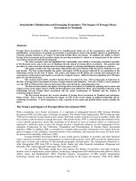
Fig. 1 Representative screening for GSTM1 and GSTP1 Polymerase chain reaction products. Agarose gel of the PCR products for detection of
GSTM1 deletion polymorphism [a] GSTM1 verified by PCR analysis. b Agarose gel of the PCR products for detection of GSTP1 polymorphisms. c
GSTP1 validation by sequencing: (1) The wild allele homozygote AA, (2) heterozygote AG and (3) variant allele homozygote GG genotypes
rank comparison test. Multiple logistic regression analysis
was performed to assess the association between GST polymorphisms with aggressiveness of bladder cancer. Odds Ratios (OR) and their 95% Confidence Intervals (95% CI)
were used to calculate the results. The wild type of all genotypes was used as the reference group. Interactions between
GSTM1 and GSTP1 polymorphisms and aggressiveness
bladder cancer phenotypes were analyzed using Spearman
correlation analysis. In all tests, the values p ≤ 0.05 were
considered as statistically significant.
Results
Characteristics of the study population
In the current study, 93 patients with urinary bladder carcinoma were genotyped for two polymorphisms in two
important genes of the glutathione-s-transferase family involved in xenobiotic metabolism. The distribution of the
clinicopathological characteristics of the bladder cancer
patients is presented in Table 1. Patients age ranges from
34 to 93 years with median age of 64 ± 12, the median
follow-up time of 10.10 months (ranging 0–139 months)
and preponderance of male over female in the ratio 5:1.
Genotype distributions of the GSTM1and GSTP1
polymorphisms in patients
Polymerase chain reaction-based and Sanger gene
sequencing-base assays were undertaken to assess the
contribution of genetic polymorphism in GSTM1 and
GSTP1 to the susceptibility of bladder cancer (Fig. 1).
Lack of amplification products for the GSTM1 gene
was considered as a homozygous null genotype (−/−).
Our data revealed that a total of 44 bladder cancer patients out of 93 (47.31%) had a GSTM1-deleted genotype (−/−). GSTM1 specific bands showing on agarose
gel electrophoresis was seen in 45 out of 93 patients
(48.38%). No further investigations were carried out to
discriminate between heterozygous deletion (+/−) and
wild-type (+/+) GSTM1 variants hence both heterozygous deletion and wild-type variants are considered
GSTM1 present (Fig. 1a).
As for the GSTP1 frequencies, amplified PCR products
containing GSTP1 were visualized on agarose gels (Fig. 1b)
and the resultant DNA fragments were subjected to Sanger
sequencing using BigDye terminator v3.1 (Life technologies).
The GSTP1 wild allele homozygote (AA), heterozygote
(AG) and variant allele homozygote (GG) genotypes were
36/93 (38.70%), 36/93 (38.70%) and 6/93 (6.45%) respectively (Fig. 1c). Merging both AG/GG genetic variants represent 45.16% (42/93) of the total analyzed cases, Table 2.
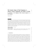
A higher frequency within our cohort was found between those carrying GSTM1 null and GSTP1 recessive homozygote / heterozygote AG/GG 23 (24.73%),
whereas the lower percentage was with GSTM1 null
and the GSTP1 wild allele 14 (15.05%) shown in Fig. 2.
Albarakati et al. BMC Cancer
(2019) 19:991
Page 5 of 11
Table 2 The distribution (count and percentage) of GSTM1 and
GSTP1 genotypes in the patients with bladder cancer
GSTM1
GSTP1
N
%
Present
45
(48.38)
Null
44
(47.31)
AA
36
(38.70)
GG
6
(6.45)
AG
36
(38.70)
AG/GG
42
(45.16)
No statistical significant was found between GSTs different groups.
Effect of GSTM1and GSTP1 polymorphisms on patients’
survival
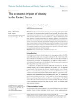
Kaplan-Meier curve showed that GSTM1 null genotype was associated with poor overall survival in
comparison to GSTM1 present genotype, log rank p =
0.038 (Fig. 3a). As for GSTP1, though it is not statistically significant, patients harboring the wild type allele
GSTP1 AA have tendency for better survival in comparison to patients with GSTP1 AG/GG genotype (Log
rank, p = 0.234). GSTP1 AG carriers had the worst
overall survival compared to GSTP1 AA or GG genotypes carriers (Fig. 3b, c. However, the associations
were not statistically significant (log-rank test; p =
0.40). When merging GSTM1 survival and GSTP1
polymorphisms (Fig. 3d), there was trend towards
Fig. 2 Distribution of the GSTM1 + GSTP1 variants in bladder cancer
patients. The distribution of patients carrying GSTM1 null and GSTP1
recessive homozygote/ heterozygote AG/GG was 23 of 93 (24.73%).
whereas the lowest was GSTM1 null and the GSTP1 wild allele
14 (15.05%)
poorer survival for patients with combined GSTM1
null and GSTP1 AG/GG (Log rank, p = 0.146).
Relationships between GST genotypes, HER2 status and
survival outcomes
Published data, including our own, revealed that bladder
cancer exhibit high ratios of the Human Epidermal
growth factor Receptor 2 (HER2) gene amplification,
after breast and gastric cancers, and also demonstrates
frequent overexpression of HER2 protein [33, 34]. Recently published data revealed that bladder cancer possess the highest frequency mutation in HER2 gene
across 38 types of tumors analyzed [31]. Furthermore,
HER2 is considered among the prognostic factors, along
with staging and grading system, in urothelial bladder
cancer [36]. In the current study we sought to investigate the relationship between GSTM1 and GSTP1 polymorphisms in respect to HER2 status of the same
cohort. HER2 protein expression and gene amplification
data [33] were available for 89 patients out of our 93
bladder cancer patients. Histograms showed the frequency of expression patterns of HER2 protein receptors
in our cohort (Additional file 1: Figure S1). To establish
the relationship between GST genotypes and HER2 status, bright field double in situ hybridization (BDISH)
and immunohistochemistry (IHC) data were used to
analyze HER2 gene amplification and protein expression
within the GSTP1/ GSTM1 analyzed cohort. Our data
indicated no association between HER2 protein level
and both GSTP1 (p = 0.07) and GSTM1 (p = 0.75) polymorphic status (Table 3). However, HER2 gene amplification was significantly associated with the GSTP1 AA,
AG & GG variants (p = 0.03). Such a relationship was
not established for amplified HER2 gene and GSTM1
null/present variants (Table 3).
Interestingly, Kaplan-Meier survival curve for GSTM1
status adjusted to HER2 gene status (amplified or nonamplified) showed a significant impact on patients’ overall
survival. Figure 4a, illustrates that poor overall survival
was associated with combining GSTM1 null and amplified
HER2 gene (Log rank, p = 0.05), though this was not the
case with non-amplified HER2 patients (Fig. 4b). To further confirm the observed relationship between amplified
HER2 gene and GSTM1 null, we sought to analyze the relationship between HER2 protein level and GSTM1 genotype. Similarly, survival curve (Fig. 4c) showed poor
survival for patients carrying GSTM1 null variant with
high HER2 protein expression (Log rank, p = 0.041) compared to GSTM1 null/ low HER2 protein expression
counterpart (Fig. 4D). This synergistic effect of combined
GSTM1 genotype and increased HER2 status indicated a
possible interaction between the two genes in bladder carcinogenesis. On the other hand, no difference in overall
survival was observed in patients harboring combined
Albarakati et al. BMC Cancer
(2019) 19:991
Page 6 of 11
Fig. 3 Kaplan-Meier survival curves demonstrating the overall survival of: a GSTM1 null and present genotypes were evaluated in bladder cancer
patients. b GSTP1 genotypes, AA, AG and GG. c GSTP1 AA and combined AG/GG. d Merging GSTM1 and GSTP1 overall survival. All P values tested
by log-rank test. Patients alive at the last follow-up or lost to follow-up were censored in the survival comparison analysis
GSTP1 polymorphism and altered HER2 gene/protein
levels (Additional file 2: Fig. S2A - 2D). The study cohort
was then stratified into two groups based on the type of
tumour (MIBC and NMIBC) and statistical analysis was
performed to to determine which variables were independently associated with the patients’ outcome. In a multivariate analysis polymorphic GSTs gene expression has no
independent prognostic value on bladder cancer overall
survival. Similarly, No independent prognostic value of
HER2 status was observed on overall survival (Table 4).
Table 3 Interaction between GSTM1 and GSTP1 polymorphisms
and HER2 status
GSTM1
GSTP1
(AA, AG & GG)
GSTP1
(AA & AG/GG)
P value
P value
P value
HER2 Gene
0.42
0.03
0.08
HER2 Protein
0.75
0.11
0.07
Considering the small number of patients in each group
(MIBC = 52, NMIBC = 28), it is meaningful to further explore its prognostic value in a large population size.
GSTM1 and GSTP1 polymorphisms and
clinicopathological parameters
Multiple logistic regression analysis was performed to
assess the association between GSTs polymorphisms
with patients’ clinical characteristics including tumor
grade/ stage, muscle invasion, lymph node invasion, vascular invasion and metastasis. No association was observed between GSTM1 polymorphism and patients’
clinicopathological characteristics. Similarly, no correlation was reported between GSTP1 gene variants and
patients’ clinicopathological features (Table 5).
Discussion
Globally, bladder cancer is a leading cause of mortality
[37, 38]. It has long been perceived that bladder cancer
is a result of occupational and environmental exposure
Albarakati et al. BMC Cancer
(2019) 19:991
Page 7 of 11
Fig. 4 Kaplan-Meier survival curves demonstrating the overall survival of GSTM1 adjusted with HER2 status. a GSTM1 genotypes with HER2 gene
amplification. b GSTM1 genotypes with HER2 gene Non-amplification. c GSTM1 genotypes with HER2 Protein expression. d GSTM1 genotypes with
No HER2 Protein expression
to carcinogens and tobacco smoking, however, the exact
mechanisms of bladder carcinogenesis remain unclear.
Recent findings suggested that genetic factors contribute
potentially, through mutations in key genes, in the etiology and pathogenesis of bladder cancer [7, 8, 39].
Glutathione S-Transferases (GSTs) are members of a
large gene family of cytosolic phase II xenobiotic metabolizing enzymes involved in catalyzing and detoxifying a
variety of carcinogens including reactive electrophilic
compounds [11]. Members of the GST family play an
important role in cellular defense through conjugation
of xenobiotics with sulfhydryl group and promoting their
excretion at later stage [11, 40]. It has been proposed
that polymorphisms in members of GST of carcinogendetoxifying gene family as well as in NAT2 confer increased risk of bladder cancer [39]. Moreover, increased
expression of GST family members, especially GSTP1 and
GSTM1, was reported in several human solid tumors
and is believed to confer resistance to various platinum-
base chemotherapy drugs and metformin through regulation of many genes and molecular pathways [41, 42].
Mechanistically, it is believed that polymorphisms in
genes involved in drug-metabolizing enzymes may result
in drastic changes in carcinogens biotransformation
leading to increased cancer susceptibility [2].
In our investigation we examined the frequency of
GSTP1 and GSTM1 variants in a cohort of 93 bladder
cancer patient from Saudi Arabia. We also evaluated the
association between GSTP1 and GSTM1 gene polymorphisms with a set of clinical and pathological parameters
as well as the prognostic value of both genes polymorphisms in bladder cancer patients.
The frequency and distribution of GSTM1 and GSTP1
gene variants was represented in Table 2. In our study,
the ratio of GSTM1 present and null is equally distributed in our cohort 48.38 and 47.31% respectively. This
data is in agreement with previous report on the frequency of the GSTM1 null genotype in the Caucasian
Albarakati et al. BMC Cancer
(2019) 19:991
Page 8 of 11
Table 4 Multivariate analyses compared with patients’ clinicopathological parameters, GSTs and HER2 status for bladder cancer
overall survival
Variable
NMIBC
MIBC
Hazard
ratio
95% Confidence Interval
Group age (≤60/> 60 Years)
0.81
− 0.144
0.564
0.215
1.64
Lower bound Upper bound P value
Hazard
ratio
95% Confidence Interval
Lower bound Upper bound P value
− 0.035
0.480
0.087
Gender (F/M)
2.77
−0.519
0.206
0.356
1.45
−0.137
0.398
0.319
Tumor grade (High/Low)
0.46
−0.558
0.167
0.255
0.52
−0.433
0.113
0.232
Tumor subtypes (Transitional/Squamous)
1.75
−0.383
0.363
0.953
1.41
−0.173
0.367
0.459
Tumor shape (nonpapillary/papillary)
1.48
−0.099
0.594
0.142
1.07
−0.207
0.335
0.626
Lymph node (present/absent)
1.88
−0.655
0.015
0.059
0.98
−0.581
−0.093
0.009
Vascular invasion (present/absent)
–
–
–
–
0.66
−0.496
0.026
0.075
Metastasis (present/absent)
0.36
−0.530
0.205
0.345
1.72
−0.496
0.033
0.083
Smoking (yes/no)
1.41
−0.914
0.839
0.788
0.94
−0.638
0.284
0.380
Family history of cancer (yes/no)
–
–
–
–
0.38
−0.578
0.338
0.546
GSTM1 status (present/null)
1.21
−0.400
0.374
0.940
0.95
−0.510
0.002
0.052
GSTP1 status (AA/AG + GG)
0.55
−0.422
0.350
0.837
0.71
−0.035
0.549
0.081
HER2 gene status (non-amplified/amplified)
2.38
−0.457
0.297
0.642
0.54
−0.214
0.345
0.627
HER2 protein status (no expression/expression) 2.14
−0.459
0.310
0.669
1.04
−0.151
0.390
0.366
Table 5 Association between GSTM1 and GSTP1 polymorphisms and clinicopathological features
GSTM1
GSTP1
Null
Group age (Years)
Gender
Race
Tumor Grade
Cancer Type
Subtypes
Present
AA
N
%
N
%
≤60
17
18.2%
20
21.5%
> 60
27
29.0%
24
25.8%
Male
37
39.7%
37
39.7%
Female
7
7.5%
8
8.6%
Asian
35
37.6%
40
43.0%
African
8
8.6%
5
5.3%
High Grade
29
31.1%
26
27.9%
Low Grade
10
10.7%
17
18.2%
MIBC
28
30.1%
23
24.7%
NMIBC
10
10.7%
16
17.2%
Transitional
32
34.4%
39
41.9%
Squamous
3
3.2%
0
0.0%
Transitional/ Squamous
9
9.6%
5
5.3%
Lymph Node
Positive
13
13.9%
7
7.5%
Negative
30
32.2%
35
37.6%
Vascular Invasion
Positive
10
10.7%
7
7.5%
Negative
32
34.4%
35
37.6%
Metastasis
Positive
11
11.8%
9
9.6%
Negative
31
33.3%
33
35.4%
Survival
Alive
26
27.9%
37
39.7%
Deceased
18
19.3%
8
8.6%
AG/GG
P value
N
%
N
%
0.51
16
17.2%
16
17.2%
19
20.4%
26
27.9%
0.81
0.32
0.18
0.17
0.08
0.14
0.41
0.60
0.01*
32
34.4%
33
35.4%
4
4.3%
9
9.6%
31
33.3%
38
40.8%
5
5.3%
4
4.3%
23
24.7%
26
27.9%
12
12.9%
11
11.8%
18
19.3%
21
22.5%
12
12.9%
14
15.0%
30
32.2%
35
37.6%
0
0.0%
3
3.2%
5
5.3%
4
4.3%
8
8.6%
10
10.7%
26
27.9%
31
33.3%
6
6.4%
9
9.6%
28
30.1%
31
33.3%
7
7.5%
13
13.9%
27
29.0%
28
30.1%
29
31.1%
26
27.9%
7
7.5%
16
17.2%
P value
0.49
0.22
0.54
0.67
1.00
0.23
0.93
0.60
0.27
0.072
Albarakati et al. BMC Cancer
(2019) 19:991
population [43]. In an independent study, Kang et al, revealed that the frequency of the GSTM1 null genotype
was 59.1% in patients with muscle invasive bladder cancer (MIBC) [44]. Nonetheless, it is well documented that
the prevalence of GSTM1 null genotype varies significantly among populations from different ethnic groups
[45]. As for GSTP1 gene polymorphism when we considered patients holding at least one copy of the dominant
allele, data indicated that the frequency of AA and AG
genotypes were found to be significantly high in our
study group with a combined ratio of 77.4% for both genotypes compared to the GG genotype (6.45%). The reported frequency of GSTP1 AA/AG genotypes is around
67% of the Iranian patients [26] and Indian patients [46].
However, a slight high frequency, approximately 80%, of
GSTP1 AA/AG variants was observed in in the Caucasian population with bladder cancer [47].
We next sought to evaluate the association between
polymorphism of the GSTP1 and GSTM1 genes and patients’ outcome. Our results indicated a significant association between the null GSTM1 genotype and poor
overall survival among bladder cancer patients. The association between GSTs and poor survival was previously
highlighted in many cancer types including bladder cancer [48–50]. As for GSTP1 genotypes, our data show
trend for better survival for patients with the wild allele
homozygote AA in comparison to heterozygote AG and
variant allele homozygote GG genotypes or to GG/AG
combined though data are not significant. When GSTP1
GG/AG and GSTM1 null genotypes were present together, poor overall survival increased in comparison to
GSTP1 alone.
The accumulating data suggested that genetic polymorphism of GSTs leads to reduced detoxification potential which may result in increased DNA adduct levels
in the target tissues and eventual mutations in the driver
genes leading carcinogenesis. Therefore, the association
of GSTP1/ GSTM1 variants with highly malignant disease and poor prognosis in cancer patients was suggested [50].
Previous studies on patients from different ethnic origins revealed that individuals with the null GSTM1 were
at high risk of developing bladder cancer [26, 51–54].
This association was also seen between GSTM1 null and
other cancers such as breast [50], lung [55] and colorectal cancers [35]. Anwar et al. showed significantly higher
GSTM1 null distribution in bladder cancer patients than
in healthy individuals [51]. The distribution of the null
GSTM1 in our cohort did not show any significant difference in comparison to the wild-type allele which may
indicate that the null genotype is not the only factor in
determining the increased risk and aggressiveness of
bladder cancer but is certainly one of many combined
genetic factors that contribute to the pathogenesis of the
Page 9 of 11
disease. To-Figueras et al. suggested a relation between
GSTM1 null genotype and p53 mutation in increasing
the risk of lung cancer susceptibility among smokers
[55]. In an early observation by Ryk et al. the investigators demonstrated that the carriers of the variant allele
of the GSTP1 Ile105Val polymorphism were characterized by frequent mutations in the tumor suppressor gene
p53 and high-grade/ high stage tumors in bladder cancer
[56]. In an independent investigation we performed high
throughput mutational analysis of 50 oncogenes and
tumor suppressor genes using cancer hotspot panel
(CHP, v.2). Our data indicated that high proportion (~
82%) of our bladder cancer cohort harbor p53 mutation
(data not published) which may suggest the involvement
of p53 mutation in association with GSTP1 in the risk of
bladder cancer development and drug resistance. This
suggestion is valid knowing that GSTP1 gene contains a
functional canonical p53 binding motif and the capacity
of p53 to transcriptionally activate the human GSTP1
gene [57].
In the same context and for the first time we investigated the relationship between different GSTP1/GSTM1
variants and Human Epidermal growth factor Receptor
2 (HER2) gene/ protein status in bladder cancer patients.
Our data indicated that patients with high HER2 protein
expression/ gene amplification and null GSTM1 genotype had significant poor survival compared to patients
with low HER2 expression and null GSTM1 genotype,
suggesting that combining HER2 status with GSTM1
genotype may have a prognostic value for bladder cancer
patients. The exact mechanism of the influence of
GSTM1 and HER2 on bladder cancer is yet to be elucidated. Together, our data showed that GSTM1 gene deletion either alone or in combination with HER2 may
serve as markers for bladder cancer prognosis.
We observed no association between the GSTP1 Ile105Val genotype, GSTM1 genotype alone or in combination
with HER2 status and patients’ clinicopathological features. This is consistent with previous published reports
[29, 58], and disagree with Safarinejad et al [26] who
found a significant increase in tumor grade and stage of
bladder cancer patients carrying GSTP1 Val/Val genotype and GSTM1/GSTT1 double null genotypes.
Conclusions
The present study revealed that GSTM1 null genotype is
significantly associated with poor overall survival in urinary bladder cancer patients. Furthermore, combined
GSTM1 deletion and amplified HER2 gene might be considered as the worse prognostic genotype combination in
bladder cancer. To the best of our knowledge, this is the
first study to investigate the association between GSTs
genes polymorphisms and HER2 status in Saudi bladder
cancer patients. One of the limitations of the current
Albarakati et al. BMC Cancer
(2019) 19:991
investigation is scarcity of the sample size and clinical data
used for correlation analysis. Therefore, further analyses
using larger sample size are needed to investigate the
functional significance of combined GSTM1 deletion and
HER2 on bladder cancer prognosis. Furthermore, larger
epidemiological studies are needed to assess the relationship between these genes and response to therapies
(chemotherapy and anti-HER2 therapy) which may support their use as potential predictive biomarkers for bladder cancer treatment.
Supplementary information
Supplementary information accompanies this paper at />1186/s12885-019-6244-6.
Additional file 1: Figure S1. Histograms showed the frequency of
expression patterns of HER2 protein receptors in 93 of bladder cancer by
IHC.
Additional file 2: Figure S2. Kaplan-Meier survival curves demonstrating the overall survival of GSTP1 adjusted with HER2 status. (A) GSTP1 genotypes with HER2 gene amplification. (B) GSTP1 genotypes with HER2
gene Non-amplification. (C) GSTP1 genotypes with HER2 Protein expression. (D) GSTP1 genotypes with No HER2 Protein expression.
Abbreviations
BDISH: Bright field double in situ hybridization; GSTM1: Glutathione STransferase mu (μ); GSTP1: Glutathione S-Transferase pi (π); GSTs: Glutathione
S-Transferases; HER2: Human epidermal growth factor receptor-2;
IHC: Immunohistochemistry; TNM: Tumor, node and metastasis
Acknowledgements
The authors would like to acknowledge King Abdullah International Medical
Research Center (KAIMRC) for their financial support to cover the publication
fees. Data from this manuscript was presented as a poster presentation at
the NCRI cancer conference 04-06 November 2018, Glasgow, United Kingdom ( />Authors’ contributions
NA participated in revising the clinicopathological follow up data, data
analysis and interpretation, designing images, tables and drafted the
manuscript. DK performed the PCR and sequencing experiments. AD
participated in study design and critically corrected the manuscript. J M
collected patients’ samples. TN designed the study, participated in retrieving
and revising the clinicopathological follow up data, helped in data analysis
and interpretation, and revising manuscript. All authors read and approved
the final manuscript.
Funding
The authors would like to acknowledge King Abdullah International Medical
Research Centre, Kingdom of Saudi Arabia, for the financial support (protocol
number# SF17/001/J). The funding body has no role in study design, data
collection and analysis, interpretation of data; in the writing of the
manuscript.
Availability of data and materials
The datasets used and/or analyzed during the current study are available
from the corresponding author on reasonable request.
Ethics approval and consent to participate
The study was approved by the Ethics Committee of King Abdulaziz
University Hospital, Jeddah, Saudi Arabia (Ref#149–14). Written informed
consents were taken from all participants in this study and both clinical and
follow up data were retrieved according to the permission and guidelines of
the Ethical Committee.
Page 10 of 11
Consent for publication
Not applicable.
Competing interests
The authors declare that they have no competing interests.
Author details
King Abdullah International Medical Research Center, King Saud bin
Abdulaziz University for Health Sciences, Ministry of the National Guard Health Affairs, Jeddah, Kingdom of Saudi Arabia. 2King Fahd Medical
Research Center, King Abdulaziz University, Jeddah, Saudi Arabia. 3Centre of
Excellence in Genomic Medicine Research and Medical Laboratory
Technology Department, Faculty of Applied Medical Sciences, King Abdulaziz
University, Jeddah, Saudi Arabia. 4Department of Pathology King Abdulaziz
University, Jeddah, Saudi Arabia. 5King Faisal Specialist Hospital & Research
Center, Jeddah, Saudi Arabia.
1
Received: 7 May 2019 Accepted: 7 October 2019
References
1. Bray F, Ferlay J, Soerjomataram I, Siegel RL, Torre LA, Jemal A. Global cancer
statistics 2018: GLOBOCAN estimates of incidence and mortality worldwide
for 36 cancers in 185 countries. CA Cancer J Clin. 2018;68(6):394–424.
2. Sanli O, Dobruch J, Knowles MA, Burger M, Alemozaffar M, Nielsen ME, et al.
Bladder cancer. Nature Reviews Disease Primers. 2017;3:17022.
3. Youssef RF, Lotan Y. Predictors of outcome of non-muscle-invasive and
muscle-invasive bladder cancer. TheScientificWorldJournal. 2011;11:369–81.
4. Boada LD, Henríquez-Hernández LA, Navarro P, Zumbado M, AlmeidaGonzález M, Camacho M, et al. Exposure to polycyclic aromatic
hydrocarbons (PAHs) and bladder cancer: evaluation from a geneenvironment perspective in a hospital-based case-control study in the
Canary Islands (Spain). Int J Occup Environ Health. 2015;21(1):23–30.
5. Kellen E, Zeegers M, Paulussen A, Vlietinck R, Vlem EV, Veulemans H, et al.
Does occupational exposure to PAHs, diesel and aromatic amines interact
with smoking and metabolic genetic polymorphisms to increase the risk on
bladder cancer?; the Belgian case control study on bladder cancer risk.
Cancer Lett. 2007;245(1–2):51–60.
6. Ferrís J, Garcia J, Berbel O, Ortega JA. Constitutional and occupational risk factors
associated with bladder cancer. Actas Urologicas Espanolas. 2013;37(8):513–22.
7. Brennan P, Bogillot O, Cordier S, Greiser E, Schill W, Vineis P, et al. Cigarette
smoking and bladder cancer in men: a pooled analysis of 11 case-control
studies. Int J Cancer. 2000;86(2):289–94.
8. Saint-Jacques N, Brown P, Nauta L, Boxall J, Parker L, Dummer TJB. Estimating
the risk of bladder and kidney cancer from exposure to low-levels of arsenic in
drinking water, Nova Scotia, Canada. Environ Int. 2018;110:95–104.
9. Glaser AP, Fantini D, Shilatifard A, Schaeffer EM, Meeks JJ. The evolving
genomic landscape of urothelial carcinoma. Nature Rev Urol. 2017;14:215.
10. Guengerich FP. Metabolism of chemical carcinogens. Carcinogenesis. 2000;
21(3):345–51.
11. Strange RC, Spiteri MA, Ramachandran S, Fryer AA. Glutathione-S-transferase
family of enzymes. Mutat Res. 2001;482(1–2):21–6.
12. McIlwain CC, Townsend DM, Tew KD. Glutathione S-transferase polymorphisms:
cancer incidence and therapy. Oncogene. 2006;25(11):1639–48.
13. Sau A, Pellizzari Tregno F, Valentino F, Federici G, Caccuri AM. Glutathione
transferases and development of new principles to overcome drug
resistance. Arch Biochem Biophys. 2010;500(2):116–22.
14. Lin HJ, Han C-Y, Bernstein DA, Hsiao W, Lin BK, Hardy S. Ethnic distribution
of the glutathione transferase mu 1-1 (GSTM1) null genotype in 1473
individuals and application to bladder cancer susceptibifity. Carcinogenesis.
1994;15(5):1077–81.
15. Benhamou S, Lee WJ, Alexandrie AK, Boffetta P, Bouchardy C,
Butkiewicz D, et al. Meta- and pooled analyses of the effects of
glutathione S-transferase M1 polymorphisms and smoking on lung
cancer risk. Carcinogenesis. 2002;23(8):1343–50.
16. Ates NA, Tamer L, Ates C, Ercan B, Elipek T, Ocal K, et al. Glutathione S-transferase
M1, T1, P1 genotypes and risk for development of colorectal cancer. Biochem
Genet. 2005;43(3–4):149–63.
17. Singh M, Shah PP, Singh AP, Ruwali M, Mathur N, Pant MC, et al. Association
of genetic polymorphisms in glutathione S-transferases and susceptibility to
head and neck cancer. Mutat Res. 2008;638(1–2):184–94.
Albarakati et al. BMC Cancer
(2019) 19:991
18. Yin X, Chen J. Is there any association between glutathione S-transferases
M1 and glutathione S-transferases T1 Gene polymorphisms and endometrial
Cancer risk? A Meta-analysis. Int J Prev Med. 2017;8:47.
19. Agudo A, Sala N, Pera G, Capella G, Berenguer A, Garcia N, et al. No association
between polymorphisms in CYP2E1, GSTM1, NAT1, NAT2 and the risk of gastric
adenocarcinoma in the European prospective investigation into cancer and
nutrition. Cancer Epidemiol Biomarkers Prev. 2006;15(5):1043–5.
20. Gorukmez O, Yakut T, Gorukmez O, Sag SO, Topak A, Sahinturk S, et al. Glutathione
S-transferase T1, M1 and P1 genetic polymorphisms and susceptibility to colorectal
Cancer in Turkey. Asian Pac J Cancer Prev. 2016;17(8):3855–9.
21. Piao JM, Shin MH, Kweon SS, Kim HN, Choi JS, Bae WK, et al.
Glutathione-S-transferase (GSTM1, GSTT1) and the risk of
gastrointestinal cancer in a Korean population. World J Gastroenterol.
2009;15(45):5716–21.
22. Saint-Ruf C, Malfoy B, Scholl S, Zafrani B, Dutrillaux B. GST pi gene is
frequently coamplified with INT2 and HSTF1 proto-oncogenes in human
breast cancers. Oncogene. 1991;6(3):403–6.
23. Ali-Osman F, Akande O, Antoun G, Mao JX, Buolamwini J. Molecular cloning,
characterization, and expression in Escherichia coli of full-length cDNAs of
three human glutathione S-transferase pi gene variants. Evidence for
differential catalytic activity of the encoded proteins. J Biol Chem. 1997;
272(15):10004–12.
24. Tew KD, Manevich Y, Grek C, Xiong Y, Uys J, Townsend DM. The role of
glutathione S-transferase P in signaling pathways and S-glutathionylation in
Cancer. Free Radic Biol Med. 2011;51(2):299–313.
25. Zhang Y, Yuan Y, Chen Y, Wang Z, Li F, Zhao Q. Association between GSTP1
Ile105Val polymorphism and urinary system cancer risk: evidence from 51
studies. Onco Targets Ther. 2016;9:3565–9.
26. Safarinejad MR, Safarinejad S, Shafiei N, Safarinejad S. Association of genetic
polymorphism of glutathione S-transferase (GSTM1, GSTT1, GSTP1) with
bladder cancer susceptibility. Urol Oncol. 2013;31(7):1193–203.
27. Kellen E, Hemelt M, Broberg K, Golka K, Kristensen VN, Hung RJ, et al.
Pooled analysis and meta-analysis of the glutathione S-transferase P1 Ile
105Val polymorphism and bladder cancer: a HuGE-GSEC review. Am J
Epidemiol. 2007;165(11):1221–30.
28. Fontana L, Delort L, Joumard L, Rabiau N, Bosviel R, Satih S, et al. Genetic
polymorphisms in CYP1A1, CYP1B1, COMT, GSTP1 and NAT2 genes and
association with bladder cancer risk in a French cohort. Anticancer Res.
2009;29(5):1631–5.
29. Yu Y, Li X, Liang C, Tang J, Qin Z, Wang C, et al. The relationship between
GSTA1, GSTM1, GSTP1, and GSTT1 genetic polymorphisms and bladder
cancer susceptibility: a meta-analysis. Medicine. 2016;95(37):e4900.
30. Matic M, Pekmezovic T, Djukic T, Mimic-Oka J, Dragicevic D, Krivic B, et al. GSTA1,
GSTM1, GSTP1, and GSTT1 polymorphisms and susceptibility to smoking-related
bladder cancer: a case-control study. Urol Oncol. 2013;31(7):1184–92.
31. Meric-Bernstam F, Johnson AM, Dumbrava EEI, Raghav K, Balaji K, Bhatt M,
et al. Advances in HER2-targeted therapy: novel agents and opportunities
beyond breast and gastric Cancer. Clin Cancer Res. 2019;25(7):2033–41.
32. Tan TZ, Rouanne M, Tan KT, Huang RY, Thiery JP. Molecular subtypes of
Urothelial bladder Cancer: results from a Meta-cohort analysis of 2411
tumors. Eur Urol. 2019;75(3):423–32.
33. Nedjadi T, Al-Maghrabi J, Assidi M, Dallol A, Al-Kattabi H, Chaudhary A, et al.
Prognostic value of HER2 status in bladder transitional cell carcinoma
revealed by both IHC and BDISH techniques. BMC Cancer. 2016;16:653.
34. Akbani R, Ng PK, Werner HM, Shahmoradgoli M, Zhang F, Ju Z, et al. A pan-cancer
proteomic perspective on the Cancer genome atlas. Nat Commun. 2014;5:3887.
35. Khabaz MN, Nedjadi T, Gari MA, Al-Maghrabi JA, Atta HM, Bakarman M, et al.
GSTM1 gene polymorphism and the risk of colorectal cancer in a Saudi Arabian
population. Genet Mol Res. 2016;15(1). />36. Zhao J, Xu W, Zhang Z, Song R, Zeng S, Sun Y, et al. Prognostic role of
HER2 expression in bladder cancer: a systematic review and meta-analysis.
Int Urol Nephrol. 2015;47(1):87–94.
37. Antoni S, Ferlay J, Soerjomataram I, Znaor A, Jemal A, Bray F. Bladder Cancer
incidence and mortality: a global overview and recent trends. Eur Urol.
2017;71(1):96–108.
38. Felsenstein KM, Theodorescu D. Precision medicine for urothelial bladder
cancer: update on tumour genomics and immunotherapy. Nat Rev Urol.
2018;15(2):92–111.
39. Knowles MA, Hurst CD. Molecular biology of bladder cancer: new insights
into pathogenesis and clinical diversity. Nat Rev Cancer. 2015;15(1):25–41.
Page 11 of 11
40. Lang M, Pelkonen O. Metabolism of xenobiotics and chemical
carcinogenesis. IARC Sci Publ. 1999;148:13–22.
41. Sawers L, Ferguson MJ, Ihrig BR, Young HC, Chakravarty P, Wolf CR, et al.
Glutathione S-transferase P1 (GSTP1) directly influences platinum drug
chemosensitivity in ovarian tumour cell lines. Br J Cancer. 2014;111(6):1150–8.
42. Allocati N, Masulli M, Di Ilio C, Federici L. Glutathione transferases:
substrates, inihibitors and pro-drugs in cancer and neurodegenerative
diseases. Oncogenesis. 2018;7(1):8.
43. Carless MA, Lea RA, Curran JE, Appleyard B, Gaffney P, Green A, et al. The
GSTM1 null genotype confers an increased risk for solar keratosis
development in an Australian Caucasian population. J Invest Dermatol.
2002;119(6):1373–8.
44. Kang HW, Song PH, Ha YS, Kim WT, Kim YJ, Yun SJ, et al. Glutathione Stransferase M1 and T1 polymorphisms: susceptibility and outcomes in
muscle invasive bladder cancer patients. Eur J Cancer (Oxford, England:
1990). 2013;49(14):3010–9.
45. Yu C, Hequn C, Longfei L, Long W, Zhi C, Feng Z, et al. GSTM1 and GSTT1
polymorphisms are associated with increased bladder cancer risk: evidence
from updated meta-analysis. Oncotarget. 2017;8(2):3246–58.
46. Srivastava DS, Mishra DK, Mandhani A, Mittal B, Kumar A, Mittal RD.
Association of genetic polymorphism of glutathione S-transferase M1, T1, P1
and susceptibility to bladder cancer. Eur Urol. 2005;48(2):339–44.
47. Harries LW, Stubbins MJ, Forman D, Howard GC, Wolf CR. Identification of
genetic polymorphisms at the glutathione S-transferase pi locus and
association with susceptibility to bladder, testicular and prostate cancer.
Carcinogenesis. 1997;18(4):641–4.
48. Shiga H, Heath EI, Rasmussen AA, Trock B, Johnston PG, Forastiere AA, et al.
Prognostic value of p53, glutathione S-transferase pi, and thymidylate
synthase for neoadjuvant cisplatin-based chemotherapy in head and neck
cancer. Clin Cancer Res. 1999;5(12):4097–104.
49. Ruano-Ravina A, Garcia-Basteiro AL, Perez-Rios M, Gomez-Mosquera A,
Cerdeira-Carames S, Barros-Dios JM. Lung cancer survival and deletion of
GSTM1 and GSTT1 genes. A case-series from Spain. Tumori. 2013;99(4):445–51.
50. Zhang J, Wu Y, Hu X, Wang B, Wang L, Zhang S, et al. GSTT1, GSTP1, and
GSTM1 genetic variants are associated with survival in previously untreated
metastatic breast cancer. Oncotarget. 2017;8(62):105905–14.
51. Anwar WA, Abdel-Rahman SZ, El-Zein RA, Mostafa HM, Au WW. Genetic
polymorphism of GSTM1, CYP2E1 and CYP2D6 in Egyptian bladder cancer
patients. Carcinogenesis. 1996;17(9):1923–9.
52. Katoh T, Inatomi H, Kim H, Yang M, Matsumoto T, Kawamoto T. Effects of
glutathione S-transferase (GST) M1 and GSTT1 genotypes on urothelial
cancer risk. Cancer Lett. 1998;132(1–2):147–52.
53. Srivastava DS, Kumar A, Mittal B, Mittal RD. Polymorphism of GSTM1 and
GSTT1 genes in bladder cancer: a study from North India. Arch Toxicol.
2004;78(8):430–4.
54. Shao J, Gu M, Zhang Z, Xu Z, Hu Q, Qian L. Genetic variants of the cytochrome
P450 and glutathione S-transferase associated with risk of bladder cancer in a
south-eastern Chinese population. Int J Urol. 2008;15(3):216–21.
55. To-Figueras J, Gene M, Gomez-Catalan J, Galan C, Firvida J, Fuentes M, et al.
Glutathione-S-Transferase M1 and codon 72 p53 polymorphisms in a
northwestern Mediterranean population and their relation to lung cancer
susceptibility. Cancer Epidemiol Biomarkers Prev. 1996;5(5):337–42.
56. Ryk C, Berggren P, Kumar R, Hemminki K, Larsson P, Steineck G, et al.
Influence of GSTM1, GSTT1, GSTP1 and NAT2 genotypes on the p53
mutational spectrum in bladder tumours. Int J Cancer. 2005;113(5):761–8.
57. Lo HW, Stephenson L, Cao X, Milas M, Pollock R, Ali-Osman F. Identification
and functional characterization of the human glutathione S-transferase P1
gene as a novel transcriptional target of the p53 tumor suppressor gene.
Mol Cancer Res. 2008;6(5):843–50.
58. Grando JP, Kuasne H, Losi-Guembarovski R, Sant'ana Rodrigues I, Matsuda
HM, Fuganti PE, et al. Association between polymorphisms in the
biometabolism genes CYP1A1, GSTM1, GSTT1 and GSTP1 in bladder cancer.
Clin Exp Med. 2009;9(1):21–8.
Publisher’s Note
Springer Nature remains neutral with regard to jurisdictional claims in
published maps and institutional affiliations.