The value of miR-155 as a biomarker for the diagnosis and prognosis of lung cancer: A systematic review with meta-analysis
Bạn đang xem bản rút gọn của tài liệu. Xem và tải ngay bản đầy đủ của tài liệu tại đây (1.99 MB, 10 trang )
Shao et al. BMC Cancer
(2019) 19:1103
/>
RESEARCH ARTICLE
Open Access
The value of miR-155 as a biomarker for
the diagnosis and prognosis of lung cancer:
a systematic review with meta-analysis
Chuchu Shao1,2†, Fengming Yang1,2†, Zhiqiang Qin3†, Xinming Jing1,2, Yongqian Shu1,2*
and Hua Shen1,2*
Abstract
Background: Recently, a growing number of studies have reported the coorelation between miR-155 and the
diagnosis and prognosis of lung cancer, but results of these researches were still controversial due to insufficient
sample size. Thus, we carried out the systematic review and meta-analysis to figure out whether miR-155 could be
a screening tool in the detection and prognosis of lung cancer.
Methods: A meta-analysis of 13 articles with 19 studies was performed by retrieving the PubMed, Embase and
Web of Science. We screened all correlated literaters until December 1st, 2018. For the diagnosis analysis of miR-155
in lung cancer, sensitivity (SEN), specificity (SPE), positive likelihood ratio (PLR), negative likelihood ratio (NLR),
diagnostic odds ratio (DOR) and area under the ROC curve (AUC) were pooled to evaluate the accuracy of miRNA155 in the diagnosis of lung cancer. For the prognosis analysis of miR-155 in lung cancer, the pooled HRs and 95%
CIs of miR-155 for overall survival/disease free survival/progression-free survival (OS/DFS/PFS) were calculated. In
addition, Subgroup and meta-regression analyses were performed to distinguish the potential sources of
heterogeneity between studies.
Results: For the diagnostic analysis of miR-155 in lung cancer, the pooled SEN and SPE were 0.82 (95% CI: 0.72–
0.88) and 0.78 (95% CI: 0.71–0.84), respectively. Besides, the pooled PLR was 3.75 (95% CI: 2.76–5.10), NLR was 0.23
(95% CI: 0.15–0.37), DOR was 15.99 (95% CI: 8.11–31.52) and AUC was 0.87 (95% CI: 0.84–0.90), indicating a
significant value of miR-155 in the lung cancer detection. For the prognostic analysis of miR-155 in lung cancer, upregulated miRNA-155 expression was not significantly associated with a poor OS (pooled HR = 1.26, 95% CI: 0.66–
2.40) or DFS/PFS (pooled HR = 1.28, 95% CI: 0.82–1.97).
Conclusions: The present meta-analysis demonstrated that miR-155 could be a potential biomarker for the
detection of lung cancer but not an effective biomarker for predicting the outcomes of lung cancer. Furthermore,
more well-designed researches with larger cohorts were warranted to confirm the value of miR-155 for the
diagnosis and prognosis of lung cancer.
Keywords: Lung cancer, miR-155, Diagnosis, Prognosis, Biomarker
* Correspondence: ;
†
Chuchu Shao, Fengming Yang and Zhiqiang Qin contributed equally to this
work.
1
Department of Oncology, The First Affiliated Hospital of Nanjing Medical
University, 300 Guangzhou Road, Nanjing 210029, People’s Republic of China
Full list of author information is available at the end of the article
© The Author(s). 2019 Open Access This article is distributed under the terms of the Creative Commons Attribution 4.0
International License ( which permits unrestricted use, distribution, and
reproduction in any medium, provided you give appropriate credit to the original author(s) and the source, provide a link to
the Creative Commons license, and indicate if changes were made. The Creative Commons Public Domain Dedication waiver
( applies to the data made available in this article, unless otherwise stated.
Shao et al. BMC Cancer
(2019) 19:1103
Background
Lung cancer, as the dominant reason of cancer-associated
deaths, remains a serious global public health issue to human beings [1]. Due to lack of effective early screening
tools and therapeutic techniques, the clinical outcome of
lung cancer patients remains very poor [2]. Thus, a growing number of researchers are commited to finding useful
non-invasive biomarkers for cancer detection or predict
outcomes, specially in the early stages [3, 4]. However, not
all biomarkers have appropirate sensitivity and specificity
at the same time like AFP (alpha-fetoprotein), which has
been widely applied in hepatocellular carcinoma detection
clinically and monitoring development and prognosis of
the disease at any time. Consequently, it is imperative to
identify a comprehensive biomarker which coluld be used
to screen in the early stage of lung cancer or predict clinical outcomes in advance to provide guidance for cancer
therapy.
Numerous studies have indicated that microRNAs
(miRNAs) are emerging potential biomarkers for cancer
detection, predicting clinical outcomes and monitoring
disease conditions. MiRNAs refer to short, high conserved, noncoding RNAs that regulate the downstream
gene expression in a post-transcriptional manner [5]. Increasing evidences revealed that miRNAs participate in
diverse biological processess including cellular multiplication, apoptosis, differentiation, invasion, metastasis,
etc. [6]. Moreover, miRNAs are easy to isolate from human body fluids (serum, plasma, etc) combined with excellent stability and non-invasive advantages [7]. Hence,
miRNAs might be promising biomarkers in the cancer
for early diagnosis, prognosis or clinical treatment responses prediction.
Notably, miR-155 was widely studied as an oncogene
involved in multiple cancers [8–12]. Recently, several
studies showed that aberrant expression of miR-155 was
tied to the diagnosis and prognosis of lung cancer. However, due to different sample sizes, ethnicities and detection methods, these articles showed conflicting results
[13]. Hence, this comprehensive meta-analysis was carried out based on previous studies to elaborate the value
of miR-155 for lung cancer diagnosis and prognosis.
Materials and methods
Search strategy
The systematic literature search was carried out based on
PubMed, Embase and other similar databases for eligible
original literatures until December 1st, 2018. The relevant
keywords “miR-155”, “microRNA-155”, “miRNA-155” and
“lung cancer”, “NSCLC”, “lung”, and “prognosis” or “diagnosis” or “detection” or “variants” were used. The MeSH
terminology and relevant keywords were randomly combined in order to ensure acquiring the most comprehensive
data. In addition, we also sifted through the reference lists
Page 2 of 10
of original articles and manually searched from relevant reviews for additional literatures.
Inclusion and exclusion criteria
In order to screen out eligible studies, specific criteria
were used: (1) Research focus on pathological diagnosed
lung cancer patients; (2) Detection of miR-155 expression in plasma, serum or other human body fluids; (3)
Sufficient data of assessing the coorelation between miR155 over-expression and poor overall survival (OS), disease free survival (DFS) and progression-free survival
(PFS) in lung cancer patients; (4) Available data of true
positive (TP), false positive (FP), false negative (FN), true
negative (TN) or clear sample size combined with sensitivity (SEN) and specificity (SPE) to calculate the area
under the ROC curve (AUC) for diagnostic analysis. In
addition, the criteria for patient exclusion were as follows: (1) Studies with no case-control; (2) Non-English
or Chinese studies; (3) No data available for lung cancer
diagnosis and prognosis; (4) Duplicates or the same samples used in previous publications.
Data extraction
Two researchers extracted data from all the included
studies (SCC and YFM), the uncertain results were
assessed by another investigator (QZQ). The extracted
data include following information: first author’s name,
country, year of publication, ethnicity of the population
studied; number of patients and controls; assay type;
diagnostic results of SEN, SPE, TP, FP, FN, and TN; or
prognostic outcomes including HRs of elevated miR-155
expression for OS/DFS/PFS. Moreover, if not directly
available from each article, data was extracted from the
Kaplan-Meier curve using the previously described
method to infer HR with 95% CI.
Quality assessment
Two researchers (SCC and YFM) in our institution
assessed whether each included literature met the quality
standards separately. Then, another researcher (QZQ)
reevaluated and make a unified conclusion if there is a
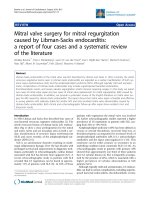
discrepancy between first two researchers. For diagnostic
meta-analysis, the quality assessment was conducted following the guidelines of the the Quality assessment of
diagnostic accuracy studies 2 (QUADAS-2) [14]. This
tool include 4 domains to evaluate the risk and applicability of bias, which are refined into 14 specific questions. Each item has a rating of “Yes”, “No” or “Unclear”,
corresponding to the scores of − 1, 1 and 0, respectively
(Fig. 2). For prognostic meta-analysis, the quality of involved studies were evaluated with the NewcastleOttawa Scale (NOS), which is the tool most commonly
used to assess the quality of non-randomized research
[15, 16]. By scoring one by one, the total quality score
Shao et al. BMC Cancer
(2019) 19:1103
Page 3 of 10
ranges from 0 to 9. Studies with a final score > 6 were
considered high-quality.
analysis was carried out with the statistical software
STATA (version 12.0) [21].
Statistical analysis
Results
For diagnostic accuracy studies, the SEN, SPE, PLR, NLR
and corresponding 95% CI from included studies were
pooled to initially assess the diagnostic value of circulating
miR-155 in lung cancer. The summary receiver operating
characteristic (SROC) curve was then drawn based on the
original data, and the area under the SROC curve (AUC)
was calculated to comprehensively determine the diagnostic accuracy of miR-155, taking into account the trade-off
between SEN and SPE. To assess the heterogeneity across
studies, the X2-based Q-statistic and I2 statistic were utilized. The I2 square value typically fluctuates within a
range of 0 (unobserved heterogeneity) to 100% (maximum
heterogeneity). P value < 0.05 or I2 > 50% was recognized
statistically significant [17]. If the studies were proved to
be homogenous, a fixed-effect model would be utilized for
further analysis. If not, the random-effect model would be
utilized [18]. Subsequently, subgroup and meta-regression
analyses were carried out to find the potential sources of
heterogeneity. Finally, the publication bias of all the included diagnostic accuracy studies was assessed by Deeks’
funnel plots (significant at P < 0.05) [19].
For prognostic meta-analyses, a combination of the
pooled HR and 95% CI was calculated to elucidate the link
between high expression of miR-155 and cooresponding
OS/DFS/PFS of lung cancer patients. Cochran’s Q test
and I2 statistics were applied to evaluate the heterogeneity
of the pooled results [20]. In addition, we used Begg’s and
Egger’s tests to assess publication bias. All above statistical
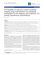
Litereture search results
Fig. 1 Flow chart of selection process
Based on a systematic search on the above databases,
363 records related to miR-155 in lung cancer were initially identified. Then, 245 duplicates were deleted following the inclusion and exclusion criteria described
previously. Eighty-seven articles were subsequently removed after a quick skim through the titles and abstracts. As a result, the remaining 31 articles were all
downloaded to obtain valid information individually.
After reading the full texts carefully, 12 studies were elimated due to lack of available diagnostic or prognostic
related data. Ultimately, this meta-analysis included 13
articles covering 19 cohort studies [22–34]. Among
them, 6 articles with 8 studies focused on the miR-155
expression for lung cancer diagnostic accuracy, whereas
7 articles including 11 studies related to the correlation
of miR-155 and lung cancer prognosis. (Fig. 1).
Studies characteristics and quality assessment
In 8 eligible studies for diagnostic analysis, 457 cases
and 342 controls were identified as presented on Table 1.
Among these 8 studies, three ethnic groups were analyzed, in which six from Asians, one from Africans, and
the remaining from Caucasians. All included studies detected miR-155 expression through qRT-PCR using
SYBR or Tagman reagent. The results of QUADAS-2
quality assessment were shown in Fig. 2a and 2b. Most
studies were consistent with the criteria in QUADAS-2,
Shao et al. BMC Cancer
(2019) 19:1103
Page 4 of 10
Table 1 Characteristics and methodology assessment of 8 studies included in the diagnosis meta-analysis
First author
Year
Country
Ethnicity
Case/Control
Assay type
SEN (%)
SPE(%)
TP
FP
FN
TN
Feng Gao [22]
2013
China
Asian
36/32
SYBR
72.20
68.70
26
8
10
22
Dongfang Tang (1) [23]
2013
China
Asian
62/60
TaqMan
59.70
75.00
37
15
25
45
Dongfang Tang (2) [23]
2013
China
Asian
34/32
TaqMan
67.60
65.60
23
11
11
21
Qing Geng (1) [24]
2014
China
Asian
25/25
SYBR
87.00
87.00
22
3
3
22
Qing Geng (2) [24]
2014
China
Asian
126/60
SYBR
86.00
84.00
108
10
18
50
Amal A [25].
2013
Egypt
African
65/37
SYBR
95.40
62.20
62
14
3
23
Carina Roth [26]
2011
Germany
Caucasian
35/28
TaqMan
87.70
88.90
31
3
4
25
Dali Zheng [27]
2011
China
Asian
74/68
SYBR
80.36
83.93
59
11
15
57
indicating that the enrolled studies are suitable for quantitative synthesis.
In the 7 included articles for prognosis, a total of 1382
participants were identified for assessing OS/DFS/PFS,
respectively. The characteristics of these enrolled literatures were presented on Table 2. The included population were classified into Asians and Caucasians from five
different countries, including China, France, America,
Japan and Norway. In addtion, the detailed quality assessment for each study scored following the guidlines
of NOS is shown in Table 3.
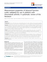
Diagnosis meta-analysis
Pooled diagnostic value of miR-155 in lung cancer
The forest plots results were presented in Fig. 3a and 3b
as follows: the pooled SEN and SPE were 82% (95% CI:
78–88%) and 78% (95% CI: 71–84%). The PLR and NLR
were 3.75 (95% CI: 2.76–5.10) and 0.23 (95% CI: 0.15–
0.37) respectively (Fig. 3c and 3d). Meanwhile, the pooled
DOR was 15.99 (95% CI: 8.11–31.52) (Fig. 5a) and the area
under SROC (AUC) was 0.85 (95% CI: 0.82–0.88) (Fig. 6a).
All above data demonstrated the relatively high diagnostic
value of miR-155 in lung cancer.
Fig. 2 QUADAS-2 quality assessment. Investigators’ assessment regarding each domain for included studies: (a) The graph and (b) summary
Shao et al. BMC Cancer
(2019) 19:1103
Page 5 of 10
Table 2 The main features of 11 included studies in prognostic meta-analysis
First author
Year
Country
Ethnicity
Case
Outcome
HR (95%CIs)
P value
Mitch Raponi [28]
2009
America
Caucasian
54
OS
2.30 (1.00–5.60)
0.060
Motonobu Saito (1) [29]
2011
Japan
Caucasian
89
PFS
2.37 (1.27–4.42)
0.006
Motonobu Saito (2) [29]
2011
Japan
Caucasian
37
PFS
1.60 (0.73–3.52)
0.245
Motonobu Saito (3) [29]
2011
Japan
Asian
191
PFS
1.33 (0.77–2.29)
0.309
Yi Gao [30]
2014
China
Asian
162
OS
2.31 (1.48–3.61)
< 0.001
Johannes Voortman [31]
2010
France
Caucasian
637
OS
0.91 (0.72–1.13)
0.390
Tom Donnem (1) [32]
2011
Norway
Caucasian
191 (SCC)
PFS
0.45 (0.21–0.96)
0.039
Tom Donnem (2) [32]
2011
Norway
Caucasian
95 (AC)
PFS
1.87 (1.01–3.48)
0.047
Ce´ line Sanfiorenzo [33]
2013
France
Caucasian
52
DFS
0.94 (0.15–5.74)
0.008
Xinying Xue (1) [34]
2016
China
Asian
80
OS
0.52 (0.24–1.14)
0.045
Xinying Xue (2) [34]
2016
China
Asian
80
DFS
0.83 (0.30–2.31)
0.054
OS: overall survival; DFS: disease free survival; PFS: progression-free survival;SCC:squamous cell carcinoma;AC:Adenocarcinoma
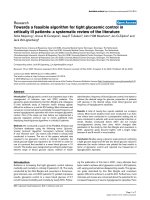
Subgroup analysis
To distinguish the potential origins of heterogeneity between studies, a subgroup analysis was perfomed based on
Assay type. The pooled results of this subgroup analysis
were shown in Fig. 4. It can be observed that studies based
on SYBR qPCR method showed similar results: the SEN
was 86% (95% CI: 77–91%), SPE was 79% (95% CI: 71–
86%), PLR was 4.11 (95% CI: 2.99–5.65) and NLR was
0.18 (95% CI: 0.12–0.28), respectively. The summary DOR
was 22.69 (95% CI: 13.90–37.04) (Fig. 5b) and AUC was
0.89 (95% CI: 0.86–0.91) (Fig. 6b).
Prognosis meta-analysis
The main outcome of the prognostic meta-analysis was to
evaluate the correlation between miR-155 expression and
OS/DFS/PFS of lung cancer patients. In the 4 studies evaluating OS, the pooled HR and its 95% CIs were calculated
using a random-effect model with a result of 1.26 (95% CI:
0.66–2.40) (Fig. 7a). Meanwhile, for 7 studies evaluating
DFS/PFS, the combined HR with 95% CIs was 1.28 (95% CI:
0.82–1.97) (Fig. 7b). To sum up, the results given above
proved that there was not significant correction between
over-expression of miRNA-155 and poor OS or DFS/PFS.
Publication bias and meta-regression analyses
The potential publication bias across the enrolled diagnostic studies was accessed by the Deeks’ funnel plot test
whereas the prognostic studies evaluated using Begg’s
funnel plot and Egger’s test. The Deeks’ funnel plot was
symmetry and reached a P value of 0.951 above 0.05, indicating there is no obvious publication bias in these included studies. The P values of Begg’s tests for OS and
DFS/PFS were 0.497 and 0.453. The results of Egger’s
test (OS: P = 0.785, DFS/PFS: P = 0.264, respectively)
also proved no existence of publication bias. These results indicated that the data were reliable in the current
meta-analysis.
Discussion
As a malignant tumor with extremely high mortality,
lung cancer has gaining great attention and extensive researches during recent decades. With the development
of surgical techniques, concurrent radiotherapy and
Table 3 Newcastle–Ottawa quality assessments scale
First author
Year
Quality indicators from Newcastle–Ottawa Scale
Scores
1
2
3
4
5
6
7
8
Raponi [28]
2009
★
★
–
–
★★
★
★
★
7
Saito [29]
2011
★
★
–
★
★★
★
★
★
8
Yi G [30]
2014
★
★
–
–
★★
★
★
★
7
Voortman [31]
2010
–
–
–
★
★★
★
★
★
6
Donnem [32]
2011
★
–
–
–
★★
★
★
★
6
Sanfiorenzo [33]
2013
★
★
–
★
★★
★
★
★
8
Xue [34]
2016
★
★
–
★
★★
★
★
–
7
1. Representativeness of the exposed cohort; 2. Selection of the non-exposed cohort; 3. Ascertainment of exposure; 4. Outcome of interest not present at start of
study; 5. Control for important factor or additional factor; 6. Assessment of outcome; 7. Follow-up long enough for outcomes to occur; 8. Adequacy of follow up
of cohorts
Shao et al. BMC Cancer
(2019) 19:1103
Page 6 of 10
Fig. 3 Forest plots of sensitivity (a), specificity (b), positive likelihood ratios (c) and negative likelihood ratios (d) for miR-155 in the diagnosis of
lung cancer
chemotherapy, and imaging examination technology
have greatly improved the prognosis of lung cancer patients. Nevertheless, the most effective way to improve
the survival of lung cancer patients lies in early diagnosis
and targeted treatment. Therefore, a large amount of researchers are committed to finding suitable non-invasive
biomarkers to predict the diagnosis or prognosis of lung
cancer, and provide directions for clinical treatment of
lung cancer .
As vital regulators of various biological processes in cancer, miRNAs were regarded as perfect non-invasive biomarkers for human cancers [35, 36]. MiR-155 was widely
studied to participate in the occurrence and progression of
diverse cancers, including lung cancer [37]. Several researches suggested that up-regulated miR-155 is positively
correlated with the pathogenesis of lung cancer, indicating
that miR-155 acts as an oncogene in lung cancer [37, 38].
Zang et al. revealed that miR-155 was involved in the drug
resistance of lung cancer. In their study, miR-155 was
shown to modulate celluar poptosis and DNA damage via
Apaf-1 regulated pathways to decrease the sensitivity of
lung cancer cells to cisplatin [38]. Moreover, another research conducted by Katrien et al. found that miR-155 increases resistance to chemotherapy in lung cancer cells by
forming a feedback loop with TP53 [39]. In particular, they
also found that over-expression of miR-155 is significantly
linked to poor OS of lung cancer patients. These results indicated that miR-155 has the potential to be an ideal biomarker for lung cancer. In addition to the above studies
focused on the molecular mechanism of miR-155 regulation in lung cancer cells, accumulating cohort studies have
reported the coorelationship between miR-155 levels in different individuals with lung cancer diagnosis or prognosis
to determine whether miR-155 acts as an ideal biomarker
[40, 41]. However, these results have not been corroborated
and even contradictory. Thus, this meta-analysis appears to
be necessary to figure out the diagnostic and prognostic
value of miR-155 for lung cancer.
In the diagnositic meta-analysis, the total DOR with 95%
CI of miR-155 was 15.99 (95% CI: 8.11–31.52). In addition,
AUC and corresponding 95% CI were 0.85 (95% CI: 0.82–
0.88), indicating that miR-155 could act as a moderate
Shao et al. BMC Cancer
(2019) 19:1103
Page 7 of 10
Fig. 4 Subgroup analysis based on Assay type of sensitivity (a), specificity (b), positive likelihood ratios (c) and negative likelihood ratios (d) for
miR-155 by SYBR in the diagnosis of lung cancer
Fig. 5 Forest plots of the diagnostic odds ratio (DOR) for miR-155 in the diagnosis of lung cancer. (a). All studies; (b). The studies based on SYBR
Shao et al. BMC Cancer
(2019) 19:1103
Page 8 of 10
Fig. 6 Summary receiver operating characteristic curves (sROC) from the hierarchical summary receiver operating characteristic model generated
from the 8 studies that found that miR-155 was a diagnostic marker for lung cancer. (a). All studies; (b). The studies based on SYBR
marker in the lung cancer diagnosis compared to healthy
individuals. Subgroup analysis of Assay type revealed that
studies based on SYBR had a higher DOR of 22.69 (95% CI:
13.90–37.04) and the higher AUC of 0.89 (95% CI: 0.86–
0.91), which might be the possible sources of heterogeneity.
Nowadays, several tumor biomarkers have been applied for
detecting early lung cancer clinically, such as CA-125, CEA,
CYFRA21-1, NSE and so on. However, limited sensitivity
and specificity of these existing biomakers restricted their
diagnostic accuracy. Based on the 6 included articles, miR155 can be stably detected in the plasma of lung cancer
patients with marked differences when compared with
control samples, suggesting it can serve as a serum-based
biomarker for lung cancer detection individually. As Currently, miR-155 hasn’t been applied as a clinical diagnostic
tool in patients that had not previously been diagnosed with
lung cancer. And clinical detection of lung cancer usually
involves not only a single miRNA, but a combination of
miRNAs.What’s more, miR-155 could be combined with
traditional biomarkers for the diagnosis of lung cancer, so
as to improve the diagnosis accuracy in the future.
By the way, as polymorphisms in genes encoding miRNAs may alter the expression of the corresponding
miRNA and thus confer susceptibility to multiple
Fig. 7 Forest plots of the studies that evaluated the hazard ratios of high miR-155 expression. (a). The studies based on OS; (b). The studies based
on DFS/PFS
Shao et al. BMC Cancer
(2019) 19:1103
diseases such as cancers, it might be meaningful to investigate the association between polymorphisms in
genes encoding miR-155 and lung cancer susceptibility.
For example, previous study published by Xie et al. identified that rs767649 (A > T) in regulatory regions of miR155 was associated with the increased risk and poor
prognosis of lung cancer [42]. What’s more, they found
four target genes of miR-155 including HBP1, TJP1,
SMAD5 and PRKAR1A involved in the oxidative stress
process of lung cancer. Given that miR-155 is a typical
oncogene in lung cancer, more well-designed studies in
the future could confirm its diagnostic value, and more
importantly, further researches colud focus on gene
polymorphisms encoding miR-155, which can manually
regulate miRNA levels, leading to changes in cancerassociated downstream protein signaling pathways.
On the other hand, the prognostic meta-analysis suggested that up-regulated miR-155 might not be associated with poor clinical outcomes of lung cancer patients,
which was 1.26-fold higher risk for poor OS and 1.28fold higher risk for poor DFS/PFS. These results might
caused by different genetic backgrounds, environmental
exposures and detection methods. Recently, accumulating studies worldwide have shown that expression levels
of miRNAs in different individuals have significant predictive value in cancers. Currently, the detection of miRNAs in tissue samples has been applied to current
tumor prognosis studies, but the detection of serum/
plasma samples and other human body fluids appears to
be more portable, non-invasive, and can effectively
assess survival prognosis at any time before or after
treatment. It can even play a role in the patient’s lifelong disease surveillance and is of great help to clinical
thearapy. This meta-analysis found that miR-155 has no
obvious prognostic effect on lung cancer, which is inconsistent with results of some previous prognostic
studies, while the result is consistent with the prognostic
value of miR-155 of NSCLC reported in a meta-analysis
published by Lamichhane SR et al. in 2018 [43]. However, the sample size included in our meta-analysis is
larger than previous mata-analysis, more researches with
sufficient data will be needed to verify this result.
Ultimately, several limitations still existed in this metaanalysis as follows: (1) Racial factors were not comprehensive enough, and the population is too monotonous. For
example, the diagnostic meta-analysis is mainly for Asians
and Africans while the prognostic meta-analysis only focused on Caucasians and Asians. Therefore, more researchers should pay attention to the impact of racial
factors in the subsequent studies. (2) Unpublished studies
may contain negative results, but we are not available include them, which potentially lead to lack of credibility in
the data. (3) We only included articles published in English and Chinese, but did not cover articles in other
Page 9 of 10
languages. (4) The sample size was still relatively small, including only 19 studies, which may undermine the reliability of our findings. Therefore, more well-designed
studies based on larger samples and sufficient data are
required to verify the diagnostic and prognostic value of
circulating miR-155 in lung cancer. (5) Adjusted estimates
could not be performed in our meta analysis without
enough data for the adjustment by other covariates such
as TNM stage, histological type, mean of age, gender and
so on.. Therefore, further high-quality researches in the
risk of lung cancer might be performed to draw more
accuracy results in subsequent years.
Conclusion
To summarize, our meta-analysis demonstrated for the
first time that circulating miR-155 is promising to be a
novel biomarker for diagnosis of lung cancer. However,
miR-155 is not an effective biomarker for predicting the
prognosis of lung cancer. Together, these findings provide important evidence for further development of future non-invasive methods for diagnosing lung cancer.
Further large-scale relevant studies with better designs
and more comprehensive data support will help to clarify the diagnostic and prognostic value of miR-155 in
lung cancer.
Abbreviations
AUC: Area under the ROC curve; DFS: Disease free survival; DOR: Diagnostic
odds ratio; FN: False negative; FP: False positive; HR: Hazard ratio;
miRNAs: MicroRNAs; NLR: Negative likelihood ratio; NOS: Newcastle-Ottawa
Scale; OS: Overall survival; PFS: Progression-free survival; PLR: Positive
likelihood ratio; QUADAS: Quality assessment of diagnostic accuracy studies;
TN: True negative; TP: True positive
Acknowledgements
Not applicable.
Authors’ contribution
SYQ and SH proposed the conjecture and design of this study. SCC and YFM
conducted the collection of materials and data management. Analysis and
interpretation of the data were performed by QZQ and YFM. The writing and
revision of the manuscript were done by SCC and JXM. All authors have
checked the full text carefully and approved the final draft.
Funding
This study was supported by grants from the Natural Science Foundation of
China (no. 81874230) in collecting and analyzing data.
Availability of data and materials
The data that support the findings of this study are available from the
corresponding author upon reasonable request.
Ethics approval and consent to participate
Not applicable.
Consent for publication
Not applicable.
Competing interests
The authors declare that they have no competing interests.
Shao et al. BMC Cancer
(2019) 19:1103
Author details
1
Department of Oncology, The First Affiliated Hospital of Nanjing Medical
University, 300 Guangzhou Road, Nanjing 210029, People’s Republic of China.
2
Department of Oncology, The First Affiliated Hospital of Nanjing Medical
University, Nanjing, China. 3Department of Urology, Nanjing First Hospital,
Nanjing Medical University, Nanjing, China.
Received: 16 May 2019 Accepted: 27 October 2019
References
1. Siegel RL, Miller KD, Jemal A. Cancer statistics, 2018. CA Cancer J Clin. 2018;
68(1):7–30.
2. Heng WS, Gosens R, Kruyt F. Lung cancer stem cells: origin, features,
maintenance mechanisms and therapeutic targeting. Biochem Pharmacol.
2019;160:121–33.
3. Tsukamoto M, Iinuma H, Yagi T, Matsuda K, Hashiguchi Y. Circulating
Exosomal MicroRNA-21 as a biomarker in each tumor stage of colorectal
Cancer. Oncology. 2017;92(6):360–70.
4. Mabert K, Cojoc M, Peitzsch C, Kurth I, Souchelnytskyi S, Dubrovska A.
Cancer biomarker discovery: current status and future perspectives. Int J
Radiat Biol. 2014;90(8):659–77.
5. Mohr AM, Mott JL. Overview of microRNA biology. Semin Liver Dis.
2015;35(1):3–11.
6. Rupaimoole R, Slack FJ. MicroRNA therapeutics: towards a new era for
the management of cancer and other diseases. Nat Rev Drug Discov.
2017;16(3):203–22.
7. Armand-Labit V, Pradines A. Circulating cell-free microRNAs as clinical
cancer biomarkers. Biomol Concepts. 2017;8(2):61–81.
8. Zhang XF, Tu R, Li K, Ye P, Cui X. Tumor suppressor PTPRJ is a target of miR155 in colorectal Cancer. J Cell Biochem. 2017;118(10):3391–400.
9. Khoshinani HM, Afshar S, Pashaki AS, Mahdavinezhad A, Nikzad S, Najafi R,
Amini R, Gholami MH, Khoshghadam A, Saidijam M. Involvement of miR155/FOXO3a and miR-222/PTEN in acquired radioresistance of colorectal
cancer cell line. Jpn J Radiol. 2017;35(11):664–72.
10. Zuo J, Yu Y, Zhu M, Jing W, Yu M, Chai H, Liang C, Tu J. Inhibition of miR155, a therapeutic target for breast cancer, prevented in cancer stem cell
formation. Cancer Biomark. 2018;21(2):383–92.
11. Qu Y, Zhang H, Sun W, Han Y, Li S, Qu Y, Ying G, Ba Y. MicroRNA-155
promotes gastric cancer growth and invasion by negatively regulating
transforming growth factor-beta receptor 2. Cancer Sci. 2018;109(3):618–28.
12. Wang J, Guo J, Fan H. MiR-155 regulates the proliferation and apoptosis of
pancreatic cancer cells through targeting SOCS3. Eur Rev Med Pharmacol
Sci. 2019;23(12):5168–75.
13. Hou Y, Wang J, Wang X, Shi S, Wang W, Chen Z. Appraising MicroRNA-155
as a noninvasive diagnostic biomarker for Cancer detection: a Meta-analysis.
Medicine (Baltimore). 2016;95(2):e2450.
14. Whiting PF, Rutjes AW, Westwood ME, Mallett S, Deeks JJ, Reitsma JB,
Leeflang MM, Sterne JA, Bossuyt PM. QUADAS-2: a revised tool for the
quality assessment of diagnostic accuracy studies. Ann Intern Med.
2011;155(8):529–36.
15. Lo CK, Mertz D, Loeb M. Newcastle-Ottawa scale: comparing reviewers' to
authors' assessments. BMC Med Res Methodol. 2014;14:45.
16. Roysri K, Chotipanich C, Laopaiboon V, Khiewyoo J. Quality assessment of
research articles in nuclear medicine using STARD and QUADAS-2 tools.
Asia Ocean J Nucl Med Biol. 2014;2(2):120–6.
17. Raponi M, Dossey L, Jatkoe T, Wu X, Chen G, Fan H, Beer DG. MicroRNA
classifiers for predicting prognosis of squamous cell lung cancer. Cancer
Res. 2009;69(14):5776–83.
18. Higgins JP, Thompson SG. Quantifying heterogeneity in a meta-analysis.
Stat Med. 2002;21(11):1539–58.
19. Deeks JJ, Macaskill P, Irwig L. The performance of tests of publication bias
and other sample size effects in systematic reviews of diagnostic test
accuracy was assessed. J Clin Epidemiol. 2005;58(9):882–93.
20. Higgins JP, Thompson SG, Deeks JJ, Altman DG. Measuring inconsistency in
meta-analyses. BMJ. 2003;327(7414):557–60.
21. Zamora J, Abraira V, Muriel A, Khan K, Coomarasamy A. Meta-DiSc: a
software for meta-analysis of test accuracy data. BMC Med Res
Methodol. 2006;6:31.
22. Gao F, Chang J, Wang H, Zhang G. Potential diagnostic value of miR-155 in
serum from lung adenocarcinoma patients. Oncol Rep. 2014;31(1):351–7.
Page 10 of 10
23. Tang D, Shen Y, Wang M, Yang R, Wang Z, Sui A, Jiao W, Wang Y.
Identification of plasma microRNAs as novel noninvasive biomarkers for
early detection of lung cancer. Eur J Cancer Prev. 2013;22(6):540–8.
24. Geng Q, Fan T, Zhang B, Wang W, Xu Y, Hu H. Five microRNAs in plasma as
novel biomarkers for screening of early-stage non-small cell lung cancer.
Respir Res. 2014;15:149.
25. Abd-El-Fattah AA, Sadik NA, Shaker OG, Aboulftouh ML. Differential
microRNAs expression in serum of patients with lung cancer, pulmonary
tuberculosis, and pneumonia. Cell Biochem Biophys. 2013;67(3):875–84.
26. Roth C, Kasimir-Bauer S, Pantel K, Schwarzenbach H. Screening for
circulating nucleic acids and caspase activity in the peripheral blood as
potential diagnostic tools in lung cancer. Mol Oncol. 2011;5(3):281–91.
27. Zheng D, Haddadin S, Wang Y, Gu LQ, Perry MC, Freter CE, Wang MX.
Plasma microRNAs as novel biomarkers for early detection of lung cancer.
Int J Clin Exp Pathol. 2011;4(6):575–86.
28. Raponi M, Dossey L, Jatkoe T, Wu X, Chen G, Fan H, Beer DG. MicroRNA
classifiers for predicting prognosis of squamous cell lung cancer. Cancer
Res. 2009;69(14):5776–83.
29. Saito M, Schetter AJ, Mollerup S, Kohno T, Skaug V, Bowman ED, Mathe EA,
Takenoshita S, Yokota J, Haugen A, et al. The association of microRNA
expression with prognosis and progression in early-stage, non-small cell
lung adenocarcinoma: a retrospective analysis of three cohorts. Clin Cancer
Res. 2011;17(7):1875–82.
30. Yi G, Shengling F, Wenyang J, Binfeng L, Yitao T, Xiangning F. Relationship
between expression of miR-155 and prognosis in pIII stage non-small cell
lung cancer. Chinese Journal of Lung Cancer. 2014;05:417–23.
31. Voortman J, Goto A, Mendiboure J, Sohn JJ, Schetter AJ, Saito M, Dunant A,
Pham TC, Petrini I, Lee A, et al. MicroRNA expression and clinical outcomes
in patients treated with adjuvant chemotherapy after complete resection of
non-small cell lung carcinoma. Cancer Res. 2010;70(21):8288–98.
32. Donnem T, Eklo K, Berg T, Sorbye SW, Lonvik K, Al-Saad S, Al-Shibli K,
Andersen S, Stenvold H, Bremnes RM, et al. Prognostic impact of MiR155 in non-small cell lung cancer evaluated by in situ hybridization. J
Transl Med. 2011;9:6.
33. Sanfiorenzo C, Ilie MI, Belaid A, Barlesi F, Mouroux J, Marquette CH, Brest P,
Hofman P. Two panels of plasma microRNAs as non-invasive biomarkers for
prediction of recurrence in resectable NSCLC. PLoS One. 2013;8(1):e54596.
34. Xue X, Liu Y, Wang Y, Meng M, Wang K, Zang X, Zhao S, Sun X, Cui L,
Pan L, et al. MiR-21 and MiR-155 promote non-small cell lung cancer
progression by downregulating SOCS1, SOCS6, and PTEN. Oncotarget.
2016;7(51):84508–19.
35. Wang J, Yan F, Zhao Q, Zhan F, Wang R, Wang L, Zhang Y, Huang X.
Circulating exosomal miR-125a-3p as a novel biomarker for early-stage
colon cancer. Sci Rep. 2017;7(1):4150.
36. Switlik W, Karbownik MS, Suwalski M, Kozak J, Szemraj J. miR-30a-5p
together with miR-210-3p as a promising biomarker for non-small cell lung
cancer: a preliminary study. Cancer Biomark. 2018;21(2):479–88.
37. Higgs G, Slack F. The multiple roles of microRNA-155 in oncogenesis. J Clin
Bioinforma. 2013;3(1):17.
38. Zang YS, Zhong YF, Fang Z, Li B, An J. MiR-155 inhibits the sensitivity of
lung cancer cells to cisplatin via negative regulation of Apaf-1 expression.
Cancer Gene Ther. 2012;19(11):773–8.
39. Van Roosbroeck K, Fanini F, Setoyama T, Ivan C, Rodriguez-Aguayo C,
Fuentes-Mattei E, Xiao L, Vannini I, Redis RS, D'Abundo L, et al. Combining
anti-Mir-155 with chemotherapy for the treatment of lung cancers. Clin
Cancer Res. 2017;23(11):2891–904.
40. Liu K, Zhao K, Wang L, Sun E. Prognostic value of microRNA-155 in human
carcinomas: An updated meta-analysis. Clin Chim Acta. 2018;479:171–80.
41. Wang F, Zhou J, Zhang Y, Wang Y, Cheng L, Bai Y, Ma H. The value of
MicroRNA-155 as a prognostic factor for survival in non-small cell lung
Cancer: a Meta-analysis. PLoS One. 2015;10(8):e136889.
42. Xie K, Ma H, Liang C, Wang C, Qin N, Shen W, Gu Y, Yan C, Zhang K, Dai N,
et al. A functional variant in miR-155 regulation region contributes to lung
cancer risk and survival. Oncotarget. 2015;6(40):42781–92.
43. Lamichhane SR, Thachil T, De Ieso P, Gee H, Moss SA, Milic N. Prognostic
role of MicroRNAs in human non-small-cell lung Cancer: a systematic review
and Meta-analysis. Dis Markers. 2018;2018:8309015.
Publisher’s Note
Springer Nature remains neutral with regard to jurisdictional claims in
published maps and institutional affiliations.