Biosynthesis of silver nanoparticles using Bacillus sp. and evaluation of its antibacterial activity
Bạn đang xem bản rút gọn của tài liệu. Xem và tải ngay bản đầy đủ của tài liệu tại đây (432.48 KB, 8 trang )
Int.J.Curr.Microbiol.App.Sci (2017) 6(3): 2496-2503
International Journal of Current Microbiology and Applied Sciences
ISSN: 2319-7706 Volume 6 Number 3 (2017) pp. 2496-2503
Journal homepage:
Original Research Article
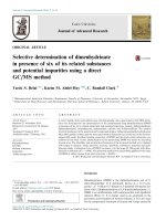
/>
Biosynthesis of Silver Nanoparticles using Bacillus sp. and
Evaluation of its Antibacterial Activity
M. Durairasu1, V. Indra1, N. Arunagirinathan2, J. Hemapriya3 and S. Vijayanand4*
1
Department of Zoology, Presidency College, Chennai, Tamilnadu, India
Department of Microbiology, Presidency College, Chennai, Tamilnadu, India
3
Department of Microbiology, DKM College, Vellore, Tamilnadu, India
4
Department of Biotechnology, Thiruvalluvar University, Vellore, Tamilnadu, India
2
*Corresponding author
ABSTRACT
Keywords
Antibacterial activity,
Bactericidal,
Bacillus sp. DRI-6,
Silver Nanoparticles.
Article Info
Accepted:
20 February 2017
Available Online:
10 March 2017
Nanotechnology has recently emerged as an elementary discipline of science that
explores the interaction of synthetic and biological materials. Nanotechnology is
currently employed as a tool to exploit the darkest avenues of medical sciences to
combat dreadful diseases caused by drug resistant microbes. Silver nanoparticles
(Ag NPs) have been well known for its inhibitory and bactericidal effects. Silver
Nanoparticles was synthesized by ecofriendly biogenic approach mediated by
using the culture supernatant of Bacillus sp. DRI-6. The biogenic silver
nanoparticles were characterized by UV-visible spectroscopy, X-ray diffraction
(XRD), scanning electron microscopy (SEM) and Transmission electron
microscopy (TEM). Ag NPs exhibited maximum antibacterial activity against
E.coli and Pseudomonas sp.
Introduction
Nanotechnology has recently emerged as an
elementary division of science that explores
the interaction at cellular level between
synthetic and biological entities with the help
of nanoparticles. „Nano‟ is a Greek word
synonymous to dwarf meaning extremely
small (Kushwaha et al., 2015). The word
“nano” is used to indicate one billionth of a
meter or 10 -9. Nanoparticles are clusters of
atoms in the size range of 1–100 nm. A wide
range of nanophasic and nanostructured
particles are being fabricated globally with
the aim of developing clean, nontoxic and
eco-friendly technologies. Use of ambient
biological resources in nanotechnology is
rapidly acquiring significant importance
owing to its alarming success and simplicity
(Sinha et al., 2009). Nanobiotechnology, the
combination
of
biotechnology
and
nanotechnology greatly focuses on the
development of the environmental benign
biogenic approach and technology for
synthesis of nanomaterials (Sahayaraj and
Rajesh, 2011).
Nanobiotechnology combines biological
principles with physical and chemical
approaches to produce nano-sized particles
2496
Int.J.Curr.Microbiol.App.Sci (2017) 6(3): 2496-2503
with specific functions, representing an
economic substitute for chemical and physical
methods
of
nanoparticles
formation.
Biosynthesis of NP‟S can be divided into
intracellular and extracellular (Ahmad et al.,
2005).
Among
them,
the
metallic
nanoparticles are considered to be the most
promising ones, as they contain significant
antibacterial and antifungal properties due to
their large surface area to volume ratio, which
is of great interest to researchers due to the
growing microbial resistance against metal
ions, antibiotics and the development of
resistant strains (Gong et al., 2007).
Silver nanoparticles (Ag NPs) have several
important applications in the field of
biolabelling, sensors, antimicrobial agents and
filters. They are capable of purifying drinking
water, degrading pesticides and killing human
pathogenic bacteria (Bhainsa and D‟Souza,
2006). Recently, biological synthesis of silver
nanoparticles has received a special attention
due to environmental friendly green synthesis
and easy to scale-up. Many researchers
demonstrated that the green synthesis of silver
nanoparticles
including
bacteria,
actinomycetes, fungi and plants (Lavanya et
al., 2013). The recent advances in researches
on metal nanoparticles appear to revive the
use of silver nanoparticles (Ag NPs) for
antimicrobial applications. Ag NPs have
strong inhibitory and bactericidal effects as
well as a broad spectrum of antimicrobial
activities for bacteria, fungi, and virus since
ancient times (Lok et al., 2006). The
mechanism of inhibition by silver ions on
microorganisms is partially known. It is
believed that DNA loses its replication ability
and cellular proteins become inactivated upon
silver ion treatment (Gupta et al., 2008).
Furthermore, higher concentrations of Ag+
ions have been shown to interact with
cytoplasmic components and nucleic acids
(Kim, 2007; Kumar et al., 2008). In the
present study, the ecofriendly biosynthesis of
silver nanoparticles using the culture
supernatant of Bacillus sp. Strain DRI-6 was
mediated. Synthesized nanoparticles were
characterized by UV-Visible spectroscopy,
XRD, FTIR, SEM and TEM analysis.
Furthermore, the antimicrobial activity of
synthesized silver nanoparticles against S.
aureus, Klebsiella pneumoniae, E.coli and
Pseudomonas sp. was evaluated.
Materials and Methods
Bacterial Strain Used
The bacterial strain used in this study was
isolated
from
environmental
samples
including contaminated water samples,
effluent samples and soil samples collected
from in and around Kanchipuram. Based on
the morphological, cultural, biochemical
characteristics and 16 s rDNA sequencing, the
isolate was identified as Bacillus sp. strain
DRI-6.
Synthesis of Ag NP’s from Culture
Supernatant of Bacillus sp. Strain DRI-6
The aqueous solution of 1 mM silver nitrate
(AgNO3) was prepared and used for the
synthesis of silver nanoparticles. 15 ml of
culture supernatant of Bacillus sp. strain DRI6 was added into 200 ml of aqueous solution
of 1 mM silver nitrate for reduction into Ag+
ions and kept for 15-20 minutes. Culture
supernatant acts as reducing and stabilizing
agent. The prepared Ag NP‟s were further
characterized (Karthika et al., 2015).
Characterization of synthesized Ag NP’s
The techniques used for characterization were
as follows:
UV-VIS spectroscopy
Biogenic synthesis of Ag NP‟s solution with
the culture supernatant of Bacillus sp. strain
2497
Int.J.Curr.Microbiol.App.Sci (2017) 6(3): 2496-2503
DRI-6
was
observed
by
UV–Vis
spectroscopy. Samples were monitored as a
function of time of reaction using Shimadzu
1601 spectrophotometer in the 300–800 nm
range operated at a resolution of 1 nm. The
double distilled water used as a blank
reference.
Fourier Transform Infra-Red
Spectroscopy (FTIR)
Klebsiella pneumoniae, Escherichia coli, and
Pseudomonas aeruginosa by disc diffusion
method. The synthesized nanoparticles were
diluted with distilled water (15 μg/ml) and
placed onto each wells and incubated for 24
hours. Following incubation, the zone of
inhibition against nanoparticle was observed
and measured (Karthika et al., 2015).
Results and Discussion
The
purified
suspension
of
silver
nanoparticles was freeze dried to obtain dried
powder. Then, the dried nanoparticle samples,
prepared as KBr discs were analyzed by FTIR Spectrometer for the detection of different
functional groups from the region of 4004000 cm-1.
X- Ray Diffraction (XRD) Analysis
Purified and dried pellet of synthesized Ag
NP‟s were subjected to XRD analysis. For
XRD studies, dried NPs were coated on XRD
grid, and the spectra were recorded by using
Phillips PW 1830 instrument operating at a
voltage of 40 kV and a current of 30 mA with
Cu Kα1 radiation.
Scanning Electron Microscopy (SEM) and
Transmission Electron Microscopy (TEM)
The particle size and morphology of the silver
nanoparticles were examined using Scanning
electron microscopic observations. SEM
measurements were performed on a JEOL
JSM 6390 instrument operated at an
accelerating voltage at 15kV. The shape and
size of Ag NP‟s was determined by
transmission electron microscopy. The images
were obtained at a bias voltage of 200 kV
used to analyze samples.
Antibacterial activity of Ag Nanoparticles
The antibacterial effect of Ag NP‟s was
examined against Staphylococcus aureus,
Nanobiotechnology combines biological
principles with physical and chemical
procedures to generate nano-sized particles
with specific functions. Nanobiotechnology
represents an economic alternative for
chemical
and physical
methods
of
nanoparticles formation (Ahmad et al., 2005).
The biosynthesis of metallic nanoparticles is
an active and pronounced area of research in
nanotechnology. The synthesis of metal
nanoparticles depends on the nitrate reductase
enzyme present in the microbes. The
mechanism
of
the
biosynthesized
nanoparticles involves the reduction of silver
ions by the electron shuttle enzymatic metal
reduction process. NADH and NADHdependent enzymes are important factors in
the biosynthesis of metal nanoparticles
(Kalimuthu et al., 2008). The microbes are
known to secrete the cofactor NADH, and
NADH-dependent enzymes like nitrate
reductase might be responsible for the
bioreduction of metal ions and the subsequent
formation of silver nanoparticles.
Biogenic Synthesis of Ag NPs using the
culture supernatant of Bacillus sp. DRI-6
Biogenic synthesis of silver nanoparticles was
carried out by using the culture supernatant of
Bacillus sp. Strain DRI-6. On mixing the
culture supernatant of Bacillus sp. with silver
nitrate solution (1 mM), a change in the color
from pale yellow to dark brown was
observed. Similarly, Kushwaha et al. (2015)
reported the biosynthesis and characterization
2498
Int.J.Curr.Microbiol.App.Sci (2017) 6(3): 2496-2503
of Ag NPs from E. coli. The brown color
confirms the reduction of Ag+ which indicates
the formation of Ag nanoparticles. Various
microbes are known to reduce metal ions to
the metals. The formation of extracellular
silver nanoparticles by photoautotrophic
cyanobacterium Plectonema boryanum had
been described (Langke et al., 2007).
Characterization
Nanoparticles
of
Biogenic
Ag
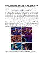
UV-vis spectrophotometer Analyses
The corresponding UV-Vis absorption
spectrum showed absorption in the form of a
sharp peak between 200-250 nm which
indicates the synthesis of silver nanoparticles
(Fig 1). The absorption behavior arises due to
surface Plasmon resonance (SPR), which
originates from coherent oscillations of
electrons in the conduction band of
nanoparticles induced by the electromagnetic
field. Similar results were reported with the
silver nanoparticles synthesized with the
culture supernatant of Bacillus licheniformis
and Streptomyces sp. JAR1 (Kalimuthu et al.,
2008; Chauhan et al., 2013).
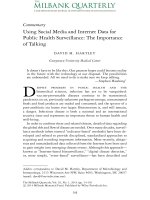
FTIR of Ag Nanoparticles
The FTIR spectroscopy is used to probe the
chemical composition of the surface and
capping agents for the synthesis of NPs (Fig
2). The synthesized Ag NPs showed the
presence of bands due to heterocyclic amine,
O-H free bond (3280 cm-1), alkanes, O-H
bend (2916 cm-1), Carboxylic acid, OH (very
broad) (2812 cm-1), arene, = C-H and
Carboxylic acid derivative, C-O-H bending
(1417 cm-1). Hence, it proves that synthesized
Ag NPs have been synthesized with the
culture supernatant of Bacillus sp. Strain DRI6 involved in the biological reduction of the
AgNO3.
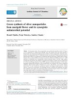
X-ray Diffractometer of Ag Nanoparticles
The crystal structure of the AgNPs was
analyzed by X-ray diffractometer. X-ray
diffraction is a very important method to
characterize the structure of crystalline
material and used for the lattice parameters
analysis of single crystals, or the phase,
texture or even stress analysis of samples. Xray diffractogram of the synthesized Ag NPs
showed distinct diffraction peaks at 38.30°,
44.44°, 64.61° and 76.88° which were
indexed to the planes 111, 200, 220 and 311
respectively (Fig 3). The sharp peaks and
absence of unidentified peaks confirmed the
crystallinity and higher purity of prepared
NPs.
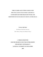
SEM & TEM Analysis
The morphology and size details of the
nanoparticles were analyzed by SEM
analyses.
The
formation
of
silver
nanoparticles as well as their morphological
dimensions in the SEM study demonstrated
that the average size was from 30 ± 3 nm with
inter particle distance, whereas the shapes
were slightly oval to spherical (Fig 4). TEM
images revealed that the morphology of Ag
NPs are nearly spherical and some nonspherical in nature having particle size less
than 100 nm (Fig 5).
Antibacterial
Nanoparticles
activity
of
Silver
Exploration of nanoparticles (NPs) as
medicines / therapeutical agents is one of the
major significance of nanomedicine (Kim et
al., 2010; Irache et al., 2011). Ag NPs
synthesized using Bacillus sp. DRI-6 exerted
maximum antibacterial activity against E.coli
(17 mm) and Klebsiella pneumoniae (13 mm)
(Table 1). Similar study was carried out by
Sadhasivam et al. (2010).
2499
Int.J.Curr.Microbiol.App.Sci (2017) 6(3): 2496-2503
Table.1 Antibacterial activity of biogenic Ag NPs against the selected bacterial isolates
S. No
Bacterial strains
1.
2.
3.
4.
Staphylococcus aureus
Klebsiella pneumoniae
Pseudomonas aeruginosa
Escherichia coli
Zone of
Inhibition
9 ± 0.5 mm
13 ± 0.4 mm
8 ± 0.6 mm
17 ± 0.8 mm
Fig.1 UV-Vis absorption spectrum of Ag Nanoparticles
SILVER
Abs
11
10
9
8
7
6
5
4
3
2
1
0
-1
200
300
400
500
600
700
800
Fig.2 FT IR analysis of biogenic Ag Nanoparticles
102
100
90
80
70
%T 60
50
40
30
20
18
4000
3500
3000
2500
2000
cm-1
2500
1500
1000
500
450
nm
Int.J.Curr.Microbiol.App.Sci (2017) 6(3): 2496-2503
Fig.3 XRD Analysis of Biogenic Ag Nanoparticles
Fig.4 SEM micrographs of biogenic Ag nanoparticles
Fig.5 TEM micrographs of biogenic Ag nanoparticles
2501
Int.J.Curr.Microbiol.App.Sci (2017) 6(3): 2496-2503
Silver ions have long been known to exert
strong inhibitory and bactericidal effects as
well as to possess a broad spectrum of
antimicrobial activities. And the acting
mechanism of silver has been known in some
extent (Rai et al., 2009). Ag+ inhibits
phosphate uptake and exchange in bacterial
cells and causes efflux of accumulated
phosphate as well as of mannitol, succinate,
glutamine, and proline (Schreurs and
Rosenberg, 1982).
Tenover (2006) proposed three different
mechanisms for the antibacterial activity of
Ag NPs. Firstly, Ag NPs attach to the surface
of the cell membrane and disturb its power
functions, such as permeability and
respiration. The binding of the particles to the
bacteria depends on the interaction of the
surface area available. With a smaller particle
size, a large surface area will have a stronger
bactericidal effect. Secondly, Ag NPs are able
to penetrate the bacteria by possibly
interacting with sulfur- and phosphoruscontaining compounds such as DNA and
cause further damage (Gibbons and Warner,
2005). Thirdly, the silver nanoparticles
release silver ions, which contribute to the
bactericidal effect (Feng et al., 2000).
References
Ahmad, A., S. Senapati, M. I. Khan, R.
Kumar and M. Sastry (2005). Extra -/
intracellular, biosynthesis of gold
nanoparticles by an alkalotolerant
fungus, Trichothecium. J. Biomed.
Nanotechnol., 1: 47-53.
Bhainsa, K.C. and S.F. D‟Souza (2006).
Extracellular biosynthesis of silver
nanoparticles
using
the
fungus
Aspergillus fumigatus. Colloids Surf B
Biointerfaces. 47: 160-164.
Chauhan, R., A.Kumar and J. Abraham.
(2013). Biological Approach to the
Synthesis of Silver Nanoparticles with
Streptomyces sp JAR1 and its
Antimicrobial Activity. Sci Pharm., 81:
607-62.
Feng, Q.L., J. Wu, G.Q. Chen, F.Z. Cui, T.N.
Kim, J.O. Kim. (2000). A mechanistic
study of the antibacterial effect of silver
ions on E. coli and Staphylococcus
aureus. J. Biomed. Mater Res., 2000;
52: 662–668.
Gibbins, B. and L. Warner. (2005). The role
of antimicrobial silver nanotechnology.
Med Device Diagnostic Indust Mag., 1:
1–2.
Gong, J., Y. Liang, Y. Huang, Y. Chen, J.
Jiang, J. Shen and R. Yu (2007).
Ag/SiO2 core-shell nanoparticle-based
surface-enhanced Raman probes for
immunoassay of cancer marker using
silica-coated magnetic nanoparticles as
separation tools. Biosensors and
Bioelectronics, 22: 1501–1507.
Gupta, P., M. Bajpai, S.K. Bajpai. (2008).
Investigation of antibacterial properties
of silver nanoparticle-loaded poly
(acrylamide-co-itaconic
acid)-grafted
cotton fabric. J Cotton Sci., 12: 280–
286.
Irache, J.M., I. Esparza, C. Gamazo, M.
Agüeros and S. Espuelas (2011).
Nanomedicine: novel approaches in
human and veterinary therapeutics. Vet.
Parasitol., 180: 47- 71.
Kalimuthu, K., R.S. Babu, D. Venkataraman,
M. Bilal and S. Gurunathan. (2008).
Biosynthesis of silver nanocrystals by
Bacillus licheniformis. Colloids Surf B
Biointerfaces. 65: 150–153.
Kim, B.Y., J.T. Rutka and W.C. Chan. (2010)
Nanomedicine. N. Engl. J. Med., 363:
2434- 2443.
Kim, J.S. (2007). Antibacterial activity of Ag+
ion-containing silver nanoparticles
prepared using the alcohol reduction
method. J Ind Eng Chem., 13: 718–722.
Kumar, A., P. Kumar-Vemula, P.M. Ajayan
and G. John. (2008). Silver-nanoparticle
2502
Int.J.Curr.Microbiol.App.Sci (2017) 6(3): 2496-2503
embedded antimicrobial paints based on
vegetable oil. Nat Mater 2008; 7: 236–
241.
Kushwaha, A., V. K. Singh, J. Bhartariya, P.
Singh and K. Yasmeen (2015). Isolation
and identification of E. coli bacteria for
the synthesis of silver nanoparticles:
Characterization of the particles and
study of antibacterial activity. Euro. J.
Exp. Biol., 2015, 5(1): 65-70.
Lavanya, M., S.V.Veenavardhini, G.H. Gim,
M. N. Kathiravan and S.W. Kim (2015).
Synthesis,
Characterization
and
Evaluation of Antimicrobial Efficacy of
Silver Nanoparticles using Paederia
foetida L. leaf extract. Int. Res. J.
Biological Sci., 2(3), 28-34.
Lengke, F.M., E.M. Fleet and G. Southam.
(2007).
Biosynthesis
of
silver
nanoparticles
by
filamentous
cyanobacteria a from a silver (I) nitrate
complex, Langmuir, 23, 2694-2699.
Lok, C.N, C.M. Ho, R. Chen, Q.Y. He, W.Y.
Yu, H. Sun, P.K. Tam, J.F. Chiu and
C.M. Chen (2006). Proteomic analysis
of the mode of antibacterial action of
silver nanoparticles. J Proteome Res.,
5:916–924.
Rai, M., A.Yadav and A. Gade (2009). Silver
nanoparticles as a new generation of
microbials. Biotechnol Adv., 27:76–83.
Sadhasivam, S., P. Shanmugam, K.S. and
Yun. (2010). Biosynthesis of silver
nanoparticles
by
Streptomyces
hygroscopicus
and
antimicrobial
activity against medically important
pathogenic microorganisms. Colloids
Surf B Biointerfaces. 81: 358–362.
Schreurs, W.J.A. and H. Rosenberg (1982).
Effect of silver ions on transport and
retention of phosphate by Escherichia
coli. J Bacteriol., 152:7–13.
Sinha, S., I. Pan, P. Chanda and S K. Sen
(2009). Nanoparticles fabrication using
ambient biological resources. J. Appl.
Biosci., 19: 1113 – 1130.
Tenover, F.C. (2006). Mechanisms of
antimicrobial resistance in bacteria. Am
J Med., 119: 3-10.
D. Karthika., K. Vadakkan, R. Ashwini, A.
Shyamala,
J.
Hemapriya
and
S.Vijayanand.
(2015).
Prodigiosin
mediated
biosynthesis
of
silver
nanoparticles and evaluation of its
antibacterial activity. Int. J. Curr.
Microbiol. Appl. Sci., 3(10): 868-874.
How to cite this article:
Durairasu, M., V. Indra, N. Arunagirinathan, J. Hemapriya and Vijayanand, S. 2017.
Biosynthesis of Silver Nanoparticles using Bacillus Sp. and Evaluation of its Antibacterial
Activity. Int.J.Curr.Microbiol.App.Sci. 6(3): 2496-2503.
doi: />
2503