PD-L1/PD-1 crosstalk in colorectal cancer: Are we targeting the right cells?
Bạn đang xem bản rút gọn của tài liệu. Xem và tải ngay bản đầy đủ của tài liệu tại đây (1.11 MB, 9 trang )
Cantero-Cid et al. BMC Cancer (2018) 18:945
/>
RESEARCH ARTICLE
Open Access
PD-L1/PD-1 crosstalk in colorectal cancer:
are we targeting the right cells?
Ramón Cantero-Cid1,2,3†, José Casas-Martin1,2†, Enrique Hernández-Jiménez1,2,4†, Carolina Cubillos-Zapata1,2,4,
Aníbal Varela-Serrano1,2, José Avendaño-Ortiz1,2, Marta Casarrubios1,2, Karla Montalbán-Hernández1,2,
Ignacio Villacañas-Gil1,2, Laura Guerra-Pastrián5, Begoña Peinado3, Cristóbal Marcano3, Luis A Aguirre1,2*
and Eduardo López-Collazo1,2,4*
Abstract
Background: The analysis of tumour-infiltrating immune cells within patients’ tumour samples in colorectal cancer
(CRC) has become an independent predictor of patient survival. The tumour microenvironment and the immune
checkpoints, such as PD-L1/PD-1, are relevant to the prognoses and also appear to be relevant for further CRC therapies.
Methods: We analysed the presence and features of the infiltrated monocyte/macrophage and lymphocyte populations
in both tumour and peritumour samples from patients with CRC (n = 15).
Results: We detected a large number of CD14+ monocytes/macrophages with an alternative phenotype (CD64+CD163+)
and CD4+ lymphocytes that infiltrated the tumour, but not the peritumour area. The monocytes/macrophages expressed
PD-L1, whereas the lymphocytes were PD-1+; however, we did not find high PD-L1 levels in the tumour cells. Coculture
of circulating naïve human monocytes/macrophages and lymphocytes with tumour cells from patients with proficient
mismatch repair CRC induced both an alternative phenotype with higher expression of PD-L1 in CD14+ cells and the Tcell exhaustion phenomenon. The addition of an α-PD-1 antibody restored lymphocyte proliferation.
Conclusion: These results emphasise the interesting nature of immune checkpoint shifting therapies, which have potential
clinical applications in the context of colorectal cancer.
Keywords: Colorectal cancer, Immune checkpoints, MMR status, PD-L1/PD-1, T-cell exhaustion
Background
Colorectal cancer (CRC) is the third-most common
cancer worldwide and the second in Europe [1–3].
Current treatments include tumour surgery in the early
stages, followed by chemo- and radiotherapies for
patients in advanced stages [4]. Although the 5-year life
expectancy is close to 90% with early detection, up to
40% of patients experience recurrence, mainly in the
form of regional or distant metastases [5], which has
driven oncologists to search for further preventive treatments such as immunotherapies [6].
Immunotherapy is a rapidly expanding field, and significant effort is being made to improve the antitumour
* Correspondence: ;
†
Ramón Cantero-Cid, José Casas-Martin and Enrique Hernández-Jiménez
contributed equally to this work.
1
The Innate Immune Response Group, IdiPAZ, La Paz University Hospital,
Madrid, Spain
Full list of author information is available at the end of the article
immune response by shifting immune checkpoint (IC)
molecules [7, 8]. IC molecules are key regulators of T cell
activation and self-tolerance [9], offering a new avenue of
potential therapeutic targets in immune response diseases.
ICs comprise a heterogeneous superfamily of molecules
that either costimulate or inhibit T cell responses to mediate immune tolerance and mitigate collateral tissue damage when the immune system is responding to pathogenic
infection [10]. Pathological conditions affecting both the
adaptive and the innate systems, such as cancer progression, have been observed to evolve by altering the expression of these proteins. The surface ICs might act as
ligands of lymphocyte receptors, modulating the duration
and range of the adaptive immune response, as both
stimulatory and adaptive response inhibitors. Within the
inhibitory ICs, members of the B7 superfamily of molecules, and especially the ligand of programmed cell death
© The Author(s). 2018 Open Access This article is distributed under the terms of the Creative Commons Attribution 4.0
International License ( which permits unrestricted use, distribution, and
reproduction in any medium, provided you give appropriate credit to the original author(s) and the source, provide a link to
the Creative Commons license, and indicate if changes were made. The Creative Commons Public Domain Dedication waiver
( applies to the data made available in this article, unless otherwise stated.
Cantero-Cid et al. BMC Cancer (2018) 18:945
proteins (PD-L1), emerge as promising molecules in various clinical contexts [11–13].
Unfortunately, there are still no effective immunotherapies for CRC [14]. The continuous interaction of the
gastrointestinal tract with pathogens, and hence the
continuous action of the immune system on this tissue,
represents a problem for the use of immune-based therapies [15, 16]. Nevertheless, initial studies have shown
the importance of the immune system in prognoses,
highlighting the crucial role of tumour-infiltrating cells
such as lymphocytes, natural killer cells and macrophages [17]. In addition, analyses of the type, density
and location of tumour-infiltrating immune cells within
CRC tumour samples have revealed that, in addition to
genetic mutations and tumour/node/metastasis staging,
immunological data are an independent predictor of
patient survival [18, 19]. Along these lines, there is broad
consensus in that the subset of patients with a clinical response to PD-1 therapy consist of those harbouring a
tumour microsatellite instability-high (MSI-H) phenotype,
also called deficient DNA mismatch repair (dMMR) CRC,
in contrast to proficient mismatch repair (pMMR) CRC
[20], with MLH1, MSH2, MSH6 and PMS2 as the main
MMR gene products.
Herein, we have analysed the presence and primary
features of monocytes and T-lymphocytes in both
tumour and peritumour tissues of patients with CRC, to
elucidate which are the main tumour cells involved with
ICs effects. Our data reinforce the importance of innate
immune cells in the tumour microenvironment context,
emphasising that crosstalk between tumour cells and immune components is significantly driven through the
interaction of PD-L1/PD-1 immune checkpoint shifting,
despite pMMR status.
Page 2 of 9
Table 1 Cohort description
Characteristic
Value
%
All patients (n = 15)
Age, years
73.80 ± 7.79
Sex
Male
10
66.7
Female
5
33.3
Caecum
4
26.7
Ascending colon
8
53.3
Transverse colon
3
20
Tumour location
Emergency surgery
Yes
2
13.3
No
13
86.7
Laparoscopic right haemicolectomy
13
86.7
Open right haemicolectomy
2
13.3
I
1
6.7
IIA
6
40
IIB
3
20
IIIA
0
0
IIIB
2
13.3
IIIC
1
6.7
IVA
2
13.3
IVB
0
0
Yes
6
40
No
9
60
Surgical procedure
TNMa stage
Adjuvant chemotherapy
Hepatic metastases
Methods
No
12
80
Study design
Synchronous metastases
2
13.3
Metachronous metastases
1
6.7
pMMR
13
86.6
dMMR
1
6.7
Unknown
1
6.7
Fifteen patients fulfilling the diagnostic criteria for colon
adenocarcinoma resection were surgically treated. A radical right colectomy with ileotransversostomy anastomosis was performed on all of them. Samples from the
tumours and their surrounding (peritumour) areas were
collected during the surgery. Histological diagnoses were
based on microscopic features of the carcinoma cells,
thus determining the histological type and grade. The
clinical data on the patients included in the study are
summarised in Table 1. All the patients provided informed consent to participate in the study, which was
approved by the Institutional Review Board of La Paz
University Hospital.
Microsatellites stability analyses
The status of DNA mismatch repair proteins was assessed
by performing immunohistochemistry directed against
MMR status
TNM tumour-nodes-metastasis classification
a
MLH1, PMS2, MSH2 and MSH6, over areas of infiltrative
adenocarcinoma previously selected on haematoxylineosin slides. Adequate internal and external controls were
used in each case. Protein loss was identified by a
complete absence of nuclear staining in malignant cells.
Tumours with retained expression of the four proteins
were considered stable, whereas tumours in which one or
more proteins were lost were considered unstable [21].
Whenever the interpretation was doubtful, the results
Cantero-Cid et al. BMC Cancer (2018) 18:945
Page 3 of 9
were further analysed by polymerase chain reaction with a
commercial kit, as specified by the manufacturer (Promega, MD1641), to compare tumour and nontumour
tissue areas. Tumours were considered dMMR when
more than two out of the five markers examined were
unstable [22]. The results are summarised in Table 1.
secondary host-matched conjugated antibodies were
added and incubated for another 30 min. Matched isotype
antibodies were used as negative controls. Data were acquired by flow cytometry using a BD FACSCalibur flow
cytometer (BD Biosciences) and analysed with FlowJo
vX.0.7 software (FlowJo, LLC).
Isolation and culture of cancer cells
Proliferation assays
We followed standardised protocols [23]. Briefly, fresh
tumour and peritumour tissue samples were washed in
phosphate-buffered saline (PBS) solution containing a
mixture of antibiotics (gentamicin, fungizome/amphotericin-B and penicillin/streptomycin), gently shaking for
15 min at room temperature. Next, samples were chopped
into pieces of approximately 1 mm3 and enzymatically
digested with collagenase-P (1 mg/mL, SIGMA) in PBS,
gently shaking for 30 min at 37 °C. After centrifugation,
the supernatants were sieved with a 70-μm cell strainer
and seeded on nontreated Costar plates: the tumour supernatants were cultured in selective Dulbecco’s Modified
Eagle Medium (DMEM)/F12 (enriched media with 5 mM
hydroxyethyl piperazineethanesulfonic acid (HEPES),
serum-free supplements B-27 (0.2%) and N-2 (1%),
20 ng/mL basic fibroblast growth factor (bFGF) and
10 ng/mL epidermal growth factor (EGF) supplements)
and peritumour samples in DMEM, both with 10%
foetal bovine serum (FBS) and antibiotics (gentamicin,
fungizome and penicillin/streptomycin). All the cell
cultures were performed at 37 °C in a 5% CO2 humidified incubator. Images were acquired with a Leica
CTR6000 microscope. Aliquots were taken immediately
after finishing the isolation protocol and markers expression were analysed by fluorescence-activated cell
sorting (FACS).
Due to the availability of tumour cells, seven of the 13
pMMR patients were assessed for proliferative capacity.
Peripheral blood mononuclear cells (PBMCs), isolated
from two healthy volunteers by standardised protocol
[24], were seeded in a 96-well plate (105 per well) in
complete RPMI, and cocultured or not (naïve control; φ)
with 5 × 10⁴ tumour (T) cells from CRC samples. An
α-PD-1 antibody was used to a final concentration of
5 μg/mL to block the PD-L1/PD-1 interaction. Thereafter, we stained the cultures with CFSE-fluorescein isothiocyanate (FITC) following the manufacturers’
instructions, and let them grow for 5 days before measuring CFSE dimming by FACS.
Reagents
Roswell Park Memorial Institute (RPMI) medium and
DMEM (Invitrogen) were used for the cell cultures. The
following antibodies were used for the FACS analysis:
α-CD14, α-CD4, α-CD8, α-CD3 (Immunostep); α-PanK,
α-EpCAM, α-PD-1, α-PD-L1, α-CD163, α-CD133, αCD64, α-EphBR2, α-vimentin (MiltenyiBiotec); α-CD34
(BD Pharmingen); α-CD90 and α-CD45 (Labclinics
eBioscience). The carboxyfluorescein succinimidyl ester
(CFSE) for the proliferation assays was purchased from
Thermo Fisher. To inhibit PD-L1/PD-1 interaction, an
α-PD-1 antibody was used (Bristol-Myers Squibb). All the
reagents were endotoxin-free, as assayed with the Limulus
amoebocyte lysate test (Cambrex).
Flow cytometry
For marker staining, the cells were labelled with the
specific monoclonal antibodies and incubated for 30 min
at 4 °C in the dark. For the unconjugated antibodies,
Statistical analysis
The number of experiments analysed is indicated in each
figure. For the analysis, Wilcoxon matched paired tests
were used. The statistical significance was set at p < .05,
and the analyses were conducted using Prism 5.0 software (GraphPad).
Results
Isolated tumour cells show an activated stemness-like
phenotype
Phenotyping of cells isolated from tumour and peritumour samples from patients with CRC (Fig. 1) revealed
significantly different profiles (Fig. 1a). The former had
a higher expression of some colorectal cancer (PanK,
CD133), mesenchymal (vimentin), stemness (CD34,
CD90) and immune system (CD14) markers, as expected from activated tumour cells. After isolation,
tumour cells appeared with morphological features resembling stem cell-like spheroids and aggregates (Fig.
1b), unlike the PT cells (Fig. 1c). Moreover, cells from
the tumour area showed limited expression of the
immune-checkpoint molecule PD-L1, but significantly
greater expression than those cells isolated from the
peritumour region (Fig. 1d). However, the percentages
of PD-L1+ cells were quite low in both PanK and
EpCAM (epithelial cell adhesion molecule)-positive
subpopulations (Fig. 1e).
Tumour but not peritumour areas are enriched in immune
populations
We next characterised the immune populations infiltrated in both the tumour and peritumour tissues. As
Cantero-Cid et al. BMC Cancer (2018) 18:945
Page 4 of 9
A
B
D
C
E
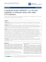
Fig. 1 Phenotypic characterisation of isolated cells from colorectal cancer samples. Percentage of surface marker expression in cells from tumour
(T, grey) vs. peritumour (PT, white) areas, immediately after isolation from patients with CRC (n = 15), as determined by FACS (a). Morphological
appearance (10×) of tumour (b) and peritumour cells (c) immediately after isolation procedure. Percentage of PD-L1+ cells within tumour (T, grey
boxes) and peritumour (PT, white boxes) areas, as measured by FACS (d). Percentage of PD-L1+ cells on PanK+ and EpCAM+ gated tumour cells
from D (e). * p < .05, ** p < .01 using a Wilcoxon test
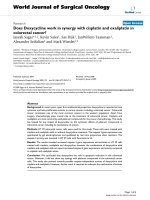
Fig. 2 Phenotype of infiltrated monocytes/macrophages in tumour and peritumour areas in colorectal cancer samples. Percentage of infiltrated
CD14+ cells within tumour (T, grey boxes) and peritumour (PT, white boxes) areas, as measured by FACS (a). Percentages of PD- L1+ (b), CD64+
(c) and CD163+ (d) cells on CD14+ gated populations in C. * p < .05, ** p < .01 using a Wilcoxon test
Cantero-Cid et al. BMC Cancer (2018) 18:945
shown in Fig. 2a, there was patent monocyte infiltration
of the tumour, but not in the surrounding tissue. Curiously,
tumour-infiltrated monocytes/macrophages expressed higher
levels of PD-L1 than those in the surrounding area (Fig. 2b).
As expected [25], CD14+ cells in tumour tissue exhibited an
M2-like alternative phenotype, as shown by their higher
expression of CD64 (Fig. 2c) and CD163 (Fig. 2d) with
respect to peritumours.
In terms of the study of the lymphoid cell lineage, we
found a large number of CD3+ cells in the tumour, but not
in the peritumour samples (Fig. 3a). Moreover, the peritumour CD3+ population was enriched in CD8+ cells (Fig. 3b),
whereas CD4+ were found in the majority of CD3+ tumour
cells (Fig. 3c). Interestingly, most of these CD4+ lymphocytes
were also PD-1+ in both areas studied (Fig. 3d).
PD-L1/PD1 crosstalk controls T-cell proliferation
Finally, we explored the crosstalk among the various immune lineages and the pMMR tumour cells in coculture
conditions. In this context, after 120 h of interaction,
CD14+ monocytes/macrophages showed an alternative
phenotype (high expression of CD64 and CD163; Fig. 4a,
b) as well as higher levels of PD-L1 than naïve controls
(Fig. 4c), which did not have any contact with tumour
cells. We also noticed that CD4+ (Fig. 5a), but not CD8+
(Fig. 5b), lymphocytes increased their expression of surface PD-1. Furthermore, CD4+ T-lymphocytes from five
of these pMMR tumours significantly increased their
proliferative capacity when an α-PD-1 antibody was
added to the coculture (Fig. 5c, d).
Page 5 of 9
Discussion
Although cells isolated from CRC patients’ tumours
exhibited proper stem cell marker expression, which
differs from those with peritumour origin, we did not
detect a significantly associated expression of PD-L1.
Hence, these data suggest that tumour cells, per se, are
not the primary source of PD-L1 in tumour samples.
This suggestion is in line with other authors’ reports
showing that methylation of the PD-L1 gene in CRC
cells can result in low transcription and translation of
PD-L1 in these cells [26].
Due to the increased relevance of immune system
components in the tumour microenvironment [27], we
first studied the presence and phenotype of infiltrated
CD14+ monocytes/macrophages, and CD4+ or CD8+
T-lymphocytes, noting that the tumour zones were significantly enriched in CD14+ cells. Our results suggest
that not only lymphocytes but also monocytes/macrophages play an important role in the evolution of CRC.
These cells serve as the first line of host defence and
are equipped to recognise and respond to tumour cells
by triggering inflammation. However, we confirmed
that tumour-infiltrating monocytes/macrophages exhibited an alternative, M2-like activation [28] in the samples. Thus, their protective influence might be
suppressed in this context, given that tumours counteract the cytotoxic and proinflammatory activities (M1
phenotype) of tumour-associated macrophages (TAMs)
in their local environments by neutralising them via
multiple mechanisms, such as the production of certain
tumour cytokines and prostaglandins [29]. In fact,
Fig. 3 Characterisation of infiltrated T-lymphocytes in tumour and peritumour areas in colorectal cancer samples. Percentage of CD3+ cells within
tumour (T, grey boxes) and peritumour (PT, white boxes) areas from patients with CRC (n = 15), as analysed by FACS (a). Percentage of CD8+ cells (b)
and CD4+ cells (c) on CD3+ gated populations in A. Expression of PD-1 on CD4+ gated populations in C (d). * p < .05, ** p < .01 using a Wilcoxon test
Cantero-Cid et al. BMC Cancer (2018) 18:945
Page 6 of 9
Fig. 4 Monocyte/macrophage phenotype in cocultures with colorectal cancer stem cells and lymphocytes. Expression of CD163 (a), CD64 (b) and
PD-L1 (c) in CD14+ naïve monocytes/macrophages (φ, white boxes) vs. monocytes/macrophages cocultured (T, grey boxes) for 5 days with
isolated tumour cells from colorectal cancer tumour samples and lymphocytes. * p < .05, ** p < .01 using a Wilcoxon test
findings on the importance of this polarisation process
have not been only reported for CRC progression [30] but
also for other several cancers, such as glioblastoma [31],
as a consequence of M2-macrophage production of important factors that augment tumour growth (e.g., IL-10).
Furthermore, these infiltrated CD14+ cells exhibited
high PD-L1 expression, which allowed them to interact
with CD3+PD-1+ T-cells, and might provoke the
phenomenon known as ‘T-cell exhaustion’ thus impairing the T-cell response to tumour expansion. PD-L1
expression in CRC cells appears to be paradoxically
associated with a high number of CD8+ cells [32], and it
correlates with early tumour stages. Nevertheless, we
found no significant PD-L1 levels on cancer cells or
CD3+CD8+ cells in our tumour samples. In fact, our data
showed a significant number of infiltrated CD4+ T-cells
expressing PD-1, which strongly suggests an interaction
with the CD14+PD-L1+ resident population. These findings are in line with those of Llosa et al. [33], who postulated that CD4+ T-cells infiltrated populations, in both
pMMR and dMMR CRC tumours, might play an important role in PD-L1/PD-1 axis function through the T-cell
exhaustion phenomenon. In addition, we have recently reported a similar behaviour in some other clinical contexts,
such as sepsis [34] and obstructive sleep apnoea [35], leading to T-cell exhaustion and the progression of illness due
to the inability of T-cells to work steadily.
It is noteworthy that when an α-PD-1 was added to
cocultures of naïve immune lineages and pMMR tumour
cells, an increase in CD4+ cell proliferation was observed, suggesting that the T-cell exhaustion mechanism
due to PD-L1/PD-1 crosstalk had been abolished [36,
37]. These data are in agreement with the observed positive outcome of patients with CRC who were treated
with new α-PD-1 drugs such as pembrolizumab, nivolumab and other related antagonists [38], and match with
the observed predisposition of tumour-infiltrating lymphocytes (TILs) and TAMs in dMMR tumours to respond to immune checkpoint blockade therapy (ICBT)
[33, 39]. The benefits of α-PD-1 antibody therapy for
pMMR and dMMR tumours remain controversial [40].
Whilst a majority of authors concur on defining dMMR
as the sole tumour type able to respond to this therapy
[41], there are also various studies stating its relevance
against pMMR tumour progression [32, 42]. In this
sense, the most common argument for dMMR performance is based precisely on immune-infiltrated population
activity in highly mutated MMR-deficiency tumour
microenvironment (TME) [43]. However, it could apply
also to pMMR (although to a lesser extent of specific
TME cases due to their associated gene stability) in which
up-regulators of PD-L1 expression (e.g., IFNγ) are present
in higher proportions [41]. In addition, the responsiveness
to α-PD-1 observed in these pMMR tumours might be
Cantero-Cid et al. BMC Cancer (2018) 18:945
A
Page 7 of 9
B
C
D
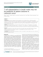
Fig. 5 Proliferative properties of T-lymphocytes cocultured with monocytes/macrophages and colorectal cancer stem cells. Expression of PD-1 by CD4+ (a)
and CD8+ (b) populations within naïve lymphocytes (φ, white boxes) vs. lymphocytes cocultured (T, grey boxes) for 5 days with isolated tumour cells from
colorectal tumour samples and monocytes/macrophages. Representative gating strategy to analyse the proliferation of CD4+ T-lymphocytes (c) and their
proliferation in the presence (+) or not (−) of an α-PD-1 antibody, as measured by CFSE dimming (d). * p < .05, ** p < .01 using a Wilcoxon test
linked to alternative mechanisms rather than to a deficiency of DNA repairing enzymes. For all these cases,
other features should also be taken into consideration
when mechanisms for PD-L1/PD-1 crosstalk are dug
into. These features include the generation of neo-antigens that then activate T-cell response to tumours,
changes in signalling pathways of chemokines and cytokines expression, and the loss of MHC class I molecules
in tumour cells [44]; all indicating the solid implication
of TILs and TAMs. Hence, our results reinforce the
suitability of either active or passive therapies [43] to
restore patient immunity, e.g., M1-macrophage polarisation, together with ICBT.
Finally, some recent studies have found significant
differences in PD-L1/PD-1 axis regulation among CRC
primary and metastatic lesions. In fact, epigenetic
mechanisms, PD-L1/PD-1 regulatory factors in the
TME and TILs/TAMs composition, might be quite
different in both tissues [41], making regimens for these
metastatic patients clinically challenging with a single
approach. Even though these differences need further
investigation, combinatory treatments for fighting the
complex relationships between immune surveillance
and the evolving properties in tumour cells appear to be
the most advantageous for treatment of the various CRC
subtypes. Accordingly, our findings open the possibility of
reconsidering combination therapies for pMMR tumours,
including both ICBT and driving local infiltrated immune
populations into a “naïve-like” stage to overcome tumour
resistance.
Cantero-Cid et al. BMC Cancer (2018) 18:945
Conclusion
Altogether, our results support previous findings indicating that the tumour microenvironment induces the
expression of PD-L1 in CD14+-TAMs, and that their
interaction with CD4+PD-1+ cells triggers T-cell exhaustion, thus allowing tumour propagation. In this sense,
inhibition of the crosstalk at this immune checkpoint,
PD-1/PD-L1, would abrogate this effect even in pMMR
tumours under specific conditions. Although currently
there are still no effective immune therapies for CRC,
our results indicate that this line of research should not
be abandoned, given both ICs and “naïve-like” stage
monocytes/macrophages appear to be crucial for the
development of CRC. Therefore, new strategies to avoid
such interference must be addressed in this clinical
context.
Abbreviations
CRC: colorectal cancer; DMEM/F12: Dulbecco Modified Eagle Medium/Nutrient
Mixture F12 media; dMMR: deficient mismatch repair; IC: immune checkpoint;
ICBT: immune checkpoint blocking therapy; PanK: Pan-cytokeratin (Miltenyi, Cat.
130–080-101); pMMR: proficient mismatch repair; RPMI: Roswell Park Memorial
Institute media; TAMs: tumour-associated macrophages; TILs: tumour-infiltrated
lymphocytes; TME: tumour microenvironment
Acknowledgements
The authors would like to thank Aurora Muñoz for technical assistance with
sample collection and ServingMed.com for the editing of the manuscript.
Funding
This study was supported by grants from Instituto de Salud Carlos III and
Fondos FEDER to EL-C (PI14/01234 and PIE15/00065). The funding bodies
allowed us to collect, analyse and interpret the data and write the
manuscript.
Availability of data and materials
The datasets generated and/or analysed during the current study are
available on request to the corresponding authors, and conform to the
regulations of the Institutional Review Board of La Paz University Hospital.
Authors’ contributions
RC-C and EL-C had the original idea and designed the experiments. RC-C, BP
and CM recruited and provided the patients. LGP collected pathological
anatomy data. JC-M, EH-J, AV-S, JA-O, IV-G, KM-H and LAA collected and assembled the data. JC-M, EH-J, CC-Z, AV-S, JA-O, MC, LAA and EL-C analysed
and interpreted the data. LAA and EL-C wrote the manuscript. All the authors
have contributed to and accepted the final manuscript.
Ethics approval and consent to participate
The study was approved by the Institutional Review Board of La Paz
University Hospital and was conducted according to the Declaration of
Helsinki.
Consent for publication
All the participants provided signed informed consent, including consent for
publication.
Competing interests
The authors declare no conflicts of interest.
Publisher’s Note
Springer Nature remains neutral with regard to jurisdictional claims in published
maps and institutional affiliations.
Page 8 of 9
Author details
The Innate Immune Response Group, IdiPAZ, La Paz University Hospital,
Madrid, Spain. 2Tumour Immunology Laboratory, IdiPAZ, Madrid, Spain.
3
Surgery Department, La Paz University Hospital, Madrid, Spain. 4Centre for
Biomedical Research Network, CIBEres, Madrid, Spain. 5Pathologic Anatomy
Service, La Paz University Hospital, Madrid, Spain.
1
Received: 10 October 2017 Accepted: 24 September 2018
References
1. Greenlee RT, Hill-Harmon MB, Murray T, Thun M. Cancer statistics, 2001. CA
Cancer J Clin. 2001;51(1):15–36.
2. Torre LA, Bray F, Siegel RL, Ferlay J, Lortet-Tieulent J, Jemal A. Global cancer
statistics, 2012. CA Cancer J Clin. 2015;65(2):87–108.
3. Ferlay J, Steliarova-Foucher E, Lortet-Tieulent J, Rosso S, Coebergh JW, Comber
H, Forman D, Bray F. Cancer incidence and mortality patterns in Europe:
estimates for 40 countries in 2012. Eur J Cancer. 2013;49(6):1374–403.
4. DeSantis CE, Lin CC, Mariotto AB, Siegel RL, Stein KD, Kramer JL, Alteri R,
Robbins AS, Jemal A. Cancer treatment and survivorship statistics, 2014. CA
Cancer J Clin. 2014;64(4):252–71.
5. Augestad KM, Merok MA, Ignatovic D. Tailored treatment of colorectal
Cancer: surgical, molecular, and genetic considerations. Clin Med Insights
Oncol. 2017;11:1179554917690766.
6. Jing F, Kim HJ, Kim CH, Kim YJ, Lee JH, Kim HR. Colon cancer stem cell
markers CD44 and CD133 in patients with colorectal cancer and
synchronous hepatic metastases. Int J Oncol. 2015;46(4):1582–8.
7. Brahmer JR, Tykodi SS, Chow LQ, Hwu WJ, Topalian SL, Hwu P, Drake CG,
Camacho LH, Kauh J, Odunsi K, et al. Safety and activity of anti-PD-L1
antibody in patients with advanced cancer. N Engl J Med. 2012;366(26):
2455–65.
8. Topalian SL, Hodi FS, Brahmer JR, Gettinger SN, Smith DC, McDermott DF,
Powderly JD, Carvajal RD, Sosman JA, Atkins MB, et al. Safety, activity, and
immune correlates of anti-PD-1 antibody in cancer. N Engl J Med. 2012;
366(26):2443–54.
9. Greenwald RJ, Freeman GJ, Sharpe AH. The B7 family revisited. Annu Rev
Immunol. 2005;23:515–48.
10. Pardoll DM. The blockade of immune checkpoints in cancer
immunotherapy. Nat Rev Cancer. 2012;12(4):252–64.
11. El Annan J, Goyal S, Zhang Q, Freeman GJ, Sharpe AH, Dana R.
Regulation of T-cell chemotaxis by programmed death-ligand 1 (PD-L1)
in dry eye-associated corneal inflammation. Invest Ophthalmol Vis Sci.
2010;51(7):3418–23.
12. Mataki N, Kikuchi K, Kawai T, Higashiyama M, Okada Y, Kurihara C,
Hokari R, Kawaguchi A, Nagao S, Kondo T, et al. Expression of PD-1,
PD-L1, and PD-L2 in the liver in autoimmune liver diseases. Am J
Gastroenterol. 2007;102(2):302–12.
13. Wu P, Wu D, Li L, Chai Y, Huang J. PD-L1 and survival in solid tumors: a
meta-analysis. PLoS One. 2015;10(6):e0131403.
14. Sun X, Suo J, Yan J. Immunotherapy in human colorectal cancer: challenges
and prospective. World J Gastroenterol. 2016;22(28):6362–72.
15. Markman JL, Shiao SL. Impact of the immune system and immunotherapy
in colorectal cancer. J Gastrointest Oncol. 2015;6(2):208–23.
16. Giannakis M, Mu XJ, Shukla SA, Qian ZR, Cohen O, Nishihara R, Bahl S, Cao Y,
Amin-Mansour A, Yamauchi M, et al. Genomic correlates of immune-cell
infiltrates in colorectal carcinoma. Cell Rep. 2016;17(4):1206.
17. Deschoolmeester V, Baay M, Lardon F, Pauwels P, Peeters M. Immune cells
in colorectal Cancer: prognostic relevance and role of MSI. Cancer
Microenviron. 2011;4(3):377–92.
18. Galon J, Costes A, Sanchez-Cabo F, Kirilovsky A, Mlecnik B, Lagorce-Pages C,
Tosolini M, Camus M, Berger A, Wind P, et al. Type, density, and location of
immune cells within human colorectal tumors predict clinical outcome.
Science. 2006;313(5795):1960–4.
19. Kocian P, Sedivcova M, Drgac J, Cerna K, Hoch J, Kodet R, Bartunkova J,
Spisek R, Fialova A. Tumor-infiltrating lymphocytes and dendritic cells in
human colorectal cancer: their relationship to KRAS mutational status and
disease recurrence. Hum Immunol. 2011;72(11):1022–8.
20. Emambux S, Tachon G, Junca A, Tougeron D. Results and challenges of
immune checkpoint inhibitors in colorectal cancer. Expert Opin Biol Ther.
2018:1–13.
Cantero-Cid et al. BMC Cancer (2018) 18:945
21. Funkhouser WK, Jr., Lubin IM, Monzon FA, Zehnbauer BA, Evans JP, Ogino S,
Nowak JA: Relevance, pathogenesis, and testing algorithm for mismatch
repair-defective colorectal carcinomas: a report of the association for
molecular pathology J Mol Diagn 2012, 14(2):91–103.
22. Boland CR, Thibodeau SN, Hamilton SR, Sidransky D, Eshleman JR, Burt RW,
Meltzer SJ, Rodriguez-Bigas MA, Fodde R, Ranzani GN, et al. A National
Cancer Institute workshop on microsatellite instability for cancer detection
and familial predisposition: development of international criteria for the
determination of microsatellite instability in colorectal cancer. Cancer Res.
1998;58(22):5248–57.
23. Cammareri P, Lombardo Y, Francipane MG, Bonventre S, Todaro M,
Stassi G. Isolation and culture of colon cancer stem cells. Methods Cell
Biol. 2008;86:311–24.
24. del Fresno C, Garcia-Rio F, Gomez-Pina V, Soares-Schanoski A, FernandezRuiz I, Jurado T, Kajiji T, Shu C, Marin E, Gutierrez del Arroyo A, et al. Potent
phagocytic activity with impaired antigen presentation identifying
lipopolysaccharide-tolerant human monocytes: demonstration in isolated
monocytes from cystic fibrosis patients. J Immunol. 2009;182(10):6494–507.
25. Jurado-Camino T, Cordoba R, Esteban-Burgos L, Hernandez-Jimenez E,
Toledano V, Hernandez-Rivas JA, Ruiz-Sainz E, Cobo T, Siliceo M, Perez de
Diego R, et al. Chronic lymphocytic leukemia: a paradigm of innate immune
cross-tolerance. J Immunol. 2015;194(2):719–27.
26. Goltz D, Gevensleben H, Dietrich J, Dietrich D. PD-L1 (CD274) promoter
methylation predicts survival in colorectal cancer patients.
Oncoimmunology. 2017;6(1):e1257454.
27. Casey SC, Amedei A, Aquilano K, Azmi AS, Benencia F, Bhakta D, Bilsland AE,
Boosani CS, Chen S, Ciriolo MR, et al. Cancer prevention and therapy
through the modulation of the tumor microenvironment. Semin Cancer
Biol. 2015;35(Suppl):S199–223.
28. Biswas SK, Lopez-Collazo E. Endotoxin tolerance: new mechanisms,
molecules and clinical significance. Trends Immunol. 2009;30(10):475–87.
29. del Fresno C, Otero K, Gomez-Garcia L, Gonzalez-Leon MC, Soler-Ranger L,
Fuentes-Prior P, Escoll P, Baos R, Caveda L, Garcia F, et al. Tumor cells
deactivate human monocytes by up-regulating IL-1 receptor associated
kinase-M expression via CD44 and TLR4. J Immunol. 2005;174(5):3032–40.
30. Li C, Luo X, Lin Y, Tang X, Ling L, Wang L, Jiang Y. A higher frequency of
CD14+ CD169+ monocytes/macrophages in patients with colorectal
Cancer. PLoS One. 2015;10(10):e0141817.
31. Zhou W, Ke SQ, Huang Z, Flavahan W, Fang X, Paul J, Wu L, Sloan AE,
McLendon RE, Li X, et al. Periostin secreted by glioblastoma stem cells
recruits M2 tumour-associated macrophages and promotes malignant
growth. Nat Cell Biol. 2015;17(2):170–82.
32. Droeser RA, Hirt C, Viehl CT, Frey DM, Nebiker C, Huber X, Zlobec I,
Eppenberger-Castori S, Tzankov A, Rosso R, et al. Clinical impact of
programmed cell death ligand 1 expression in colorectal cancer. Eur J Cancer.
2013;49(9):2233–42.
33. Llosa NJ, Cruise M, Tam A, Wicks EC, Hechenbleikner EM, Taube JM, Blosser
RL, Fan H, Wang H, Luber BS, et al. The vigorous immune
microenvironment of microsatellite instable colon cancer is balanced by
multiple counter-inhibitory checkpoints. Cancer Discov. 2015;5(1):43–51.
34. Avendano-Ortiz J, Maroun-Eid C, Martin-Quiros A, Toledano V, CubillosZapata C, Gomez-Campelo P, Varela-Serrano A, Casas-Martin J, LlanosGonzalez E, Alvarez E, et al. PD-L1 overexpression during endotoxin
tolerance impairs the adaptive immune response in septic patients via
HIF1alpha. J Infect Dis. 2018;217(3):393–404.
35. Cubillos-Zapata C, Avendano-Ortiz J, Hernandez-Jimenez E, Toledano V,
Casas-Martin J, Varela-Serrano A, Torres M, Almendros I, Casitas R,
Fernandez-Navarro I, et al. Hypoxia-induced PD-L1/PD-1 crosstalk impairs Tcell function in sleep apnoea. Eur Respir J. 2017;50(4).
36. Koelzer VH, Canonica K, Dawson H, Sokol L, Karamitopoulou-Diamantis E,
Lugli A, Zlobec I. Phenotyping of tumor-associated macrophages in
colorectal cancer: impact on single cell invasion (tumor budding) and
clinicopathological outcome. Oncoimmunology. 2016;5(4):e1106677.
37. Riella LV, Paterson AM, Sharpe AH, Chandraker A. Role of the PD-1 pathway
in the immune response. Am J Transplant. 2012;12(10):2575–87.
38. Boland PM, Ma WW. Immunotherapy for Colorectal Cancer. Cancers
(Basel). 2017:9(5).
39. Marginean EC, Melosky B. Is there a role for programmed death Ligand-1 testing
and immunotherapy in colorectal Cancer with microsatellite instability? Part II-the
challenge of programmed death Ligand-1 testing and its role in microsatellite
instability-high colorectal Cancer. Arch Pathol Lab Med. 2018;142(1):26–34.
Page 9 of 9
40. Passardi A, Canale M, Valgiusti M, Ulivi P. Immune checkpoints as a target
for colorectal Cancer treatment. Int J Mol Sci. 2017;18(6).
41. Inaguma S, Lasota J, Felisiak-Golabek A, Kowalik A, Wang Z, Zieba S, Kalisz J,
Ikeda H, Miettinen M. Histopathological and genotypic characterization of
metastatic colorectal carcinoma with PD-L1 (CD274)-expression: possible
roles of tumour micro environmental factors for CD274 expression. J Pathol
Clin Res. 2017;3(4):268–78.
42. Li Y, Liang L, Dai W, Cai G, Xu Y, Li X, Li Q, Cai S. Prognostic impact of
programed cell death-1 (PD-1) and PD-ligand 1 (PD-L1) expression in cancer
cells and tumor infiltrating lymphocytes in colorectal cancer. Mol Cancer.
2016;15(1):55.
43. Gang W, Wang JJ, Guan R, Yan S, Shi F, Zhang JY, Li ZM, Gao J, Fu XL.
Strategy to targeting the immune resistance and novel therapy in colorectal
cancer. Cancer Med. 2018;7(5):1578–603.
44. Liu SS, Yang YZ, Jiang C, Quan Q, Xie QK, Wang XP, He WZ, Rong YM, Chen
P, Yang Q, et al. Comparison of immunological characteristics between
paired mismatch repair-proficient and -deficient colorectal cancer patients. J
Transl Med. 2018;16(1):195.