N-cadherin in cancer metastasis, its emerging role in haematological malignancies and potential as a therapeutic target in cancer
Bạn đang xem bản rút gọn của tài liệu. Xem và tải ngay bản đầy đủ của tài liệu tại đây (1.16 MB, 16 trang )
Mrozik et al. BMC Cancer (2018) 18:939
/>
REVIEW
Open Access
N-cadherin in cancer metastasis, its
emerging role in haematological
malignancies and potential as a therapeutic
target in cancer
Krzysztof Marek Mrozik1,2, Orest William Blaschuk3, Chee Man Cheong1,2,
Andrew Christopher William Zannettino1,2,4† and Kate Vandyke1,2*†
Abstract
In many types of solid tumours, the aberrant expression of the cell adhesion molecule N-cadherin is a hallmark of
epithelial-to-mesenchymal transition, resulting in the acquisition of an aggressive tumour phenotype. This transition
endows tumour cells with the capacity to escape from the confines of the primary tumour and metastasise to
secondary sites. In this review, we will discuss how N-cadherin actively promotes the metastatic behaviour of
tumour cells, including its involvement in critical signalling pathways which mediate these events. In addition, we
will explore the emerging role of N-cadherin in haematological malignancies, including bone marrow homing and
microenvironmental protection to anti-cancer agents. Finally, we will discuss the evidence that N-cadherin may be
a viable therapeutic target to inhibit cancer metastasis and increase tumour cell sensitivity to existing anti-cancer
therapies.
Keywords: N-cadherin, Cancer, Metastasis, Haematological malignancies, Therapeutic target
Background
Cancer metastasis is a leading cause of cancer-related
mortality. The metastasis of cancer cells within primary
tumours is characterised by localised invasion into the
surrounding microenvironment, entry into the vasculature and subsequent spread to permissive distant organs
[1, 2]. In many epithelial cancers, metastasis is facilitated
by the genetic reprogramming and transitioning of cancer cells from a non-motile, epithelial phenotype into a
migratory, mesenchymal-like phenotype, a process
known as epithelial-to-mesenchymal transition (EMT)
[3, 4]. A common feature of EMT is the loss of epithelial
cadherin (E-cadherin) expression and the concomitant
up-regulation or de novo expression of neural cadherin
* Correspondence:
†
Andrew Christopher William Zannettino and Kate Vandyke contributed
equally to this work.
1
Myeloma Research Laboratory, Adelaide Medical School, Faculty of Health
and Medical Sciences, The University of Adelaide, Adelaide, Australia
2
Cancer Theme, South Australian Health and Medical Research Institute,
Adelaide, Australia
Full list of author information is available at the end of the article
(N-cadherin). This so-called “cadherin switch” is associated with increased migratory and invasive behaviour [5,
6] and inferior patient prognosis [7–10]. A major consequence of E-cadherin down-regulation is the loss of
stable epithelial cell-cell adhesive junctions, apico-basal
cell polarity and epithelial tissue structure, thereby facilitating the release of cancer cells from the primary
tumour site [11, 12]. In contrast to the
migration-suppressive role of E-cadherin, N-cadherin
endows tumour cells with enhanced migratory and invasive capacity, irrespective of E-cadherin expression [13].
Thus, the acquisition of N-cadherin appears to be a critical step in epithelial cancer metastasis and disease
progression.
In this review, we will discuss how N-cadherin promotes the metastatic behaviour of tumour cells by directly mediating cell-cell adhesion, and by its
involvement in modulating critical signalling pathways
implicated in metastatic events. In addition, we will discuss the emerging relevance of N-cadherin in haematological malignancies, namely leukaemias and multiple
© The Author(s). 2018 Open Access This article is distributed under the terms of the Creative Commons Attribution 4.0
International License ( which permits unrestricted use, distribution, and
reproduction in any medium, provided you give appropriate credit to the original author(s) and the source, provide a link to
the Creative Commons license, and indicate if changes were made. The Creative Commons Public Domain Dedication waiver
( applies to the data made available in this article, unless otherwise stated.
Mrozik et al. BMC Cancer (2018) 18:939
Page 2 of 16
myeloma. Finally, we will review the emerging evidence
that N-cadherin may be a viable therapeutic target to inhibit cancer metastasis and overcome resistance to
anti-cancer agents.
Structure and formation of the N-cadherin
adhesive complex
N-cadherin is a member of the calcium-dependent adhesion molecule family of classical cadherins which directly
mediate homotypic and heterotypic cell-cell adhesion.
N-cadherin is a classical type I cadherin consisting of 5
extracellular domains linked to a functional intracellular
domain. The engagement between N-cadherin monomers on opposing cells occurs by reciprocal insertion of
a tryptophan residue side-chain on its first extracellular
domain (EC1) into the hydrophobic pocket of the partner N-cadherin EC1 (trans adhesion). In addition, the
stabilisation of N-cadherin-mediated adhesion requires
the clustering of adjacent monomers on the surface of
the same cell, involving the His-Ala-Val (HAV) motif on
EC1 and a recognition sequence on the second extracellular domain (EC2) of the lateral N-cadherin monomer
(cis adhesion) [14–16]. The membrane expression and
lateral clustering of N-cadherin is dependent upon p120
catenin, which localises N-cadherin at cholesterol-rich
microdomains [17, 18]. The initial ligation of N-cadherin
extracellular domains triggers the activation of the Rho
GTPase family member Rac, which stimulates localised
actin filament assembly and the formation of membrane
protrusions at points of cell-cell contact [19, 20]. The
subsequent activation of the Rho GTPase family member
RhoA, at the expense of Rac function, facilitates the
maturation of N-cadherin-based cell-cell junctions by
triggering the sequestration of β-catenin to the cadherin
intracellular domain [21, 22]. β-catenin serves as a critical link to α-catenin which accumulates at nascent
cell-cell junctions and suppresses actin branching. In
addition, α-catenin facilitates the anchorage of the
N-cadherin-catenin complex to the actin cytoskeleton
via actin-binding proteins such as cortactin and
α-actinin, thereby promoting the maturation of cell-cell
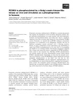
contacts [23, 24] (Fig. 1). Notably, the adhesive function
of N-cadherin is regulated by post-translational modifications of the N-cadherin-catenin complex. For instance,
the stability of the N-cadherin-catenin complex is highly
dependent on the phosphorylation status of N-cadherin
and the associated catenins, which is regulated by tyrosine kinases, such as Fer and Src, and the tyrosine phosphatase PTP1B [25, 26]. In addition, branched
N-glycosylation of N-cadherin EC2 and third extracellular domain regulates N-cadherin-dependent cell adhesion, at least in part, by controlling the lateral clustering
of N-cadherin monomers [27].
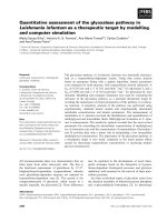
Fig. 1 Schematic representation of the N-cadherin-catenin adhesive
complex. The extracellular domains of N-cadherin monomers
engage in trans and cis interactions with partner monomers,
facilitated by p120-catenin (p120), resulting in a lattice-like
arrangement. Interaction between monomers on opposing cells
occurs via a reciprocal insertion of tryptophan side-chains (W) on
the first extracellular domain (EC1) (trans adhesion). Clustering of Ncadherin monomers on the same cell occurs via a His-Ala-Val (HAV)
adhesion motif on EC1 and a recognition sequence on the second
extracellular domain (EC2) of the partner monomer (cis adhesion)
(inset). Activation of RhoA sequesters β-catenin (β-cat) and results in
accumulation of α-catenin (α-cat) to the N-cadherin intracellular
domain. This promotes anchorage of the N-cadherin-catenin
complex to the actin cytoskeleton via actin-binding proteins,
thereby stabilising cell-cell contacts. Initial ligation of N-cadherin
extracellular domains also triggers PI3K/Akt signalling which
inactivates the pro-apoptotic protein Bad, resulting in activation of
the anti-apoptotic protein Bcl-2
The functional role of N-cadherin in solid tumour
metastasis
N-cadherin expression is spatiotemporally regulated
throughout development and adulthood. In development, N-cadherin plays an important role in morphogenetic processes during the formation of cardiac and
neural tissues, and is involved in osteogenesis, skeletal
myogenesis and maturation of the vasculature [28–32].
In adulthood, N-cadherin is expressed by numerous cell
types including neural cells, endothelial cells, stromal
Mrozik et al. BMC Cancer (2018) 18:939
cells and osteoblasts, and is integral to synapse function,
vascular stability and bone homeostasis [30, 33–36].
While N-cadherin is typically absent or expressed at low
levels in normal epithelial cells, the aberrant expression
of N-cadherin in epithelial cancer cells is a
well-documented feature of epithelial malignancies, such
as breast, prostate, urothelial and pancreatic cancer, and
is associated with disease progression [37–40]. In a similar manner, the up-regulation of N-cadherin expression
is a feature of melanoma progression [41–43]. Whilst
the aberrant expression of N-cadherin in epithelial tissues is not considered to be oncogenic, or a promoter of
solid tumour growth [44–46], increased expression of
N-cadherin in cancer is widely associated with tumour
aggressiveness. Indeed, many studies have demonstrated
a significant correlation between elevated N-cadherin
levels in epithelial, and some non-epithelial solid tumours, and clinicopathologic features such as increased
localised tumour invasion and distant metastasis, and inferior patient prognosis [7, 8, 47–81] (Table 1). Multivariate analyses have also identified that elevated
N-cadherin expression is independently associated with
inferior patient prognosis in several epithelial malignancies including prostate, lung and bladder cancer [8, 55,
56, 60, 62, 63, 67, 72, 78, 80] (Table 1). The aggressive
phenotype and inferior prognosis associated with
up-regulated N-cadherin expression in solid tumours is
also supported by a recent meta-analysis incorporating
patients with various epithelial malignancies [82].
Beyond the prognostic implications of aberrant
N-cadherin expression, the relationship between
N-cadherin and metastasis is not merely associative. Indeed, there is a wealth of evidence that increased
N-cadherin expression enhances the migratory and invasive
capacity of multiple epithelial cancer cell types in vitro [83–
87]. The ability of N-cadherin to promote epithelial tumour
metastasis in vivo was initially demonstrated using the
MCF-7 breast cancer cell line, following injection into the
mammary fat pad of nude mice. In contrast to wild-type
cells, MCF-7 cells ectopically expressing N-cadherin
formed tumour metastases in several organs including the
liver, pancreas and lymph nodes [88]. Similarly, N-cadherin
expression in the mammary epithelium in the transgenic
MMTV-PyMT murine breast cancer model resulted in a
three-fold increase in the number of pulmonary metastatic
foci without affecting the onset or growth of the primary
tumour [45]. Using an orthotopic mouse model of pancreatic cancer, the over-expression of N-cadherin in BxPC-3
cells increased the formation of disseminated tumour nodules throughout the abdominal cavity and induced the formation of N-cadherin-expressing lung micro-metastases
[85]. Consistent with these findings, enforced expression of
N-cadherin in androgen-responsive prostate cancer cells
promoted invasion of underlying muscle and lymph node
Page 3 of 16
metastasis following subcutaneous injection in castrated
mice [89]. Notably, N-cadherin also potentiates the invasiveness of melanoma cells. To this end, studies have demonstrated that N-cadherin promotes the capacity of
melanoma cells to migrate on monolayers of dermal fibroblasts and undergo trans-endothelial migration in vitro [86,
90, 91]. Moreover, N-cadherin silencing has been shown to
attenuate the ability of intravenously injected melanoma
cells to extravasate and form lung metastases in immunocompromised mice [92].
To appreciate how N-cadherin, a cell adhesion molecule, may actively promote cancer cell migration, it is
important to consider that the N-cadherin-catenin complex mediates both cell-cell adhesion and pro-metastatic
cell signalling. Moreover, the adhesive function and
migration-related signalling capacity of N-cadherin can
occur simultaneously, or as antagonistic events, adding
further complexity to its role in cancer metastasis. In the
following section, we describe three key mechanisms by
which N-cadherin has been shown to actively promote
the migratory capacity of tumour cells: facilitation of
collective cell migration, augmentation of fibroblast
growth factor-receptor (FGFR) signalling and modulation of canonical Wnt signalling.
N-cadherin promotes collective cell migration
The migration of cells as sheets, clusters or strands, a
process termed collective cell migration, frequently occurs throughout development and in adulthood. For instance,
collective
cell
migration
occurs
in
embryogenesis, during gastrulation and neural crest cell
migration, and in adult tissues, during wound healing
and angiogenesis [93, 94]. In addition, collective cell migration facilitates the invasion of epithelial cells through
the localised tumour host microenvironment, thereby
promoting metastasis [95]. During this process, collectively migrating cells maintain physical interconnectivity,
collective cell polarity and co-ordinated cytoskeletal activity, resulting in a ‘leader-follower’-type cellular arrangement. This promotes more efficient directional
migration, in response to a chemotactic gradient, than
that of an individual migrating cell [93, 96]. Adhesive
complexes are integral to the co-ordinated behaviour of
collectively migrating cells by mediating adhesion, signal
transduction and mechanotransduction between adjacent cells [94, 97]. Notably, studies have demonstrated
that N-cadherin expression by epithelial cancer cells
promotes their capacity for collective migration. For instance, N-cadherin has been shown to promote the ability of lung or ovarian cancer cells to form aggregates
and collectively invade three-dimensional (3D) collagen
matrices or penetrate peritoneal mesothelium-like cell
layers in vitro [87, 98]. Similarly, studies in transformed
canine kidney epithelial cells (MDCK cells) have shown
Mrozik et al. BMC Cancer (2018) 18:939
Page 4 of 16
Table 1 Association of increased N-cadherin expression in cancer with clinicopathologic features and survival
Cancer type
Cohort information
& treatment details
No. of
patients
N-cadherin detection
method
Association with
clinicopathologic features
Association
with survival
Reference
Pre-metastatic; resected
574
IHC
High grade & LN metastasis
Shorter PFS (U)
[47]
Early-stage invasive
1902
IHC
Earlier development
of distant metastasis
n/a
[48]
Primary inoperable
and LN negative
275
IHC
n.s.
Shorter OS (U)
[49]
Invasive; no prior therapy
94
IHC
High grade, late
stage & LN metastasis
n/a
[50]
Clinically localised;
radical prostatectomy
104
IHC
Poor differentiation,
seminal vesicle invasion
& pelvic LN metastasis
Shorter time to
biochemical failure
(U), clinical
recurrence
(M) & skeletal
metastasis (U)
[8]
Castration-resistant;
transurethral resection
26
IHC
Higher Gleason
score & metastasis
n/a
[51]
Localised; no therapy prior to
radical prostatectomy
157
IHC
Later stage, higher PSA &
Gleason score, seminal vesicle
invasion and LN metastasis
n/a
[52]
Blood from cancer
follow-up patients
179
Serum ELISA (sN-cad)
Higher PSA
n/a
[53]
Radical prostatectomy,
metformin-treated
49
IHC
n/a
Increased recurrence
[54]
Adenocarcinoma & squamous
cell carcinoma; no therapy
prior to surgery
68
IHC
Higher TNM stage
& poor differentiation
Shorter OS (M)
[55]
Primary adenocarcinoma;
no therapy prior to surgery
147
IHC
n/a
Shorter OS (M)
[56]
qPCR
LN metastasis
n/a
[57]
Epithelial cancers
Breast cancer
Prostate cancer
Lung cancer
Surgical resection of adenocarcinoma; 57
no prior therapy
Urothelial
cancers
Liver cancer
Head & neck
No post-operative surgery
186
IHC
Higher TNM stage & metastasis
n/a
[58]
Adenocarcinoma & squamous
cell carcinoma; blood collected
prior to or up to 3 weeks after
platinum-based therapy
43
IF (on CTCs)
n/a
Shorter PFS
[59]
Radical cystecomy with pelvic LN
dissection, clinically nonmetastatic
bladder cancer
433
IHC
Higher clinical & pathologic tumour Shorter RFS
stage, LN metastasis & LN stage,
(M), OS (U) &
lymphovascular invasion
cancer-specific
survival (U)
[60]
Invasive bladder cancer
undergoing radical
cystectomy; no prior treatment
30
qPCR
n/a
Shorter OS
[61]
Transurethral resection
of non-muscle-invasive
bladder cancer
115
IHC
Higher incidence
of intravesical recurrence
Shorter intravesical
RFS (M)
[62]
Clinically-localised upper
urinary tract carcinoma
undergoing nephroureterectomy;
cisplatin- based therapy
in late-stage patients
59
IHC
n/a
Intravesical
and extravesical
RFS (M)
[63]
Resection of hepatocellular
carcinoma
100
IHC
Higher histologic grade, multifocal
tumours & vascular invasion
Shorter
disease-free
and OS
[64]
Surgical resection of
hepatocellular carcinoma
57
IHC
n.s.
Increased
recurrencerate within
2 years of resection
[65]
Surgical resection of intrahepatic
cholangiocarcinoma
(no prior therapy); adjuvant
therapy in patients with recurrence
96
IHC
Higher recurrence
of vascular invasion
Shorter OS
[66]
Surgical specimen of
119
IHC
Greater tumour
Shorter OS (M)
[67]
Mrozik et al. BMC Cancer (2018) 18:939
Page 5 of 16
Table 1 Association of increased N-cadherin expression in cancer with clinicopathologic features and survival (Continued)
Cancer type
cancer
Cohort information
& treatment details
No. of
patients
N-cadherin detection
method
HNSCC, patients
are +/− LN metastasis
Association with
clinicopathologic features
Association
with survival
Reference
size, higher clinical
stage & LN metastasis
Laryngeal, oripharyngeal & oral
cancer; blood collected following
HNSCC resection
10
IF
n/a
Shorter OS
[68]
Radical surgery for laryngeal
cancer; adjuvant
therapy in 60% of cases
50
(on CTCs) IHC
Higher grade
Increased relapse
[69]
Nasopharyngeal cancer
122
IHC
LN involvement,
distant metastasis
& later clinical stage
Shorter OS
(nuclear N-cadherin)
[70]
Colorectal cancer; no
therapy prior to surgery
37
qPCR
Local invasion, Dukes
staging & vascular invasion
n/a
[71]
Colorectal cancer; no
therapy prior to surgery
102
IHC
Larger tumour size, poor
differentiation, tumour invasion,
LN metastasis & distant metastasis
Shorter OS
(M) & shorter
disease-free survival
[72]
Colon carcinoma; no
therapy prior to surgery
90
IHC
Greater depth of tumour
invasion & higher
TNM stage
n/a
[73]
Gastric cancer surgery with
LN metastasis; no prior therapy
89
IHC (on LN)
LN involvement, higher
pathological stage,
lymphatic invasion
& venous invasion
Shorter OS
[74]
Curative surgery for gastric
adenocarcinoma; no prior
therapy, stage II patients
received adjuvant therapy
146
IHC
Haematogenous recurrence
Shorter survival
[75]
Renal cancer
Blood collected from
metastatic renal cell
carcinoma patients
with prior
nephrectomy and therapy
14
IF (on CTCs; also CK-)
n/a
Shorter PFS
[76]
Ovarian cancer
Surgical specimens of
high-grade serous carcinoma
167
IHC
n/a
Shorter PFS
and OS (U)
[77]
Gallbladder
cancer
Adenocarcinoma
(+/− surgery)
80
IHC
Poor differentiation,
larger tumour size,
TNM stage, invasion
& LN metastasis
Shorter OS (M)
[78]
Squamous cell/adenosquamous
carcinoma (+/− surgery)
46
IHC
Larger tumour size,
invasion and LN metastasis
Shorter OS (M)
[78]
Gastrointestinal
tract cancer
Non-epithelial solid cancers
Melanoma
Removal of primary
melanoma, various
stages of disease
394
IHC
Increased Breslow thickness
Distant metastasis-free [7]
survival (M; p = 0.13)
Sarcoma
Surgical resection of
osteosarcoma
107
qPCR
Later stage and
distant metastasis
Shorter survival
[79]
Blood collected from a variety
of bone & soft tissue sarcoma
patients
73
Serum ELISA (sN-cad)
Larger tumour size
& higher grade
Shorter disease-free
survival (M) & OS (U)
[80]
Blood collected from
newly- diagnosed patients;
no prior therapy
84
Serum ELISA (sN-cad)
n/a
Shorter PFS and OS
[81]
Bone marrow aspirate from
newly-diagnosed patients;
no prior therapy
14
qPCR (on CD38+/CD138 n/a
+ tumour cells)
Shorter PFS
[81]
Haematological malignancies
Multiple
myeloma
All clinicopathologic and survival data shown is positively associated with increased N-cadherin expression. All data is statistically significant (P < 0.05), unless
otherwise indicated. Abbreviations: PFS Progression-free survival, RFS Recurrence-free survival, OS Overall survival, U Univariate analysis, M Multivariate analysis,
IHC Immunohistochemistry, qPCR Quantitative PCR, IF Immunofluorescence, ELISA Enzyme-linked immunosorbent assay, sN-cad Soluble N-cadherin, PSA Prostate
specific antigen, LN Lymph node, TNM Tumour, node and metastases, CTCs Circulating tumour cells, CK Cytokeratin, n/a Not applicable, n.s. Not significant
Mrozik et al. BMC Cancer (2018) 18:939
that N-cadherin promotes aggregate formation which allows directional collective cell migration in a 3D collagen matrix. In these cells, deletion of the entire
N-cadherin intracellular domain, or the β-catenin binding domain alone, resulted in greater individual cell detachment and migration from cell clusters, highlighting
the importance of the N-cadherin-actin cytoskeleton
interaction in collective cell migration. Moreover,
over-expression of an N-cadherin mutant in which the
extracellular domain was fused to the anti-binding domain of α-catenin hindered the movement of follower
cells, demonstrating that dynamic N-cadherin-actin linkage is required for efficient collective cell migration [99].
In addition to maintaining multi-cellular aggregates of
tumour cells, studies in N-cadherin-expressing
non-tumour cells have demonstrated that N-cadherin
also promotes collective cell migration by polarising
Rho-family GTPase signalling (e.g. Rac1 and cdc42),
known to co-ordinate cytoskeletal remodelling in collectively migrating cells [100, 101]. For example, models
of arterial smooth muscle wound-healing and neural
crest migration have shown that the asymmetric distribution of N-cadherin-mediated cell-cell adhesion at the
lateral and posterior aspects of leader cells promotes directional cell alignment and increased cdc42 and Rac1
activity and protrusion formation at the free leading cell
edge, resulting in enhanced migration [102, 103]. Mechanistically, studies in mouse embryonic fibroblasts have
demonstrated that N-cadherin-adhesive complexes at
the rear of cells suppress localised integrin-α5 activity,
thereby polarising integrin and Rac activity towards the
free leading edge of the cell [104]. Indeed, functional inhibition of N-cadherin in transformed mammary cells
has been shown to reduce integrin-α5-dependent cell
migration on fibronectin in vitro [105]. In a similar manner, silencing of N-cadherin expression in melanoma
cells perturbs α2β1-integrin-dependent collagen matrix
invasion in vitro [106]. Reciprocally, integrin signalling
at focal adhesions has been shown to regulate the ability
of HeLa cells to engage in N-cadherin-based connections and to promote collective cell migration [107].
Given that integrins play an important role in the activation of Rho signalling [108, 109], it is plausible that
N-cadherin may polarise Rho-family GTPase signalling
via intercommunication with integrins, thereby promoting the collective migration of cancer cells (Fig. 2a).
N-cadherin augments fibroblast growth factor receptor
signalling
Functional interaction between the extracellular domains
of N-cadherin and receptor-tyrosine kinase FGFRs was
first recognised as a mechanism by which N-cadherin promoted axonal outgrowth of rat cerebellar neuronal cells.
These studies identified that the fourth extracellular
Page 6 of 16
domain of N-cadherin (EC4) trans-activated FGFRs to
promote neurite outgrowth independent of FGF ligands,
suggesting that N-cadherin can act as a surrogate ligand
of FGFRs [33, 110]. The physical interaction of
N-cadherin and FGFRs has also been shown in breast and
pancreatic cancer cells [111–114]. Evidence that FGFR
plays a functional role in N-cadherin-mediated cancer metastasis has been demonstrated in BT-20 and PyMT breast
cancer cells, whereby FGFR inhibition reduced the in vitro
migratory capacity of N-cadherin-expressing cells, but not
N-cadherin-negative cells [45, 84]. In addition, FGF-2 increased the invasiveness of N-cadherin-expressing MCF-7
human breast cancer cells, but not control MCF-7 cells
[88]. To this end, it has been shown that N-cadherin potentiates FGF-2-activated FGFR-1 signalling by attenuating ligand-induced FGFR-1 internalisation, thereby
stabilising FGFR-1 expression [111, 113]. In turn, the sustained activation of down-stream MEK/ERK signalling results in increased production of the extracellular matrix
(ECM)-degrading enzyme matrix metalloproteinase-9
(MMP-9) and enhanced breast cancer cell invasiveness
[88, 111]. In addition, the interaction of N-cadherin and
FGFR is also likely to promote metastasis by activation of
the phosphatidylinositide-3 kinase/Akt (PI3K/Akt) signalling pathway in some cancer cell types. For example, studies suggest that the invasiveness of N-cadherin-expressing
ErbB2/Neu breast cancer cells following FGFR activation
is mediated by PI3K/Akt signalling. N-cadherin potentiates FGFR-Akt signalling and sensitivity to FGFR inhibition in ErbB2/Neu cells, suggesting the involvement of an
N-cadherin-FGFR-PI3K/Akt signalling axis in breast cancer cell invasion [115] (Fig. 2b).
Two lines of evidence suggest that N-cadherin-FGFR-1
interactions promote the invasive behaviour in both collectively migrating and individual cancer cells. Firstly,
N-cadherin-FGFR-1 interactions have been shown to
occur over most of the cell membrane, but are excluded
from sites of cell-cell adhesion, suggesting that the interaction is independent of N-cadherin-mediated cellular adhesion [112]. Secondly, blocking antibodies directed at the
FGFR-1-interacting domain of N-cadherin (EC4) have been
shown to inhibit N-cadherin-mediated migration, but not
N-cadherin-mediated aggregation, of human breast cancer
cells [116]. Thus, it would appear that N-cadherin-mediated
cell-cell adhesion and N-cadherin-mediated cell migration
via FGFR-1 are independent and mutually exclusive events.
Further studies are warranted to identify whether
N-cadherin potentiates FGFR-1 signalling in other epithelial
malignancies such as pancreatic cancer.
N-cadherin modulates canonical Wnt signalling
In addition to stabilising cadherin-mediated cell-cell adhesion, β-catenin plays a central role in the canonical
Wnt signalling pathway. Canonical Wnt signalling
Mrozik et al. BMC Cancer (2018) 18:939
Page 7 of 16
A
B
C
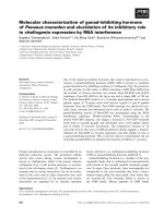
Fig. 2 Schematic representation of cell signalling events modulated by increased N-cadherin expression in the context of cell migration. a In
addition to mediating cellular aggregation, N-cadherin may facilitate the collective migration of tumour cells by excluding focal adhesions and
Rac1 activity, and promoting RhoA activity, at sites of N-cadherin-mediated cell-cell contact. The asymmetric distribution of N-cadherin adhesive
complexes polarises integrin function and Rac1 activity towards the free edges of cells, thereby directing focal adhesion and lamellipodia
formation away from the cell cluster and promoting cell migration. Similar to Rac1, N-cadherin-mediated cell-cell adhesion promotes cdc42
activity at the free edges of cells, resulting in filipodia formation. b Functional interaction between the extracellular domains of N-cadherin and
FGFR-1 potentiates FGF-2-activated FGFR-1 signalling by attenuating ligand-induced receptor internalisation. The resulting augmentation of
down-stream MEK/ERK and PI3K/Akt signalling promotes the metastatic behaviour of cancer cells by increasing the production of invasionfacilitating molecules such as matrix metalloproteinases (MMPs). c N-cadherin-mediated adhesive complexes and Wnt/β-catenin signalling are
thought to compete for the same cellular pool of β-catenin. While N-cadherin sequesters β-catenin from the nucleus, the N-cadherin adhesive
complex provides a reservoir of β-catenin which, upon Wnt activation, becomes available for nuclear translocation and TCF/LEF-mediated gene
transcription (e.g. CD44 and MMP genes), resulting in the loss of N-cadherin-mediated cellular adhesion in cancer cells
promotes the cytoplasmic accumulation and nuclear
translocation of β-catenin, which activates T cell factor/
lymphoid enhancer factor (TCF/LEF)-mediated transcription of genes [117–119] that encode tumour invasion and metastasis-promoting molecules (e.g. MMPs
and CD44) [120–126]. It has been proposed that cadherins and the canonical Wnt signalling pathway may compete for the same cellular pool of β-catenin, with
cadherins sequestering β-catenin from the nucleus,
thereby attenuating Wnt signalling [127, 128]. Indeed,
enforced expression of N-cadherin in colon carcinoma
cells resulted in the relocation of nuclear β-catenin to
the plasma membrane and attenuated LEF-responsive
trans-activation [129]. Alternatively, studies suggest that
the N-cadherin-β-catenin complex may provide a stable
pool of β-catenin available for TCF/LEF-mediated gene
transcription in cancer cells [91, 130]. To this end, disruption of N-cadherin-mediated adhesion in leukaemic
cells was found to increase TCF/LEF reporter activity
[131]. Thus, given β-catenin is essential in the stabilisation of N-cadherin-mediated cellular adhesion (discussed
earlier), it is feasible that the ability of N-cadherin to
modulate TCF/LEF-mediated gene transcription may
play an important role in individual cell migration, at
the expense of collective cell migration (Fig. 2c).
Trans-endothelial migration is an important process in
the haematogenous dissemination of cancer cells to distant
sites [132]. Notably, studies suggest that N-cadherin promotes the trans-endothelial migration of cancer cells. To
this end, N-cadherin silencing has been shown to reduce
Mrozik et al. BMC Cancer (2018) 18:939
the ability of melanoma cells to undergo trans-endothelial
migration in vitro [91]. Studies have demonstrated that
N-cadherin-mediated melanoma cell adhesion to endothelial cells promotes trans-endothelial migration by modulating canonical Wnt signalling. β-catenin co-localises with
N-cadherin during the initial stages of melanoma cell adhesion to endothelial cells; however, during transendothelial
migration, the tyrosine kinase Src is activated and subsequently phosphorylates the N-cadherin cytoplasmic domain, thereby dissociating the N-cadherin-β-catenin
complex. β-catenin is then translocated to the nucleus of
melanoma cells and activates TCF/LEF-mediated gene transcription, resulting in up-regulation of the adhesion molecule CD44 [91, 133]. Studies using epithelial cancer cells
suggest that CD44 binding to E-selectin on endothelial cells
activates intracellular signalling pathways that lead to disassembly of endothelial junctions, thereby facilitating
trans-endothelial migration [134–136]. In line with these
studies, CD44 expression in melanoma cells has been
shown to promote endothelial gap formation and
trans-endothelial migration in vitro [137]. Moreover,
N-cadherin knock-down in human melanoma cells reduces
extravasation and lung nodule formation following intravenous injection in immuno-compromised mice [92]. Notably, while N-cadherin-expressing tumour cells have been
detected in the circulation of patients with various epithelial
cancers [59, 68, 76], and CD44 has been shown to promote
diapedesis in breast cancer cells [134, 138], a role for
N-cadherin in the trans-endothelial migration of epithelial
cancer cells has not been directly demonstrated to date.
The emerging role of N-cadherin in
haematological malignancies
We have thus far summarised the functional role and
clinical implications of aberrant N-cadherin expression
in the context of solid tumour metastasis. There is now
emerging evidence suggesting that N-cadherin plays a
role in haematological malignancies, including leukaemia
and multiple myeloma (MM). These cancers account for
approximately 10% of all cancer cases and are typically
characterised by the abnormal proliferation of malignant
white blood cells within the bone marrow (BM) and the
presence of tumour cells within the circulation. Specialised compartments, or ‘niches’, within the BM microenvironment play critical roles in housing and
maintaining pools of quiescent haematopoietic stem cells
(HSCs), and in regulating HSC self-renewal and differentiation [139, 140]. Notably, N-cadherin is expressed by
various cell types associated with the HSC niche, including osteoblasts and stromal cells in the endosteal niche,
and endothelial cells and pericytes in the perivascular
niche [32, 36, 141, 142]. In the following section, we discuss the potential implications of aberrant N-cadherin
expression in haematological cancer cells; namely, BM
Page 8 of 16
homing and BM microenvironment-mediated protection
to chemotherapeutic agents.
Leukaemia
Leukaemias are thought to arise by the malignant transformation of HSCs into leukaemic stem cells (LSCs)
which occupy and modify BM HSC niches [143–146].
Adhesive interactions between LSCs and the BM microenvironment activate signalling cascades which contribute to LSC self-renewal and survival, and the capacity to
evade the cytotoxic effects of chemotherapeutic agents
[147, 148]. Indeed, therapeutic targeting of adhesion
molecules to disrupt interactions with the niche represents a potential strategy to eliminate LSCs [149].
Studies have demonstrated that N-cadherin is expressed
in a subpopulation of primitive HSCs [36], but its precise
role within the HSC niche in normal haematopoiesis is
controversial. To this end, the over-expression of
N-cadherin in HSCs has been shown to increase HSC
lodgement to BM endosteal surfaces in irradiated mice,
enhance HSC self-renewal following serial BM transplantation and promote HSC quiescence in vitro [150]. However, other studies have reported that deletion of
N-cadherin in HSCs or osteoblastic cells has no effect on
haematopoiesis or HSC quiescence, self-renewal or
long-term repopulating activity [141, 151, 152].
While these studies suggest that N-cadherin function
may be dispensable in HSC niche maintenance, emerging
evidence implicates N-cadherin in the function of the LSC
niche. Studies have reported that N-cadherin is expressed
on primitive sub-populations of leukaemic cells including
patient-derived CD34+ CD38− chronic myeloid leukaemia
(CML) cells and CD34+ CD38− CD123+ acute myeloid leukaemia (AML) cells, suggesting that N-cadherin is a marker
of LSCs [130, 153, 154]. Similar to solid tumours,
N-cadherin is thought to facilitate engagement of leukaemic
cancer cells with cells of the surrounding BM microenvironment. For example, treatment of primary human CD34+
CML cells with the N-cadherin blocking antibody GC-4
significantly reduced their adhesion to human BM stromal
cells (BMSCs) [130]. Similarly, GC-4 treatment of a
BCR-ABL-positive mouse acute lymphoblastic leukaemia
(ALL) cell line was found to inhibit their ability to adhere
to mouse fibroblasts [155]. Pre-clinical mouse models also
suggest that N-cadherin may promote BM homing, engraftment and self-renewal of AML cells in vivo [156,
157]. Thus, N-cadherin represents a potential target to inhibit LSC interactions with the BM microenvironment.
N-cadherin-mediated cell adhesive interactions promote
microenvironmental protection of leukaemic cells to anticancer agents
Adhesive interactions between leukaemic cells and
BMSCs confer sub-populations of leukaemic cells with
Mrozik et al. BMC Cancer (2018) 18:939
resistance to anti-cancer agents, leading to disease relapse
[158, 159]. As such, there is growing interest in targeting
molecules involved in leukaemic cell-BMSC interactions
to enhance leukaemic sensitivity to anti-cancer agents
[130, 160]. The role of N-cadherin in the microenvironmental protection of leukaemic cells to anti-cancer agents
was first demonstrated in studies showing that
N-cadherin expression was associated with resistance to
treatment with a farnesyltransferase inhibitor in the murine lymphoblastic leukaemia cell line, B-1, when grown in
co-culture with fibroblasts. Enforced N-cadherin expression in B-1 cells also conferred farnesyltransferase
inhibitor-resistance when grown in the presence of fibroblasts [155]. Notably, these findings are in line with reports showing that N-cadherin is up-regulated in solid
tumour cancer cells resistant to anti-cancer agents [161–
164] and androgen deprivation therapy [51, 165]. Direct
demonstration that N-cadherin-mediated cell-cell adhesion facilitated microenvironmental protection of leukaemic cells to anti-cancer agents was provided in
co-culture experiments with primary human CD34+ CML
cells and BMSCs. Disruption of CML cell-BMSC adhesion, using an N-cadherin antagonist peptide (containing
the HAV sequence) or the N-cadherin function-blocking
antibody GC-4 increased CML cell sensitivity to the tyrosine kinase inhibitor imatinib [130, 131]. An association
between response to chemotherapy and LSC expression of
N-cadherin has also been reported in AML patients. To
this end, studies suggest that AML patients exhibiting a
higher proportion of N-cadherin-expressing BM-derived
CD34+ CD38− CD123+ LSCs at diagnosis are less responsive to induction chemotherapy [153]. While the precise
mechanism by which N-cadherin-mediated adhesion confers drug-resistance in leukaemic cells is unclear, studies
in solid tumour cells suggest that N-cadherin-mediated
adhesion increases activity of the anti-apoptotic protein
Bcl-2, by PI3K/Akt-mediated inactivation of the
pro-apoptotic protein Bad [86, 162, 166].
MM
MM is characterised by the uncontrolled proliferation of
transformed immunoglobulin-producing plasma cells
(PCs) within the BM. Data from our group, and others,
suggest that N-cadherin gene and protein expression is
elevated in CD138+ BM-derived PCs in approximately
50% of newly-diagnosed MM patients compared with
BM PCs from healthy individuals and is associated with
poor prognosis [81, 167] (Table 1). Notably, the expression of the N-cadherin gene, CDH2, is up-regulated in
MM patients harbouring the high-risk t(4;14)(p16;q32)
translocation [167, 168]. This translocation encompasses
15–20% of all MM patients and is universally characterised by the dysregulated expression of the oncogenic
histone methyltransferase MMSET (also known as
Page 9 of 16
NSD2) [169–171]. In addition, CDH2 expression is also
up-regulated in more than 50% of MM patients in the
hyperdiploidy-related sub-group [167].
N-cadherin promotes MM PC BM homing
The progression of MM disease is underscored by MM
PC egress from the primary BM environment and dissemination via the peripheral circulation to distal medullary sites [172]. Functionally, N-cadherin is thought to
play a role in MM PC extravasation and homing to the
BM. Following intravenous inoculation, the BM-homing
capacity of the human MM PC line NCI-H929 in
immuno-deficient mice was significantly attenuated by
N-cadherin silencing in tumour cells, resulting in increased numbers of residual circulating tumour cells
[167]. In addition, N-cadherin knock-down in the murine MM cell line 5TGM1 significantly inhibited adhesion
to BM endothelial cell monolayers in vitro, although
N-cadherin knock-down or GC-4 antibody-mediated
blocking of N-cadherin did not affect the
trans-endothelial migration capacity of MM PCs in vitro
[167, 173]. Taken together, these data suggest that
N-cadherin may promote BM homing of circulating
MM PC by facilitating their adhesion to the vasculature,
without affecting the rate of subsequent diapedesis.
N-cadherin mediates cell-cell adhesion between MM PCs
and the BM microenvironment
Adhesive interactions between MM PCs and the BM
microenvironment are critical in the permissiveness of
the BM to the development of MM disease. These include cell-cell interactions which support MM PC
growth and resistance to anti-cancer agents, and promote the inhibition of osteoblast differentiation, thereby
contributing to MM PC-mediated bone loss [174, 175].
In addition to endothelial cell adhesion, in vitro studies
have demonstrated that N-cadherin mediates the adhesion of human MM PCs to osteoblasts and stromal cells,
which constitute the endosteal MM niche [167, 176]. In
a functional context, N-cadherin-mediated adhesion between MM PCs and pre-osteoblastic cells has been
shown to inhibit osteoblast differentiation, suggesting
that N-cadherin may contribute to MM-related bone
loss in the clinical setting [167]. Studies have also shown
that treatment of human MM PC lines in co-culture
with stromal cells or osteoblasts with the N-cadherin
blocking antibody GC-4 induced a significant expansion
of MM PCs in vitro [176]. Thus, it has been proposed
N-cadherin may maintain the proliferative quiescence of
MM PC in contact with cells of the endosteal MM niche
[176]. In light of the role of N-cadherin in mediating
leukaemic cell resistance to anti-cancer agents [130, 131,
155], these findings may provide a rationale to
Mrozik et al. BMC Cancer (2018) 18:939
investigate whether N-cadherin-mediated adhesion potentiates resistance to anti-cancer agents in MM.
N-cadherin as a therapeutic target in cancer
As N-cadherin is widely implicated in cancer metastasis,
the utility of N-cadherin antagonists as therapeutic drugs
is being investigated in the oncology setting. Notably,
N-cadherin-targeting agents have been shown to inhibit
cell adhesion and to modulate cell signalling. Interestingly,
studies have also shown that N-cadherin-targeting agents
affect both tumour cells and tumour-associated vasculature. Here, we describe the current repertoire of
N-cadherin antagonists that have displayed efficacy as
anti-cancer agents in vivo.
Monoclonal antibodies
Several monoclonal antibodies directed against N-cadherin
have been investigated for their ability to block
N-cadherin-dependent tumour migration and invasion in
vitro and metastasis in vivo. The mouse monoclonal antibody, designated GC-4, binds to the EC1 domain of
N-cadherin monomers and subsequently blocks
N-cadherin-mediated adhesion [36, 167, 177, 178]. GC-4
has been shown to suppress N-cadherin-mediated Akt signalling [61, 166], and inhibit the migration and invasion of
melanoma, bladder, ovarian and breast cancer cells in vitro
[61, 87, 88, 91]. In addition, pre-treatment of AML cells
with GC-4 has been shown to inhibit BM homing of circulating tumour cells in vivo [156]. Thus, as N-cadherin plays
a role in trans-endothelial migration and BM homing of circulating tumour cells in melanoma and MM, in addition to
AML [91, 156, 167, 173], treatment with GC-4 may by
therapeutically relevant in the context of limiting the metastatic dissemination of tumour cells in these cancers. Additionally, GC-4-mediated blocking of N-cadherin
engagement between human CD34+ CML cells and stromal
cells increased tumour cell sensitivity to imatinib, demonstrating a potential therapeutic strategy to overcome tyrosine kinase inhibitor resistance [131]. Two additional
monoclonal antibodies, 1H7 (targeting N-cadherin EC1–3)
and 2A9 (targeting N-cadherin EC4), have shown efficacy
in a subcutaneous xenograft prostate cancer mouse model,
whereby both antibodies reduced the growth of established
tumours and inhibited localised muscle invasion and distant lymph node metastasis [89].
ADH-1
The lateral clustering of N-cadherin monomers (cis adhesion) is essential in the stabilisation and maturation of
nascent N-cadherin-mediated adhesive junctions between neighbouring cells [14, 16]. Peptides containing
the classical cadherin motif, HAV, are likely to compete
with the HAV motif on N-cadherin EC1 for binding to a
recognition sequence on EC2 of an adjacent N-cadherin
Page 10 of 16
monomer, thereby inhibiting the lateral clustering of
N-cadherin monomers [179]. On the basis that a HAV
motif located on FGFR-1 is required for FGF-2 binding
[112], it is feasible that peptides containing a HAV motif
may also inhibit FGFR signalling. This concept led to
the development of ADH-1 (N-Ac-CHAVC-NH2), a
stable cyclic peptide harbouring a HAV motif, which
similarly inhibited N-cadherin-dependent function [180].
In vitro, ADH-1 has been shown to induce apoptosis in
a range of tumour cell types, and inhibits tumour cell
migration at sub-cytotoxic concentrations, with cell sensitivity proportional to relative N-cadherin expression
[181–183]. The efficacy of ADH-1 as an anti-cancer
agent has been demonstrated in a number of pre-clinical
mouse models including pancreatic, breast, colon, ovarian
and lung cancer [181, 184]. In addition to inhibiting primary tumour growth, pre-clinical studies also suggest that
ADH-1 may inhibit localised tumour invasion and dissemination via the circulation [173, 181]. For example, studies
using a mouse model of MM reported that daily ADH-1
treatment commencing immediately prior to, but not after,
intravenous inoculation of MM PCs resulted in inhibition
of tumour development [173]. Notably, ADH-1 has also
been identified as a vascular-disrupting agent, suggesting
the compound may have effects on both tumour cells
and tumour-associated vasculature [184, 185]. In phase
I clinical trials, ADH-1 was shown to have an acceptable toxicity profile with no maximum tolerated dose
achieved. ADH-1 treatment was associated with disease
control in approximately 25% of patients with advanced
chemotherapy-refractory solid tumours, independent of
tumour N-cadherin expression status [186, 187].
The therapeutic efficacy of ADH-1 as an anti-cancer
agent has been most extensively evaluated in the melanoma setting. Pre-clinical studies suggest that ADH-1
synergistically enhances melanoma tumour response to
melphalan [188, 189]. These studies showed that ADH-1
enhances the permeability of tumour vasculature and increases melphalan delivery to the tumour microenvironment, as evidenced by increased formation of
melphalan-DNA adducts in tumours. However, the combinatorial effects of ADH-1 and melphalan were not replicated in phase I/II clinical trials [190, 191]. In contrast
to other tumour settings, studies have also suggested
that ADH-1 may stimulate tumour growth in some
mouse models of melanoma [188, 189]. These effects
were associated with activation of pro-growth and survival intracellular signalling pathways including Akt signalling and the down-stream mTOR signalling pathway
in vitro and in vivo [189]. These data suggest that
ADH-1 may act as an N-cadherin agonist in certain
tumour contexts. However, to date, ADH-1-mediated activation of tumour cell proliferation and signalling has
not been reported in the clinical setting.
Mrozik et al. BMC Cancer (2018) 18:939
Conclusions
The up-regulation or ‘de novo’ expression of N-cadherin
has significant negative implications in metastasis-related
cancer relapse and progression, as well as overall survival
of cancer patients. In addition to its prognostic significance in cancer, N-cadherin actively promotes the metastatic capacity of tumour cells. Here, we have described
three distinct mechanisms by which N-cadherin endows
tumour cells with increased migratory capacity: facilitation
of collective cell migration, augmentation of FGFR-1 signalling and modulation of canonical Wnt signalling. Unfortunately, our understanding of how N-cadherin
influences cancer cell metastasis, and tumorigenesis in
general, remains incomplete. Studies in cardiomyocytes,
stromal cells and epithelial cancer-like cells have ascribed
focal adhesion-like properties to N-cadherin including
mechanotransduction and traction-force transmission
[192–195]. Indeed, whether a ‘traction and propulsion’-type system, via homotypic N-cadherin mediated cell-cell
contacts, is utilised by cancer cells to facilitate migration
is intriguing and warrants further investigation. Moreover,
there is an emerging body of evidence demonstrating that
N-cadherin is expressed and is functionally relevant in the
context of numerous haematological malignancies including lymphoblastic and myelogenous leukaemias, and MM.
Functionally, pre-clinical studies have demonstrated
that N-cadherin promotes the BM homing capacity of
circulating MM and leukaemic cells, thereby facilitating
metastatic dissemination and intramedullary tumour
colonisation [156, 167, 173]. Given N-cadherin is
expressed by circulating tumour cells in several epithelial cancers [59, 68, 76] and facilitates trans-endothelial
migration in melanoma cells [91, 133], it is tempting to
speculate that N-cadherin may also promote tumour
cell extravasation in non-haematological malignancies.
Studies also suggest that N-cadherin facilitates engagement of LSCs with the tumour microenvironment and
promotes leukaemic cell resistance to anti-cancer
agents [130, 131, 155]. On the basis of observations in
epithelial cancers, N-cadherin may mediate drug resistance in leukaemic cells, at least in part, by activation of
the pro-survival protein Bcl-2 [89, 162, 166], or modulation of Sonic Hedgehog signalling [196], widely implicated in cancer stem cell function and maintenance
[197]. Interestingly, N-cadherin expression is induced in
solid tumour cells resistant to standard anti-cancer agents
including tyrosine kinase inhibitors [161–164]. However,
it remains to be determined whether N-cadherin functionally contributes to microenvironmental cell adhesion
mediated-drug resistance in these cancers.
Given the established role of N-cadherin in cancer,
N-cadherin is continually being investigated as a therapeutic target. To date, peptides and mouse monoclonal
antibodies have demonstrated some efficacy in the
Page 11 of 16
pre-clinical setting, by inhibiting cancer metastasis, enhancing cancer cell sensitivity to chemotherapeutic
agents and delaying castration resistance in prostate cancer. However, the challenge remains to develop
N-cadherin antagonists which are effective anti-cancer
agents in the clinical setting. The humanisation of
N-cadherin-blocking antibodies such as GC-4 may represent one such approach to utilise N-cadherin as a
therapeutic target. Moreover, the development of
next-generation N-cadherin-targeting small molecules
with enhanced stability over existing peptide inhibitors
show promise as potent inhibitors of N-cadherin function [198–200]. It remains to be seen whether these
compounds have efficacy as anti-cancer agents. Undoubtedly, further exploration of N-cadherin as a therapeutic target to inhibit metastasis and overcome drug
resistance is warranted.
Abbreviations
ALL: Acute lymphoblastic leukaemia; AML: Acute myeloid leukaemia;
BM: Bone marrow; BMSC: Bone marrow stromal cell; CML: Chronic myeloid
leukaemia; EC1: First extracellular domain; EC2: Second extracellular domain;
EC4: Fourth extracellular domain ; ECM: Extracellular matrix; EMT: Epithelialto-mesenchymal transition; FGFR: Fibroblast growth factor-receptor;
HAV: Histidine-alanine-valine; HSC: Haematopoietic stem cell; LSC: Leukaemic
stem cells; MDCK: Transformed canine kidney epithelial cells; MM: Multiple
myeloma; PC: Plasma cell; PI3K: Phosphatidylinositide-3 kinase; TCF/LEF: T cell
factor/lymphoid enhancer factor
Funding
This work was supported by the Cancer Australia Priority-driven Collaborative
Cancer Research Scheme, co-funded by the Leukaemia Foundation. KV was
supported by a research fellowship awarded by the Cancer Council SA Beat
Cancer Project on behalf of its donors and the State Government of South
Australia, through the Department of Health.
Authors’ contributions
KMM, OWB, CMC, ACW and KV contributed to manuscript conception and
design. KMM performed literature search and wrote the manuscript. All
authors critically edited the manuscript, and read and approved the final
manuscript.
Ethics approval and consent to participate
Not applicable.
Consent for publication
Not applicable.
Competing interests
The authors declare that they have no competing interests.
Publisher’s Note
Springer Nature remains neutral with regard to jurisdictional claims in
published maps and institutional affiliations.
Author details
1
Myeloma Research Laboratory, Adelaide Medical School, Faculty of Health
and Medical Sciences, The University of Adelaide, Adelaide, Australia. 2Cancer
Theme, South Australian Health and Medical Research Institute, Adelaide,
Australia. 3Division of Urology, Department of Surgery, McGill University,
Montreal, Canada. 4Centre for Cancer Biology, University of South Australia,
Adelaide, Australia.
Mrozik et al. BMC Cancer (2018) 18:939
Received: 27 June 2018 Accepted: 21 September 2018
References
1. Valastyan S, Weinberg RA. Tumor metastasis: molecular insights and
evolving paradigms. Cell. 2011;147(2):275–92.
2. Friedl P, Alexander S. Cancer invasion and the microenvironment: plasticity
and reciprocity. Cell. 2011;147(5):992–1009.
3. Thiery JP, Acloque H, Huang RY, Nieto MA. Epithelial-mesenchymal
transitions in development and disease. Cell. 2009;139(5):871–90.
4. De Craene B, Berx G. Regulatory networks defining EMT during cancer
initiation and progression. Nat Rev Cancer. 2013;13(2):97–110.
5. Wheelock MJ, Shintani Y, Maeda M, Fukumoto Y, Johnson KR. Cadherin
switching. J Cell Sci. 2008;121(Pt 6):727–35.
6. Gheldof A, Berx G. Cadherins and epithelial-to-mesenchymal transition. Prog
Mol Biol Transl Sci. 2013;116:317–36.
7. Lade-Keller J, Riber-Hansen R, Guldberg P, Schmidt H, Hamilton-Dutoit SJ,
Steiniche T. E- to N-cadherin switch in melanoma is associated with
decreased expression of phosphatase and tensin homolog and cancer
progression. Br J Dermatol. 2013;169(3):618–28.
8. Gravdal K, Halvorsen OJ, Haukaas SA, Akslen LA. A switch from E-cadherin to
N-cadherin expression indicates epithelial to mesenchymal transition and is
of strong and independent importance for the progress of prostate cancer.
Clin Cancer Res. 2007;13(23):7003–11.
9. Araki K, Shimura T, Suzuki H, Tsutsumi S, Wada W, Yajima T, Kobayahi T,
Kubo N, Kuwano H. E/N-cadherin switch mediates cancer progression via
TGF-beta-induced epithelial-to-mesenchymal transition in extrahepatic
cholangiocarcinoma. Br J Cancer. 2011;105(12):1885–93.
10. Aleskandarany MA, Negm OH, Green AR, Ahmed MA, Nolan CC, Tighe PJ,
Ellis IO, Rakha EA. Epithelial mesenchymal transition in early invasive breast
cancer: an immunohistochemical and reverse phase protein array study.
Breast Cancer Res Treat. 2014;145(2):339–48.
11. Kourtidis A, Lu R, Pence LJ, Anastasiadis PZ. A central role for cadherin
signaling in cancer. Exp Cell Res. 2017;358(1):78–85.
12. Perl AK, Wilgenbus P, Dahl U, Semb H, Christofori G. A causal role for Ecadherin in the transition from adenoma to carcinoma. Nature. 1998;
392(6672):190–3.
13. Hazan RB, Qiao R, Keren R, Badano I, Suyama K. Cadherin switch in tumor
progression. Ann N Y Acad Sci. 2004;1014:155–63.
14. Harrison OJ, Jin X, Hong S, Bahna F, Ahlsen G, Brasch J, Wu Y, Vendome J,
Felsovalyi K, Hampton CM, et al. The extracellular architecture of adherens
junctions revealed by crystal structures of type I cadherins. Structure. 2011;
19(2):244–56.
15. Shapiro L, Fannon AM, Kwong PD, Thompson A, Lehmann MS, Grubel G,
Legrand JF, Als-Nielsen J, Colman DR, Hendrickson WA. Structural basis of
cell-cell adhesion by cadherins. Nature. 1995;374(6520):327–37.
16. Yap AS, Brieher WM, Pruschy M, Gumbiner BM. Lateral clustering of the
adhesive ectodomain: a fundamental determinant of cadherin function.
Curr Biol. 1997;7(5):308–15.
17. Taulet N, Comunale F, Favard C, Charrasse S, Bodin S, Gauthier-Rouviere C.
N-cadherin/p120 catenin association at cell-cell contacts occurs in
cholesterol-rich membrane domains and is required for RhoA activation and
myogenesis. J Biol Chem. 2009;284(34):23137–45.
18. Davis MA, Ireton RC, Reynolds AB. A core function for p120-catenin in
cadherin turnover. J Cell Biol. 2003;163(3):525–34.
19. Yap AS, Kovacs EM. Direct cadherin-activated cell signaling: a view from the
plasma membrane. J Cell Biol. 2003;160(1):11–6.
20. Ratheesh A, Priya R, Yap AS. Coordinating rho and Rac: the regulation of
Rho GTPase signaling and cadherin junctions. Prog Mol Biol Transl Sci. 2013;
116:49–68.
21. Charrasse S, Meriane M, Comunale F, Blangy A, Gauthier-Rouviere C. Ncadherin-dependent cell-cell contact regulates rho GTPases and beta-catenin
localization in mouse C2C12 myoblasts. J Cell Biol. 2002;158(5):953–65.
22. Comunale F, Causeret M, Favard C, Cau J, Taulet N, Charrasse S, GauthierRouviere C. Rac1 and RhoA GTPases have antagonistic functions during Ncadherin-dependent cell-cell contact formation in C2C12 myoblasts. Biol
Cell. 2007;99(9):503–17.
23. Niessen CM, Leckband D, Yap AS. Tissue organization by cadherin adhesion
molecules: dynamic molecular and cellular mechanisms of morphogenetic
regulation. Physiol Rev. 2011;91(2):691–731.
Page 12 of 16
24. Pokutta S, Weis WI. Structure and mechanism of cadherins and catenins in
cell-cell contacts. Annu Rev Cell Dev Biol. 2007;23:237–61.
25. McLachlan RW, Yap AS. Not so simple: the complexity of phosphotyrosine
signaling at cadherin adhesive contacts. J Mol Med (Berl). 2007;85(6):545–54.
26. Lilien J, Balsamo J. The regulation of cadherin-mediated adhesion by
tyrosine phosphorylation/dephosphorylation of beta-catenin. Curr Opin Cell
Biol. 2005;17(5):459–65.
27. Guo HB, Johnson H, Randolph M, Pierce M. Regulation of homotypic cellcell adhesion by branched N-glycosylation of N-cadherin extracellular EC2
and EC3 domains. J Biol Chem. 2009;284(50):34986–97.
28. Takeichi M. The cadherins: cell-cell adhesion molecules controlling animal
morphogenesis. Development. 1988;102(4):639–55.
29. Radice GL, Rayburn H, Matsunami H, Knudsen KA, Takeichi M, Hynes RO.
Developmental defects in mouse embryos lacking N-cadherin. Dev Biol.
1997;181(1):64–78.
30. Fontana F, Hickman-Brecks CL, Salazar VS, Revollo L, Abou-Ezzi G, Grimston
SK, Jeong SY, Watkins M, Fortunato M, Alippe Y, et al. N-cadherin regulation
of bone growth and homeostasis is Osteolineage stage-specific. J Bone
Miner Res. 2017;32(6):1332–42.
31. George-Weinstein M, Gerhart J, Blitz J, Simak E, Knudsen KA. N-cadherin
promotes the commitment and differentiation of skeletal muscle precursor
cells. Dev Biol. 1997;185(1):14–24.
32. Paik JH, Skoura A, Chae SS, Cowan AE, Han DK, Proia RL, Hla T. Sphingosine
1-phosphate receptor regulation of N-cadherin mediates vascular
stabilization. Genes Dev. 2004;18(19):2392–403.
33. Williams EJ, Furness J, Walsh FS, Doherty P. Activation of the FGF receptor
underlies neurite outgrowth stimulated by L1, N-CAM, and N-cadherin.
Neuron. 1994;13(3):583–94.
34. Tanaka H, Shan W, Phillips GR, Arndt K, Bozdagi O, Shapiro L, Huntley GW,
Benson DL, Colman DR. Molecular modification of N-cadherin in response
to synaptic activity. Neuron. 2000;25(1):93–107.
35. Alimperti S, Mirabella T, Bajaj V, Polacheck W, Pirone DM, Duffield J, Eyckmans
J, Assoian RK, Chen CS. Three-dimensional biomimetic vascular model reveals a
RhoA, Rac1, and N-cadherin balance in mural cell-endothelial cell-regulated
barrier function. Proc Natl Acad Sci U S A. 2017;114(33):8758–63.
36. Puch S, Armeanu S, Kibler C, Johnson KR, Muller CA, Wheelock MJ, Klein G.
N-cadherin is developmentally regulated and functionally involved in early
hematopoietic cell differentiation. J Cell Sci. 2001;114(Pt 8):1567–77.
37. Tomita K, van Bokhoven A, van Leenders GJ, Ruijter ET, Jansen CF,
Bussemakers MJ, Schalken JA. Cadherin switching in human prostate cancer
progression. Cancer Res. 2000;60(13):3650–4.
38. Choi Y, Lee HJ, Jang MH, Gwak JM, Lee KS, Kim EJ, Kim HJ, Lee HE, Park SY.
Epithelial-mesenchymal transition increases during the progression of in situ
to invasive basal-like breast cancer. Hum Pathol. 2013;44(11):2581–9.
39. Lascombe I, Clairotte A, Fauconnet S, Bernardini S, Wallerand H, Kantelip B,
Bittard H. N-cadherin as a novel prognostic marker of progression in
superficial urothelial tumors. Clin Cancer Res. 2006;12(9):2780–7.
40. Nakajima S, Doi R, Toyoda E, Tsuji S, Wada M, Koizumi M, Tulachan SS, Ito D,
Kami K, Mori T, et al. N-cadherin expression and epithelial-mesenchymal
transition in pancreatic carcinoma. Clin Cancer Res. 2004;10(12 Pt 1):4125–33.
41. Hsu MY, Wheelock MJ, Johnson KR, Herlyn M. Shifts in cadherin profiles
between human normal melanocytes and melanomas. J Investig Dermatol
Symp Proc. 1996;1(2):188–94.
42. Hao L, Ha JR, Kuzel P, Garcia E, Persad S. Cadherin switch from E- to Ncadherin in melanoma progression is regulated by the PI3K/PTEN pathway
through twist and snail. Br J Dermatol. 2012;166(6):1184–97.
43. Watson-Hurst K, Becker D. The role of N-cadherin, MCAM and beta3 integrin
in melanoma progression, proliferation, migration and invasion. Cancer Biol
Ther. 2006;5(10):1375–82.
44. Knudsen KA, Sauer C, Johnson KR, Wheelock MJ. Effect of N-cadherin
misexpression by the mammary epithelium in mice. J Cell Biochem. 2005;
95(6):1093–107.
45. Hulit J, Suyama K, Chung S, Keren R, Agiostratidou G, Shan W, Dong X,
Williams TM, Lisanti MP, Knudsen K, et al. N-cadherin signaling potentiates
mammary tumor metastasis via enhanced extracellular signal-regulated
kinase activation. Cancer Res. 2007;67(7):3106–16.
46. Su Y, Li J, Shi C, Hruban RH, Radice GL. N-cadherin functions as a growth
suppressor in a model of K-ras-induced PanIN. Oncogene. 2016;35(25):3335–41.
47. Saadatmand S, de Kruijf EM, Sajet A, Dekker-Ensink NG, van Nes JG, Putter H,
Smit VT, van de Velde CJ, Liefers GJ, Kuppen PJ. Expression of cell adhesion
molecules and prognosis in breast cancer. Br J Surg. 2013;100(2):252–60.
Mrozik et al. BMC Cancer (2018) 18:939
48. Aleskandarany MA, Soria D, Green AR, Nolan C, Diez-Rodriguez M, Ellis IO,
Rakha EA. Markers of progression in early-stage invasive breast cancer: a
predictive immunohistochemical panel algorithm for distant recurrence risk
stratification. Breast Cancer Res Treat. 2015;151(2):325–33.
49. Bock C, Kuhn C, Ditsch N, Krebold R, Heublein S, Mayr D, Doisneau-Sixou S,
Jeschke U. Strong correlation between N-cadherin and CD133 in breast
cancer: role of both markers in metastatic events. J Cancer Res Clin Oncol.
2014;140(11):1873–81.
50. Ning Q, Liu C, Hou L, Meng M, Zhang X, Luo M, Shao S, Zuo X, Zhao X.
Vascular endothelial growth factor receptor-1 activation promotes migration
and invasion of breast cancer cells through epithelial-mesenchymal
transition. PLoS One. 2013;8(6):e65217.
51. Jennbacken K, Tesan T, Wang W, Gustavsson H, Damber JE, Welen K. Ncadherin increases after androgen deprivation and is associated with
metastasis in prostate cancer. Endocr Relat Cancer. 2010;17(2):469–79.
52. Drivalos A, Chrisofos M, Efstathiou E, Kapranou A, Kollaitis G, Koutlis G,
Antoniou N, Karanastasis D, Dimopoulos MA, Bamias A. Expression of
alpha5-integrin, alpha7-integrin, Epsilon-cadherin, and N-cadherin in
localized prostate cancer. Urol Oncol. 2016;34(4):165.e111–68.
53. Derycke L, De Wever O, Stove V, Vanhoecke B, Delanghe J, Depypere H,
Bracke M. Soluble N-cadherin in human biological fluids. Int J Cancer. 2006;
119(12):2895–900.
54. Ge R, Wang Z, Wu S, Zhuo Y, Otsetov AG, Cai C, Zhong W, Wu CL, Olumi
AF. Metformin represses cancer cells via alternate pathways in N-cadherin
expressing vs. N-cadherin deficient cells. Oncotarget. 2015;6(30):28973–87.
55. Hui L, Zhang S, Dong X, Tian D, Cui Z, Qiu X. Prognostic significance of
twist and N-cadherin expression in NSCLC. PLoS One. 2013;8(4):e62171.
56. Xu J, Lv W, Hu Y, Wang L, Wang Y, Cao J, Hu J. Wnt3a expression is
associated with epithelial-mesenchymal transition and impacts prognosis of
lung adenocarcinoma patients. J Cancer. 2017;8(13):2523–31.
57. Mo D, Yang D, Xiao X, Sun R, Huang L, Xu J. MiRNA-145 suppresses lung
adenocarcinoma cell invasion and migration by targeting N-cadherin.
Biotechnol Lett. 2017;39(5):701–10.
58. Yang Z, Wang H, Xia L, Oyang L, Zhou Y, Zhang B, Chen X, Luo X, Liao Q,
Liang J. Overexpression of PAK1 correlates with aberrant expression of EMT
markers and poor prognosis in non-small cell lung Cancer. J Cancer. 2017;
8(8):1484–91.
59. Nel I, Jehn U, Gauler T, Hoffmann AC. Individual profiling of circulating tumor
cell composition in patients with non-small cell lung cancer receiving
platinum based treatment. Transl Lung Cancer Res. 2014;3(2):100–6.
60. Abufaraj M, Haitel A, Moschini M, Gust K, Foerster B, Ozsoy M, D'Andrea D,
Karakiewicz PI, Roupret M, Briganti A, et al. Prognostic role of N-cadherin
expression in patients with invasive bladder Cancer. Clin Genitourin Cancer.
2017. />61. Wallerand H, Cai Y, Wainberg ZA, Garraway I, Lascombe I, Nicolle G, Thiery
JP, Bittard H, Radvanyi F, Reiter RR. Phospho-Akt pathway activation and
inhibition depends on N-cadherin or phospho-EGFR expression in invasive
human bladder cancer cell lines. Urol Oncol. 2010;28(2):180–8.
62. Muramaki M, Miyake H, Terakawa T, Kumano M, Sakai I, Fujisawa M.
Expression profile of E-cadherin and N-cadherin in non-muscle-invasive
bladder cancer as a novel predictor of intravesical recurrence following
transurethral resection. Urol Oncol. 2012;30(2):161–6.
63. Muramaki M, Miyake H, Terakawa T, Kusuda Y, Fujisawa M. Expression profile
of E-cadherin and N-cadherin in urothelial carcinoma of the upper urinary
tract is associated with disease recurrence in patients undergoing
nephroureterectomy. Urology. 2011;78(6):1443.e1447–12.
64. Zhou SJ, Liu FY, Zhang AH, Liang HF, Wang Y, Ma R, Jiang YH, Sun NF.
MicroRNA-199b-5p attenuates TGF-beta1-induced epithelialmesenchymal transition in hepatocellular carcinoma. Br J Cancer. 2017;
117(2):233–44.
65. Seo DD, Lee HC, Kim HJ, Min HJ, Kim KM, Lim YS, Chung YH, Lee YS, Suh
DJ, Yu E, et al. Neural cadherin overexpression is a predictive marker for
early postoperative recurrence in hepatocellular carcinoma patients. J
Gastroenterol Hepatol. 2008;23(7 Pt 1):1112–8.
66. Yao X, Wang X, Wang Z, Dai L, Zhang G, Yan Q, Zhou W.
Clinicopathological and prognostic significance of epithelial mesenchymal
transition-related protein expression in intrahepatic cholangiocarcinoma.
Onco Targets Ther. 2012;5:255–61.
67. Zhang J, Cheng Q, Zhou Y, Wang Y, Chen X. Slug is a key mediator of
hypoxia induced cadherin switch in HNSCC: correlations with poor
prognosis. Oral Oncol. 2013;49(11):1043–50.
Page 13 of 16
68. Weller P, Nel I, Hassenkamp P, Gauler T, Schlueter A, Lang S, Dountsop P,
Hoffmann AC, Lehnerdt G. Detection of circulating tumor cell
subpopulations in patients with head and neck squamous cell carcinoma
(HNSCC). PLoS One. 2014;9(12):e113706.
69. Mezi S, Chiappetta C, Carletti R, Nardini A, Cortesi E, Orsi E, Piesco G, Di
Gioia C. Clinical significance of epithelial-to-mesenchymal transition in
laryngeal carcinoma: its role in the different subsites. Head Neck. 2017;39(9):
1806–18.
70. Luo WR, Wu AB, Fang WY, Li SY, Yao KT. Nuclear expression of N-cadherin
correlates with poor prognosis of nasopharyngeal carcinoma.
Histopathology. 2012;61(2):237–46.
71. Ye Z, Zhou M, Tian B, Wu B, Li J. Expression of lncRNA-CCAT1, E-cadherin
and N-cadherin in colorectal cancer and its clinical significance. Int J Clin
Exp Med. 2015;8(3):3707–15.
72. Yan X, Yan L, Liu S, Shan Z, Tian Y, Jin Z. N-cadherin, a novel prognostic
biomarker, drives malignant progression of colorectal cancer. Mol Med Rep.
2015;12(2):2999–3006.
73. Liu CC, Cai DL, Sun F, Wu ZH, Yue B, Zhao SL, Wu XS, Zhang M, Zhu XW,
Peng ZH, et al. FERMT1 mediates epithelial-mesenchymal transition to
promote colon cancer metastasis via modulation of beta-catenin
transcriptional activity. Oncogene. 2017;36(13):1779–92.
74. Okubo K, Uenosono Y, Arigami T, Yanagita S, Matsushita D, Kijima T,
Amatatsu M, Uchikado Y, Kijima Y, Maemura K, et al. Clinical significance of
altering epithelial-mesenchymal transition in metastatic lymph nodes of
gastric cancer. Gastric Cancer. 2017;20(5):802–10.
75. Kamikihara T, Ishigami S, Arigami T, Matsumoto M, Okumura H, Uchikado Y,
Kita Y, Kurahara H, Kijima Y, Ueno S, et al. Clinical implications of N-cadherin
expression in gastric cancer. Pathol Int. 2012;62(3):161–6.
76. Nel I, Gauler TC, Bublitz K, Lazaridis L, Goergens A, Giebel B, Schuler M,
Hoffmann AC. Circulating tumor cell composition in renal cell carcinoma.
PLoS One. 2016;11(4):e0153018.
77. Quattrocchi L, Green AR, Martin S, Durrant L, Deen S. The cadherin switch in
ovarian high-grade serous carcinoma is associated with disease progression.
Virchows Arch. 2011;459(1):21–9.
78. Yi S, Yang ZL, Miao X, Zou Q, Li J, Liang L, Zeng G, Chen S. N-cadherin and
P-cadherin are biomarkers for invasion, metastasis, and poor prognosis of
gallbladder carcinomas. Pathol Res Pract. 2014;210(6):363–8.
79. Han K, Zhao T, Chen X, Bian N, Yang T, Ma Q, Cai C, Fan Q, Zhou Y, Ma B.
microRNA-194 suppresses osteosarcoma cell proliferation and metastasis in
vitro and in vivo by targeting CDH2 and IGF1R. Int J Oncol. 2014;45(4):1437–49.
80. Niimi R, Matsumine A, Iino T, Nakazora S, Nakamura T, Uchida A, Sudo A.
Soluble neural-cadherin as a novel biomarker for malignant bone and soft
tissue tumors. BMC Cancer. 2013;13(1):309.
81. Vandyke K, Chow AW, Williams SA, To LB, Zannettino AC. Circulating Ncadherin levels are a negative prognostic indicator in patients with multiple
myeloma. Br J Haematol. 2013;161(4):499–507.
82. Luo Y, Yu T, Zhang Q, Fu Q, Hu Y, Xiang M, Peng H, Zheng T, Lu L, Shi H.
Up-regulated N-cadherin expression is associated with poor prognosis in
epithelial-derived solid tumors: a meta-analysis. Eur J Clin Investig. 2018.
/>83. Islam S, Carey TE, Wolf GT, Wheelock MJ, Johnson KR. Expression of Ncadherin by human squamous carcinoma cells induces a scattered
fibroblastic phenotype with disrupted cell-cell adhesion. J Cell Biol. 1996;
135(6 Pt 1):1643–54.
84. Nieman MT, Prudoff RS, Johnson KR, Wheelock MJ. N-cadherin promotes
motility in human breast cancer cells regardless of their E-cadherin
expression. J Cell Biol. 1999;147(3):631–44.
85. Shintani Y, Hollingsworth MA, Wheelock MJ, Johnson KR. Collagen I
promotes metastasis in pancreatic cancer by activating c-Jun NH(2)-terminal
kinase 1 and up-regulating N-cadherin expression. Cancer Res. 2006;66(24):
11745–53.
86. Li G, Satyamoorthy K, Herlyn M. N-cadherin-mediated intercellular
interactions promote survival and migration of melanoma cells. Cancer Res.
2001;61(9):3819–25.
87. Klymenko Y, Kim O, Loughran E, Yang J, Lombard R, Alber M, Stack MS.
Cadherin composition and multicellular aggregate invasion in organotypic
models of epithelial ovarian cancer intraperitoneal metastasis. Oncogene.
2017;36(42):5840–51.
88. Hazan RB, Phillips GR, Qiao RF, Norton L, Aaronson SA. Exogenous
expression of N-cadherin in breast cancer cells induces cell migration,
invasion, and metastasis. J Cell Biol. 2000;148(4):779–90.
Mrozik et al. BMC Cancer (2018) 18:939
89. Tanaka H, Kono E, Tran CP, Miyazaki H, Yamashiro J, Shimomura T, Fazli L,
Wada R, Huang J, Vessella RL, et al. Monoclonal antibody targeting of Ncadherin inhibits prostate cancer growth, metastasis and castration
resistance. Nat Med. 2010;16(12):1414–20.
90. Sandig M, Voura EB, Kalnins VI, Siu CH. Role of cadherins in the
transendothelial migration of melanoma cells in culture. Cell Motil
Cytoskeleton. 1997;38(4):351–64.
91. Qi J, Chen N, Wang J, Siu CH. Transendothelial migration of melanoma cells
involves N-cadherin-mediated adhesion and activation of the beta-catenin
signaling pathway. Mol Biol Cell. 2005;16(9):4386–97.
92. Na YR, Lee JS, Lee SJ, Seok SH. Interleukin-6-induced twist and N-cadherin
enhance melanoma cell metastasis. Melanoma Res. 2013;23(6):434–43.
93. Etienne-Manneville S. Neighborly relations during collective migration. Curr
Opin Cell Biol. 2014;30:51–9.
94. Barriga EH, Mayor R. Adjustable viscoelasticity allows for efficient collective
cell migration. Semin Cell Dev Biol. 2018. />2018.05.027.
95. Clark AG, Vignjevic DM. Modes of cancer cell invasion and the role of the
microenvironment. Curr Opin Cell Biol. 2015;36:13–22.
96. Mayor R, Etienne-Manneville S. The front and rear of collective cell
migration. Nat Rev Mol Cell Biol. 2016;17(2):97–109.
97. De Pascalis C, Etienne-Manneville S. Single and collective cell migration: the
mechanics of adhesions. Mol Biol Cell. 2017;28(14):1833–46.
98. Kuriyama S, Yoshida M, Yano S, Aiba N, Kohno T, Minamiya Y, Goto A,
Tanaka M. LPP inhibits collective cell migration during lung cancer
dissemination. Oncogene. 2016;35(8):952–64.
99. Shih W, Yamada S. N-cadherin-mediated cell-cell adhesion promotes cell
migration in a three-dimensional matrix. J Cell Sci. 2012;125(Pt 15):3661–70.
100. Ridley AJ. Rho GTPase signalling in cell migration. Curr Opin Cell Biol. 2015;
36:103–12.
101. Combedazou A, Gayral S, Colombie N, Fougerat A, Laffargue M, Ramel D.
Small GTPases orchestrate cell-cell communication during collective cell
movement. Small GTPases. 2017. />1366965.
102. Sabatini PJ, Zhang M, Silverman-Gavrila R, Bendeck MP, Langille BL.
Homotypic and endothelial cell adhesions via N-cadherin determine
polarity and regulate migration of vascular smooth muscle cells. Circ Res.
2008;103(4):405–12.
103. Theveneau E, Marchant L, Kuriyama S, Gull M, Moepps B, Parsons M, Mayor
R. Collective chemotaxis requires contact-dependent cell polarity. Dev Cell.
2010;19(1):39–53.
104. Ouyang M, Lu S, Kim T, Chen CE, Seong J, Leckband DE, Wang F, Reynolds
AB, Schwartz MA, Wang Y. N-cadherin regulates spatially polarized signals
through distinct p120ctn and beta-catenin-dependent signalling pathways.
Nat Commun. 2013;4:1589.
105. Wasil LR, Shair KH. Epstein-Barr virus LMP1 induces focal adhesions and
epithelial cell migration through effects on integrin-alpha5 and N-cadherin.
Oncogenesis. 2015;4:e171.
106. Siret C, Terciolo C, Dobric A, Habib MC, Germain S, Bonnier R, Lombardo D,
Rigot V, Andre F. Interplay between cadherins and alpha2beta1 integrin
differentially regulates melanoma cell invasion. Br J Cancer. 2015;113(10):
1445–53.
107. Yano H, Mazaki Y, Kurokawa K, Hanks SK, Matsuda M, Sabe H. Roles played
by a subset of integrin signaling molecules in cadherin-based cell-cell
adhesion. J Cell Biol. 2004;166(2):283–95.
108. Moreno-Layseca P, Streuli CH. Signalling pathways linking integrins with cell
cycle progression. Matrix Biol. 2014;34:144–53.
109. Grande-Garcia A, Echarri A, Del Pozo MA. Integrin regulation of
membrane domain trafficking and Rac targeting. Biochem Soc Trans.
2005;33(Pt 4):609–13.
110. Williams EJ, Williams G, Howell FV, Skaper SD, Walsh FS, Doherty P.
Identification of an N-cadherin motif that can interact with the fibroblast
growth factor receptor and is required for axonal growth. J Biol Chem.
2001;276(47):43879–86.
111. Suyama K, Shapiro I, Guttman M, Hazan RB. A signaling pathway leading to
metastasis is controlled by N-cadherin and the FGF receptor. Cancer Cell.
2002;2(4):301–14.
112. Sanchez-Heras E, Howell FV, Williams G, Doherty P. The fibroblast growth
factor receptor acid box is essential for interactions with N-cadherin and all
of the major isoforms of neural cell adhesion molecule. J Biol Chem. 2006;
281(46):35208–16.
Page 14 of 16
113. Rachagani S, Macha MA, Ponnusamy MP, Haridas D, Kaur S, Jain M, Batra SK.
MUC4 potentiates invasion and metastasis of pancreatic cancer cells
through stabilization of fibroblast growth factor receptor 1. Carcinogenesis.
2012;33(10):1953–64.
114. Cavallaro U, Niedermeyer J, Fuxa M, Christofori G. N-CAM modulates
tumour-cell adhesion to matrix by inducing FGF-receptor signalling. Nat
Cell Biol. 2001;3(7):650–7.
115. Qian X, Anzovino A, Kim S, Suyama K, Yao J, Hulit J, Agiostratidou G,
Chandiramani N, McDaid HM, Nagi C, et al. N-cadherin/FGFR promotes
metastasis through epithelial-to-mesenchymal transition and stem/
progenitor cell-like properties. Oncogene. 2014;33(26):3411–21.
116. Kim JB, Islam S, Kim YJ, Prudoff RS, Sass KM, Wheelock MJ, Johnson KR. Ncadherin extracellular repeat 4 mediates epithelial to mesenchymal
transition and increased motility. J Cell Biol. 2000;151(6):1193–206.
117. Murillo-Garzon V, Kypta R. WNT signalling in prostate cancer. Nat Rev Urol.
2017;14(11):683–96.
118. Valenta T, Hausmann G, Basler K. The many faces and functions of betacatenin. EMBO J. 2012;31(12):2714–36.
119. Klaus A, Birchmeier W. Wnt signalling and its impact on development and
cancer. Nat Rev Cancer. 2008;8(5):387–98.
120. Qi J, Yu Y, Akilli Ozturk O, Holland JD, Besser D, Fritzmann J, WulfGoldenberg A, Eckert K, Fichtner I, Birchmeier W. New Wnt/beta-catenin
target genes promote experimental metastasis and migration of colorectal
cancer cells through different signals. Gut. 2016;65(10):1690–701.
121. Stein U, Arlt F, Walther W, Smith J, Waldman T, Harris ED, Mertins SD,
Heizmann CW, Allard D, Birchmeier W, et al. The metastasis-associated gene
S100A4 is a novel target of beta-catenin/T-cell factor signaling in colon
cancer. Gastroenterology. 2006;131(5):1486–500.
122. Zeilstra J, Joosten SP, Dokter M, Verwiel E, Spaargaren M, Pals ST. Deletion
of the WNT target and cancer stem cell marker CD44 in Apc(min/+) mice
attenuates intestinal tumorigenesis. Cancer Res. 2008;68(10):3655–61.
123. Crawford HC, Fingleton BM, Rudolph-Owen LA, Goss KJ, Rubinfeld B, Polakis
P, Matrisian LM. The metalloproteinase matrilysin is a target of beta-catenin
transactivation in intestinal tumors. Oncogene. 1999;18(18):2883–91.
124. Chen L, Li M, Li Q, Wang CJ, Xie SQ. DKK1 promotes hepatocellular
carcinoma cell migration and invasion through beta-catenin/MMP7
signaling pathway. Mol Cancer. 2013;12:157.
125. Lowy AM, Clements WM, Bishop J, Kong L, Bonney T, Sisco K, Aronow B,
Fenoglio-Preiser C, Groden J. Beta-catenin/Wnt signaling regulates
expression of the membrane type 3 matrix metalloproteinase in gastric
cancer. Cancer Res. 2006;66(9):4734–41.
126. Takahashi M, Tsunoda T, Seiki M, Nakamura Y, Furukawa Y. Identification of
membrane-type matrix metalloproteinase-1 as a target of the beta-catenin/
Tcf4 complex in human colorectal cancers. Oncogene. 2002;21(38):5861–7.
127. Heuberger J, Birchmeier W. Interplay of cadherin-mediated cell adhesion
and canonical Wnt signaling. Cold Spring Harb Perspect Biol. 2010;2(2):
a002915.
128. Nelson WJ, Nusse R. Convergence of Wnt, beta-catenin, and cadherin
pathways. Science. 2004;303(5663):1483–7.
129. Sadot E, Simcha I, Shtutman M, Ben-Ze'ev A, Geiger B. Inhibition of betacatenin-mediated transactivation by cadherin derivatives. Proc Natl Acad Sci
U S A. 1998;95(26):15339–44.
130. Zhang B, Li M, McDonald T, Holyoake TL, Moon RT, Campana D, Shultz L,
Bhatia R. Microenvironmental protection of CML stem and progenitor cells
from tyrosine kinase inhibitors through N-cadherin and Wnt-beta-catenin
signaling. Blood. 2013;121(10):1824–38.
131. Eiring AM, Khorashad JS, Anderson DJ, Yu F, Redwine HM, Mason CC, Reynolds
KR, Clair PM, Gantz KC, Zhang TY, et al. Beta-catenin is required for intrinsic but
not extrinsic BCR-ABL1 kinase-independent resistance to tyrosine kinase
inhibitors in chronic myeloid leukemia. Leukemia. 2015;29(12):2328–37.
132. Shenoy AK, Lu J. Cancer cells remodel themselves and vasculature to
overcome the endothelial barrier. Cancer Lett. 2016;380(2):534–44.
133. Qi J, Wang J, Romanyuk O, Siu CH. Involvement of Src family kinases in Ncadherin phosphorylation and beta-catenin dissociation during transendothelial
migration of melanoma cells. Mol Biol Cell. 2006;17(3):1261–72.
134. Zen K, Liu DQ, Guo YL, Wang C, Shan J, Fang M, Zhang CY, Liu Y. CD44v4 is
a major E-selectin ligand that mediates breast cancer cell transendothelial
migration. PLoS One. 2008;3(3):e1826.
135. Tremblay PL, Auger FA, Huot J. Regulation of transendothelial migration of
colon cancer cells by E-selectin-mediated activation of p38 and ERK MAP
kinases. Oncogene. 2006;25(50):6563–73.
Mrozik et al. BMC Cancer (2018) 18:939
136. Hu Y, Szente B, Kiely JM, Gimbrone MA Jr. Molecular events in transmembrane
signaling via E-selectin. SHP2 association, adaptor protein complex formation
and ERK1/2 activation. J Biol Chem. 2001;276(51):48549–53.
137. Zhang P, Fu C, Bai H, Song E, Dong C, Song Y. CD44 variant, but not
standard CD44 isoforms, mediate disassembly of endothelial VE-cadherin
junction on metastatic melanoma cells. FEBS Lett. 2014;588(24):4573–82.
138. Kang SA, Hasan N, Mann AP, Zheng W, Zhao L, Morris L, Zhu W, Zhao YD,
Suh KS, Dooley WC, et al. Blocking the adhesion cascade at the
premetastatic niche for prevention of breast cancer metastasis. Mol Ther.
2015;23(6):1044–54.
139. Adams GB, Scadden DT. The hematopoietic stem cell in its place. Nat
Immunol. 2006;7(4):333–7.
140. Wilson A, Trumpp A. Bone-marrow haematopoietic-stem-cell niches. Nat
Rev Immunol. 2006;6(2):93–106.
141. Greenbaum AM, Revollo LD, Woloszynek JR, Civitelli R, Link DC. N-cadherin
in osteolineage cells is not required for maintenance of hematopoietic stem
cells. Blood. 2012;120(2):295–302.
142. Tillet E, Vittet D, Feraud O, Moore R, Kemler R, Huber P. N-cadherin
deficiency impairs pericyte recruitment, and not endothelial differentiation
or sprouting, in embryonic stem cell-derived angiogenesis. Exp Cell Res.
2005;310(2):392–400.
143. Lapidot T, Sirard C, Vormoor J, Murdoch B, Hoang T, Caceres-Cortes J,
Minden M, Paterson B, Caligiuri MA, Dick JE. A cell initiating human acute
myeloid leukaemia after transplantation into SCID mice. Nature. 1994;
367(6464):645–8.
144. Petzer AL, Eaves CJ, Lansdorp PM, Ponchio L, Barnett MJ, Eaves AC.
Characterization of primitive subpopulations of normal and leukemic cells
present in the blood of patients with newly diagnosed as well as
established chronic myeloid leukemia. Blood. 1996;88(6):2162–71.
145. Colmone A, Amorim M, Pontier AL, Wang S, Jablonski E, Sipkins DA.
Leukemic cells create bone marrow niches that disrupt the behavior of
normal hematopoietic progenitor cells. Science. 2008;322(5909):1861–5.
146. Schepers K, Pietras EM, Reynaud D, Flach J, Binnewies M, Garg T, Wagers AJ,
Hsiao EC, Passegue E. Myeloproliferative neoplasia remodels the endosteal
bone marrow niche into a self-reinforcing leukemic niche. Cell Stem Cell.
2013;13(3):285–99.
147. Wang X, Huang S, Chen JL. Understanding of leukemic stem cells and their
clinical implications. Mol Cancer. 2017;16(1):2.
148. Zhou HS, Carter BZ, Andreeff M. Bone marrow niche-mediated survival of
leukemia stem cells in acute myeloid leukemia: Yin and Yang. Cancer Biol
Med. 2016;13(2):248–59.
149. Jin L, Hope KJ, Zhai Q, Smadja-Joffe F, Dick JE. Targeting of CD44 eradicates
human acute myeloid leukemic stem cells. Nat Med. 2006;12(10):1167–74.
150. Hosokawa K, Arai F, Yoshihara H, Iwasaki H, Hembree M, Yin T, Nakamura Y,
Gomei Y, Takubo K, Shiama H, et al. Cadherin-based adhesion is a potential
target for niche manipulation to protect hematopoietic stem cells in adult
bone marrow. Cell Stem Cell. 2010;6(3):194–8.
151. Kiel MJ, Acar M, Radice GL, Morrison SJ. Hematopoietic stem cells do not
depend on N-cadherin to regulate their maintenance. Cell Stem Cell. 2009;
4(2):170–9.
152. Bromberg O, Frisch BJ, Weber JM, Porter RL, Civitelli R, Calvi LM.
Osteoblastic N-cadherin is not required for microenvironmental support and
regulation of hematopoietic stem and progenitor cells. Blood. 2012;120(2):
303–13.
153. Zhi L, Wang M, Rao Q, Yu F, Mi Y, Wang J. Enrichment of N-cadherin and
Tie2-bearing CD34+/CD38-/CD123+ leukemic stem cells by chemotherapyresistance. Cancer Lett. 2010;296(1):65–73.
154. Qiu S, Jia Y, Xing H, Yu T, Yu J, Yu P, Tang K, Tian Z, Wang H, Mi Y, et al. Ncadherin and Tie2 positive CD34(+)CD38(−)CD123(+) leukemic stem cell
populations can develop acute myeloid leukemia more effectively in NOD/
SCID mice. Leuk Res. 2014;38(5):632–7.
155. Zhang B, Groffen J, Heisterkamp N. Increased resistance to a
farnesyltransferase inhibitor by N-cadherin expression in Bcr/Abl-P190
lymphoblastic leukemia cells. Leukemia. 2007;21(6):1189–97.
156. Marjon KD, Termini CM, Karlen KL, Saito-Reis C, Soria CE, Lidke KA, Gillette
JM. Tetraspanin CD82 regulates bone marrow homing of acute myeloid
leukemia by modulating the molecular organization of N-cadherin.
Oncogene. 2016;35(31):4132–40.
157. Zhi L, Gao Y, Yu C, Zhang Y, Zhang B, Yang J, Yao Z. N-cadherin aided in
maintaining the characteristics of leukemic stem cells. Anat Rec (Hoboken).
2016;299(7):990–8.
Page 15 of 16
158. Jacamo R, Chen Y, Wang Z, Ma W, Zhang M, Spaeth EL, Wang Y, Battula VL,
Mak PY, Schallmoser K, et al. Reciprocal leukemia-stroma VCAM-1/VLA-4dependent activation of NF-kappaB mediates chemoresistance. Blood. 2014;
123(17):2691–702.
159. Kurtova AV, Balakrishnan K, Chen R, Ding W, Schnabl S, Quiroga MP, Sivina
M, Wierda WG, Estrov Z, Keating MJ, et al. Diverse marrow stromal cells
protect CLL cells from spontaneous and drug-induced apoptosis:
development of a reliable and reproducible system to assess stromal cell
adhesion-mediated drug resistance. Blood. 2009;114(20):4441–50.
160. Hsieh YT, Gang EJ, Geng H, Park E, Huantes S, Chudziak D, Dauber K,
Schaefer P, Scharman C, Shimada H, et al. Integrin alpha4 blockade
sensitizes drug resistant pre-B acute lymphoblastic leukemia to
chemotherapy. Blood. 2013;121(10):1814–8.
161. Latifi A, Abubaker K, Castrechini N, Ward AC, Liongue C, Dobill F, Kumar J,
Thompson EW, Quinn MA, Findlay JK, et al. Cisplatin treatment of primary
and metastatic epithelial ovarian carcinomas generates residual cells with
mesenchymal stem cell-like profile. J Cell Biochem. 2011;112(10):2850–64.
162. Yamauchi M, Yoshino I, Yamaguchi R, Shimamura T, Nagasaki M, Imoto S,
Niida A, Koizumi F, Kohno T, Yokota J, et al. N-cadherin expression is a
potential survival mechanism of gefitinib-resistant lung cancer cells. Am J
Cancer Res. 2011;1(7):823–33.
163. Zhang X, Liu G, Kang Y, Dong Z, Qian Q, Ma X. N-cadherin expression is
associated with acquisition of EMT phenotype and with enhanced invasion
in erlotinib-resistant lung cancer cell lines. PLoS One. 2013;8(3):e57692.
164. Wu Y, Ginther C, Kim J, Mosher N, Chung S, Slamon D, Vadgama JV.
Expression of Wnt3 activates Wnt/beta-catenin pathway and promotes EMTlike phenotype in trastuzumab-resistant HER2-overexpressing breast cancer
cells. Mol Cancer Res. 2012;10(12):1597–606.
165. Sun Y, Wang BE, Leong KG, Yue P, Li L, Jhunjhunwala S, Chen D, Seo K,
Modrusan Z, Gao WQ, et al. Androgen deprivation causes epithelialmesenchymal transition in the prostate: implications for androgendeprivation therapy. Cancer Res. 2012;72(2):527–36.
166. Tran NL, Adams DG, Vaillancourt RR, Heimark RL. Signal transduction from
N-cadherin increases Bcl-2. Regulation of the phosphatidylinositol 3-kinase/
Akt pathway by homophilic adhesion and actin cytoskeletal organization. J
Biol Chem. 2002;277(36):32905–14.
167. Groen RW, de Rooij MF, Kocemba KA, Reijmers RM, de Haan-Kramer A,
Overdijk MB, Aalders L, Rozemuller H, Martens AC, Bergsagel PL, et al. Ncadherin-mediated interaction with multiple myeloma cells inhibits
osteoblast differentiation. Haematologica. 2011;96(11):1653–61.
168. Dring AM, Davies FE, Fenton JA, Roddam PL, Scott K, Gonzalez D, Rollinson
S, Rawstron AC, Rees-Unwin KS, Li C, et al. A global expression-based
analysis of the consequences of the t(4;14) translocation in myeloma. Clin
Cancer Res. 2004;10(17):5692–701.
169. Keats JJ, Reiman T, Maxwell CA, Taylor BJ, Larratt LM, Mant MJ, Belch AR,
Pilarski LM. In multiple myeloma, t(4;14)(p16;q32) is an adverse prognostic
factor irrespective of FGFR3 expression. Blood. 2003;101(4):1520–9.
170. Keats JJ, Maxwell CA, Taylor BJ, Hendzel MJ, Chesi M, Bergsagel PL, Larratt
LM, Mant MJ, Reiman T, Belch AR, et al. Overexpression of transcripts
originating from the MMSET locus characterizes all t(4;14)(p16;q32)-positive
multiple myeloma patients. Blood. 2005;105(10):4060–9.
171. Lauring J, Abukhdeir AM, Konishi H, Garay JP, Gustin JP, Wang Q, Arceci RJ,
Matsui W, Park BH. The multiple myeloma associated MMSET gene
contributes to cellular adhesion, clonogenic growth, and tumorigenicity.
Blood. 2008;111(2):856–64.
172. Ghobrial IM. Myeloma as a model for the process of metastasis: implications
for therapy. Blood. 2012;120(1):20–30.
173. Mrozik KM, Cheong CM, Hewett D, Chow AW, Blaschuk OW, Zannettino AC,
Vandyke K. Therapeutic targeting of N-cadherin is an effective treatment for
multiple myeloma. Br J Haematol. 2015;171(3):387–99.
174. Noll JE, Williams SA, Purton LE, Zannettino AC. Tug of war in the
haematopoietic stem cell niche: do myeloma plasma cells compete for the
HSC niche? Blood Cancer J. 2012;2:e91.
175. Katz BZ. Adhesion molecules--the lifelines of multiple myeloma cells. Semin
Cancer Biol. 2010;20(3):186–95.
176. Sadler NM, Harris BR, Metzger BA, Kirshner J. N-cadherin impedes
proliferation of the multiple myeloma cancer stem cells. Am J Blood Res.
2013;3(4):271–85.
177. Volk T, Volberg T, Sabanay I, Geiger B. Cleavage of A-CAM by endogenous
proteinases in cultured lens cells and in developing chick embryos. Dev
Biol. 1990;139(2):314–26.
Mrozik et al. BMC Cancer (2018) 18:939
178. Harrison OJ, Corps EM, Berge T, Kilshaw PJ. The mechanism of cell adhesion
by classical cadherins: the role of domain 1. J Cell Sci. 2005;118(Pt 4):711–21.
179. Blaschuk OW. N-cadherin antagonists as oncology therapeutics. Philos Trans
R Soc B Biol Sci. 2015;370(1661):20140039.
180. Williams E, Williams G, Gour BJ, Blaschuk OW, Doherty P. A novel family of
cyclic peptide antagonists suggests that N-cadherin specificity is
determined by amino acids that flank the HAV motif. J Biol Chem. 2000;
275(6):4007–12.
181. Shintani Y, Fukumoto Y, Chaika N, Grandgenett PM, Hollingsworth MA,
Wheelock MJ, Johnson KR. ADH-1 suppresses N-cadherin-dependent
pancreatic cancer progression. Int J Cancer. 2008;122(1):71–7.
182. Li H, Price DK, Figg WD. ADH1, an N-cadherin inhibitor, evaluated in
preclinical models of angiogenesis and androgen-independent prostate
cancer. Anti-Cancer Drugs. 2007;18(5):563–8.
183. Lammens T, Swerts K, Derycke L, De Craemer A, De Brouwer S, De Preter K,
Van Roy N, Vandesompele J, Speleman F, Philippe J, et al. N-cadherin in
neuroblastoma disease: expression and clinical significance. PLoS One. 2012;
7(2):e31206.
184. Kelland L. Drug evaluation: ADH-1, an N-cadherin antagonist targeting
cancer vascularization. Curr Opin Mol Ther. 2007;9(1):86–91.
185. Erez N, Zamir E, Gour BJ, Blaschuk OW, Geiger B. Induction of apoptosis in
cultured endothelial cells by a cadherin antagonist peptide: involvement of
fibroblast growth factor receptor-mediated signalling. Exp Cell Res. 2004;
294(2):366–78.
186. Perotti A, Sessa C, Mancuso A, Noberasco C, Cresta S, Locatelli A, Carcangiu ML,
Passera K, Braghetti A, Scaramuzza D, et al. Clinical and pharmacological phase
I evaluation of Exherin (ADH-1), a selective anti-N-cadherin peptide in patients
with N-cadherin-expressing solid tumours. Ann Oncol. 2009;20(4):741–5.
187. Yarom N, Stewart D, Malik R, Wells J, Avruch L, Jonker DJ. Phase I clinical
trial of Exherin (ADH-1) in patients with advanced solid tumors. Curr Clin
Pharmacol. 2013;8(1):81–8.
188. Augustine CK, Yoshimoto Y, Gupta M, Zipfel PA, Selim MA, Febbo P,
Pendergast AM, Peters WP, Tyler DS. Targeting N-cadherin enhances
antitumor activity of cytotoxic therapies in melanoma treatment. Cancer
Res. 2008;68(10):3777–84.
189. Turley RS, Tokuhisa Y, Toshimitsu H, Lidsky ME, Padussis JC, Fontanella A,
Deng W, Augustine CK, Beasley GM, Davies MA, et al. Targeting N-cadherin
increases vascular permeability and differentially activates AKT in melanoma.
Ann Surg. 2015;261(2):368–77.
190. Beasley GM, McMahon N, Sanders G, Augustine CK, Selim MA, Peterson B,
Norris R, Peters WP, Ross MI, Tyler DS. A phase 1 study of systemic ADH-1 in
combination with melphalan via isolated limb infusion in patients with locally
advanced in-transit malignant melanoma. Cancer. 2009;115(20):4766–74.
191. Beasley GM, Riboh JC, Augustine CK, Zager JS, Hochwald SN, Grobmyer SR,
Peterson B, Royal R, Ross MI, Tyler DS. Prospective multicenter phase II trial
of systemic ADH-1 in combination with melphalan via isolated limb
infusion in patients with advanced extremity melanoma. J Clin Oncol. 2011;
29(9):1210–5.
192. Cosgrove BD, Mui KL, Driscoll TP, Caliari SR, Mehta KD, Assoian RK, Burdick
JA, Mauck RL. N-cadherin adhesive interactions modulate matrix
mechanosensing and fate commitment of mesenchymal stem cells. Nat
Mater. 2016;15(12):1297–306.
193. Ganz A, Lambert M, Saez A, Silberzan P, Buguin A, Mege RM, Ladoux B. Traction
forces exerted through N-cadherin contacts. Biol Cell. 2006;98(12):721–30.
194. Chopra A, Tabdanov E, Patel H, Janmey PA, Kresh JY. Cardiac myocyte
remodeling mediated by N-cadherin-dependent mechanosensing. Am J
Physiol Heart Circ Physiol. 2011;300(4):H1252–66.
195. Lee E, Ewald ML, Sedarous M, Kim T, Weyers BW, Truong RH, Yamada S.
Deletion of the cytoplasmic domain of N-cadherin reduces, but does not
eliminate, traction force-transmission. Biochem Biophys Res Commun. 2016;
478(4):1640–6.
196. Su Y, Li J, Witkiewicz AK, Brennan D, Neill T, Talarico J, Radice GL. N-cadherin
haploinsufficiency increases survival in a mouse model of pancreatic cancer.
Oncogene. 2012;31(41):4484–9.
197. Cochrane CR, Szczepny A, Watkins DN, Cain JE. Hedgehog signaling in the
maintenance of Cancer stem cells. Cancers (Basel). 2015;7(3):1554–85.
198. Burden-Gulley SM, Gates TJ, Craig SE, Lou SF, Oblander SA, Howell S, Gupta
M, Brady-Kalnay SM. Novel peptide mimetic small molecules of the HAV
motif in N-cadherin inhibit N-cadherin-mediated neurite outgrowth and cell
adhesion. Peptides. 2009;30(12):2380–7.
Page 16 of 16
199. Devemy E, Blaschuk OW. Identification of a novel N-cadherin antagonist.
Peptides. 2008;29(11):1853–61.
200. Doro F, Colombo C, Alberti C, Arosio D, Belvisi L, Casagrande C, Fanelli R,
Manzoni L, Parisini E, Piarulli U, et al. Computational design of novel
peptidomimetic inhibitors of cadherin homophilic interactions. Org Biomol
Chem. 2015;13(9):2570–3.