Tailored exercise interventions to reduce fatigue in cancer survivors: Study protocol of a randomized controlled trial
Bạn đang xem bản rút gọn của tài liệu. Xem và tải ngay bản đầy đủ của tài liệu tại đây (1014.72 KB, 19 trang )
Twomey et al. BMC Cancer (2018) 18:757
/>
STUDY PROTOCOL
Open Access
Tailored exercise interventions to reduce
fatigue in cancer survivors: study protocol
of a randomized controlled trial
Rosie Twomey1, Tristan Martin1, John Temesi1, S. Nicole Culos-Reed1,2 and Guillaume Y. Millet1*
Abstract
Background: Cancer-related fatigue (CRF) is a common and distressing symptom of cancer and/or cancer
treatment that persists for years after treatment completion in approximately one third of cancer survivors. Exercise
is beneficial for the management of CRF, and general exercise guidelines for cancer survivors are available. There
are multiple potential pathways by which exercise improves CRF, and cancer survivors with CRF are diverse with
respect to cancer type, treatments and experienced side effects. While the general exercise guidelines are likely
sufficient for most cancer survivors, tailoring of exercise interventions may be more effective in those with
persistent fatigue. The primary aim of this research is to investigate the effect of a traditional vs. tailored exercise
intervention on CRF severity in cancer survivors with persistent CRF.
Methods/design: Cancer survivors (≥ 3 months and ≤ 5 years since primary treatment) who score ≤ 34 on the
Functional Assessment of Chronic Illness Therapy Fatigue Scale (FACIT-F) will be randomly allocated to one of two
parallel treatment arms: traditional (active control) and tailored exercise. Participants in the traditional exercise
group will engage in aerobic and resistance exercise that is consistent with exercise guidelines for cancer survivors.
The tailored exercise group will be prescribed an intervention designed to address individual deficits identified at
baseline, such as loss of muscular strength, cardiorespiratory deconditioning or sleep disturbance. Participants will
be assessed before and after the intervention for CRF severity and other patient-reported outcomes, neuromuscular
function and fatigue in response to whole-body exercise, sleep quantity and quality, physical activity levels,
cardiorespiratory fitness and blood biomarkers.
Discussion: To our knowledge, this will be the first study to compare the effects of a traditional vs. tailored exercise
intervention on CRF severity in cancer survivors with persistent CRF. Using physiological, behavioural and patientreported outcomes, this study will add to the current knowledge about both the factors contributing to CRF, and
the potential reduction in CRF severity with an exercise intervention.
Trial registration: The study is registered at ClinicalTrials.gov (NCT03049384), February, 2017.
Keywords: Central fatigue, Peripheral fatigue, Sleep, Transcranial magnetic stimulation
* Correspondence:
1
Faculty of Kinesiology, University of Calgary, 2500 University Dr NW, Calgary,
AB T2N 1N4, Canada
Full list of author information is available at the end of the article
© The Author(s). 2018 Open Access This article is distributed under the terms of the Creative Commons Attribution 4.0
International License ( which permits unrestricted use, distribution, and
reproduction in any medium, provided you give appropriate credit to the original author(s) and the source, provide a link to
the Creative Commons license, and indicate if changes were made. The Creative Commons Public Domain Dedication waiver
( applies to the data made available in this article, unless otherwise stated.
Twomey et al. BMC Cancer (2018) 18:757
Background
Fatigue, characterised by a subjective sense of tiredness, is a
common and distressing symptom associated with cancer
or cancer treatment [1]. Cancer-related fatigue (CRF) differs
from fatigue experienced by healthy individuals (e.g. following intense or prolonged exercise, or sleep deprivation), in
that it is non-transient and less likely to be relieved by rest.
With chronic fatigue, which is a hallmark of multiple pathologies, daily activities can be limited or associated with undue effort [2]. This is the case for CRF, which has been
defined from an experiential perspective as a distressing,
persistent sense of physical, emotional, and/or cognitive
tiredness or exhaustion that is not proportional to recent
activity and interferes with usual functioning [3]. Because
there is no objective surrogate measure of CRF in humans,
its severity is assessed as a patient-reported outcome. A
number of CRF scales have been used for this purpose, and
the Functional Assessment of Chronic Illness Therapy Fatigue Scale (FACIT-F) [4], a 13-item self-report questionnaire which delineates the physical and functional
consequences of CRF, is widely recommended [5]. Alongside self-report, measures of physiological, behavioural and/
or psychosocial ‘performance’ can supplement our understanding of fatigue as a symptom [6].
The majority of cancer patients will experience CRF during primary treatment, and this often improves or returns
to baseline levels after treatment completion. However,
one-third of cancer survivors are estimated to have clinically significant CRF which persists for months and years
after cancer treatment [7]. This estimate of CRF is predominantly from breast cancer survivors [8], and it may be
higher in other tumor groups (e.g. [9]). Considering the incidence of cancer (for example, 206,200 new cases were estimated for 2017 in Canada) and 60% five-year survival
estimates (from people diagnosed between 2006 and 2008)
[10], the prevalence of cancer survivors with CRF is likely
to increase. CRF results in increased utilization of health
care resources [11], impacts return to work, and reduces
the capability to work [12]. Furthermore, CRF leads to a reduction in the health-related quality of life (HRQL) of cancer survivors [13]. Accordingly, refining or developing
evidence-based interventions to alleviate CRF and its impact on functioning and HRQL is a priority for future research [14, 15].
Although the etiology of CRF is under investigation, recent evidence indicates a number of possible mechanisms.
In the past decade, the pathogenic processes associated
with CRF during or after treatment have been investigated
using blood biomarkers and genomic variates [16, 17]. This
research has led to the hypothesis that CRF involves multiple (and interacting) biological processes that result from
cancer and/or cancer treatment [16]. These include alterations in the cellular immune response, hypothalamic–pituitary–adrenal axis dysfunction and inflammation [18, 19]. A
Page 2 of 19
number of physiological alterations and a resultant decline
in physical function may be related to CRF, where cancer
cachexia [20], other neuromuscular complications [21] and
cardiorespiratory deconditioning [22] may contribute.
These may be induced by cancer and/or cancer treatment
and compounded by physical inactivity during/after treatment (where physical inactivity may also be a behavioural
mediator of other alterations such as weight gain). CRF is
also influenced by psychosocial factors, such as self-efficacy
[23]. Furthermore, cancer survivors with CRF may also be
experiencing chronic sleep disturbance as part of a
multi-symptom cluster [24]. Given this range of potential
contributing factors, investigating, and indeed treating the
cause of CRF is challenging. There is minimal evidence of
efficacy for any pharmacological treatment for CRF after
cancer treatment [25]. Clinicians are guided to screen for a
primary cause in the event that CRF is secondary to, for example, anemia, mood disorders or pain [26, 27]. However,
where CRF is not resolved in this way (i.e. with treatment
of a co-morbidity) and does not recover in the first few
months after treatment, it may persist as a primary symptom indefinitely.
A number of behavioural interventions have been investigated for the improvement of CRF in adults (e.g. [28]), but
there is widespread agreement the most beneficial intervention is exercise ([29–31]). However, a systematic review of
several meta-analyses recently highlighted that although
effects are in the direction of benefit, the magnitude of the
effect of exercise on CRF varies substantially, such that caution is warranted when drawing definitive conclusions about
exercise and CRF [32]. CRF is often a secondary rather than
primary outcome of randomized controlled trials, and participant level of CRF at baseline is not necessarily clinically
relevant, meaning that the effect of exercise on CRF may actually be diluted [32]. In addition, exercise interventions
may not be optimally designed with an improvement in
CRF as the target. Nevertheless, due to numerous health
benefits, exercise should be part of standard care for cancer
survivors, and the American College of Sports Medicine
(ACSM) has published specific recommendations for exercise and CRF [33]. In accordance with other published
guidelines [34–37], a combination of aerobic and resistance
training is recommended, and the health-related physical
activity guidelines for the general population are considered
appropriate for most cancer survivors (with modifications
as necessary for cancer-related side effects). However, it has
also been suggested that this ‘one size fits all’ approach may
be too generic, particularly in regards to the treatment of
persisting side effects such as CRF [38].
Given the lack of pharmacological targets for CRF, and
the wider evidence base for exercise as medicine in
chronic diseases [39], the potential for improvement in
CRF with exercise warrants further evaluation. CRF has
been reported in many tumour groups, following a range
Twomey et al. BMC Cancer (2018) 18:757
of (often multi-modal) cancer treatments, meaning that
it is likely that cancer survivors present with diverse
physiological profiles (and/or deficits) prior to beginning
an exercise intervention. Therefore, improvement in specific physiological parameters on an individual basis
(such as cardiorespiratory fitness [40]) is a potential target for optimizing the improvement in CRF with exercise. By prescribing an intervention that is tailored to
the individual and based on a range of pre-intervention
data, the effectiveness of the intervention for the improvement in CRF may be enhanced. The initial data
could also include an assessment of baseline sleep quality and quantity, where the potential reciprocal relationship between CRF and sleep disturbance deserves
attention because an improvement in CRF with exercise
may be related to improvements in sleep [23]. To date,
the effect of exercise on sleep in cancer survivors has
primarily been assessed using self-report questionnaires,
despite differences when compared to objective measures such as actigraphy [41]. Few studies have identified
cancer survivors with both CRF and sleep disturbance at
baseline prior to an exercise intervention [42]. As such,
the relationship between sleep disturbance, CRF and exercise is yet to be fully elucidated [43].
Although some physiological parameters have been associated with CRF, the neuromuscular correlates of CRF
have received less attention. Fatigue has been considered
under two distinct domains: the perceptions of fatigue
(subjective sensations as in CRF) and performance fatigability (objective changes in task performance) [44, 45].
These domains are not mutually exclusive, and investigating performance fatigability may inform the discussion
about the improvement of CRF with exercise [46]. One
method used to investigate performance fatigability involves firstly measuring the force-generating capacity of a
muscle or muscle group, as part of an assessment of
neuromuscular function. Next, the participant performs a
motor task that results in a reduction in force-generating
capacity, i.e. neuromuscular fatigue. In combination with
electrical and magnetic stimulation techniques, the central
and peripheral contributions to neuromuscular fatigue
can be appraised [47]. One such technique is transcranial
magnetic stimulation (TMS), a non-invasive and safe
method of brain stimulation that is widely used to stimulate the motor cortex in clinical settings [48]. TMS has
been used to assess central alterations and fatigue in other
chronic conditions [49–51], but to our knowledge, has not
yet been used as an investigative tool in cancer survivors.
The measurement of neuromuscular function or fatigue in cancer survivors is relatively rare, with < 10
studies published on the topic [52–59]. The majority of
these investigated participants with advanced cancer,
who were on [56–58] or off [52–54] active treatment.
To summarise the findings, these studies provide
Page 3 of 19
evidence that (i) cancer survivors with CRF are unable
to sustain submaximal sustained [52–54] or intermittent
[55, 56] isometric contractions for as long as matched
controls; (ii) early task disengagement occurs with less
evidence of peripheral fatigue (i.e. less disturbance to
muscle contractile properties) [52, 54, 55] and (iii) central mechanisms may contribute more to the decision to
terminate exercise in cancer survivors with CRF compared to matched controls [52, 53]. The evidence for the
greater contribution of central mechanisms in these
studies is mostly indirectly inferred [e.g. from electromyography or finding (ii)], and only one study [59] used
the interpolated twitch technique to measure a reduction in voluntary activation [60] that is the current
standard for the measurement of central fatigue. The
majority of these studies used measurements in the
upper limb [52–55, 58] and/or sustained isometric contractions as the motor task [52–54, 57, 59]. This is of
limited functional relevance in regards to daily activities,
particularly locomotion. In order to investigate dynamic
exercise involving large muscle groups, our lab has developed an ergometer that allows a comprehensive
measurement of neuromuscular fatigue before, during
and immediately after (cycling) [61], which could be
used to inform current understanding of the link between the neuromuscular system and CRF.
In summary, while generic exercise recommendations
are likely sufficient for the majority of cancer survivors
after treatment, tailoring of interventions may be warranted in cancer survivors with persistent CRF. Indeed, it
has previously been suggested that interventions should
be tailored according to the specific health outcome [62].
Here, we propose a comprehensive pre-intervention assessment including a number of pathways by which exercise may alleviate CRF, via improvements in physiological
parameters or sleep. Based on objective assessments including cardiorespiratory fitness, neuromuscular function
and sleep (actigraphy), a tailored exercise intervention will
be designed based on individual deficits or areas for improvement. The primary aim of this research is to investigate the effect of a traditional vs. tailored 12-week exercise
intervention on self-reported CRF severity (FACIT-F
score) in cancer survivors with persistent CRF. We
hypothesize that there will be an improvement in CRF
after the exercise intervention in both groups, and that
the improvement will be greater in the tailored vs. traditional exercise intervention.
Methods/design
This study is a prospective randomized controlled trial with
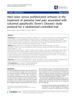
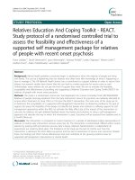
a two-armed parallel design and 1:1 allocation ratio. Flow
through the study is presented in Fig. 1. This study has
been approved by the Health Research Ethics Board of Alberta Cancer Committee (HREBA.CC-16-10-10). The
Twomey et al. BMC Cancer (2018) 18:757
Page 4 of 19
not unique to a specific tumour group or
cancer-treatment type. Furthermore, demographic and
clinical characteristics do not moderate exercise-induced
improvements in other patient-reported outcomes such
as physical function and HRQL [64]. Our intention is to
offer this program as widely as possible to a heterogeneous group of cancer survivors who are experiencing
CRF after treatment. Therefore, male and female cancer
survivors aged 18–75, following any cancer diagnosis
and cancer-treatment type, will be recruited for the
study. Potential participants will be screened for study
inclusion using a score of CRF severity using the
FACIT-F questionnaire [4] (where a score of ≤34 is the
recommended cut-off point for the diagnosis of CRF
[65]). Additional inclusion and exclusion criteria are
presented in Table 1.
Recruitment
Screening &
Lab Visit #1
Lab Visit #2
Randomization
n=52
Traditional Exercise
Tailored Exercise
Lab Visit #3
Lab Visit #4
Follow-up
(6 month)
Follow-up
(12 month)
Fig. 1 Flowchart of the study design
approved study will be reviewed annually by the HREBA.CC until completion. Any amendment to the protocol
which may impact on the conduct of the study will require
formal modification and approval by the HREBA.CC prior
to implementation, and will be described transparently in
subsequent reports. The study will be performed according
to the Declaration of Helsinki. The study was registered at
ClinicalTrials.gov before recruitment of the first participant
(NCT03049384, first posted in February, 2017). The research will be conducted at a single site (Faculty of Kinesiology, University of Calgary, Alberta, Canada). This study
protocol is written in accordance with the SPIRIT guidelines [63] (SPIRIT Checklist provided in Additional file 1).
Study population
The majority of research in exercise and cancer has been
conducted in female breast cancer survivors due to high
prevalence and survival estimates [10]. However, CRF is
Sample size
An a priori sample size estimation was performed using
G*Power 3 (v3.1.2–3.1.9 [66]). The hypothesis was treated
as a time × treatment (within-between) interaction for the
primary outcome measure of score on the FACIT-F. A
standardized mean difference of 0.44 was used, as reported
in a 2012 Cochrane review for comparisons of CRF following an exercise intervention or control, from a total of 539
cancer survivors after anti-cancer therapy [29]. Using an α
level of 0.05, a 1 - β of 0.8, the total sample size was calculated as 44. Potential loss to attrition was estimated as 15%,
based on previous reports from supervised exercise interventions in cancer survivors post-treatment (e.g. [67]), and
thus the total sample size was calculated as 52.
Recruitment
Participants will primarily be recruited via the Alberta
Cancer Registry (Alberta Health Services, Canada). Data
extraction criteria include age (≥ 18 and ≤ 75 years), diagnosed with any invasive cancer from 2013 to 2016, and
postal codes within 20 km of the University of Calgary.
From the resulting extraction, equal numbers of males
and females will be randomly sampled and sent a letter
confidentially and anonymously via the Registry. Those
contacted will be under no obligation to respond to the
research team. Participants will also be recruited via liaising with clinicians and/or advertising at cancer centres
local to the University of Calgary. When interested participants contact the study co-ordinator (via phone or email),
they will be informed about the main aspects of the research and asked about the time since their last cancer
treatment, and fatigue severity. If potentially eligible, participants will be provided with the participant information
sheet and encouraged to ask questions about the risks and
benefits of participation. Once the participant has had
time to review the information, the first visit to the laboratory (Lab Visit #1) will be scheduled.
Twomey et al. BMC Cancer (2018) 18:757
Table 1 Inclusion and Exclusion Criteria
Inclusion Criteria Aged ≥ 18 and ≤ 75 years
FACIT-F score ≤ 34
Completion of primary treatment in ≥ 3 months
and ≤ 5 years from enrolment
Approval received from CSEP-CEP and/or physician
Command of the English language and ability to
understand instructions related to the study
procedures
Exclusion Criteria Contraindication to experimental procedures and/or
exercise
Previously diagnosed as having sleep apnea or
anemia
Currently participating in a structured exercise
program or another clinical trial
Participant is pregnant
CSEP-CEP Canadian Society for Exercise Physiology Certified Exercise
Physiologist, FACIT-F Functional Assessment of Chronic Illness Therapy-Fatigue,
TMS transcranial magnetic stimulation
Randomization
Due to a continuous enrolment strategy and relatively
small sample size, participants will be allocated to groups
using a dynamic allocation procedure (minimization, performed using an open-source, online minimization program (MinimPy Program 0.3) [68]. In this process, the
first participant is allocated randomly to one of two treatment groups. Each newly enrolled participant is hypothetically allocated to each treatment group and an imbalance
score is calculated given unweighted baseline categorical
variables: sex (male, female), age group (18–39, 40–49,
50–59, 60–75) and cancer type (breast, prostate, lung,
colorectal, other). The participant will be allocated to the
preferred group (least imbalance) with a 1:1 allocation ratio, and a randomisation weighting of 0.75 in order to
avoid the potential introduction of bias associated with
pure minimization [69]. Participants will be randomized
following completion of all baseline assessments (i.e. after
Lab Visit #2, see below), by a researcher who is independent of the recruitment, enrolment and laboratory assessment process, and who has password-protected access to
the allocation procedure for the trial within the online
minimization program.
Blinding
During the assessments before the intervention, the participants and study team will be blinded to group assignment as this occurs before randomization. Following
randomization, blinding of participants and the study
team is not possible due to the recognised complexity of
blinding to an exercise intervention [29], particularly in
the case of interventions that are tailored to the individual, and where the study team will communicate about
participant wellbeing. Data files will be anonymized
using a code by an independent researcher who is
Page 5 of 19
unrelated to the study prior to processing by study
personnel. Participants will be informed that there is
equal possibility that they will be assigned to one of the
two treatment groups, and that the relative impact of
two exercise interventions is under investigation.
Laboratory assessments
The study team and/or assessors for specific methods
are extensively trained in those methods, and a number
of pilot assessments were performed prior to the study.
Regular internal inspections of the methods described
below are carried out to maintain high methodological
quality. Participants will be required to attend an exercise physiology laboratory (Faculty of Kinesiology, University of Calgary, Alberta, Canada) on four occasions.
Lab Visits #1 and #2 take place before the 12-week exercise intervention, and Lab Visits #3 and #4 take place
after the intervention. All laboratory visits will commence between 8 am and 9 am and will last 2.5–3 h.
Participants will be advised to consume breakfast 1.5 h
prior to arrival at the laboratory. Participants will be
instructed to arrive to the laboratory hydrated and to refrain from alcohol, caffeine and strenuous activity for
the preceding 24 h. Overall study time points are presented in Table 2.
Lab visit #1
Informed consent
Following initial communication about the main aspects
of the research during the recruitment process, Lab Visit
#1 will begin with an in-person discussion with the study
coordinator (20–30 min). The participant will have the
opportunity to express any concerns and/or ask questions. The participant information sheet will be reviewed
and/or explained such that the information is comprehensible, and the study coordinator will seek verbal assurance that the participant understands the research,
and that their participation is entirely voluntary. The
study co-ordinator will then obtain written informed
consent to participate.
Screening and participant information
Participants will complete a Physical Activity Readiness
Questionnaire for Everyone (PAR-Q+) and will be
screened for contraindications to TMS [70]. Participants
will be also screened for arrhythmia and hypertension,
determined during resting electrocardiography and
blood pressure measurements, respectively. Continuation with the study is conditional on the screening
process, and physician approval may be sought at this
stage. If the participant displays a normal sinus rhythm
and systolic and diastolic blood pressure of ≤144 and ≤
94 mmHg, respectively, is cleared for physical activity by
a Canadian Society for Exercise Physiology Certified
Twomey et al. BMC Cancer (2018) 18:757
Page 6 of 19
Table 2 Study Time Points
Process, Outcome or Test
Time Point
Recruitment
Pre-Intervention
Lab Visit
Intervention
Week (Total = 12)
#1
3
#2
6
9
Post-Intervention
Lab Visit
Follow-Up
Month
#3
6
12
Informed Consent
×
PAR-Q+
×a
ECG
×a
Blood Pressure
×a
×
a
×
Resting HR
×
Participant Information
×
FACIT-F
×
ERAS-r
FACT-G
#4
a
×
×
×
×
×
×
×
×
×
×
×
×
×
×
×
×
×
×
CES-D
×
×
×
BPI-sf
×
×
×
SPS
×
×
GLTEQ
×
×
ISI
×
×
Venous Blood Sample
×
Body Mass
×
Stature
×
Neuromuscular Familiarization
×
Start of 15-day Actigraphy
×
×
Sleep Diary
×
×
HRV
Maximal Exercise Test
×
×
×
×
×
×
×
×
×
×
DXA
×
×
CSA
×
×
Grip Strength
×
×
Neuromuscular Function
×
×
Intermittent Cycling Test
×
×
a
Indicates that the item is part of the screening process. BPI-sf, Brief Pain Inventory Short Form, CES-D, Center for Epidemiological Studies on Depression Scale;
CSA, cross-sectional area; DXA, dual-energy X-ray absorptiometry; ECG, electrocardiogram; ESAS-r, Edmonton Symptom Assessment System (revised version);
FACIT-F, Functional Assessment of Chronic Illness Therapy Fatigue Scale; Functional Assessment of Cancer Therapy - General (FACT-G); GLTEQ, Godin Leisure-Time
Exercise Questionnaire; HR, heart rate; HRV, heart rate variability; ISI, insomnia severity index; PAR-Q+, Physical Activity Readiness Questionnaire for Everyone; SPS,
Social Provision Scale
Exercise Physiologist (CSEP-CEP), and no further concerns are raised that would warrant physician approval,
the participant will continue to the procedures described
for below. Demographics will be self-identified using a
participant questionnaire, and will include sex, age, race,
marital status, education, employment status and household income. Clinical variables will include cancer diagnosis, time since diagnosis, time since completion of
primary treatment, treatment type (e.g. surgery, radiation
therapy, hormone therapy and/or chemotherapy) and
persisting side effects. Participants will self-identify as
non-smokers or smokers (daily or occasional). The
Alcohol Use Disorders Identification Test (AUDIT) will
be used as an indication of hazardous and harmful alcohol use (score > 8) [71].
Participant-reported outcomes
In addition to CRF severity (Lab Visit #2), patient-reported
outcomes include HRQL, depressive symptomatology, pain,
social provisions, leisure-time exercise and insomnia severity. The following questionnaires will be completed in the
order described and were chosen for their established reliability and validity with specific emphasis on use in cancer
populations. HRQL will be assessed using the Functional
Twomey et al. BMC Cancer (2018) 18:757
Assessment of Cancer Therapy – General (FACT-G) [72],
which provides scores for subscales of physical well-being,
social/family well-being, emotional well-being, functional
well-being, and additional concerns specific to cancer type.
In addition, the sum of these subscale scores will be used to
calculate total score for HRQL. Depressive symptomatology
will be assessed using the 20-item Center for Epidemiological Studies on Depression Scale (CES-D) [73]. Pain will
be evaluated using the Brief Pain Inventory Short Form
(BPI-sf) [74] which measures both pain severity and the impact of pain on functioning (interference). Social provisions
will be assessed using the Social Provision Scale (SPS) [75],
which provides scores for six sub-groups: guidance, reliable
alliance, reassurance of worth, attachment, social integration, and opportunity for nurturance. A total SPS score will
be derived from the six sub-scales. Leisure-time exercise
will be assessed using a modified Godin Leisure-Time Exercise Questionnaire (GLTEQ) [76], including the frequency
and duration of mild, moderate, strenuous, resistance and
flexibility exercise.
Page 7 of 19
adjusted on an individual basis as that which is estimated
to result in a test of 8–12 min duration. The power will be
increased at 1-min intervals until volitional exhaustion. Rating of perceived exertion (RPE) (Borg’s RPE scale [77], administered according to published instructions [78]) and
dyspnea (Borg CR-10 scale [79]) will be recorded every minute. For all cycling tests, verbal encouragement will be
provided by the same experimenters every 20–60 s [80]. A
fingertip blood sample will be collected at exercise cessation for the analysis of blood lactate (Lactate Scout+, EFK
Diagnostics, Cardiff, UK) and participants will complete a
supervised cool down. The highest 30-s average oxygen uptake will be calculated and a plateau in oxygen uptake will
be verified according to published criteria [81, 82]. Where
not achieved, the measure will be referred to as peak oxygen uptake (V_ O2peak ).
Neuromuscular familiarization
Participants will complete a 45–60 min familiarization
to the neuromuscular assessments described in detail
under Lab Visit #2.
Venous blood sample
A venous blood sample (total volume 35 mL) will be collected from the antecubital fossa by a certified phlebotomist,
between 9:30–10:30 am (≥ 2 h post-prandial). The sample
will be analyzed for whole blood count and variables including catecholamines, serotonin, cortisol, and cytokines including tumor necrosis factor-α, interferon-γ, transforming
growth factors (TGF-β1, 2 and 3), interleukins (IL-1β, 2, 4,
5, 6, 8, 10, 12 and 13), monocyte chemoattractant protein-1
and granulocyte-macrophage colony-stimulating factor.
Whole blood count will be analyzed within 2 h of collection
at the laboratory of Foothills Medical Centre. Other parameters assessed from blood serum and plasma will be centrifuged at 4 °C and 3000×g for 15 min, divided into aliquots
and stored at − 80 °C. Samples will be stored until laboratory evaluation for the current study only, and will not be
stored for use in any ancillary or future study.
Maximal exercise test
Following measurement of stature (cm) and body mass
(kg), a maximal exercise test will be conducted for the
measurement of maximal oxygen uptake ( V_ O2 max ). The
test will be conducted on a custom-built recumbent ergometer, which uses an electromagnetically-braked Velotron
system (RacerMate Inc., Seattle, WA). Seat position will be
adjusted for each participant, and self-selected cadence will
be determined (≥ 60 rpm). These details will be recorded
for replication in subsequent visits. Participants will be instrumented for the measurement of heart rate (HR) and
breath-by-breath pulmonary gas exchange and ventilation
(Quark CPET, COSMED, Rome, Italy). The starting power
output (25–50 W) and increment (10–20 W) will be
Actigraphy
Participants will be provided with a MotionWatch 8©
actigraphy system (CamNtech, UK) and instructions for
use in the study. This is an unobtrusive, waterproof,
wrist-worn device containing a light sensor and a tri-axial
accelerometer detecting acceleration in a 0.01–8 g range.
The device will record activity counts and light intensity
in 30-s epochs, as recommended by the manufacturer.
The device will be worn on the non-dominant wrist for a
continuous 15-day period, in accordance with established
recommendations [83, 84]. Participants will also be provided with a sleep diary to complete alongside the actigraphy measurement [85].
Heart rate variability
Short-term heart rate variability (HRV) will be measured
before and after the intervention. Participants will receive a
HR monitor (S810i Polar, Polar Electro, Kempele, Finland;
sampling rate 1000 Hz) to complete a 10-min HRV measurement in their own home. To control for transient variables [86], the participant will be instructed to follow a
normal sleep routine the night before the measurement,
avoid strenuous physical activity and alcohol for the preceding 24 h, complete the measurement immediately upon
waking (after visiting the washroom if necessary) and thus
prior to any food or caffeine intake, in the supine position,
under quiet and thermoneutral conditions.
Lab visit #2
Participants will return to the lab after completion of the
15-day actigraphy measurement. Participants will return
Twomey et al. BMC Cancer (2018) 18:757
the actigraph and sleep diary and complete the below assessments for Lab Visit #2.
Cancer-related fatigue
Score on the FACIT-F will be used to assess CRF severity (primary outcome). The revised Edmonton Symptom
Assessment System (ESAS-r) tiredness scale will also be
used [87, 88], as recommended for the screening of CRF
severity in the clinical practice guidelines for standard
cancer care in Alberta [89]. The questionnaires will be
completed between 8:30–9:30 am.
Body composition and bone mineral density
Participants will undergo a whole-body scan using dual
energy X-ray absorptiometry (DXA; Discovery W, Hologic, Bedford, MA), for the assessment of parameters relating to body-composition and bone mineral density.
Muscle cross-sectional area
Cross-sectional area (CSA) of the right vastus lateralis
(VL) and rectus femoris (RF) will be assessed using
real-time B-mode ultrasonography (M2540A, Phillips,
Bothell, WA). Participants will adopt a supine position
with legs relaxed and knees extended. One axial perpendicular line will be marked with indelible ink at 50% of the
distance between the greater trochanter and the lateral
epicondyle of the knee. After 20 min of rest, sufficient
water-soluble gel will be applied to the transducer to ensure that clear images are obtained with minimal and consistent pressure to avoid compression of the muscle
during examination. Participants will be asked to fully
relax the muscle while consecutive two-dimensional (2-D)
images are acquired with the probe placed perpendicular
to the skin at the anatomical site. Three images will be acquired by the same experimenter. CSA will be estimated
through manual tracing of the muscle borders using
open-source imaging program (ImageJ).
Grip strength
The grip bar of a handgrip dynamometer will be adjusted
for each participant and recorded for post-intervention
measurement. The measurement will be made in a standing position, with the elbow extended and arm parallel to,
but not touching, the side of the body. Grip strength (kg)
for the dominant hand [90] will be assessed as the highest
of three ~ 3-s maximal efforts, separated by 1-min rest.
Neuromuscular assessments
All neuromuscular data will be acquired using a PowerLab
16/35 and LabChart v8 software (ADInstruments, Bella
Vista, Australia), and further measurement details are described in later sections. For neuromuscular assessments,
three preliminary procedures will take place on an isometric chair as: (i) determination of a supramaximal femoral
Page 8 of 19
nerve electrical stimulation (FNES) intensity; (ii) determination of optimal coil position for TMS; (iii) determination of optimal stimulation intensity for TMS. On the
isometric chair, participants will also perform a set of preparatory contractions of 5-s duration, with 5 s rest between contractions. The preparatory contractions involve
five at 10%, five at 30%, three at 50% and two at 75% of a
familiarization MVC. Participants will then perform a
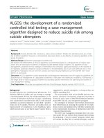
neuromuscular assessment (Fig. 2a), starting with two
MVCs with no stimulations, separated by 60 s. Where the
two MVCs differ by ≥5%, a third will be performed. All
MVCs (duration 3–5 s) will be performed with strong verbal encouragement and visual feedback of force displayed
on a large computer monitor positioned ~ 1 m in front of
the participant. Next, two MVCs with single FNES delivered during the plateau of the MVC, and within 2 s of relaxation will be performed (for the calculation of
voluntary activation with FNES [VAFNES], also see Data
Analysis). Finally, participants will perform two sets of
contractions at 100, 75 and 50% MVC, separated by 5-s
rest, with 20-s rest between sets (for the calculation of voluntary activation with TMS [VATMS], see also Data Analysis). Guidelines at 75 and 50% of the preceding MVC for
each set will be plotted without any delay using a
custom-made macroinstruction. TMS will be delivered
during each contraction when force has plateaued or stabilized on the target guideline. Participants will be
instructed to resume the contraction immediately after
TMS delivery (in order to better evaluate silent periods,
see also Data Analysis). During the voluntary contraction
at 50% MVC in the second set, FNES will also be delivered
after the TMS, once the participant has returned to the
guideline. The participant will be transferred to the cycle
ergometer shown in Fig. 2c, to repeat this neuromuscular
assessment before cycling exercise (Fig. 2a).
Incremental cycling test and neuromuscular fatigue
Following the pre-exercise assessment on the ergometer,
participants will complete an incremental cycling test
(Fig. 2d). Immediately post-exercise i.e. within 1 s from task
disengagement (or rpm < 60), a post-exercise neuromuscular assessment will be performed (Fig. 2a). The incremental
protocol involves stages of 3-min duration. Between each
3-min stage, the pedals will be locked, and an intermediate
assessment will be performed (Fig. 2b). As shown in Fig. 2b,
this involves an MVC with FNES, and a contraction at 50%
MVC with TMS and FNES. After the intermediate assessment (30 s duration), the pedals will be unlocked and the
participant will resume cycling at their target cadence at
the pre-determined higher power output for the next stage.
Increments in power output are 0.3 W·kg− 1 for the first
four stages, 0.4 W·kg− 1 for the following five stages and by
0.5 W·kg− 1 for any subsequent stages. Cadence will be the
only real-time feedback participants will receive during
Twomey et al. BMC Cancer (2018) 18:757
Page 9 of 19
a
b
c
d
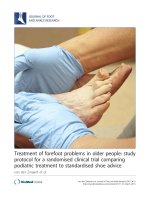
Fig. 2 A schematic illustrating: (panel a) the neuromuscular assessment performed pre- and post-exercise; (panel b) the intermediate
neuromuscular assessments performed every 3 min as part of the intermittent cycling protocol; (panel c) the cycle ergometer; and (panel d) the
intermittent cycling protocol including neuromuscular assessments. MVC, maximal voluntary contraction; 75 and 50%, the percentage of the
preceding MVC; TMS, transcranial magnetic stimulation; FNES, femoral nerve electrical stimulation
Twomey et al. BMC Cancer (2018) 18:757
cycling, and verbal instructions will be given should cadence drift by ≥4 rpm. The reliability of this incremental
protocol has previously been assessed in our laboratory in
healthy individuals [61] and cancer survivors (unpublished
data). The procedures described are covered in detail during the neuromuscular familiarization of Lab Visit #1.
Measurement of voluntary and evoked force
On the isometric chair (custom made from a Kin-Com
dynamometer frame), force will be measured during voluntary and evoked contractions using a calibrated load cell
(LC101-2 K, Omegadyne, Sunbury, OH) connected to a
noncompliant cuff attached around the right ankle, just superior to the malleoli. The load cell will be adjusted to position it directly behind the point of applied force.
Participants will sit upright with the knees and hips at 90°
flexion, secured using straps across trunk and shoulders.
On the ergometer, force will be measured during voluntary
and evoked contractions using a wireless pedal force analysis system located between the pedal and crank (PowerForce Model PF1.0.0, Radlabor GmbH, Freiburg, Germany).
The ergometer permits the pedals to be locked instantly in
a fixed position with hip angle at ~ 100° and right knee and
at ~ 90°, and the crank parallel to the ground [61]. This allows participants to perform a contraction whereby force is
measured in line with the crank. Participants will be secured
with non-compliant straps across the trunk. Force will be
sampled at 500 Hz and recorded using Imago Record (version 8.50, Radlabor GmbH). To provide real-time visual
force feedback, the PowerForce signal will be transmitted to
the PowerLab system using a National Instruments 16-bit
A/D card (NI PCI-6229, National Instruments, Austin, TX)
and connector block (BNC-2111, National Instruments).
Measurement of Electromyographic responses
Surface electromyography (EMG) will be recorded from
the right vastus lateralis (VL) and rectus femoris (RF), and
the long head of the biceps femoris (BF). The skin will be
shaved, abraded and cleaned with isopropyl alcohol wipe
to ensure a low impedance (< 10 kΩ). Two single-use
electrodes (10-mm diameter, Meditrace 100, Covidien,
Mansfield, USA) will be placed in a bipolar configuration
(inter-electrode distance of 20 mm) over the muscle belly
following SENIAM recommendations [91]. The reference
electrode will be placed over the patella. Raw EMG signal
will be analog-to-digitally converted, amplified (octal
bio-amplifier ML138, ADInstruments; common mode rejection ratio = 85 dB, gain = 500) and sampled at 2000 Hz.
EMG will be band-pass filtered (5–500 Hz).
Femoral nerve electrical stimulation
Electrical stimuli (1 ms pulse width) will be delivered
using a constant-current stimulator (DS7AH, Digitimer
Ltd., Hertfordshire, UK). The cathode (10-mm diameter,
Page 10 of 19
Meditrace 100) will be positioned over the femoral
nerve, high in the femoral triangle. The electrode will be
secured with tape and a gauze plug to apply pressure.
The anode (50 × 90 mm, Durastick Plus, DJO Global,
Vista, CA) will be placed midway between the greater
trochanter and the iliac crest. For the determination of
supramaximal FNES intensity, single FNES will be delivered beginning at 10 mA and increasing by 10 mA until
no further increase in twitch force or VL M-wave amplitude can be elicited. The intensity at this plateau will
then be increased by 30%.
Transcranial magnetic stimulation
Single TMS pulses (1-ms duration) will be delivered with a
110-mm diameter concave double-cone coil powered by a
mono-pulse magnetic stimulator (Magstim 2002, The Magstim Company Ltd., Whitland, UK) with the coil orientated
to induce a postero-anterior intracranial current flow. Optimal coil position will be defined as the location eliciting the
largest motor evoked potential (MEP) in the VL and RF and
a concurrent small MEP in the BF with stimulations delivered at 50% maximal stimulator output (MSO) during brief
contractions at 20% MVC, with 10 s rest between contractions. A standardised procedure will be used involving six
potential sites marked on a white Lycra swim cap worn by
participants. The six sites (A – F) include the vertex (A),
1 cm (B) and 2 cm (C) posterior to the vertex along the
nasion-inion line, 1 cm lateral to vertex over the left motor
cortex (D), and 1 cm (E) and 2 cm (F) posterior to D. When
optimal stimulation site is selected and marked clearly on
the swim cap, the stimulation intensity will be determined
using a standardised procedure involving stimulations at 50,
60, 70 and 80% MSO (randomized order, four stimulations
at each intensity) delivered during brief contractions to 20%
MVC [92], with 10 s rest between contractions. The intensity will be optimised for the measurement of VATMS. That
is, the intensity eliciting a maximal VL and RF MEP will be
selected (i.e. the lowest intensity resulting in an increase of
less than 5% MEP amplitude at higher stimulus intensities).
The size of the superimposed twitch (SIT) will also be examined to ensure that this intensity corresponds to a maximal SIT at 20% MVC (as the SIT can be lower at higher
TMS intensities due to co-activation of the knee flexors).
Post-intervention assessments
After the intervention, Lab Visit #3 will be completed 72–
96 h after the final exercise session. Lab Visit #3 involves the
post-intervention assessment of patient reported outcomes,
a venous blood sample, maximal exercise test (replicated
starting power output and increment) and (re)familiarization
to neuromuscular measures as described for Lab Visit #1
(see also Table 2). Lab Visit #4 will be completed 72–96 h
after Lab Visit #3. Lab Visit #4 assessments are identical to
those described for the pre-intervention Lab Visit #2,
Twomey et al. BMC Cancer (2018) 18:757
including replication of power outputs during the incremental cycling test (i.e. power outputs will be the same absolute
value, even where body mass has changed). The sleep diary
and actigraph measurement will begin after Lab Visit #4,
and collected from participants after the 15-day measurement period.
Follow-up
Six and 12 months after the exercise interventions, participants will be contacted via phone or email to
complete the FACT-F scale, ESAS-r tiredness scale and
the GLTEQ.
Treatment arms
Both exercise interventions will take place in the Thrive
Centre, a fitness facility for people affected by cancer (Faculty of Kinesiology, University of Calgary, Alberta, Canada).
The exercise intervention will be delivered in a small group
setting by exercise specialists, who have completed specific
cancer and exercise training (). Participants in both treatment arms will be supervised by the same exercise specialists. The following
prescriptions for aerobic and/or resistance exercise will be
followed only where achievable, such as where the exercise
is not voluntarily terminated prematurely due to intolerable
levels of perceived fatigue, dyspnea or muscle weakness.
Where an existing adverse effect of treatment (e.g. shoulder
dysfunction) or injury (e.g. knee replacement) limits the
performance of a movement, the movement will be restricted, modified or substituted to ensure that there is no
pain during or after exercise. In addition, the exercise specialist may adjust intensity or duration based on
observation and judgement, particularly in the case of reducing the demands to accommodate day-to-day fluctuations
in health or wellbeing in a diverse group of cancer survivors. A decision to discontinue an individual’s intervention
will be made by the research team if there is concern that
the exercise intervention is causing harm. Upon arrival to
supervised exercise sessions (i.e. before exercise), participants will be asked to indicate their fatigue levels using a
rating-of-fatigue scale, which quantifies the intensity of the
subjective feeling state at a given moment [93]. Should the
exercise intervention appear to gradually increase the rating
of fatigue over 1–2 weeks, the intensity will be reduced as
deemed necessary to ensure the overall wellbeing of the
participant. The exercise specialist and research team will
communicate regularly regarding individual participants.
Participant safety is paramount and any adverse events (related to exercise or not) will be monitored and reported according to the standardized guidelines for reportable events
from the independent ethics committee (HREBA.CC). The
reasons for dropout from the intervention will be recorded
where possible, and no further outcome data will be collected in participants who withdraw from the study.
Page 11 of 19
In both treatment arms (see Traditional Exercise Group
and Tailored Exercise Group, below), each supervised exercise session will include a low-intensity warm-up (5 min
light cycling and 5 min dynamic stretching or mobility exercises), and a cool-down which will include stretching of
major muscle groups. Adherence to the exercise intervention will be reported as the number of sessions attended as
a percentage of total sessions scheduled (with a maximum
of 36). Where the participant has a commitment known in
advance such as a medical appointment or holiday, the
missed sessions will be rescheduled or replaced at the end
of the 12-week intervention. Unanticipated cancellations
(non-attendance) will not be substituted. Exercise sessions
will primarily be offered on weekday afternoons/evenings
with flexibility to ensure that people who have returned to
work and/or are caring for dependents are logistically able
to participate.
Participants in both treatment arms will be provided with
an identical intervention booklet during the first training
session. The booklet contains weekly physical activity logs
and guidelines for ~ 10 static stretches. The physical activity
logs will be used as a self-report measure of additional
physical activity (e.g. a brisk walk) that is undertaken outside of the exercise intervention (such additional physical
activity is not restricted/prohibited). The booklet also contains educational information related to promoting participant adherence, retention, and long-term behavioural
change [94, 95]. The booklet is written in a plain language,
and includes sections on goal setting, planning for barriers,
monitoring behaviour, maintaining motivation and enhancing personal control. The information in the booklet will
be discussed verbally by the supervising exercise specialist,
to ensure comprehension and to encourage engagement
with the material. When the study is complete, participants
will be encouraged to continue attending the Thrive Centre
as a ‘drop-in’, during regular scheduled time-slots when the
facility is monitored by volunteers with specific cancer and
exercise training.
Traditional exercise group
Participants in the traditional exercise group will engage
in exercise of a duration, frequency and intensity that is
consistent with published recommendations and clinical
practice guidelines for cancer survivors (e.g. [33–36]),
and as such, compatible with health-related physical activity guidelines for the general population. The goal of
the intervention is to progress to meet guidelines of
150 min per week of moderate-intensity aerobic exercise,
and resistance training on at least two days per week.
Aerobic exercise will be performed on a stationary cycle
ergometer, rowing ergometer, treadmill and/or elliptical
trainer (participant’s preference). The aerobic exercise
duration will be progressive such that in weeks 1–4, exercise will be performed for 30 min on three supervised
Twomey et al. BMC Cancer (2018) 18:757
sessions per week. The total aerobic exercise duration
will progress from 90 min in weeks 1–4, 120 min in
weeks 5–8 and 150 min in weeks 9–12 over 3 sessions
per week. The intensity of exercise will correspond to an
RPE of 11–14, which is in line with published guidelines
[35, 96] and empirical data [97] for moderate exercise.
Participants will have been familiarized with the RPE
scale and instructions on both Lab Visits #1 and #2. The
corresponding HR and equipment resistance/speed will
be monitored and recorded during every session.
Two sessions per week (separated by ≥48 h) will include resistance training after the aerobic component,
involving exercises targeting all major muscle groups.
Participants will perform one to three sets of eight to
twelve repetitions. Within these guidelines, the principle
of progressive overload will be applied to gradually increase training volume. There will be a 1–3 min rest
period between sets, and contractions will be performed
at slow to moderate velocities [98]. Eight to ten body
mass/dumbbell exercises will be selected in 3-week
micro-cycles from a pre-determined bank of ~ 30 exercises selected by the research team. Appropriate individual modifications and progressions from a novice level
will be included. Participants will be coached in correct
technique for each movement. Exercises will be prescribed with consideration of an individual’s cancer or
cancer-treatment side effects (e.g. lymphedema, peripheral neuropathy), and awareness of increased risks based
on cancer or cancer-treatment (e.g. bone fracture in
those with previous bone metastases). Specific guidelines
in this regard will be followed where available (e.g. [33]).
Tailored exercise group
The experimental tailored exercise group will be prescribed an intervention designed specifically to address
the deficits or areas for improvement identified in Lab
Visits #1 and #2. The optimisation of the exercise intervention is based on the outcome of interest i.e. CRF. As
the mechanisms of CRF are unknown, we have chosen to
focus primarily on tailoring the intervention to improve
specific physiological parameters and/or sleep, with consideration of the whole profile of baseline assessments.
The results of an individual’s assessment will be reviewed
and discussed by the research team and exercise specialists to optimize the intervention. For transparency and to
assist with interpretation of generated data where the interventions vary between participants, the characteristics
of the individual (anonymized) tailored exercise interventions (e.g. the frequency, intensity, duration, RPE, HR
and/or the type of movement) will be made available in an
open-access repository upon completion of the study. The
design and application of this experimental exercise intervention will proceed with attention to the principles of exercise training and be evidence-based [99]. The frequency
Page 12 of 19
(three times per week) and total duration (60–90 min) of
sessions (and therefore contact and interaction with exercise instructors and other participants) are the only aspects of exercise dose that are designed to be equivalent
to the traditional exercise group. The intervention will be
adjusted at 3-week intervals based on participant feedback
and the judgement and observations of the research team/
exercise specialists. Three examples of the parameters and
resulting focus of tailored exercise interventions are provided below. A diverse group of cancer survivors will have
diverse profiles based on Lab Visits #1 and #2, and therefore the intervention may be multi-modal i.e. involve a
combination of the examples provided below.
(i) Based on a low muscular strength (force-generating
capacity in the knee extensors) at baseline (in
comparison to non-fatigued cancer survivors and
healthy adults of the same sex and similar age; data
collected in our laboratory), the exercise intervention
will focus on improving this using both neuromuscular electrical stimulation (NMES) and resistance
training with voluntary contractions. For the former,
NMES is widely applied to the quadriceps as a
(re)training modality, including in pathological conditions where muscle weakness is an issue [100, 101].
In terms of resistance training, if muscle mass is low
(on consideration of VL and RF cross-sectional area,
and in comparison to reference values for DXAderived lean mass index [102, 103]), the focus will be
on hypertrophy. High repetition multi-set resistance
training will be incorporated [104], with additional
focus on eccentric actions [105, 106]. Prescribed aerobic exercise will be minimal [107], particularly in
the case of low body mass or history of malnutrition
during treatment. Where muscle mass appears to
have been maintained, concurrent to low VA, resistance training may progress (safely) to involve sets of
low repetitions and high loads [104].
(ii) Based on substantial cardiorespiratory
deconditioning, primarily based on a low V_ O2 max
according to age-group norms [96], participants will
be prescribed interval training on at least two (of
three sessions) per week. Supervised high intensity
interval training (HIIT) results in improvements in
cardiorespiratory fitness and other outcomes in cancer survivors, and can be considered low risk in
regards to adverse events [108]. The evidence of
safety (in regards to the low risk of cardiovascular
events in particular) has been convincingly demonstrated in other clinical populations e.g. coronary
heart disease patients [109]. HIIT will be performed
on a cycle ergometer to reduce risk of muscularskeletal injury. Participants will be familiarised with
HIIT gradually, and the intensity of the work
Twomey et al. BMC Cancer (2018) 18:757
intervals will be increased over the first two weeks
depending on tolerance, to reach 85–95% of peak
HR. Due to the relatively recent adoption of HIIT
in cancer populations, there are no guidelines on
optimal HIIT prescription (e.g. work:rest ratio and
interval duration), where the effectiveness of different HIIT protocols should be tailored to ensure it is
feasible for the individual. However, the recommendations from other clinical populations will be implemented such that the typical work:rest ratio will
increase to ≥1 and work intervals ranging from 30 s
up to 4 min. For example, the 4 × 4 protocol [110]
(4 × 4 min work with 3 min active recovery at the
lowest possible intensity) will be appropriate for
many cancer survivors.
(iii)Based on substantial sleep disturbance determined
via actigraphy (e.g. those who display three of the
following criteria: total sleep time ≤ 440 min; sleep
efficiency [total sleep time as a percentage of time
in bed] ≤ 87%; sleep onset latency [an index of the
difficulty in the transition from wake to sleep] >
14 min; wake after sleep onset ≥25 min [111, 112]),
there will be a focus on exercise to improve sleep.
The identification of sleep disturbance will be
primarily based on actigraphy, but subjective
complaints of sleep disturbance will also be
considered (ISI score and subjective total sleep time
from the sleep diary) with awareness of the
misperception of sleep relative to objective
measures [113]. Although exercise is a widely
recognised intervention to improve disturbed sleep,
there are many unanswered questions in regards to
optimally prescribing exercise interventions for this
purpose (e.g. dose, mode, timing) [114]. However,
in adults, evidence suggests that exercise duration
moderates sleep outcomes for regular exercise
(where longer duration is more beneficial) [115]
and most studies have used moderate aerobic
exercise such as walking [116]. Overall, the
evidence suggests that exercise improves sleep in
cancer populations, though few studies have
investigated this in cancer survivors after treatment
who present with sleep disturbance at baseline
(reviewed in [43]). Nevertheless, the intervention
will focus on long-duration (progressing to e.g.
60 min) aerobic exercise such as walking.
A further consideration is whether the participant is at
increased health risk due to obesity (body mass index >
30 kg/m2, with additional consideration of percentage
body fat from DXA). If obese, the intervention will involve low impact activity that puts minimal stress on
joints (elliptical trainer, cycling or walking) to avoid injury, with increasing duration (progressing to > 150 min
Page 13 of 19
per week) on intensity to increase energy expenditure
and assist with weight management [117].
Data monitoring
A data monitoring committee was not included because
the trial involves a behavioural intervention (progressive
exercise) with known/minimal risks, and does not require periodic benefit–risk assessments. No independent
auditing of trial conduct is planned.
Confidentiality
In order to maintain confidentiality during and after the
trial, all study-related information will be stored securely at
the study site in areas with limited access. Furthermore, access within the study team will be the minimum required
for data analysis and quality control. Blood samples, electronic files, data sheets and completed questionnaires will
be stored using coded IDs. Digital files will be stored on
password-protected computers, in password protected
folders, and backed up on a password-protected hard drive.
Records that contain personal identifiers (such as informed
consent forms) will be stored separately from those identified by coded ID, in a locked cabinet in an office accessible
to the study co-ordinator.
Data analysis
Neuromuscular data
The potentiated mechanical response from a single electrical stimulus will be analysed for amplitude of the twitch
(Qtw,pot), maximal rate of force development and maximal
relaxation rate. Voluntary activation using femoral nerve
electrical stimulation (VAFNES) will be calculated using the
interpolated twitch technique where the amplitude of the
SIT is normalized to the corresponding Qtw,pot using the
equation VAFNES (%) = (1-(SIT/Qtw.pot)) × 100 [60]. For
TMS, an estimated resting twitch (ERT) will be calculated
by taking the y-intercept of a linear regression of the
SIT-voluntary force relationship. VATMS will be subsequently quantified using the equation VATMS (%) = (1-(SIT/
ERT)) × 100 [118]. Where regressions are not linear (defined as r < 0.9 [119]), those data will be excluded.
For the evoked EMG responses, the peak-to-peak amplitude and area under the curve of the MEP in all muscle
groups will be determined from a selection of data encompassing the biphasic wave. The selection will begin at the
first deviation from zero after any stimulation artefact,
and end on the return to zero after the biphasic wave. The
M-waves evoked in the VL and RF will be analysed using
the same method. For the assessment of corticospinal excitability, the VL and RF MEPs will be normalised to an
M-wave delivered during a contraction and nearby in
time. The silent period will be measured from stimulus
artefact to the continuous resumption of voluntary EMG,
Twomey et al. BMC Cancer (2018) 18:757
determined by an experimenter experienced in the analysis, using visual inspection of the EMG trace [120].
Heart rate variability
In the time domain (ms), the mean normal-to-normal
(NN) interval, the standard deviation of the average NN
interval (SDNN) and the square root of the mean
squared differences of successive NN intervals (RMSSD)
will be calculated. As recommended for short-term HRV
recordings [115], the spectral components analyzed in
the frequency domain (ms2) will be the very low frequency (VLF; 0.01–0.04 Hz), low-frequency (LF; 0.04–
0.15 Hz) and high-frequency (HF; 0.15–0.40 Hz). The
LF/HF ratio will also be calculated. The spectral analysis
will be performed using fast Fourier transform algorithms (Kubios HRV Standard v3.0.2).
Actigraphy
The night following Visit #1 will be excluded from the
actigraphy data analysis to mitigate any effect of acute
maximal exercise (which participants are likely to be unaccustomed to). Data will be analysed using MotionWare 1.0.27 (CamNtech, UK). Responses from the sleep
log will be used to confirm the start and end time of the
sleep window, activity onset/offset and “lights out”/
“lights on” (as determined by the light sensor). Sleep parameters calculated within the software include time in
bed, actual sleep time, actual wake time, sleep efficiency
(the percentage of time in bed spent sleeping),
sleep-onset latency (time from “lights out” to sleep onset), fragmentation index (the percentage of immobile
phases of one minute). For rest-activity cycle characteristics, the following parameters will be calculated using a
non-parametric circadian rhythm analysis option [121]:
relative amplitude (calculated from estimated lowest and
highest activity periods), inter-daily stability (the degree
of regularity of the rest-activity patterns on individual
days in the 24 h environment), intra-daily variability (the
fragmentation of periods of rest and activity), the estimated peak time of activity period, mesor (mean level),
L5 (mean activity counts in the least active 5 h period in
the average 24 h pattern) and L5 mid (the central time
of the L5 period, usually referring to the trough of the
rest-activity cycle). The mean amount of activity during
the sleep period and the activity index (percentage of
30-s epoch during both sleep and wake periods with an
activity > 0) will be calculated from extracted raw data.
An objective measure of day-time physical activity will
be computed as the number of minutes spent at sedentary, light and moderate-to-vigorous physical activity intensities over the 14-day measurement period. This will
be quantified using calibrated cut-points for MotionWatch 8© activity counts, as determined in healthy older
adults [122].
Page 14 of 19
Intended statistical analysis
Data will be analysed after data collection is complete, and
no interim statistical analysis will be performed. Descriptive
statistics will be used for demographic and clinical variables
measured at baseline for each group. Frequencies and percentages will be used for categorical variables and the mean
± standard deviation (or median and range) will be used for
continuous variables. To account for any differences in loss
to follow up between groups, the primary analysis will be
conducted on an intention-to-treat basis. For the primary
outcome of FACIT-F score between treatment arms over
time, data will be analysed with linear mixed models using
R [123] and lme4 [124]. “Treatment arm” will be included
as a fixed effect and “participant” as a random effect. Parameters will be estimated using restricted maximal likelihood. The Kenward-Roger approximation for degrees of
freedom will be used when evaluating the significance of effects. This produces optimal type I error rates (neither
anti-conservative nor overly sensitive to sample size) [125].
Secondary analyses will be performed to assess adjustments
for protocol deviations (per protocol analysis). Statistical
code will be made openly available upon publication of the
results. A minimum of a 3-point difference in FACIT-F
score will be considered clinically relevant [126]. For
secondary outcomes that are assessed pre- and
post-intervention only (e.g. neuromuscular measures, sleep
parameters, blood biomarkers), two-way mixed design
ANOVA will be used (group [tailored vs. traditional] × time
[pre- vs. post-intervention]). In this case, missing data will
be dealt with using list-wise deletion. Following a significant
interaction, pairwise comparisons will be conducted with a
Bonferroni adjustment. The threshold for rejecting the null
hypothesis will be p < 0.05. For main and interaction effects,
partial eta squared will be computed as an effect size estimate. Effect sizes for pairwise comparisons will be reported
as Cohen’s d [127, 128]. This will be supplemented with
95% confidence intervals for mean differences. Further exploratory analysis will be labelled as such in later reporting.
Dissemination and data sharing policy
The results for primary and secondary outcomes will
be disseminated regardless of the magnitude or direction of the effect. The primary research aim will be
addressed in a main publication reporting the results
of the primary analysis i.e. the effect of treatment
arm on CRF severity. Due to the number of secondary outcomes, additional publications may be warranted to provide in-depth analysis of, for example,
data related to neuromuscular fatigue, sleep or ROF.
Scientific and administrative information about the
results of the trial will be submitted to the ClinicalTrials.gov results database. Participants will be informed about their personal results in a participant
report written in plain language, within 4 weeks of
Twomey et al. BMC Cancer (2018) 18:757
Visit #4. No later than 2 years after the final
follow-up assessment, an anonymized, de-identified
dataset will be made openly available to an appropriate data archive for sharing purposes.
Discussion
To the best of our knowledge, this will be the first study
to examine and compare the effects of a traditional exercise intervention against a tailored exercise intervention
on CRF in cancer survivors. Furthermore, this will be
the first study to include a comprehensive examination
of potential pathways for the improvement in CRF with
exercise, including patient-reported outcomes such as
depressive symptomology and pain severity, alongside
objective assessments of blood biomarkers, physical activity levels, sleep and cardiorespiratory fitness. In
addition, we will examine the neuromuscular correlates
of CRF, including neuromuscular function at rest and
neuromuscular fatigue during an exercise task that is dynamic and involves large muscle groups i.e. relevant to
daily activities such as locomotion.
The most important decision about the design of
this study also represents the most significant challenge. That is, tailored exercise interventions that
are designed based on comprehensive (though not
exhaustive) pre-intervention assessments. It is not
possible to specify all possible aspects of this tailoring in advance, though a number of categories have
been detailed. The basis of the tailored intervention
is that it involves consideration of individual profiles.
For most cancer survivors, the published guidelines
offer a foundation for exercise recommendations,
given that exercise is considered as being low risk
with large potential for benefits. However, in clinical
practice and during treatment, exercise prescription
does involve tailoring based on adverse events or as
recently highlighted, co-morbidities [38]. We propose
that specific tailoring may also be necessary for a diverse group of cancer survivors with persistent CRF
after treatment completion. Targeting CRF as a
symptom, rather than a tumor group or treatment
type, will undoubtedly result in a heterogeneous cohort. Although the primary research question is
clearly defined, there are multiple degrees of complexity for later interpretation and reporting of study
results. Our solution is to make the (anonymized,
de-identified) intervention data openly accessible,
with the restriction that participant privacy/confidentiality must be maintained. To our knowledge, this is
not common practice in exercise oncology research,
but will facilitate replication of the characteristics of
individual interventions and allow further exploratory analysis in regards to the interpretation and
comparison of intervention data.
Page 15 of 19
As this is a single-site study, participants must be able
to regularly travel to the University of Calgary, such
that it is only feasible for cancer survivors who live locally to participate. We recognise that this is a limitation of the study. In terms of reducing barriers to
recruitment for people who live locally, parking costs at
the site will be reimbursed. However, participants must
also have the time available to participate in an exercise
intervention. As the time post-treatment is up to five
years, it is anticipated that a number of potential participants will be struggling with CRF after having returned
to work and/or while also caring for dependents. To
help with overcoming this barrier, exercise sessions will
be offered with a large degree of flexibility in regards to
the day of the week and the exercise time, which will
also be an important factor in regards to participant
retention.
In this study, the traditional exercise group was designed as an active control, and considered to be an
appropriate comparator to the (experimental) tailored
exercise group. A ‘no exercise’ wait-list control was
not included in the study design due to the established benefit of exercise on CRF [29–31]. We did
not consider it necessary to confirm that an improvement in CRF with exercise was superior to an improvement due to, for example, an additional 3–
4 months in the passage of time since cancer treatment. Due to the difficulties in blinding, the active
control has been designed to not only be broadly
consistent with published guidelines, but to be
equivalent to the experimental group on non-specific
conditions (that may influence the primary outcome,
which is a perceptual construct) such as expectancy,
social support during exercise or attention from the
exercise specialist [129]. The continuous enrolment
strategy is due to the anticipated difficulty in recruiting a single large cohort of eligible and interested
cancer survivors who meet the CRF-severity criteria
and are able to commit to the intervention. However,
parallel groups will also control for potentially
confounding variables such as seasonal variations in
Calgary, Alberta.
In summary, although there is evidence for the benefits of exercise for CRF, it is important to design interventions specifically targeting this distressing
symptom, such that potential benefits are optimised.
If a tailored intervention confers some benefit above
a more general exercise program in cancer survivors
with persistent CRF, referral to a clinical exercise
physiologist should be considered as a treatment option given the suggestion that the profession can assist oncologists in the management of fatigue [130].
To our knowledge, this will be the first study to compare the effects of a traditional vs. tailored exercise
Twomey et al. BMC Cancer (2018) 18:757
intervention on CRF in fatigued cancer survivors.
Using physiological, behavioural and patient-reported
outcomes, this study will add to the current knowledge about both the factors contributing to CRF, and
the potential reduction in CRF severity with an exercise intervention, with the ultimate objective of improving the quality of life of cancer survivors.
Additional file
Additional file 1: SPIRIT 2013 Checklist: Recommended items to address
in a clinical trial protocol. (DOC 125 kb)
Abbreviations
ACSM: American College of Sports Medicine; AUDIT: Alcohol Use Disorders
Identification Test; BF: Biceps femoris; BPI-sf: Brief Pain Inventory Short Form;
CES-D: Center for Epidemiological Studies on Depression Scale; CRF: Cancerrelated fatigue; CSA: Cross-sectional area; CSEP-CEP: Canadian Society for
Exercise Physiology Certified Exercise Physiologist; DXA: Dual-energy X-ray absorptiometry; ECG: Electrocardiogram; EMG: Electromyography; ESASr: Edmonton Symptom Assessment System (revised version); FACITF: Functional Assessment of Chronic Illness Therapy Fatigue Scale; FACTG: Functional Assessment of Cancer Therapy – General; FNES: Femoral nerve
electrical stimulation; GLTEQ: Godin Leisure-Time Exercise Questionnaire;
HR: Heart rate; HRQL: Health-related quality of life; HRV: Heart rate variability;
ISI: Insomnia severity index; MVC: Maximal voluntary contraction; PAR-Q
+: Physical Activity Readiness Questionnaire for Everyone; RF: Rectus femoris;
RMS: Root mean square; RPE: Rating of perceived exertion; SIT: Superimposed
twitch; SP: Silent period; SPS: Social Provision Scale; TMS: Transcranial magnetic
_ 2 max : Maximal
stimulation; VA: Voluntary activation; VL: Vastus lateralis; VO
_ 2peak : Peak oxygen uptake
oxygen uptake; VO
Funding
This research is funded by the Canadian Cancer Society (grant #704208–1).
The funder will not have a role in the study design, execution, analyses,
interpretation of the data, or the decision to submit results.
Availability of data and materials
Not applicable (the current manuscript contains no data). See ‘Dissemination
and Data Sharing Policy’ for information about the data generated as a result
of this ongoing study.
Authors’ contributions
GYM (principle investigator, trial sponsor), NCR and JT obtained funding for the
research. All authors contributed to the design of the study. RT is the study
co-ordinator and is responsible for participant recruitment, data collection, and
drafting of the manuscript. RT and TM are responsible for data analysis. NCR
designed the intervention booklet materials and the online cancer and exercise
training. NCR also founded and directs the Thrive Centre. All authors edited and
approved the final manuscript.
Ethics approval and consent to participate
This study has been approved by the Health Research Ethics Board of
Alberta Cancer Committee (HREBA.CC-16-1010). Written informed consent is
obtained from each participant prior to participation in the study (see Lab
Visit #1 for more information on the informed consent process).
Consent for publication
Not applicable.
Competing interests
The authors declare that they have no competing interests.
Publisher’s Note
Springer Nature remains neutral with regard to jurisdictional claims in
published maps and institutional affiliations.
Page 16 of 19
Author details
1
Faculty of Kinesiology, University of Calgary, 2500 University Dr NW, Calgary,
AB T2N 1N4, Canada. 2Department of Oncology, Cumming School of
Medicine, University of Calgary, 2500 University Dr NW, Calgary, AB T2N 1N4,
Canada.
Received: 7 March 2018 Accepted: 11 July 2018
References
1. Williams LA, Bohac C, Hunter S, Cella D. Patient and health care provider
perceptions of cancer-related fatigue and pain. Support Care Cancer. 2016;
24:4357–63.
2. Kuppuswamy A. The fatigue conundrum. Brain. 2017;140:2240–5.
3. Berger AM, Mooney K, Alvarez-Perez A, Breitbart WS, Carpenter KM, Cella D,
et al. Cancer-related fatigue, version 2.2015. J Natl Compr Cancer Netw.
2015;13:1012–39.
4. Yellen SB, Cella DF, Webster K, Blendowski C, Kaplan E. Measuring fatigue and
other anemia-related symptoms with the functional assessment of Cancer
therapy (FACT) measurement system. J Pain Symptom Manag. 1997;13:63–74.
5. Minton O, Stone P. A systematic review of the scales used for the
measurement of cancer-related fatigue (CRF). Ann Oncol. 2008;20:17–25.
6. Dantzer R, Meagher MW, Cleeland CS. Translational approaches to treatmentinduced symptoms in cancer patients. Nat Rev Clin Oncol. 2012;9:414–26.
7. Jones JM, Olson K, Catton P, Catton CN, Fleshner NE, Krzyzanowska MK, et
al. Cancer-related fatigue and associated disability in post-treatment cancer
survivors. J Cancer Surviv. 2016;10:51–61.
8. Abrahams HJG, Gielissen MFM, Schmits IC, Verhagen CA, Rovers MM, Knoop
H. Risk factors, prevalence, and course of severe fatigue after breast cancer
treatment: a meta-analysis involving 12 327 breast cancer survivors. Ann
Oncol. 2016;27:965–74.
9. Spratt DE, Sakae M, Riaz N, Lok BH, Essandoh S, Hsu M, et al. Time course
and predictors for cancer-related fatigue in a series of oropharyngeal cancer
patients treated with chemoradiation therapy. Oncologist. 2012;17:569–76.
10. Canadian Cancer Society’s Advisory Committee on Cancer Statistics.
Canadian Cancer Statistics 2017. Toronto: Canadian Cancer Society; 2017.
cancer.ca/Canadian-CancerStatistics-2017-EN.pdf. Accessed 22 Feb 2018
11. Goldstein D, Bennett BK, Webber K, Boyle F, de Souza PL, Wilcken NRC, et al.
Cancer-related fatigue in women with breast cancer: outcomes of a 5-year
prospective cohort study. J Clin Oncol. 2012;30:1805–12.
12. Islam T, Dahlui M, Majid H, Nahar A, Mohd Taib N, Su T, et al. Factors
associated with return to work of breast cancer survivors: a systematic
review. BMC Public Health. 2014;14:S8.
13. Bower JE, Ganz PA, Desmond KA, Rowland JH, Meyerowitz BE, Belin TR.
Fatigue in breast cancer survivors: occurrence, correlates, and impact on
quality of life. J Clin Oncol. 2000;18:743–53.
14. Barsevick AM, Irwin MR, Hinds P, Miller A, Berger A, Jacobsen P, et al.
Recommendations for high-priority research on cancer-related fatigue in
children and adults. J Natl Cancer Inst. 2013;105:1432–40.
15. Minton O, Berger A, Barsevick A, Cramp F, Goedendorp M, Mitchell SA, et al.
Cancer-related fatigue and its impact on functioning. Cancer. 2013;119:2124–30.
16. Saligan LN, Olson K, Filler K, Larkin D, Cramp F, Sriram Y, et al. The biology
of cancer-related fatigue: a review of the literature. Support Care Cancer.
2015;23:2461–78.
17. Tariman J, Dhorajiwala S. Genomic variants associated with cancer-related
fatigue: a systematic review. Clin J Oncol Nurs. 2016;20:537–46.
18. Bower JE, Lamkin DM. Inflammation and cancer-related fatigue:
mechanisms, contributing factors, and treatment implications. Brain Behav
Immun. 2013;30:S48–57.
19. LaVoy ECP, Fagundes CP, Dantzer R. Exercise, inflammation, and fatigue in
cancer survivors. Exerc Immunol Rev. 2016;22:82–93.
20. Argilés JM, Busquets S, Stemmler B, López-Soriano FJ. Cancer cachexia:
understanding the molecular basis. Nat Rev Cancer. 2014;14:754–62.
21. Grisold W, Grisold A, Löscher WN. Neuromuscular complications in cancer. J
Neurol Sci. 2016;367:184–202.
22. Jones LW, Eves ND, Haykowsky M, Freedland SJ, Mackey JR. Exercise
intolerance in cancer and the role of exercise therapy to reverse
dysfunction. Lancet Oncol. 2009;10:598–605.
23. McAuley E, White SM, Rogers LQ, Motl RW, Courneya KS. Physical activity
and fatigue in breast cancer and multiple sclerosis: psychosocial
mechanisms. Psychosom Med. 2010;72:88–96.
Twomey et al. BMC Cancer (2018) 18:757
24. Roscoe JA, Kaufman ME, Matteson-Rusby SE, Palesh OG, Ryan JL, Kohli S, et
al. Cancer-related fatigue and sleep disorders. Oncologist. 2007;12:35–42.
25. Minton O, Richardson A, Sharpe M, Hotopf M, Stone P. A systematic review
and meta-analysis of the pharmacological treatment of cancer-related
fatigue. J Natl Cancer Inst. 2008;100:1155–66.
26. Runowicz CD, Leach CR, Henry NL, Henry KS, Mackey HT, Cowens-Alvarado
RL, et al. American Cancer Society/American Society of Clinical Oncology
breast Cancer survivorship care guideline. CA Cancer J Clin. 2016;66:43–73.
27. Bower JE, Bak K, Berger A, Breitbart W, Escalante CP, Ganz PA, et al.
Screening, assessment, and management of fatigue in adult survivors of
cancer: an American Society of Clinical Oncology clinical practice guideline
adaptation. J Clin Oncol. 2014;32:1840–50.
28. Goedendorp MM, Gielissen MF, Verhagen CA, Bleijenberg G.
Psychosocial interventions for reducing fatigue during cancer
treatment in adults. Cochrane Database Syst Rev. 2009:CD006953.
/>29. Cramp F, Byron-Daniel J. Exercise for the management of cancer-related
fatigue in adults. Cochrane Database Syst Rev. 2012;11:CD006145.
30. Mustian KM, Alfano CM, Heckler C, Kleckner AS, Kleckner IR, Leach CR, et al.
Comparison of pharmaceutical, psychological, and exercise treatments for
cancer-related fatigue: a meta-analysis. JAMA Oncol. 2017;3:961–8.
31. Meneses-Echávez JF, González-Jiménez E, Ramírez-Vélez R. Supervised
exercise reduces cancer-related fatigue: a systematic review. J. Physiother.
2015;61:3–9.
32. Kelley GA, Kelley KS. Exercise and cancer-related fatigue in adults: a
systematic review of previous systematic reviews with meta-analyses. BMC
Cancer. 2017;17:693.
33. Schmitz KH, Courneya KS, Matthews C, Demark-Wahnefried W, DA GO, Pinto
BM, et al. American College of Sports Medicine roundtable on exercise
guidelines for cancer survivors. Med. Sci. Sport. Exerc. 2010;42:1409–26.
34. Campbell A, Stevinson C, Crank H. The BASES expert statement on exercise
and cancer survivorship. J Sports Sci. 2012;30:949–52.
35. Hayes SC, Spence RR, Galvão DA, Newton RU. Australian Association for
Exercise and Sport Science position stand: optimising cancer outcomes
through exercise. J Sci Med Sport. 2009;12:428–34.
36. Segal R, Zwaal C, Green E, Tomasone JR, Loblaw A, Petrella T, et al. Exercise
for people with cancer: a clinical practice guideline. Curr Oncol. 2017;24:40.
37. Rock CL, Doyle C, Demark-Wahnefried W, Meyerhardt J, Courneya KS,
Schwartz AL, et al. Nutrition and physical activity guidelines for cancer
survivors. CA Cancer J Clin. 2012;62:242–74.
38. van der Leeden M, Huijsmans RJ, Geleijn E, de Rooij M, Konings IR, Buffart
LM, et al. Tailoring exercise interventions to comorbidities and treatmentinduced adverse effects in patients with early stage breast cancer
undergoing chemotherapy: a framework to support clinical decisions.
Disabil Rehabil. 2018;40:486–96.
39. Pedersen BK, Saltin B. Exercise as medicine - evidence for prescribing exercise as
therapy in 26 different chronic diseases. Scand J Med Sci Sports. 2015;25:1–72.
40. Kalter J, Kampshoff CS, Chinapaw MJM, van Mechelen W, Galindo-Garre F,
Schep G, et al. Mediators of exercise effects on HRQoL in cancer survivors
after chemotherapy. Med Sci Sport Exerc. 2016;48:1859–65.
41. Kay DB, Buysse DJ, Germain A, Hall M, Monk TH. Subjective-objective sleep
discrepancy among older adults: associations with insomnia diagnosis and
insomnia treatment. J Sleep Res. 2015;24:32–9.
42. Mercier J, Savard J, Bernard P. Exercise interventions to improve sleep in
cancer patients: a systematic review and meta-analysis. Sleep Med Rev.
2016;36:43–56.
43. Medysky ME, Temesi J, Culos-Reed SN, Millet GY. Exercise, sleep and cancerrelated fatigue: are they related? Neurophysiol Clin. 2017;47:111–22.
44. Enoka RM, Duchateau J. Translating fatigue to human performance. Med Sci
Sports Exerc. 2016;48:2228–38.
45. Kluger BM, Krupp LB, Enoka RM. Fatigue and fatigability in neurologic
illnesses: proposal for a unified taxonomy. Neurology. 2013;80:409–16.
46. Twomey R, Aboodarda SJ, Kruger R, Culos-Reed SN, Temesi J, Millet GY.
Neuromuscular fatigue during exercise: methodological considerations,
etiology and potential role in chronic fatigue. Neurophysiol Clin
Neurophysiol. 2017;47:95–110.
47. Millet GY, Bachasson D, Temesi J, Wuyam B, Féasson L, Vergès S, et al. Potential
interests and limits of magnetic and electrical stimulation techniques to assess
neuromuscular fatigue. Neuromuscul Disord. 2012;22:181–6.
48. Rossi S, Hallett M, Rossini PM, Pascual-Leone A. Safety, ethical
considerations, and application guidelines for the use of transcranial
Page 17 of 19
49.
50.
51.
52.
53.
54.
55.
56.
57.
58.
59.
60.
61.
62.
63.
64.
65.
66.
67.
68.
69.
70.
71.
magnetic stimulation in clinical practice and research. Clin Neurophysiol.
2009;120:2008–39.
Schwenkreis P, Voigt M, Hasenbring M, Tegenthoff M, Vorgerd M, Kley RA.
Central mechanisms during fatiguing muscle exercise in muscular dystrophy
and fibromyalgia syndrome: a study with transcranial magnetic stimulation.
Muscle Nerve. 2011;43:479–84.
Mhalla A, de Andrade DC, Baudic S, Perrot S, Bouhassira D. Alteration of
cortical excitability in patients with fibromyalgia. Pain. 2010;149:495–500.
Kuppuswamy A, Clark EV, Turner IF, Rothwell JC, Ward NS. Post-stroke
fatigue: a deficit in corticomotor excitability? Brain. 2015;138:136–48.
Yavuzsen T, Davis MP, Ranganathan VK, Walsh D, Siemionow V, Kirkova J, et
al. Cancer-related fatigue: central or peripheral? J Pain Symptom Manag.
2009;38:587–96.
Kisiel-Sajewicz K, Siemionow V, Seyidova-Khoshknabi D, Davis MP, Wyant
A, Ranganathan VK, et al. Myoelectrical manifestation of fatigue less
prominent in patients with cancer related fatigue. PLoS One. 2013;8:
e83636.
Kisiel-Sajewicz K, Davis MP, Siemionow V, Seyidova-Khoshknabi D, Wyant A,
Walsh D, et al. Lack of muscle contractile property changes at the time of
perceived physical exhaustion suggests central mechanisms contributing to
early motor task failure in patients with cancer-related fatigue. J Pain
Symptom Manag. 2012;44:351–61.
Cai B, Allexandre D, Rajagopalan V, Jiang Z, Siemionow V, Ranganathan VK,
et al. Evidence of significant central fatigue in patients with cancer-related
fatigue during repetitive elbow flexions till perceived exhaustion. PLoS One.
2014;9:e115370.
Alt CA, Gore EM, Montagnini ML, Ng AV. Muscle endurance, cancer-related fatigue,
and radiotherapy in prostate cancer survivors. Muscle Nerve. 2011;43:415–24.
Monga U, Jaweed M, Kerrigan AJ, Lawhon L, Johnson J, Vallbona C, et al.
Neuromuscular fatigue in prostate cancer patients undergoing radiation
therapy. Arch Phys Med Rehabil. 1997;78:961–6.
Bruera E, Brenneis C, Michaud M, Jackson PI, MacDonald RN. Muscle
electrophysiology in patients with advanced breast cancer. J Natl Cancer
Inst. 1988;80:282–5.
Neil SE, Klika RJ, Garland SJ, McKenzie DC, Campbell KL. Cardiorespiratory
and neuromuscular deconditioning in fatigued and non-fatigued breast
cancer survivors. Support Care Cancer. 2013;21:873–81.
Merton PA. Voluntary strength and fatigue. J Physiol. 1954;123:553–64.
Doyle-Baker D, Temesi J, Medyski ME, Holash RJ, Millet GY. An innovative
ergometer to measure neuromuscular fatigue immediately after cycling.
Med. Sci. Sport. Exerc. 2018;50:375–87.
Brown JC, Huedo-Medina TB, Pescatello LS, Pescatello SM, Ferrer RA,
Johnson BT. Efficacy of exercise interventions in modulating cancer-related
fatigue among adult cancer survivors: a meta-analysis. Cancer Epidemiol
Biomark Prev. 2011;20:123–33.
Chan A-W, Tetzlaff JM, Altman DG, Laupacis A, Gøtzsche PC, Krleža-Jerić K,
et al. SPIRIT 2013 statement: defining standard protocol items for clinical
trials. Ann Intern Med. 2013;158:200.
Buffart LM, Kalter J, Sweegers MG, Courneya KS, Newton RU, Aaronson NK,
et al. Effects and moderators of exercise on quality of life and physical
function in patients with cancer: an individual patient data meta-analysis of
34 RCTs. Cancer Treat Rev. 2017;52:91–104.
van Belle S, Paridaens R, Evers G, Kerger J, Bron D, Foubert J, et al.
Comparison of proposed diagnostic criteria with FACT-F and VAS for
cancer-related fatigue: proposal for use as a screening tool. Support Care
Cancer. 2005;13:246–54.
Faul F, Erdfelder E, Lang A-G, Buchner A. G*power 3: a flexible statistical
power analysis program for the social, behavioral, and biomedical sciences.
Behav Res Methods. 2007;39:175–91.
Irwin ML, Cartmel B, Harrigan M, Li F, Sanft T, Shockro L, et al. Effect of the
LIVESTRONG at the YMCA exercise program on physical activity, fitness,
quality of life, and fatigue in cancer survivors. Cancer. 2017;123:1249–58.
Saghaei M, Saghaei S. Implementation of an open-source customizable
minimization program for allocation of patients to parallel groups in clinical
trials. J Biomed Sci Eng. 2011;4:734–9.
Taves DR. Minimization: a new method of assigning patients to treatment
and control groups. Clin Pharmacol Ther. 1974;15:443–53.
Rossi S, Hallett M, Rossini PM, Pascual-Leone A. Screening questionnaire
before TMS: an update. Clin Neurophysiol. 2011;122:1686.
Saunders JB, Aasland OG, Babor TF, de la Fuente JR, Grant M. Development
of the alcohol use disorders identification test (AUDIT): WHO collaborative
Twomey et al. BMC Cancer (2018) 18:757
72.
73.
74.
75.
76.
77.
78.
79.
80.
81.
82.
83.
84.
85.
86.
87.
88.
89.
90.
91.
92.
93.
94.
95.
96.
97.
98.
99.
project on early detection of persons with harmful alcohol consumption--II.
Addiction. 1993;88:791–804.
Cella DF, Tulsky DS, Gray G, Sarafian B, Linn E, Bonomi A, et al. The
functional assessment of Cancer therapy scale: development and validation
of the general measure. J Clin Oncol. 1993;11:570–9.
Radloff LS. The CES-D scale: a self-report depression scale for research in the
general population. Appl Psychol Meas. 1977;1:385–401.
Cleeland CS, Ryan KM. Pain assessment: global use of the brief pain
inventory. Ann Acad Med Singap. 1994;23:129–38.
Cutrona CE, Russel D. The provisions of social relationships and adaptation
to stress. In: Jones WH, Perlman D, editors. Adv. Pers. Relationships.
Greenwich: JAI Press; 1987. p. 37–67.
Godin G, Shephard RJ. A simple method to assess exercise behavior in the
community. Can J Appl Sport Sci. 1985;10:141–6.
Borg G. An introduction to Borg’s RPE-scale. Ithaca: Mouvement Publications; 1985.
Borg G. Borg’s perceived exertion and pain scales. Leeds: Human Kinetics; 1998.
Mador MJ, Rodis A, Magalang UJ. Reproducibility of Borg scale measurements of
dyspnea during exercise in patients with COPD. Chest. 1995;107:1590–7.
Andreacci JL, LeMura LM, Cohen SL, Urbansky EA, Chelland SA, Von
Duvillard SP. The effects of frequency of encouragement on performance
during maximal exercise testing. J Sports Sci. 2002;20:345–52.
Taylor HL, Buskirk E, Henschel A. Maximal oxygen intake as an objective
measure of cardio-respiratory performance. J Appl Physiol. 1955;8:73–80.
Gordon D, Caddy O, Merzbach V, Gernigon M, Baker J, Scruton A, et al. Prior
knowledge of trial number influences the incidence of plateau at VO2max. J
Sports Sci Med. 2015;14:47–53.
Sadeh A. The role and validity of actigraphy in sleep medicine: an update.
Sleep Med Rev. 2011;15:259–67.
van Someren EJ. Improving actigraphic sleep estimates in insomnia and
dementia: how many nights? J Sleep Res. 2007;16:269–75.
Morgenthaler TI, Lee-Chiong T, Alessi C, Friedman L, Aurora RN, Boehlecke
B, et al. Practice parameters for the clinical evaluation and treatment of
circadian rhythm sleep disorders. An American Academy of sleep medicine
report. Sleep. 2007;30:1445–59.
Laborde S, Mosley E, Thayer JF. Heart rate variability and cardiac vagal tone
in psychophysiological research – recommendations for experiment
planning, data analysis, and data reporting. Front Psychol. 2017;8:213.
Chang VT, Hwang SS, Feuerman M. Validation of the Edmonton symptom
assessment scale. Cancer. 2000;88:2164–71.
Selby D, Cascella A, Gardiner K, Do R, Moravan V, Myers J, et al. A single set of
numerical cutpoints to define moderate and severe symptoms for the Edmonton
symptom assessment system. J Pain Symptom Manag. 2010;39:241–9.
Alberta Health Services - Cancer Care. Cancer-Related Fatigue. Clin. Pract.
Guidel. Supp-008 Version 1. Edmonton: Alberta Health Services; 2017.
Veale JF. Edinburgh handedness inventory – short form: a revised version
based on confirmatory factor analysis. Laterality. 2014;19:164–77.
Hermens HJ, Freriks B, Merletti R, Hägg GG, Stegeman D, Blok J, Rau G,
Disselhorst-Klug C. [SENIAM 8]: European Recommendations for Surface
ElectroMyoGraphy. Roessingh Research and Development; 1999. ISBN: 9075452-15-2.
Temesi J, Gruet M, Rupp T, Verges S, Millet GY. Resting and active motor thresholds
versus stimulus-response curves to determine transcranial magnetic stimulation
intensity in quadriceps femoris. J Neuroeng Rehabil. 2014;11:40.
Micklewright D, St Clair Gibson A, Gladwell V, Al Salman A. Development
and validity of the rating-of-fatigue scale. Sport. Med. 2017;47:2375–93.
Culos-Reed S, Cancer CL. Exercise: training manual for fitness professionals
4th edition [manual]. Calgary: University of Calgary; 2016.
Culos-Reed S, Leach H, Danyluk J. BEAUTY Program Manual of Operations
[Manual]. BEAUTY Dissem. Funded by Can. Breast Cancer Found. Calgary:
University of Calgary; 2014.
American College of Sports Medicine. ACSM’s guidelines for exercise testing
and prescription. Baltimore: Lippincott Williams & Wilkins; 2009.
Scherr J, Wolfarth B, Christle JW, Pressler A, Wagenpfeil S, Halle M.
Associations between Borg’s rating of perceived exertion and physiological
measures of exercise intensity. Eur J Appl Physiol. 2013;113:147–55.
American College of Sports Medicine. American College of Sports Medicine
position stand. Progression models in resistance training for healthy adults.
Med. Sci. Sport. Exerc. 2009;41:687–708.
Winters-Stone KM, Neil SE, Campbell KL. Attention to principles of exercise
training: a review of exercise studies for survivors of cancers other than
breast. Br J Sports Med. 2014;48:987–95.
Page 18 of 19
100. Jones S, Man WD-C, Gao W, Higginson IJ, Wilcock A, Maddocks M.
Neuromuscular electrical stimulation for muscle weakness in adults with
advanced disease. Cochrane Database Syst Rev. 2016;10:CD009419.
101. Maffiuletti NA. Physiological and methodological considerations for the use
of neuromuscular electrical stimulation. Eur J Appl Physiol. 2010;110:223–34.
102. Hinton BJ, Fan B, Ng BK, Shepherd JA. Dual energy X-ray absorptiometry
body composition reference values of limbs and trunk from NHANES 1999–
2004 with additional visualization methods. PLoS One. 2017;12:e0174180.
103. Kelly TL, Wilson KE, Heymsfield SB. Dual energy X-ray absorptiometry body
composition reference values from NHANES. PLoS One. 2009;4:e7038.
104. ACSM. Progression models in resistance training for healthy adults. Med. Sci.
Sport. Exerc. 2009;41:687–708.
105. Schoenfeld BJ, Ogborn DI, Vigotsky AD, Franchi MV, Krieger JW. Hypertrophic effects
of concentric vs. eccentric muscle actions. J Strength Cond Res. 2017;31:2599–608.
106. Mike J, Kerksick CM, Kravitz L. How to incorporate eccentric training into a
resistance training program. Strength Cond J. 2015;37:5–17.
107. Coffey VG, Hawley JA. The molecular bases of training adaptation. Sport
Med. 2007;37:737–63.
108. Toohey K, Pumpa K, McKune A, Cooke J, Semple S. High-intensity exercise
interventions in cancer survivors: a systematic review exploring the impact
on health outcomes. J Cancer Res Clin Oncol. 2018;144:1–12.
109. Rognmo O, Moholdt T, Bakken H, Hole T, Molstad P, Myhr NE, et al.
Cardiovascular risk of high- versus moderate-intensity aerobic exercise in
coronary heart disease patients. Circulation. 2012;126:1436–40.
110. Weston KS, Wisløff U, Coombes JS. High-intensity interval training in
patients with lifestyle-induced cardiometabolic disease: a systematic review
and meta-analysis. Br J Sports Med. 2014;48:1227–34.
111. Natale V, Plazzi G, Martoni M. Actigraphy in the assessment of insomnia: a
quantitative approach. Sleep. 2009;32:767–71.
112. Natale V, Léger D, Martoni M, Bayon V, Erbacci A. The role of actigraphy in the
assessment of primary insomnia: a retrospective study. Sleep Med. 2014;15:111–5.
113. Harvey AG, Tang NKY. (Mis)perception of sleep in insomnia: a puzzle and a
resolution. Psychol Bull. 2012;138:77–101.
114. Kline CE. The bidirectional relationship between exercise and sleep:
implications for exercise adherence and sleep improvement. Am J Lifestyle
Med. 2014;8:375–9.
115. Kredlow MA, Capozzoli MC, Hearon BA, Calkins AW, Otto MW. The effects of
physical activity on sleep: a meta-analytic review. J Behav Med. 2015;38:
427–49.
116. Yang P-Y, Ho K-H, Chen H-C, Chien M-Y. Exercise training improves sleep
quality in middle-aged and older adults with sleep problems: a systematic
review. J Physiother. 2012;58:157–63.
117. Donnelly JE, Blair SN, Jakicic JM, Manore MM, Rankin JW, Smith BK, et al.
Appropriate physical activity intervention strategies for weight loss and prevention
of weight regain for adults. Med. Sci. Sport. Exerc. 2009;41:459–71.
118. Todd G, Taylor JL, Gandevia SC. Measurement of voluntary activation of
fresh and fatigued human muscles using transcranial magnetic stimulation.
J Physiol. 2003;551:661–71.
119. Todd G, Taylor JL, Gandevia SC. Measurement of voluntary activation based
on transcranial magnetic stimulation over the motor cortex. J Appl Physiol.
2016;121:678–86.
120. Damron LA, Dearth DJ, Hoffman RL, Clark BC. Quantification of the
corticospinal silent period evoked via transcranial magnetic stimulation. J
Neurosci Methods. 2008;173:121–8.
121. van Someren EJ, Swaab DF, Colenda CC, Cohen W, McCall WV, Rosenquist
PB. Bright light therapy: improved sensitivity to its effects on rest-activity
rhythms in Alzheimer patients by application of nonparametric methods.
Chronobiol Int. 1999;16:505–18.
122. Landry GJ, Falck RS, Beets MW, Liu-Ambrose T. Measuring physical activity in
older adults: calibrating cut-points for the MotionWatch 8. Front Aging
Neurosci. 2015;7:165.
123. R Core Team. R: A Language and Environment for Statistical Computing (Version 3.
4.0). R Found. Stat. Comput. Vienna, Austria: 2017.
124. Bates D, Mächler M, Bolker B, Walker S. Fitting linear mixed-effects models
using lme4. J Stat Softw. 2015;67:1–48.
125. Luke SG. Evaluating significance in linear mixed-effects models in R. Behav
Res Methods. 2016;49:1494–1502.
126. Cella D, Eton DT, Lai J-S, Peterman AH, Merkel DE. Combining anchor and
distribution-based methods to derive minimal clinically important
differences on the functional assessment of Cancer therapy (FACT) anemia
and fatigue scales. J Pain Symptom Manag. 2002;24:547–61.
Twomey et al. BMC Cancer (2018) 18:757
127. Cohen J. Statistical power analysis for the behavioral sciences. 2nd ed.
Hillsdale: Lawrence Erlbaum; 1998.
128. Lakens D. Calculating and reporting effect sizes to facilitate cumulative
science: a practical primer for t-tests and ANOVAs. Front Psychol. 2013;4:863.
129. Lindquist R, Wyman JF, Talley KMC, Findorff MJ, Gross CR. Design of controlgroup conditions in clinical trials of behavioral interventions. J Nurs Scholarsh.
2007;39:214–21.
130. Lucía A, Earnest C, Pérez M. Cancer-related fatigue: can exercise physiology
assist oncologists? Lancet Oncol. 2003;4:616–25.
Page 19 of 19