Impact of vitamin D on pathological complete response and survival following neoadjuvant chemotherapy for breast cancer: A retrospective study
Bạn đang xem bản rút gọn của tài liệu. Xem và tải ngay bản đầy đủ của tài liệu tại đây (1.4 MB, 11 trang )
Viala et al. BMC Cancer (2018) 18:770
/>
RESEARCH ARTICLE
Open Access
Impact of vitamin D on pathological
complete response and survival following
neoadjuvant chemotherapy for breast
cancer: a retrospective study
Marie Viala1*, Akiko Chiba2, Simon Thezenas3, Laure Delmond4, Pierre-Jean Lamy5, Sarah L. Mott6,
Mary C. Schroeder7, Alexandra Thomas8 and William Jacot1
Abstract
Background: There has been interest in the potential benefit of vitamin D (VD) to improve breast cancer
outcomes. Pre-clinical studies suggest VD enhances chemotherapy-induced cell death. Vitamin D deficiency was
associated with not attaining a pathologic complete response (pCR) following neoadjuvant chemotherapy (NAC) for
operable breast cancer. We report the impact of VD on pCR and survival in an expanded cohort.
Methods: Patients from Iowa and Montpellier registries who had serum VD level measured before or during NAC
were included. Vitamin D deficiency was defined as < 20 ng/mL. Pathological complete response was defined as no
residual invasive disease in the breast and lymph nodes. Survival was defined from the date of diagnosis to the
date of relapse (PFS) or date of death (OS).
Results: The study included 327 women. Vitamin D deficiency was associated with the odds of not attaining pCR
(p = 0.04). Fifty-four patients relapsed and 52 patients died. In multivariate analysis, stage III disease, triple-negative
(TN) subtype and the inability to achieve pCR were independently associated with inferior survival. Vitamin D
deficiency was not significantly associated with survival in the overall sample; however a trend was seen in the TN
(5-years PFS 60.4% vs. 72.3%, p = 0.3), and in the hormone receptor positive /human epidermal growth factor
receptor 2 negative (HER2-) subgroups (5-years PFS 89% vs 78%, p = 0.056).
Conclusion: Vitamin D deficiency is associated with the inability to reach pCR in breast cancer patients undergoing NAC.
Keywords: Vitamin D, Neo-adjuvant breast cancer, pCR
Background
Neoadjuvant chemotherapy (NAC) has become a standard of care in locally advanced breast cancer, especially
for patients with large tumor size, lymph node metastasis, HER2 overexpression, triple negative breast cancer
(TNBC) subtype, or inflammatory breast cancer. The
aims of NAC are to reduce the size of the tumor to increase the breast conservation rate and to initiate an
early systemic therapy especially in locally advanced
* Correspondence:
1
Department of Medical Oncology, Institut Régional Du Cancer de
Montpellier ICM, 208 Avenue des Apothicaires, Cedex-5 34298 Montpellier,
France
Full list of author information is available at the end of the article
breast cancer (LABC) to treat micrometastatic disease.
This therapeutic approach allows an in vivo assessment
of the tumor chemotherapy (CT) sensitivity using the
pathological response data [1]. Systemic treatment usually consists of sequential chemotherapy regiment with
anthracycline and taxanes, with the addition of trastuzumab for patients with HER2 amplified (HER2+) tumors.
A relationship between chemotherapy response and survival has been suggested in some trials and confirmed in
two large meta-analyses [2, 3]. Indeed, pCR is associated
with improved overall survival (OS). This association appears stronger in the HER2+/ HR- disease with a pCR
rate of approximately 40% [4]. Response after NAC in
those patients is a strong predictor of recurrence and
© The Author(s). 2018 Open Access This article is distributed under the terms of the Creative Commons Attribution 4.0
International License ( which permits unrestricted use, distribution, and
reproduction in any medium, provided you give appropriate credit to the original author(s) and the source, provide a link to
the Creative Commons license, and indicate if changes were made. The Creative Commons Public Domain Dedication waiver
( applies to the data made available in this article, unless otherwise stated.
Viala et al. BMC Cancer (2018) 18:770
survival. Triple negative breast cancer patients represent
a subgroup benefitting from NAC, with pCR rate of 20
to 40% [5–8]. In this subset of patients, obtaining pCR is
a biomarker of improved survival. On the contrary, not
attaining pCR is associated with a poor prognosis, [7].
Vitamin D (VD) has gained in interest in recent years
due to its impact on cancer.
Indeed, VD seems to play a key role in the cycle cell
pathway, especially in breast cancer. Preclinical data
have found that VD impacts the regulation of cancer cell
proliferation by intervening on the cell cycle via kinases
such as cyclines, cyclin-dependant kinases and CDK
physiological modulators [9]. In addition VD has an
anti-proliferative effect and an anti-oxidative stress,
anti-invasion and anti-angiogenesis activities [10].
Vitamin D might also have a synergistic effect on the
anti-tumoral activity of some anti-neoplastic agents,
such as anthracyclines, and taxanes [11]. This effect appears optimal when VD is administrated before or during chemotherapy [12]. Nevertheless, it has been proven
that VD deficiency is extremely frequent in the global
population, and even more prevalent in breast cancer
patients [13].
In a previous trial, we confirmed those data, and
showed that this deficit increases during NAC [14]. In
addition, a VD supplementation during NAC appears
safe and feasible [15]. Further, in a previous retrospective
multicenter study, we demonstrated a statistically significant correlation between VD level at baseline and pCR
in patients with LABC receiving NAC [16]. The objective of our present study was to confirm these results in a
larger population by evaluating in an expanded cohort
the impact of VD level on pCR following breast cancer
NAC and to further analyze the association between VD
level in this setting and survival.
Methods
Design and patients
We performed an observational, retrospective study including 327 patients treated with NAC in our Comprehensive Cancer Center in Montpellier between 2005 and
2010, and at the University of Iowa Holden Comprehensive Cancer Center between 2009 and 2015. One hundred and forty four patients were already included in a
previous study published by Chiba et al. [16], we included 183 additional patients in this study. The decision for NAC was validated in multidisciplinary boards
based on the local standard of care. Patients received sequential anthracycline and/or taxane-based chemotherapy, with the adjunction of HER2-directed therapies for
HER2+ tumors (6 to 8 cycles). After completion of
NAC, patients underwent breast surgery. Patients harboring HR+ tumors received the recommendation for
adjuvant hormonal therapy after curative surgery and
Page 2 of 11
patients with HER2+ tumors received the recommendation for adjuvant trastuzumab per standard of care guidelines. Pathological response determination was made by
institutional pathologists. Pathological complete response
was defined as no residual invasive disease in breast and
lymph nodes. Survival was defined as the date of diagnosis
to the date of relapse (progression-free-survival [PFS]) or
date of death (overall survival [OS]). This study was approved by the local institutional review boards.
Selection criteria
Women treated with NAC with available (frozen) serum
for VD level determination before or from the start of
their CT were included. We excluded patients with
metastatic disease at diagnosis, patients without an available VD serum, patients with a personal history of another cancer, or with bilateral breast cancer.
Vitamin D analysis
Vitamin D deficiency was defined as < 20 ng/mL. Serum
samples were collected at baseline of chemotherapy or
at cycle 2. At Iowa samples of plasma were tested for 25,
hydroxyl vitamin D using electrochemiluminescence immunoassay and multiplex flow immunoassay methodologies. In Montpellier, they were tested using the DiaSorin
25-Hydroxyvitamin D-125I RIA kit.
Clinical staging and pathology
Clinical breast cancer staging was determined using the
7th edition of the American Joint Committee on Cancer
(AJCC) at both institutions. At Iowa, institutional practices were to confirm lymph node involvement by biopsy
of any radiographically or clinically suspicious axillary
lymph nodes. In the French cohort, axillary ultrasound
was not routinely performed. All breast cancer was diagnosed by biopsy. Immunohistochemistry (IHC) was used
to determine estrogen receptor (ER), progesterone receptor (PR) status. For this analysis hormone receptor positivity (HR+) was defined as ≥10% expression of ER or PR on
the tumor. HER2 testing was performed as per ASCO/
CAP guidelines [17]. For equivocal HER2 results (2+) on
IHC in situ hybridization was performed. Tumors which
were HR- and HER2- were considered TNBC.
Statistical considerations
Qualitative variables were expressed in percentage with
contingency table and were compared using a Chi-2 (or
Fisher’s exact test if applicable). Quantitative variables
were expressed with the median and range, and were
compared using the Kruskal Wallis test. The pCR was
evaluated based on Sataloff and Chevalier classifications
[18]. Overall survival was measured between the date of
the diagnosis and the date of death, or the date of the
last news. Progression free survival rate was estimated
Viala et al. BMC Cancer (2018) 18:770
using a reverse Kaplan-Meier method and presented with
its 95% CI. Log rank test was used to compare the difference between the groups. The median follow-up was estimated using a reverse Kaplan-Meier method. Multivariate
analysis with logistic regression on pCR was performed to
evaluate the correlation between the different parameters.
All p-values were two-sided (significance level 5%). Statistical analyses were performed using the STATA 13 software (Stata Corporation, College Station, TX).
Results
Patients
All patients who met the inclusion criteria described in
the Methods were included. A total of 327 patients were
enrolled in our observational, retrospective, multicenter
study. Median age was 50 years old. Forty-two percent
of our cohort had a VD level below 20 ng/ml (Table 1).
There was no difference on the VD levels depending on
time of measurement (baseline or cycle 2, p = 0.18).
Eighty-five percent of tumors (n = 221) were ductal
carcinomas, 8.8% lobular carcinomas (n = 23), and 6.2%
(n = 16) was from another histological subgroup.
Pathological grade (using the Ellis and Elston-modified
SBR) II and III were recorded in 45.9% (n = 147) and
54.1% (n = 173) respectively. At diagnosis, 9.5% of patients
presented with cT1 (n = 31), 60.1% with cT2 (n = 196),
19.3% with cT3 (n = 63), and 10.1% with cT4 (n = 33).
There was a clinical lymph node involvement (cN ≥ 1)
in 52.9% of the patients (n = 171). Seventy three percent
(n = 237) of patients were diagnosed with clinical stage I
or II, and 27% (n = 88) were clinical stage III. In our cohort, 28.5% (n = 93) of tumors had HER2+ status (14.7%
[n = 48] were HR-/HER2+ and 13.8% [n = 45] were HR
+/HER2+), 43.9% (n = 143) were HR+/HER2-, and 27.6%
(n = 90) were TNBC.
Low VD level, as compared with VD sufficient level was
associated significantly with HR+/HER2- (47.1% vs 41.6%)
and TN disease status (32.4% vs 24.2%) (p = 0.02). Vitamin
D level did not differ between the HR+/HER2+ and HR-/
HER2+ subgroups. Only tumor subtype was significantly
different by VD status at the 5% level (Table 1).
Page 3 of 11
Table 1 Patient and Tumor Characteristics by Vitamin D level
p
Vitamin D level
< 20 ng/ml
≥ 20 ng/ml
Population
42% (136)
58% (191)
Median age
49.5
50
0.1
0.3
Histological type
Ductal carcinoma
83.9% (99)
85.9% (122)
Lobular carcinoma
7.6 (9)
9.9% (14)
Other
8.5% (10)
4.2% (6)
NA
(18)
(49)
HER2+
20.6% (28)
34.2% (65)
HR+/HER2-
47.1% (64)
41.6% (79)
TNBC
32.4% (44)
24.2% (46)
NA
0
1
0.02
Tumor subtypes
0.7
Tumor size
T1
12.5% (17)
9.4% (17)
T2
56.6% (77)
62.6%(119)
T3
19.1% (26)
19.5% (37)
T4
11.8% (16)
8.9% (17)
NA
0
1
0.4
Nodal status
N0
43.7% (59)
19.5% (93)
N1
46.7% (63)
43.6% (82)
N2
8.9% (12)
5.3% (10)
N3
0.7% (1)
1.6% (3)
NA
1
3
0.8
SBR grade
II
46.6% (62)
45.5% (85)
III
53.4% (71)
54.5%(102)
NA
3
4
0.96
Clinical stage
I-II
72.8% (99)
73% (138)
III
27.2% (37)
27% (51)
NA
0
2
Pathological complete response and vitamin D levels
Pathological complete response was obtained in 32.7%
(n = 107) of the patients in our cohort. Using a logistic regression model, pCR and VD level were statistically and significantly associated (p = 0.04). Vitamin D
deficiency was associated with the chance of not
obtaining pCR (73.5% non pCR vs 26.5% pCR in the
low VD group). Moreover, patients with a sufficient
VD level achieved pCR in 37.2% of cases.
Pathological complete response was significantly associated with some tumors subtypes (p < 0.01): 45.3%
of patients with HER2+ tumors achieved a pCR
(62.5% in the HR-/HER2+ and 40% in the HR+/HER2
+ subgroups, Additional file 1), 33% for TNBC tumors, and 21.7% in the HR+/HER2- subtype. In the
HR+/HER2+ subgroups (n = 45/327), VD level was
not statistically associated with pCR (p = 0.08) Additional file 2. Histopathologic grade III tumors represented 66% of pCR cases compared with 34% for the
grade II (p = 0.03) (Table 2). Patients with low clinical
stage (I or II) achieved pCR significantly more often
than those affected by higher stage disease (36.3% vs
22.7%; p = 0.02).
Viala et al. BMC Cancer (2018) 18:770
Page 4 of 11
Table 2 Correlation between pCR and clinical-pathological data:
univariate analysis
No pCR
pCR
Total
< 50
44.5% (98)
55.1% (59)
p = 0.07
≥50
55.5% (122)
44.9% (48)
Age
Tumor subtypes
HER2+
20.5% (45)
45.3% (48)
HR+/Her2-
54.5% (120)
21.7% (23)
TNBC
25% (55)
33% (35)
II
51.9% (111)
34% (36)
III
48.1% (103)
66% (70)
I-II
68.9% (151)
81.1% (86)
III
31.1% (68)
18.9% (20)
< 20 ng/mL
45.5% (100)
33.6% (36)
≥ 20 ng/mL
54.5% (120)
66.4% (71)
p < 0.01
Grade SBR
p < 0.01
Clinical stage
p = 0.02
Vitamin D level
p = 0.04
In a multivariate analysis, pCR was significantly associated with age, clinical stage, VD level, and the HER2+
subtype (Table 3).
Survival
After a median follow-up of 5.3 years, 54 patients relapsed and 52 patients died. Median OS was not
reached. Death rate was 15.9%. One- and 5 year-OS was
Table 3 Correlation between pCR and clinical-pathological data:
multivariate analysis
pCR
OR
95% CI
p
0.45
0.3–0.7
0.001
Age
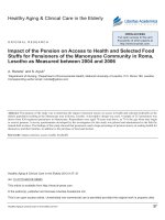
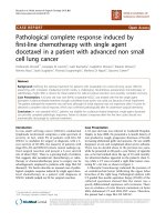
100 and 83% respectively in the VD deficient group, and
99 and 85% respectively in the VD sufficient group. No
difference was seen in terms of survival between these
two subgroups (p = 0.3, Fig. 1). Five year-OS was 89%
in patients with clinical stage I or II, compared to
72% for stage III. The difference was statistically significant (p < 0.01). There was a significant correlation
between survival and pCR. Five year-OS for patients
not obtaining pCR was 79% (95% CI 0.73–0.84), compared to 94% (95% CI 0.87–0.98) for those who obtained pCR (p = 0.0007). Ninety-one percent (95% CI
0.82–0.95) of patients with HER2+ tumors were alive
at 5 years, while 92% (95% CI 0.86–0.96) for the HR
+/HER2- subgroup, and 65% (95% CI 0.53–0.74) in the
TNBC group. The tumor subtypes constitute an independent and significant factor for survival (p = 0.00001,
Table 4).
In a multivariate analysis, clinical stage (p = 0.001), TN
subgroup (p = 0.0001) and pCR (p = 0.001) were the only
variables statistically correlated with OS (Table 5).
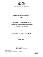
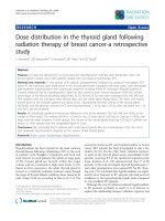
After a median follow up of 5.3 years, median PFS was
not reached. Five year-PFS was 78% (95% CI 0.73–0.83)
in our global cohort. Five year-PFS rate was 76% in the
VD deficient subgroup, whereas 80% in the VD sufficient
group. The difference did not achieve statistical significance (p = 0.2, Fig. 2). Clinical stage (84% 5-year-PFS for
stages I-II and 62% for stage III) (p = 0.00001), TNBC
subtype (62% 5-years-PFS, p = 0.00001), and pathological
response (72% 5- year-PFS for patients not achieving
pCR, versus 92% for the pCR group, p = 0.0002) were
significantly correlated with PFS. Other factors as histopathologic grade (p = 0.3), and age (p = 0.1) did not appear as significant factors correlated with pCR (Table 6).
In a multivariate analysis, clinical stage (p = 0.001),
TNBC subtype (p < 0.01) and pCR (p < 0.01) were the
only variables significantly associated with PFS (Table 7).
< 50
≥ 50
Clinical stage
I-II
III
0.34
0.2–0.6
0.0001
1.19
0.7–1.9
0.5
1.6
0.7–3.8
0.2
1.0
0.5–2.3
0.9
0.43
0.2–0.8
0.01
Histological grade (SBR)
II
III
Tumor subtypes
HER2+
HR+/Her2TNBC
VD level
< 20 ng/mL
≥ 20 ng/mL
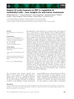
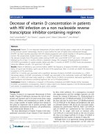
Vitamin D and survival by tumor subtypes
Regarding OS, we found no statistical difference in the
5-year survival rate for patients with HER2+ (p = 0.3)
and HR+/HER2- (p = 0.8) tumors, depending on their
VD level at diagnosis (Fig. 3a, b). Regarding the TNBC
subgroup, 5-year-OS was 59% (95% CI 0.4–0.7) in the
VD deficient group versus 70% (95% CI 0.5–0.8) in the
VD sufficient group. This trend was not statistically significant (p = 0.2, Fig. 3c).
We analyzed PFS depending on VD level and tumor
subtypes. The 5-year-PFS was of 92 and 79% in the VD
deficient and the VD sufficient group respectively for patients with HER2+ tumors (p = 0.20). Regarding the HR
+/HER2- cohort, 5-year-PFS rates were 78 and 89% respectively, this difference was approached statistical significance (p = 0.056), Fig. 4). Finally, a non-statistically
Viala et al. BMC Cancer (2018) 18:770
Page 5 of 11
Fig. 1 OS by Vitamin D level
significant trend was observed in the TNBC subgroup
(60.4% vs 72.3% respectively, p = 0.3, Fig. 5).
Survival and pCR depending on the profile subgroup
We evaluated the 5-year-OS of our cohort depending on
the NAC response and their tumor subtypes. No significant difference in terms of OS was seen in the HER2+
and HR+/HER2- subgroup. Nevertheless, in the TNBC
subgroup, the 5-year-OS was statistically significant
(93% for patients obtaining pCR, versus 47% for
non-pCR cases, p < 0.0001). Neoadjuvant chemotherapy
Table 4 Correlation between OS and clinical-pathological data
in a univariate analysis
5 years-OS (%)
95%CI
Age
response appeared as a strong and independent prognostic factor of survival in the TNBC subgroup (Fig. 6a).
Regarding PFS, 5-year-PFS rate was 77% versus 90%
in the non pCR and pCR group respectively in the
HER2+ subgroup (p = 0.03). In the HR+/HER2- cohort, 5-year-PFS rate was of 81% versus 100% in the
non pCR and pCR group respectively (p = 0.03).
Finally, in the TNBC subtype, 5-years-PFS rate for
women not achieving a pCR was 46% while it was 87%
Table 5 Correlation between OS and clinical-pathological data
in a multivariate analysis
p
Range (26–74)
0.2
Median: 49.5
< 50
86
0.79–0.91
< 50
≥ 50
82
0.76–0.88
≥ 50
< 20 ng/mL
82%
0.75–0.88
≥ 20 ng/mL
85%
0.79–0.9
0.3
VD level
≥ 20 ng/mL
89%
0.84–0.93
I-II
72%
0.61–0.80
III
no
79%
0.73–0.84
yes
94%
0.86–0.98
0.0007
90%
0.82–0.95
HR+/Her2-
92%
0.86–0.96
TNBC
65%
0.53–0.74
0.5
1.03
0.6–1.8
0.9
2.8
1.6–5.0
0.001
1.77
0.8–4.1
0.1
6.5
3.1–13.7
0.0001
0.2
0.09–0.5
0.001
0.86
0.5–1.6
0.6
HR+/HER2TNBC
pCR
no
yes
0.4
SBR grade
0.7–2.3
Tumor subtypes
HER2+
0.00001
HER2+
1.2
Clinical stage
III
Tumor subtypes
p
VD level
I-II
pCR
95%CI
< 20 ng/mL
0.00001
Clinical stage
HR
Age (years)
SBR grade
II
86%
0.79–0.91
II
III
83%
0.76–0.88
III
Viala et al. BMC Cancer (2018) 18:770
Page 6 of 11
Fig. 2 PFS by Vitamin D level in the full cohort
for those achieving pCR (p = 0.0009, Fig. 6b). Pathological complete response appears as a strong and independent prognostic factor of survival, especially in
the TNBC subgroup.
Discussion
We performed a retrospective, observational, multicenter study which included 327 breast cancer patients
treated by NAC. We evaluated specifically their VD level
at the beginning of NAC and its impact on pCR and
survival. Notably, we did not have post-NAC serial evaluations of VD levels during the 5-years follow-up.
Breast cancer patients are more frequently affected by
a VD deficiency than the general population. Seventy to
80% of these patients have VD level below the lower
limit of normal at breast cancer diagnosis, and that proportion even increases during NAC [13, 14, 19]. Our
study confirms that patients treated by NAC frequently
have deficient VD level. In fact, almost half of our cohort (42%) had baseline VD level below 20 ng/mL. Our
population appears less deficient than that reported in
Table 6 Correlation between PFS and clinical-pathological data
in a univariate analysis
Table 7 Correlation between PFS and clinical-pathological data
in a multivariate analysis
5 years-PFS (%)
95%CI
p
0.1
Age
< 50
82
0.75–0.88
< 50
≥ 50
75
0.67–0.81
≥ 50
< 20 ng/mL
76
0.67–0.82
≥ 20 ng/mL
80
0.73–0.85
0.2
VD level
≥ 20 ng/mL
84
0.78–0.89
I-II
62
0.51–0.72
III
no
72
0.65–0.78
yes
92
0.84–0.96
0.0002
84
0.74–0.90
HR+/HER2-
84
0.77–0.90
TNBC
62
0.51–0.72
0.2
0.9
0.6–1.5
0.8
2.4
1.4–3.9
0.001
1.6
0.30–1.21
0.2
4.3
1.42–4.80
0.002
0.25
0.12–0.50
0.0001
0.94
0.52–1.70
0.8
HR+/HER2TNBC
pCR
no
yes
0.3
SBR grade
0.84–2.3
Tumor subtypes
HER2+
0.00001
HER2+
1.4
Clinical stage
III
Tumor subtypes
p
VD level
I-II
pCR
95%CI
< 20 ng/mL
0.00001
Clinical stage
HR
Age
SBR grade
II
79
0.71–0.85
II
III
78
0.71–0.84
III
Viala et al. BMC Cancer (2018) 18:770
Page 7 of 11
Fig. 3 a OS depending on the Vitamin D level in the HER2+ tumor subtype. b OS depending on Vitamin D level in the HR+/HER2- tumors
subtypes. c OS depending on the Vitamin D level in the TN tumor subtypes
other series (74–80% VD deficiency rate) [14, 19], however the deficiency rate is highly dependent of geographic and lifestyle variables [20]. The TNBC subtype
appears to be the most affected subgroup. This result is
consistent with the report published by Yao et al. [21].
Considering the VD implication in the tumorigenesis
process (proliferation, apoptosis, and angiogenesis), it
could be hypothesized that this deficiency might have a
clinical impact on tumor response to treatment.
Few studies have evaluated the association between VD
and pCR. Most of these studies did not show a significant
correlation between these two factors. In the NEOZOTAC
trial, a large proportion of patients were affected with low
VD level at diagnosis, and even lower VD levels at the end
of NAC. No correlation was seen between VD level and
pCR, nevertheless, patients with sufficient VD level had a
better pathological response than the others, even if this result did not achieved statistical significance [22]. Clark et al.
Viala et al. BMC Cancer (2018) 18:770
Page 8 of 11
Fig. 4 PFS depending on the VD level in the HR+/HER2- tumor subtype
studied, in a smaller trial, the relationship between VD and
chemotherapy response. Once again, no correlation was
found, but one explanation can be linked to the absence of
HER2+ patients in this study [23]. Indeed, this subgroup of
patients are the one responding the most frequently to
chemotherapy, with the higher pCR rate, especially since
the addition of trastuzumab and other HER2-directed therapies [2]. The lack of HER2+ patients in the study by Clark
et al, limits interpretation of these results.
Our study confirms the significant correlation between
VD level and pCR. Lower VD level significantly decreases the probability of attaining pCR. These data are
consistent with our previous study [16], and validated in
this expanded cohort. This results may be explained by
the potential effect of VD on chemotherapeutic agents
such as taxanes and anthracyclines, both of which form
the backbone of breast cancer treatment [11, 24].
Tumor subtypes, histological grade and clinical stage,
as expected were also associated with pCR and were
found to be independent predictive factors of pCR in
our population [25].
Fig. 5 PFS depending on the Vitamin D level in the TN tumor subtypes
In our study, pCR was achieved in 32.4% of patients,
which is higher than in the meta-analyses previously reported [2, 3, 26] (16–22% pCR rates). However, this difference may be considered altogether with the respective
proportions of the biological subgroups. Additionally, our
cohort is more recent than the Cortazar study, and likely
benefit from improved systemic therapies, such as
anti-HER2 targeted therapies and the more wide-spread
use of taxanes. Consistent with previously reported literature, pCR was attained more frequently in the HER2
+/HR- (60%) subtype (40% for the HR+/HER2+ one),
followed by the TNBC subtype (33%) and finally the HR
+/HER2- (21%) subtype.
In our cohort we observed a good prognosis, with a
median PFS and OS not reached after a median
5.3 years of follow up. In the meta-analysis by Cortazar et al., pCR was suggested as a surrogate endpoint
due to its correlation with survival, achieving pCR being associated with an improved survival, and a decrease risk of recurrence [2, 3]. In our study, pCR
and survival are strongly associated, confirming its
Viala et al. BMC Cancer (2018) 18:770
Page 9 of 11
Fig. 6 a OS depending on the pathological response in the different tumors subtypes. b PFS depending on pathological response in the
different tumor subtypes
role as a prognostic factor, but with variable magnitude depending on tumor subtypes at this early
follow-up time-point.
In the population not achieving pCR, the HR+/HER2subgroup experienced the best prognosis, followed by
HER2+ then TNBC patients. Nevertheless, for patients
achieving pCR, no statistical difference was seen in the different subgroups. Pathological complete response appears
as a strong prognostic factor in the TNBC subgroup. The
initial general poor prognosis of this subtype is altered for
patients achieving pCR (5 years-OS 93% versus 47%), as it
has been initially reported by Liedtke et al. [7].
Other studies found more frequent deficiency of VD in
this subgroup [21, 27]. In our study, no correlation was
found between VD level and survival in this subgroup, however it appears to be a trend for a better
survival in the VD sufficient group (5-year-OS of 60%
in the VD deficient group versus 70% in the normal
VD level one, p = 0.2, Fig. 3c; (5-year-PFS of 60.4%
versus 72.3% in the low and normal VD level group
respectively, p = 0.3, Fig. 4). Similar trend was seen in
the study by Al-Azhri et al. [10]. This lack of statistical significance could be explained by the relatively
small number of patients in our TNBC cohort. In the
same article, Al-Azhri et al demonstrated that TNBC
was mostly associated with a low level of VD receptor
(VDR), due to a down regulation mechanism. VDR
functionality is necessary for VD mediated anti-cancer
activity. Indeed, in vitro, the reintroduction of VDR
restored the anti-proliferative action of VD [10].
Thus, it is possible that appropriate VD levels are of
greater impact in VDR functional tumors.
In addition, our analysis showed a near-significant correlation between VD level and PFS in the HR+/HER2subgroup. It is likely that with further follow-up this
finding will achieve significance at the 5% level. Some
Viala et al. BMC Cancer (2018) 18:770
meta-analyses previously confirmed a positive association between sufficient VD level and better survival,
nevertheless, no specific data was specifically available
for the HR+/HER2- subgroup [28–30]. One way to explain this link could be based on the discovery of new
pathways associated with VD, modulating the activity of
HR+ breast cancer cells. Indeed, Krishnan et al, showed
on in vitro and in vivo models that VD might decrease
the expression of aromatase, and so decrease the
synthesis of estrogen [31]. Thus the inhibition of estrogen synthesis and signaling by calcitriol, and its
anti-inflammatory actions may play an important role in
inhibiting HR+ breast cancer.
Conclusion
In our retrospective observational study, VD level appears
correlated with pCR in breast cancer patients treated with
NAC. Pathological complete response is a validated,
strong and independent prognostic factor of survival, especially in the TNBC population. No significant correlation was yet seen between VD level and overall survival.
Nevertheless, a trend was seen in PFS in the HR+/HERsubgroup and in OS in the TNBC subgroup. Considering
the natural history of the different breast cancer subgroups, the actualization of survival with a longer
follow-up will allow the evaluation of the presence of similar correlations in the other breast cancer subtypes. Further studies are warranted in a larger cohort population in
order to evaluate the link between VD level and survival.
An interventional prospective study in this population to
analyze the impact of VD supplementation on pCR and
survival, eventually stratified by tumoral VDR expression
would be warranted. Notably, this intervention is highly
actionable and relatively inexpensive which could offer an
opportunity for an easily applicable and value-based improvement in breast cancer outcomes.
Additional files
Additional file 1: pCR rate depending on the HER2+ subtypes. (DOCX 13 kb)
Additional file 2: pCR rate depending on the VD level at baseline in the
two HER2+ subgroups: a HR+/HER2+. b HR-/HER2+. (DOCX 15 kb)
Abbreviations
AJCC: American joint committee on Cancer; CT: Chemotherapy; ER: Estrogen
receptor; HER2: Human epidermal receptor 2; HR: Hormone receptor;
IHC: Immunohistochemistry; LABC: Locally advanced breast cancer;
NAC: Neoadjuvant chemotherapy; OS: Overall survival; pCR: Pathological
complete response; PFS: Progression-free-survival; PR: Progesterone receptor;
SBR: Scarff, Bloom and Richardson; TNBC: Triple negative breast cancer;
VD: Vitamin D
Funding
This study was funding through the GELFUC (Groupement des Entreprises
Françaises dans la Lutte contre le Cancer) Languedoc-Roussillon. None of the
funding sources were involved in the design of the study, nor the collection,
analysis and interpretation of data nor the writing of the manuscript.
Page 10 of 11
Availability of data and materials
The datasets used and/or analysed during the current study are available
from the corresponding author on reasonable request.
Authors’ contributions
MV was involved in the conception of the study, acquisition and analysis of
the data, and wrote the first draft of the manuscript. LD was involved in the
acquisition of the data. WJ was involved in the conception and design of
the study. MV, WJ, ST contributed to data analysis and interpretation of data.
WJ, AC, AT, SM critically revised the manuscript for important intellectual
content. PJL and MS participated in analyzing the results and drafting the
manuscript. All authors read and approved the final manuscript.
Ethics approval and consent to participate
This study was reviewed and approved by the Montpellier Cancer Institute
Institutional Review Board (ICM-CORT-2016-25). Considering the retrospective,
non-interventional nature of this study, no specific consent was deemed
necessary by the clinical research review board of the Montpellier Cancer
Institute Internal and according to the French regulation.
Consent for publication
Not applicable.
Competing interests
The authors declare that they have no competing interests.
Publisher’s Note
Springer Nature remains neutral with regard to jurisdictional claims in
published maps and institutional affiliations.
Author details
1
Department of Medical Oncology, Institut Régional Du Cancer de
Montpellier ICM, 208 Avenue des Apothicaires, Cedex-5 34298 Montpellier,
France. 2Division of Surgical Oncology, Department of Surgery, Wake Forest
University School of Medicine, Winston-Salem, USA. 3Biometry unit, Institut
Régional Du Cancer de Montpellier ICM, Montpellier, France. 4Department of
Surgical Oncology, Institut Régional Du Cancer de Montpellier ICM,
Montpellier, France. 5Imagenome-labosud, Clinique BeauSoleil, Montpellier,
France. 6Holden Comprehensive Cancer Center, University of Iowa, Iowa City,
USA. 7College of Pharmacy, University of Iowa, Iowa City, USA. 8Department
of Internal Medicine Wake Forest University School of Medicine,
Winston-Salem, USA.
Received: 23 January 2018 Accepted: 22 July 2018
References
1. Mieog JSD, van der Hage JA, van de Velde CJH. Preoperative chemotherapy
for women with operable breast cancer. Cochrane Database Syst Rev. 2007:
CD005002. />2. Cortazar P, Zhang L, Untch M, et al. Pathological complete response and
long-term clinical benefit in breast cancer: the CTNeoBC pooled analysis.
Lancet Lond Engl. 2014;384:164–72. />3. von Minckwitz G, Untch M, Blohmer J-U, et al. Definition and impact of
pathologic complete response on prognosis after neoadjuvant chemotherapy
in various intrinsic breast cancer subtypes. J Clin Oncol Off J Am Soc Clin
Oncol. 2012;30:1796–804. />4. Gianni L, Pienkowski T, Im Y-H, et al. Efficacy and safety of neoadjuvant
pertuzumab and trastuzumab in women with locally advanced,
inflammatory, or early HER2-positive breast cancer (NeoSphere): a
randomised multicentre, open-label, phase 2 trial. Lancet Oncol. 2012;13:25–
32. />5. Wu K, Yang Q, Liu Y, et al. Meta-analysis on the association between
pathologic complete response and triple-negative breast cancer after
neoadjuvant chemotherapy. World J Surg Oncol. 2014;12:95. />10.1186/1477-7819-12-95.
6. Barton MK. Bevacizumab in neoadjuvant chemotherapy increases the
pathological complete response rate in patients with triple-negative breast
cancer. CA Cancer J Clin. 2014;64:155–6. />
Viala et al. BMC Cancer (2018) 18:770
7.
8.
9.
10.
11.
12.
13.
14.
15.
16.
17.
18.
19.
20.
21.
22.
23.
24.
25.
Liedtke C, Mazouni C, Hess KR, et al. Response to neoadjuvant therapy and
long-term survival in patients with triple-negative breast cancer. J Clin
Oncol Off J Am Soc Clin Oncol. 2008;26:1275–81. />JCO.2007.14.4147.
Cortazar P, Geyer CE. Pathological complete response in neoadjuvant
treatment of breast cancer. Ann Surg Oncol. 2015;22:1441–6. />10.1245/s10434-015-4404-8.
Verlinden L, Verstuyf A, Convents R, et al. Action of 1,25(OH)2D3 on the cell
cycle genes, cyclin D1, p21 and p27 in MCF-7 cells. Mol Cell Endocrinol.
1998;142:57–65.
Al-Azhri J, Zhang Y, Bshara W, et al. Tumor expression of vitamin D receptor
and breast Cancer histopathological characteristics and prognosis. Clin
Cancer Res. 2017;23:97–103. />Hershberger PA, Yu WD, Modzelewski RA, et al. Calcitriol (1,25dihydroxycholecalciferol) enhances paclitaxel antitumor activity in vitro and
in vivo and accelerates paclitaxel-induced apoptosis. Clin Cancer Res Off J
Am Assoc Cancer Res. 2001;7:1043–51.
Light BW, Yu WD, McElwain MC, et al. Potentiation of cisplatin antitumor
activity using a vitamin D analogue in a murine squamous cell carcinoma
model system. Cancer Res. 1997;57:3759–64.
Goodwin PJ, Ennis M, Pritchard KI, et al. Prognostic effects of 25hydroxyvitamin D levels in early breast cancer. J Clin Oncol Off J Am Soc
Clin Oncol. 2009;27:3757–63. />Jacot W, Pouderoux S, Thezenas S, et al. Increased prevalence of vitamin D
insufficiency in patients with breast cancer after neoadjuvant
chemotherapy. Breast Cancer Res Treat. 2012;134:709–17. />1007/s10549-012-2084-7.
Jacot W, Firmin N, Roca L, et al. Impact of a tailored oral vitamin D
supplementation regimen on serum 25-hydroxyvitamin D levels in early
breast cancer patients: a randomized phase III study. Ann Oncol Off J Eur
Soc Med Oncol. 2016;27:1235–41. />Chiba A, Raman R, Thomas A, et al. Serum vitamin D levels affect pathologic
complete response in patients undergoing neoadjuvant systemic therapy
for operable breast Cancer. Clin Breast Cancer. 2018;18:144–9. https://doi.
org/10.1016/j.clbc.2017.12.001.
Wolff AC, Hammond MEH, Hicks DG, et al. Recommendations for human
epidermal growth factor receptor 2 testing in breast cancer: American
Society of Clinical Oncology/College of American Pathologists clinical
practice guideline update. J Clin Oncol Off J Am Soc Clin Oncol. 2013;31:
3997–4013. />Sataloff DM, Mason BA, Prestipino AJ, et al. Pathologic response to
induction chemotherapy in locally advanced carcinoma of the breast: a
determinant of outcome. J Am Coll Surg. 1995;180:297–306.
Crew KD, Shane E, Cremers S, et al. High prevalence of vitamin D deficiency
despite supplementation in premenopausal women with breast cancer
undergoing adjuvant chemotherapy. J Clin Oncol Off J Am Soc Clin Oncol.
2009;27:2151–6. />Zgaga L, Theodoratou E, Farrington SM, et al. Diet, environmental factors, and
lifestyle underlie the high prevalence of vitamin D deficiency in healthy adults
in Scotland, and supplementation reduces the proportion that are severely
deficient. J Nutr. 2011;141:1535–42. />Yao S, Sucheston LE, Millen AE, et al. Pretreatment serum concentrations of
25-hydroxyvitamin D and breast cancer prognostic characteristics: a casecontrol and a case-series study. PLoS One. 2011;6:e17251. />1371/journal.pone.0017251.
Charehbili A, Hamdy NA, VTHBM S, et al. Vitamin D (25-0H D3) status and
pathological response to neoadjuvant chemotherapy in stage II/III breast
cancer: data from the NEOZOTAC trial (BOOG 10-01). Breast Edinb Scotl.
2016;25:69–74. />Clark AS, Chen J, Kapoor S, et al. Pretreatment vitamin D level and response
to neoadjuvant chemotherapy in women with breast cancer on the I-SPY
trial (CALGB 150007/150015/ACRIN6657). Cancer Med. 2014;3:693–701.
/>Chaudhry M, Sundaram S, Gennings C, et al. The vitamin D3 analog, ILX-237553, enhances the response to adriamycin and irradiation in MCF-7 breast
tumor cells. Cancer Chemother Pharmacol. 2001;47:429–36.
Alvarado-Cabrero I, Alderete-Vázquez G, Quintal-Ramírez M, et al. Incidence of
pathologic complete response in women treated with preoperative
chemotherapy for locally advanced breast cancer: correlation of histology,
hormone receptor status, Her2/Neu, and gross pathologic findings. Ann Diagn
Pathol. 2009;13:151–7. />
Page 11 of 11
26. Berruti A, Amoroso V, Gallo F, et al. Pathologic complete response as a
potential surrogate for the clinical outcome in patients with breast cancer
after neoadjuvant therapy: a meta-regression of 29 randomized prospective
studies. J Clin Oncol Off J Am Soc Clin Oncol. 2014;32:3883–91. https://doi.
org/10.1200/JCO.2014.55.2836.
27. Rainville C, Khan Y, Tisman G. Triple negative breast cancer patients
presenting with low serum vitamin D levels: a case series. Cases J. 2009;2:
8390. />28. Hu K, Callen DF, Li J, Zheng H. Circulating vitamin D and overall survival in
breast Cancer patients: a dose-response meta-analysis of cohort studies.
Integr Cancer Ther. 2017; />29. Mohr SB, Gorham ED, Kim J, et al. Meta-analysis of vitamin D sufficiency for
improving survival of patients with breast cancer. Anticancer Res. 2014;34:1163–6.
30. Vrieling A, Hein R, Abbas S, et al. Serum 25-hydroxyvitamin D and
postmenopausal breast cancer survival: a prospective patient cohort study.
Breast Cancer Res BCR. 2011;13:R74. />31. Krishnan AV, Swami S, Feldman D. The potential therapeutic benefits of
vitamin D in the treatment of estrogen receptor positive breast cancer.
Steroids. 2012;77:1107–12. />