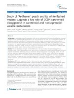
A suppressive role of guanine nucleotidebinding protein subunit beta-4 inhibited by DNA methylation in the growth of antiestrogen resistant breast cancer cells
Bạn đang xem bản rút gọn của tài liệu. Xem và tải ngay bản đầy đủ của tài liệu tại đây (1.96 MB, 13 trang )
Wang et al. BMC Cancer (2018) 18:817
/>
RESEARCH ARTICLE
Open Access
A suppressive role of guanine nucleotidebinding protein subunit beta-4 inhibited by
DNA methylation in the growth of antiestrogen resistant breast cancer cells
Bo Wang1,2, Dongping Li1,2, Rocio Rodriguez-Juarez1, Allison Farfus1, Quinn Storozynsky1, Megan Malach1,
Emily Carpenter1, Jody Filkowski1, Anne E. Lykkesfeldt3 and Olga Kovalchuk1,4*
Abstract
Background: Breast cancer is the most common malignancy in women worldwide. Although the endocrine therapy
that targets estrogen receptor α (ERα) signaling has been well established as an effective adjuvant treatment for
patients with ERα-positive breast cancers, long-term exposure may eventually lead to the development of acquired
resistance to the anti-estrogen drugs, such as fulvestrant and tamoxifen. A better understanding of the mechanisms
underlying antiestrogen resistance and identification of the key molecules involved may help in overcoming
antiestrogen resistance in breast cancer.
Methods: The whole-genome gene expression and DNA methylation profilings were performed using fulvestrantresistant cell line 182R-6 and tamoxifen-resistant cell line TAMR-1 as a model system. In addition, qRT-PCR and Western
blot analysis were performed to determine the levels of mRNA and protein molecules. MTT, apoptosis and cell cycle
analyses were performed to examine the effect of either guanine nucleotide-binding protein beta-4 (GNB4)
overexpression or knockdown on cell proliferation, apoptosis and cell cycle.
Results: Among 9 candidate genes, GNB4 was identified and validated by qRT-PCR as a potential target silenced
by DNA methylation via DNA methyltransferase 3B (DNMT3B). We generated stable 182R-6 and TAMR-1 cell lines
that are constantly expressing GNB4 and determined the effect of the ectopic GNB4 on cell proliferation, cell cycle, and
apoptosis of the antiestrogen-resistant cells in response to either fulvestrant or tamoxifen. Ectopic expression of GNB4
in two antiestrogen resistant cell lines significantly promoted cell growth and shortened cell cycle in the presence of
either fulvestrant or tamoxifen. The ectopic GNB4 induced apoptosis in 182R-6 cells, whereas it inhibited apoptosis in
TAMR-1 cells. Many regulators controlling cell cycle and apoptosis were aberrantly expressed in two resistant cell lines
in response to the enforced GNB4 expression, which may contribute to GNB4-mediated biologic and/or pathologic
processes. Furthermore, knockdown of GNB4 decreased growth of both antiestrogen resistant and sensitive breast
cancer cells.
Conclusion: GNB4 is important for growth of breast cancer cells and a potential target for treatment.
Keywords: Antiestrogen resistance, Breast cancer, DNA methylation, Fulvestrant, GNB4, Tamoxifen
* Correspondence:
1
Department of Biological Sciences, University of Lethbridge, Lethbridge, AB,
Canada
4
Hepler Hall, University of Lethbridge, 4401 University Drive, Lethbridge, AB
T1K 3M4, Canada
Full list of author information is available at the end of the article
© The Author(s). 2018 Open Access This article is distributed under the terms of the Creative Commons Attribution 4.0
International License ( which permits unrestricted use, distribution, and
reproduction in any medium, provided you give appropriate credit to the original author(s) and the source, provide a link to
the Creative Commons license, and indicate if changes were made. The Creative Commons Public Domain Dedication waiver
( applies to the data made available in this article, unless otherwise stated.
Wang et al. BMC Cancer (2018) 18:817
Background
Breast cancer is the most common malignancy found in
women worldwide and the second leading cause of
cancer-related deaths among North American women
[1]. Globally, it is estimated that 1.67 million new cases
were diagnosed, and 522,000 women died from this disease in 2012 (GLOBOCAN 2012). Approximately 60% of
these deaths were contributed by less developed countries.
Although the exact etiology of breast cancer is currently
unknown, the estrogen/estrogen-receptor (ER) signaling
may play a crucial role in the development of this disease
[2–5]. Furthermore, sustained estrogenic exposure may
also increase the risk of breast, ovarian, and endometrial
cancers [6].
Estrogen receptor alpha (ERα) and beta (ERβ), two
ligand-inducible transcription factors, are members of
the steroid/thyroid receptor superfamily that primarily
mediate estrogen’s biological function through binding
[7]. ERα and ERβ are encoded by either ESR1 or ESR2
genes that are composed of 595 and 530 amino acids,
respectively. Both eventually form an N-terminal domain
(NTD), a DNA-binding domain (DBD), and a ligandbinding domain (LBD) [8]. Sequence analysis indicates
that ERα and ERβ display ~ 97% similarity in the DBD
and 59% in the LBD, whereas they display only 16%
similarity in the NTD [8]. This implicates a functional
similarity and difference between these two ERs. For
instance, once activated via estrogen binding, both
dimerized ERs can either bind to the estrogen-response
element (ERE) in the DNA or interplay with other transcription factors, such as AP1, Sp1, and NF-κB [9], eventually influencing the transcription of genes. However, ERα
may predominantly bind to ERE elements [10], while ERβ
may primarily interact with AP1 sites [11]. Furthermore, as
demonstrated, ERα is a key player in promoting cell growth
and proliferation [12, 13], whereas ERβ plays an important
role in anti-proliferation, differentiation, and apoptosis in
human malignancies, including breast cancer [14, 15].
Because ERα is expressed in 70% of breast cancers [16],
and the proliferation of these ERα-positive breast cancers
is largely dependent on estrogen/ERα signaling [17], the
endocrine therapy that targets estrogen/ERα signaling has
been well established as an effective adjuvant treatment
for patients with ERα-positive breast cancers [18]. The
endocrine-therapy agents that are currently used for
ERα-positive breast cancer include fulvestrant (also known
as ICI 182,780 and faslodex, the ER downregulator that
selectively downregulates and/or degrades ERα), tamoxifen (the ER modulator that selectively antagonizes ERα
function), and aromatase inhibitors (e.g. letrozole and anastrozole, which inhibit estrogen production by attenuating
aromatase activity) [17, 19]. As an important adjuvant
therapy, continuing 10-year tamoxifen treatment, when
compared with 5-year exposure, has been shown to further
Page 2 of 13
reduce the risk of disease recurrence and mortality in a
randomized trial of women with ER-positive breast cancers
[20]. Unfortunately, long-term exposure may eventually
lead to the development of acquired resistance to these
drugs [21–23], which is a serious clinical problem in
hormonal therapy. However, the underlying mechanisms
are not completely understood.
In this study, we globally analyzed genomic DNA
methylation, correlated with gene expression profiling, and
identified GNB4 that was silenced by DNMT3B-mediated
DNA methylation in both fulvestrant-resistant (MCF-7/
182R-6) and tamoxifen-resistant (MCF-7/TAMR-1) breast
cancer cell lines. Ectopic expression of GNB4 enhanced
proliferation of MCF-7/182R-6 and MCF-7/TAMR-1 cell
lines in response to either fulvestrant or tamoxifen, while it
shortened G2 and S phases in the cell cycle. We also noted
that the ectopic expression of GNB4 induced apoptosis in
the MCF-7/182R-6 cell line, whereas it attenuated the
induction of apoptosis in the MCF-7/TAMR-1 cell line.
Cell-cycle and apoptosis regulators were aberrantly
expressed in these cell lines in response to the ectopic
GNB4 expression. In contrast, siRNA-mediated knockdown of GNB4 inhibited proliferation of two resistant
cell lines in the presence of either fulvestrant or tamoxifen, and induced either S phase arrest or apoptosis.
Our results provide novel insight into the role of GNB4 in
the growth of both antiestrogen-resistant and sensitive
breast cancer cells and may represent a target for treatment
of breast cancer.
Methods
Cell culture
The MCF-7/S0.5 (S05), MCF-7/182R-6 (182R-6), and
MCF-7/TAMR-1 (TAMR-1) cell sublines were developed
by Dr. Anne Lykkesfeldt (Breast Cancer Group, Cell Death
and Metabolism, Danish Cancer Society Research Center,
DK-2100, Copenhagen, Denmark). ICI 182,780 (Faslodex,
fulvestrant) and tamoxifen-resistant sublines, 182R-6 and
TAMR-1, respectively, are derived from S05 as described
elsewhere [24, 25]. These cell lines were cultured in a
DMEM/F-12 medium with 2.5 mM L-Glutamine, without
HEPES and phenol red (HyClone), and supplemented
with 1% heat-inactivated fetal bovine serum (HyClone).
Additionally, for 182R-6 and TAMR-1 sublines were
regularly supplemented with 0.1 μM ICI 182,780 and
1 μM tamoxifen, respectively. Human mammary epithelial
cells (HMEC) purchased from ThermoFisher Scientific
(Cat# A10565) were cultured in a HuMEC basal
serum-free medium (ThermoFisher Scientific) containing HuMEC supplement (ThermoFisher Scientific),
100 IU/mL penicillin, and 100 mg/mL streptomycin.
All cell lines were incubated at 37 °C in a humidified
atmosphere of 5% CO2.
Wang et al. BMC Cancer (2018) 18:817
Whole-genome gene expression profiling
Total RNA was isolated from S05, 182R-6, and TAMR-1
cells using an Illustra RNAspin mini kit according to the
manufacturer’s instructions (GE Healthcare Life Sciences).
Quantification, purity, and integrity of the RNA samples
were measured using a NanoDrop 2000c spectrophotometer (Thermo Scientific) and an Agilent 2100 bioanalyzer
(Santa Clara). RNA samples with RIN values of seven or
higher were used for further analysis. Procedures for library preparation, hybridization, detection, BeadChip statistical analysis, and data processing have been described
previously [19]. Heatmaps were generated by Dr. Yaroslav
Ilnytskyy for genes that were differentially expressed
between any of the groups (ANOVA type analysis with
p.adjusted < 0.001) and for top 1000 most variable probes
in DNA methylation.
Whole-genome DNA methylation profiling
DNA was extracted from cells using the DNeasy Blood
and Tissue Kit (QIAGEN) and treated with DNase-free
RNase (Sigma) according to the manufacturer’s protocols.
The collected DNA was bisulfite converted using the EZ
DNA Methylation Kit (Zymo Research) according to the
manufacturer’s protocols. Methylation was measured
using the Infinium assay on the Illumina platform. Data
was collected from the > 27,000 probes represented on the
HM27 microarray. These probes contain CpG dinucleotides from selected loci throughout the genome. All
steps were carried out according to the manufacturer’s
specifications and with Illumina-supplied reagents. Briefly,
bisulfate-converted samples were amplified overnight,
fragmented, and purified. The re-suspended samples were
hybridized overnight to the microarray, which harboured
millions of bead-bound 50-mer oligos. Each interrogated
loci is represented by two bead types: a methylated type
(“C” remains a “C”) and an unmethylated type (“C”
become a “T”). Hybridized chips were washed to remove
unbound and/or non-specific DNA fragments. The resulting
oligo-sample hybrid was then extracted with a biotin-linked
dideoxy cytosine and stained with streptavidin. The relative
intensity of the unmethylated bead to the methylated
bead for each allele provides a measure of relative
methylation levels.
Page 3 of 13
5′-GAA GGC TGG GGC TCA TTT-3′, and for the
reverse primer, 5’-CAG GAG GCA TTG CTG ATG AT-3′
[26]. All qRT-PCR experiments were performed in triplicate, the data was analyzed using the comparative Ct
method, and the results are shown as a fold induction of
mRNA.
Knockdown of DNMT3B
182R-6 and TAMR-1 cells grown to 80% confluency were
transiently transfected with either 200 nM Dnmt3b
siRNA (Santa Cruz Biotechnologies) or 200 nM AllStars
Negative Control siRNA (QIAGEN) using Lipofectamine
3000 (Invitrogen) according to the manufacturer’s instructions. Seventy-two hours after transfection, the total RNA
was isolated and subjected to qRT-PCR analysis using a
primer set specifically to GNB4 (Bio-Rad) according to
the manufacturer’s instructions, and the whole cellular
lysates were prepared and subjected to Western blot
analysis using antibodies against DNMT3B and GNB4.
Generation of GNB4 expression construct and GNB4
stable-expression cell lines
The coding sequence of GNB4 was amplified by RT-PCR
using total RNA isolated from HMEC. The PCR product
was then cloned into a pGEM-T easy vector (Promega),
released by digestion with EcoR I and BamH I, and
subcloned into a pEGFP-C1 vector (CloneTech) to
generate pEGFP-GNB4. Sequence identity was confirmed by automatic sequencing. Primers used here for
amplifying the GNB4 coding sequence are as follows:
GNB4-F (5′-GAG AAT TCT ATG AGC GAA CTG GAA
C-3′) and GNB4-R (5′-GGG GAT CCA TTC CAG ATT
CTA AG-3′).
182R-6 and TAMR-1 cells grown to 80% confluency
were transfected with either pEGFP-GNB4 or pEGFP-C1
using Lipofectamine 3000 (Invitrogen). 24 h after transfection, G418 was added to final concentrations of
800 μg/mL and 400 μg/mL for 182R-6 and TAMR-1 cell
lines, respectively, to kill the negative cells. The positive
cells stably expressing either GFP or GFP-GNB4 were further selected with cell sorting (University of Calgary).
MTT assay
Quantitative real-time RT-PCR (qRT-PCR)
Total RNA, isolated from the indicated cell lines with
TRIzol reagent (Invitrogen), was subjected to qRT-PCR
using an iScript™ Select cDNA Synthesis kit and an
SsoFastMT EvaGreen Supermix (Bio-Rad) with the primer
set specifically to GNB4 (PrimePCR SYBR Green Assay,
Bio-Rad) according to the manufacturer’s instructions.
Glyceraldehyde-3-phosphate dehydrogenase gene (GAPDH)
was used as the internal control to standardize and test
the RNA integrity with a sequence for the forward primer,
The MTT assay was performed as described previously
[27]. Briefly, 3.0 × 10³ 182R-6 or TAMR-1 or S05 cells
transiently transfected with either 30 nM GNB4 (Gβ4)
siRNA (Santa Cruz Biotechnology) or 30 nM AllStars
negative control siRNA (QIAGEN), or stably expressing
either GFP or GFP-GNB4 were plated in 96-well plates.
The 3-(4,5-Dimethylthiazol-2-yl)-2,5-diphenyl tetrazolium
bromide (MTT) assays were carried out using a Cell
Proliferation Kit I (Roche Diagnostics GmbH) according to
the manufacturer’s instructions. The spectrophotometric
Wang et al. BMC Cancer (2018) 18:817
Page 4 of 13
absorbance of samples was measured at 595 nm using a
microtiter plate reader (FLUOstar Omega).
Results
Cell cycle and apoptosis analyses
To explore the contribution of DNA methylation to the
development of the acquired resistance to endocrine
therapy in breast cancer, using a fulvestrant-resistant 182R-6
cell line, a tamoxifen-resistant TAMR-1 cell line, and their
parental line S05 as a model system, we performed wholegenome DNA methylation and gene-expression profilings.
We identified 284 genes as common targets of DNA
methylation in both 182R-6 and TAMR-1 cell lines
(Fig. 1a and c). Differential expression in both antiestrogenresistant cell lines was evident in 210 genes (Fig. 1b and d).
We then correlated the expression of 210 genes with their
DNA methylation status and identified nine downregulated
genes, including annexin A6 (ANXA6), dual-specificity
phosphatase 2 (DUSP2), ephrin B3 (EFNB3), guanine
nucleotide-binding protein beta-4 (GNB4), methyltransferase-like 7A (METTL7A), p8 (also known as candidate
of metastasis 1, COM1), protease serine 23 (PRSS23),
S100 calcium-binding protein A4 (S100A4), and tripartite motif-containing 4 (TRIM4). Their promoters were
hypermethylated in both 182R-6 and TAMR-1 cell lines
(Table 1), while only GNB4 was validated to be downregulated in both cell lines by qRT-PCR and Western
blot analyses (Fig. 2a). Interestingly, Western blot analysis
showed that DNMT3B was upregulated, while MeCP2
was downregulated in both cell lines. This implicates a
common role of DNMT3B in both cell lines in GNB4
promoter hypermethylation, although DNMT1 may also
play a role in the TAMR-1 cell line (Fig. 2b). To test our
hypothesis, DNMT3B was transiently knocked down using
siRNA. Seventy-two hours after transfection, DNMT3B
was downregulated by siRNA; as a result, GNB4 was
upregulated at both mRNA and protein levels in 182R-6
and TAMR-1 cell lines (Fig. 2c and d); whereas, DNMT3B
siRNA had no effect on the expression of both DNMT1
and DNMT3A (Fig. 2d). Our results suggest that GNB4
was epigenetically silenced in 182R-6 and TAMR-1 cell
lines by DNA methylation via DNMT3B.
182R-6 or TAMR-1 cells stably expressing either GFP or
GFP-GNB4 grown to 90% confluency were harvested for
cell-cycle and apoptosis analyses that were performed with
a BD FACSCanto™ II Flow Cytometer (BD Biosciences)
using a GFP-Certified Nuclear-ID Red Cell Cycle Analysis
Kit (Enzo) and an Annexin V-Cy3 Apoptosis Kit Plus
(BioVision) according to the manufacturer’s instructions.
182R-6 or TAMR-1 cells transiently transfected with
either 30 nM GNB4 siRNA (Santa Cruz Biotechnology)
or 30 nM AllStars negative control siRNA (QIAGEN),
72 h (for TAMR-1 line) or 96 h (for 182R-6 line) after
transfection, the cells were harvested for cell-cycle and
apoptosis analyses that were performed with a BD
FACSCanto™ II Flow Cytometer (BD Biosciences) using
propidium iodide staining solution and FITC Annexin V
Apoptosis Detection kit II (BD Biosciences) according to
the manufacturer’s instructions.
Western blot analysis
The indicated cells grown to 90% confluency were rinsed
twice with ice-cold PBS and scraped off the plate in a
radioimmunoprecipitation assay buffer (RIPA). We electrophoresed 30–100 μg of protein per sample on 6% or
10% SDS-PAGE and electrophoretically transferred to a
PVDF membrane (Amersham Hybond™-P, GE Healthcare) at 4 °C for 1.5 h. Blots were incubated for one hour
with 5% nonfat dry milk to block nonspecific binding
sites and then incubated with polyclonal/monoclonal
antibodies against BAX (BCL2-associated X protein),
BCL2 (B-cell CLL/Lymphoma 2), DNMT3A (DNA methyltransferase 3A), GNB4, pAKT1/2/3 (phosphorylated
AKT1/2/3) (Santa Cruz Biotechnology) or AKT1 (v-AKT
murine thymoma viral oncogene homolog 1), DNMT1
(DNA methyltransferase 1), DNMT3B, p21(Waf1/Cip1)
(Abcam) or CDK2 (cyclin-dependent kinase 2), CDK6
(cyclin-dependent kinase 6), cyclin A2, cyclin D1, cyclin
E1, ERK1/2 (extracellular signal-regulated kinase 1/2),
MeCP2 (methyl-CpG-binding protein 2), and pERK1/2
(phosphorylated ERK1/2) (Cell Signaling Technology) at
4 °C overnight. Immunoreactivity was detected using a
peroxidase-conjugated antibody and visualized by an ECL
Plus Western Blotting Detection System (GE Healthcare).
The blots were stripped before re-probing with antibody
against actin (Santa Cruz Biotechnology).
Statistical analysis
The student’s t-test was used to determine the statistical
significance between groups in GNB4 expression, cell
growth, cell cycle, and apoptosis. p < 0.05 was considered
significant.
Epigenetic silencing of GNB4 via DNMT3B-mediated DNA
methylation
Ectopic expression of GNB4 enhanced proliferation of
antiestrogen-resistant breast cancer cells in the presence
of antiestrogen drugs
Since GNB4 was downregulated in antiestrogen-resistant
breast cancer cells, we hypothesized that GNB4 may
function as an “antidrug-resistant” gene that may restore
the sensitivity of resistant cell lines to either fulvestrant
or tamoxifen. To test our hypothesis by determining the
role of GNB4 in the development of acquired fulvestrant
and tamoxifen resistance in breast cancer, we generated
GNB4 stable-expressing 182R-6 and TAMR-1 cell lines
(Fig. 3a). Surprisingly, the MTT assay showed that the ectopic expression of GNB4 further enhanced drug-resistant
Wang et al. BMC Cancer (2018) 18:817
Page 5 of 13
A
B
C
D
Fig. 1 Whole-genome DNA methylation and gene expression analyses. a, Heatmap of differentially methylated genes. DNA extracted from S05,
182R-6, and TAMR-1 cells was treated with DNase-free RNase and bisulfite converted; DNA methylation assay, data collection, and analysis were
performed as described in “Methods”. b, Heatmap of differentially expressed genes. Total RNA isolated from S05, 182R-6, and TAMR-1 cells was
subjected to gene expression profiling; the detailed procedures for library preparation, hybridization, detection, BeadChip statistical analysis, and
data processing have been described previously [19]. c and d, The number of differentially methylated genes (c) and differentially expressed
genes (d) was presented with a Venn diagram
Table 1 Genes downregulated and their promoters
hypermethylated
182R-6
TAMR-1
Symbol
Expression Methylation Symbol
Expression Methylation
ANXA6
-1.84973
ANXA6
-1.67854
1.915985
1.832057
DUSP2
-1.56577
2.003189
DUSP2
-1.52014
2.115186
EFNB3
-2.55586
1.300826
EFNB3
-1.73551
1.349243
GNB4
-1.50833
1.418815
GNB4
-1.26312
1.129974
METTL7A -1.42021
1.223772
METTL7A -1.01184
1.473379
P8
-1.55328
1.688645
P8
-1.26429
1.672422
PRSS23
-1.17273
1.018398
PRSS23
-2.00926
1.17323
S100A4
-1.8409
1.529188
S100A4
-2.17128
1.478892
TRIM4
-1.57438
1.767231
TRIM4
-1.08051
1.945837
features of these cells by significantly promoting their
proliferation (p < 0.05, Fig. 3b and c), even though they
were under treatment with either fulvestrant or tamoxifen. Interestingly, in the fulvestrant- and tamoxifen-free
medium growth conditions, GNB4 overexpression had no
effect on 182R-6 cell growth (Additional file 1: Figure S1A),
but suppressed TAMR-1 cell proliferation (Additional file 1:
Figure S1B). Our results suggest that GNB4 is a drugresistant gene in fulvestrant- and tamoxifen-resistant
breast cancer cells.
Ectopic expression of GNB4 altered cell cycle and
apoptosis of antiestrogen-resistant breast cancer cells
We next looked at the potential role of GNB4 in controlling the cell cycle and apoptosis of antiestrogen-resistant
breast cancer cells. The cell-cycle analysis indicated that
Wang et al. BMC Cancer (2018) 18:817
Page 6 of 13
A
B
D
C
Fig. 2 Silencing of GNB4 in 182R-6 and TAMR-1 cells via DNMT3B. a, Total RNA isolated from S05, 182R-6, and TAMR-1 cells was subjected to qRTPCR using a primer set specific to GNB4. Whole cellular lysate was prepared from S05, 182R-6, and TAMR-1 cells, and Western blot analysis was
performed using an antibody against GNB4. b, Whole cellular lysate was prepared from S05, 182R-6, and TAMR-1 cells, and Western blot analysis
was performed using antibodies against DNMT1, DNMT3A, DNMT3B, and MeCP2. c, 182R-6 and TAMR-1 cells were transiently transfected with
either 200 nM DNMT3B siRNA or 200 nM negative control siRNA; 72 h after transfection, total RNA isolated from these cells was subjected to qRTPCR using a primer set specific to GNB4. d, Seventy-two hours after transfection, whole cellular lysate prepared from 182R-6 and TAMR-1 cells was
transfected with either 200 nM DNMT3B siRNA or 200 nM negative control siRNA, and was subjected to Western blot analysis using antibodies
against DNMT1, DNMT3A, DNMT3B and GNB4. Asterisk indicates p < 0.03
the ectopic expression of GNB4 shortened S-phase in
182R-6 cells and G2 in TAMR-1 cells (Fig. 4a and b). The
apoptosis analysis showed that the enforced expression of
GNB4 induced apoptosis of the 182R-6 cells, whereas it
completely attenuated the induction of apoptosis in the
TAMR-1 cells (Fig. 4c and d).
To understand the mechanism underlying the GNB4mediated alterations in proliferation, cell cycle, and
apoptosis, we determined the expression of cell cycle
and apoptosis regulators in 182R-6 and TAMR-1 cells in
response to a GNB4 expression. The Western blot analysis showed that cyclin A2 was downregulated, while
CDK6 was upregulated in 182R-6 and TAMR-1 cells in
response to the ectopic expression of GNB4 (Fig. 5a).
Interestingly, GNB4 caused an induction in genes, including
those of cyclin D1 and E, CDK2, BAX, and phosphorylated
Akt1/2/3 in 182R-6 cells, whereas it attenuated the
expression of these gene in TAMR-1 cells (Fig. 5a and b).
Although BCL2 and phosphorylated ERK1/2 were elevated in 182R-6 by GNB4, they had no effect in TAMR-1
cells (Fig. 5b). Additionally, GNB4 had no effect on p21
expression in both cell lines (Fig. 5a).
Knockdown of GNB4 with siRNA suppressed proliferation
and induced apoptosis and cell cycle arrest of
antiestrogen-resistant breast cancer cells
To further confirm the role of GNB4 in the control of
cell proliferation, cell cycle and apoptosis in antiestrogen
Wang et al. BMC Cancer (2018) 18:817
A
Page 7 of 13
B
C
Fig. 3 The ectopic expression of GNB4 enhances the proliferation of antiestrogen-resistant breast cancer cells. a, 182R-6 and TAMR-1 cells were
transfected with either pEGFP-GNB4 or pEGFP-C1; after G418 selection, whole cellular lysate prepared from the positive cells was subjected to
Western blot analysis using an antibody against GNB4. b and c, MTT assay was performed using 182R-6 (b) and TAMR-1 (c) cells stably expressing
GNB4 or GFP as described in “Methods”. Asterisk indicates p < 0.05
resistant 182R-6 and TAMR-1 cells, we then knocked
down GNB4 using siRNA and measured the effect on cell
proliferation, cell cycle and apoptosis. Western blot analysis showed that 72 h after transfection, the expression of
GNB4 was attenuated in TAMR-1 cell line (Additional file 2:
Figure S2), while had no effect in 182R-6 cell line. However,
at 96 h after transfection, the expression of GNB4
was also reduced in 182R-6 cell line by GNB4 siRNA
(Additional file 3: Figure S4). As expected, knockdown of
GNB4 suppressed proliferation of 182R-6 and TAMR-1
cells in response to either fulvestrant or tamoxifen (Fig. 6a
and b). Interestingly, Knockdown of GNB4 induced
significantly apoptosis in TAMR-1 cells (Fig. 6b, middle
panel), while had no effect on that in 182R-6 cells (Fig. 6a,
middle panel). Furthermore, knockdown of GNB4 induced
S-phase arrest in 182R-6 cells (Fig. 6a, right panel), whereas
had no effect on that in TAMR-1 cells (Fig. 6b, right panel).
Moreover, knockdown of GNB4 also inhibited proliferation
of the parental S05 cells in the absence of antiestrogen
drugs (Additional file 4: Figure S3), in addition to a role in
drug resistance, may also implicating a role in the development of breast cancer.
Discussion
Although the well-established and effective endocrine
therapy has provided millions of women with ER+ breast
cancer with targeted treatment option [20, 23], most
patients with metastatic disease would unfortunately and
inevitably develop resistance to the drugs [28, 29], which
has become a major clinical challenge in the treatment
of this disease. As demonstrated, the reactivation (reestablishment) of ER and growth-factor signaling through
crosstalk has been proposed as a primary mechanism for
the development of antiestrogen resistance. A central role
of upregulated EGFR/ErbB and the sustained activation of
the EGFR/ErbB/ERK signaling pathway has been strongly
indicated in the maintenance of antiestrogen-resistant
breast cancer cell growth [30–32]. Although ERα is downregulated in tamoxifen-resistant breast cancer cell lines,
the receptor is highly activated (phosphorylated) [32].
Wang et al. BMC Cancer (2018) 18:817
100
182R-6/GNB4
20
40
60
80
0
100
20
40
60
80
*
G1
182R -6/Vector
120
200
Number
60
30
40
TAMR-1/GNB4
60
50
40
20
10
0
-10
0
150
0
PerCP-Cy5-5-A
100
PerCP-Cy5-5-A
182R-6/Vector
90
60
PE-A
0
30
PE-A
R2
0
120
30
120
PE-H
TAMR -1/Vector
FITC-A
TAMR-1/Vector
TAMR-1/GNB4
S
TAMR -1/GNB4
*
20
15
10
5
0
FITC-A
G2
Cell cycle
25
182R-6/GNB4
PE-A
PE-H
G1
150
% of early apoptotic cells
C
50
*
30
0
90
0
0
100
182R -6/GNB4
70
% of cell population
120
Debris
Aggregates
Apoptosis
Dip G1
Dip G2
Dip S
80
90
PE-A
150
100
50
TAMR-1/Vector
50
S
80
Debris
Aggregates
Apoptosis
Dip G1
Dip G2
Dip S
0
G2
Cell cycle
100
PerCP-Cy5-5-A
PerCP-Cy5-5-A
Number
100
90
80
70
60
50
40
30
20
10
0
0
100
0
0
B
% of cell population
500
300
Number
200
300
Number
200
182R-6/Vector
Debris
Aggregates
Apoptosis
Dip G1
Dip G2
Dip S
400
500
Debris
Aggregates
Apoptosis
Dip G1
Dip G2
Dip S
400
A
Page 8 of 13
Vector
GNB4
182R -6
6
4.6%
0%
% of early apoptotic cells
PE-A
PE-A
6
D
5
4
3
2
1
0
FITC-A
FITC-A
*
Vector
GNB4
TAM R-1
Fig. 4 The ectopic expression of GNB4 causes alterations in cell cycle and apoptosis. a and b, 182R-6 and TAMR-1 cells stably expressing GNB4
or GFP grown to 90% confluency were subjected to cell cycle analysis using a GFP-Certified Nuclear-ID Red Cell Cycle Analysis Kit according
to the manufacturer’s instructions. c and d, 182R-6 and TAMR-1 cells stably expressing GNB4 or GFP grown to 90% confluency were subjected
to apoptosis analysis using an Annexin V-Cy3 Apoptosis Kit Plus according to the manufacturer’s instructions. Asterisk indicates p < 0.05
Importantly, the activated ER has been shown to promote
the expression of insulin-like growth factor II (IGF2) in
tamoxifen-resistant cell lines, resulting in the reactivation
of insulin-like growth factor 1 receptor (IGF1R) signaling
that activates EGFR via c-src-dependent phosphorylation
[33]. Interestingly, the activated IGF2/IGF1R signaling
may therefore in turn contribute to the phosphorylation
of ER in tamoxifen-resistant cell lines [33], hence forming
a positive-feedback proliferation loop. In addition, changes
in the sensitivity of antiestrogen-resistant breast cancer
cells to estradiol (E2) may also play a pivotal role in the
development of antiestrogen resistance. Several lines of
evidence have demonstrated that long-term estrogen
deprivation (LTED) enhances the sensitivity of breast
cancer cells to low levels of E2 (~ 10,000-fold reduction)
[34–36]. The biological effect induced by E2 hypersensitivity,
such as proliferation, was also mediated by the activation
(phosphorylation) of ER and ERK1/2 (extracellular signalregulated kinase 1/2) signalings. More recently, several
studies highlighted a key role of protein kinases in the
Wang et al. BMC Cancer (2018) 18:817
A
Page 9 of 13
B
Fig. 5 Changes of cell cycle and apoptosis regulators in response to enforced GNB4 expression. a and b, Whole cellular lysate prepared from
182R-6 and TAMR-1 cells stably expressing GNB4 or GFP was subjected to Western blot analysis using the indicated antibodies
A
B
Fig. 6 Knockdown of GNB4 inhibits proliferation and induces cell cycle arrest and apoptosis. a and b, 182R-6 and TAMR-1 cells grown to 80%
confluency were transiently transfected with either 30 nM GNB4 siRNA or 30 nM negative control siRNA; 24 h after transfection, the cells were
replated in 96-well plate; the MTT assay was performed as described in “Methods”; 72 h (TAMR-1) or 96 h (182R-6) after transfection, the cells were
harvested for cell cycle and apoptosis analyses. Asterisk indicates p < 0.05
Wang et al. BMC Cancer (2018) 18:817
development of antiestrogen resistance, such as tyrosine
kinase FYN and aurora kinase A [37, 38]. Interestingly, the
tamoxifen-resistant breast cancer cells may derive from
cancer stem-like cells [39].
To our knowledge, this study has revealed, for the first
time, that GNB4 was epigenetically silenced in antiestrogenresistant breast cancer cells, and it has highlighted an
important role of GNB4 in the growth of antiestrogen
resistant breast cancer cells. Heterotrimeric G proteins
which are composed of an alpha subunit, a beta subunit
(e.g. GNB4), and a gamma subunit function as molecular
switches that play a crucial role in signal transduction
from a cell surface receptor to internal effectors in a G
protein-coupled receptor (GPCR) pathway [40]. Although
the GPCR signaling pathway has been extensively studied,
very little is known about GNB4. However, since the GPCR
pathway plays a pivotal role in many biologic and pathologic
processes, including tumorigenesis, it may also reflect a role
of GNB4 in these processes. Evidence has demonstrated that
GNB-isoforms are essential for chemokine-induced GPCR
downstream signaling [41]. GNB4 may be one of the key
genes that cause Charcot-Marie-Tooth disease, a heterogeneous group of the inherited neuropathies [42, 43]. GNB4
has also been linked to cancer. Haplotypes of GNB4
intron-1 have been shown to be associated with the survival
rate of patients with colorectal and urothelial bladder
carcinomas [44, 45]. We showed here that GNB4 was
downregulated in both fulvestrant- and tamoxifen-resistant
breast cancer cell lines, and that this was attributed to
DNMT3B-mediated DNA methylation, because knockdown of DNMT3B by siRNA significantly elevated the
expression of GNB4 at both mRNA and protein levels
(Fig. 2). GNB4 has been reported to be downregulated in
progressive breast cancer due to the acquired tamoxifen
resistance [46], which is consistent with our results. Importantly, we found that the ectopic expression of GNB4
remarkably promoted the proliferation of antiestrogenresistant breast cancer cells in response to antiestrogen
drugs (Fig. 3). We also noted that the enforced expression
of GNB4 caused cell cycles G2 and S to undergo phase
acceleration (Fig. 4a and b), which may contribute to the
GNB4-mediated proliferation of antiestrogen-resistant
cells (Fig. 3b and c). Interestingly, the ectopic expression of
GNB4 completely abolished apoptosis in tamoxifen-resistant
TAMR-1 cells, while it significantly induced apoptosis in
fulvestrant-resistant 182R-6 cells (Fig. 4c and d). However,
the dramatic effects of GNB4 on apoptosis in both cell lines
may all contribute to GNB4-mediated cellular proliferation
(Fig. 3b and c). Recently, several interesting studies have indicated that apoptotic cells could promote the proliferation
of surrounding cells (apoptosis-induced proliferation)
due to the mitogenic signals released by the apoptotic
cells [47–49]. siRNA-mediated GNB4 knockdown, however, suppressed proliferation of 182R-6 and TAMR-1 cells
Page 10 of 13
in the presence of antiestrogen drugs, and induced
S-phase arrest of 182R-6 cells and apoptosis of TAMR-1
cells (Fig. 6a and b), further validating a crucial role of
GNB4 in the development of antiestrogen resistance of
breast cancer cells. GNB4 siRNA had no effect on
GNB4 expression in 182R-6 cells at 72 h after transfection (Additional file 2: Figure S2), may reflect a longer
half-life time of GNB4 in this cell line, since in another
independent experiment, GNB4 was noted to be downregulated at 96 h after transfection (Additional file 3:
Figure S4A and B).
GNB4 is a key component of heterotrimeric G proteins, which play an essential role in the transduction of
GPCR-mediated signaling. Because of the crucial role of
GPCR in the activation of AKT (v-AKT murine thymoma
viral oncogene homolog) and ERK1/2 pathways [50, 51],
an increase in phosphorylated AKT and/or phosphorylated
ERK1/2 was expected to be seen in antiestrogen-resistant
cells in response to the ectopic GNB4 expression. As
expected, the phosphorylated AKT and phosphorylated
ERK1/2 were elevated in 182R-6 cells in response to the
GNB4 expression (Fig. 5b). This may contribute to
GNB4-mediated cellular proliferation (Fig. 3b). However,
the enforced expression of GNB4 caused a reduction in
the phosphorylated AKT and had no effect on the phosphorylated ERK1/2 in TAMR-1 cells (Fig. 5b). The
mechanism involved is unclear. It may be interesting to
look at the crosstalk with other pathways, such as ER.
Importantly, the GNB4-induced upregulation of BAX
in 182R-6 cells and the downregulation in TAMR-1 cells
may contribute to GNB4’s effect on apoptosis in these
cells (Fig. 5b and Fig. 4c and d). We also noted that GNB4
caused an induction in BCL2 in 182R-6 cells, while it had
no effect in TAMR-1 cells (Fig. 5b), implicating that the
functional effect of GNB4 on apoptosis in 182R-6 cells
may be due to the balance between proapoptotic (BAX)
and antiapoptotic (BCL2) proteins.
Cyclins and cyclin-dependent kinases control cell-cycle
progression and transitions. Cyclin A interacts with cyclindependent kinase 2 (CDK2) or CDK1 to form a complex
that governs the S phase of the cell cycle [52]. Cyclin D
interacts with CDK4 or CDK6 to form a complex that
controls the G1 phase of the cell cycle [49]. However,
in a complex with CDK2, cyclin E controls the S-phase
progression and G1-S transition [52, 53]. The results we
presented here showed that cyclin E and CDK2 were
upregulated in 182R-6 cells in response to GNB4 (Fig. 5a),
which may contribute to the shortened S phase (Fig. 4a).
The ectopic GNB4, however, attenuated the expression of
CDK2 and cyclin A and E in TAMR-1 cells (Fig. 5a), which
may contribute to the S-phase arrest (Fig. 4a). Although
the ectopic GNB4 caused a profound induction in both
cyclin D1 and CDK6 in 182R-6 cells (Fig. 5a), this
induction had no effect on the G1 phase of the cell cycle
Wang et al. BMC Cancer (2018) 18:817
Page 11 of 13
(Fig. 4a). The GNB4-induced upregulation of CDK6 and
downregulation of cyclin D1 had no effect on the G1
phase of TAMR-1 cells (Fig. 5a and Fig. 4b). We also noted
that the ectopic GNB4 had no effect on the expression of
p21, an inhibitor of cyclin D/CDK6 and cyclin E/CDK2
complexes [54]. However, the mechanism underlying
the shortened GNB4-induced G2 phase of the cell cycle
in TAMR-1 cells is still unclear. Although we did not
measure the expression of CDC25 and speedy/ringo C
in these cells, it has been demonstrated that these two
molecules play an important role in controlling G2-phase
progression [55, 56].
Acknowledgements
We thank Ms. Rommy Rodriguez-Juarez, and Dr. Andrey Golubov and Dr. Yaroslav
Ilnytskyy for their technical and bioinformatics support. Ms. Megan Malach was a
recipient of the AI-HS Summer Studentship. Ms. Emily Carpenter was a recipient of
the NSERC Summer Research Award.
Conclusion
In summary, although the observed reduced level of GNB4
in the two antiestrogen resistant cell lines is not the underlying cause of antiestrogen resistance, GNB4 is important
for growth of both antiestrogen resistant and antiestrogen
sensitive breast cancer cells and thereby a target for
treatment of breast cancer.
Authors’ contributions
BW, JF, AEL and OK conceived and designed the study; BW, DL, RRJ, AF, QS,
MM, EC and JF performed the experiments and collected data; BW, DL and
JF analyzed and interpreted data; BW, JF, AEL and OK drafted and revised
the manuscript; BW and OK supervised the study; All authors read and
approved the final manuscript.
Additional files
Additional file 1: Figure S1. Effect of GNB4 overexpression on cell
growth of 182R-6 and TAMR-1 cell lines (no drug treatment). A and B,
MTT assay was performed using 182R-6 (a) and TAMR-1 (b) cells stably
expressing GFP or GNB4 as described in “Methods”, using fulvestrant- and
tamoxifen-free medium. Asterisk indicates p < 0.05. (PPTX 42 kb)
Additional file 2: Figure S2. Knockdown of GNB4 in TAMR-1 and 182R-6
cells using siRNA. 182R-6 and TAMR-1 cells grown to 80% confluency
were transiently transfected with either 30 nM GNB4 siRNA or 30 nM
negative control siRNA; at 72 h after transfection, whole cellular lysates
were prepared and subjected to Western blot analysis using antibody
against GNB4. (PPTX 508 kb)
Additional file 3: Figure S4. siRNA-mediated knockdown of GNB4 in
TAMR-1 and 182R-6 cells. A, 182R-6 and TAMR-1 cells grown to 80%
confluency were transiently transfected with either 30 nM GNB4 siRNA or
40 nM negative control siRNA; At 72 and 96 h after transfection, whole
cellular lysates were prepared and subjected to Western blot analysis using
antibody against GNB4. B, a relative densitometry (GNB4/Actin) was performed
to further validate the GNB4 expression in 182R-6 cell line 96 h after transfection,
using ImageJ software. Asterisk indicates p < 0.05. (PPTX 49 kb)
Additional file 4: Figure S3. Knockdown of GNB4 using siRNA
suppresses proliferation of parental S05 cells. S05 cells grown to 80%
confluency were transiently transfected with either 30 nM GNB4 siRNA or
30 nM negative control siRNA; at 24 h after transfection, the cells were
replated in 96-well plate, MTT assay was performed as described in
“Methods”. Asterisk indicates p < 0.05. (PPTX 37 kb)
Abbreviations
AKT1: V-AKT murine thymoma viral oncogene homolog 1; BAX: BCL2-associated
X protein; BCL2: B-cell CLL/Lymphoma 2; CDK2: cyclin-dependent kinase 2;
CDK6: cyclin-dependent kinase 6; DBD: DNA-binding domain; DNMT1: DNA
methyltransferase 1; DNMT3A: DNA methyltransferase 3A; DNMT3B: DNA
methyltransferase 3B; ER: Estrogen receptor; ERK1/2: Extracellular signal-regulated
kinase 1/2; GAPDH: Glyceraldehyde-3-phosphate dehydrogenase gene;
GNB4: Guanine nucleotide-binding protein beta-4; HMEC: Human mammary
epithelial cells; LBD: Ligand-binding domain; MeCP2: Methyl-CpG-binding protein
2; MTT: The 3-(4,5-Dimethylthiazol-2-yl)-2,5-diphenyl tetrazolium bromide;
NTD: N-terminal domain; qRT-PCR: Quantitative real-time RT-PCR; siRNA: Small
interfering RNA
Funding
Study was supported by the Alberta Cancer Foundation and the CIHR grants
to Dr. Olga Kovalchuk. The funding body did not have any influence on the
design of the study, data collection, analysis or interpretation or writing the
manuscript.
Availability of data and materials
The datasets generated and/or analysed during the current study are not
publicly available due to the ongoing IP-protection issues but are available
from the corresponding author on reasonable request.
Ethics approval and consent to participate
Not applicable.
Consent for publication
All authors consented for publication of the manuscript in the journal of
BMC Cancer.
Competing interests
The authors declare that they have no competing interests.
Publisher’s Note
Springer Nature remains neutral with regard to jurisdictional claims in
published maps and institutional affiliations.
Author details
1
Department of Biological Sciences, University of Lethbridge, Lethbridge, AB,
Canada. 2Department of Biochemistry, Qiqihar Medical University, Qiqihar,
People’s Republic of China. 3Breast Cancer Group, Cell Death and
Metabolism, Danish Cancer Society Research Center, Strandboulevarden,
Copenhagen, Denmark. 4Hepler Hall, University of Lethbridge, 4401
University Drive, Lethbridge, AB T1K 3M4, Canada.
Received: 21 November 2017 Accepted: 31 July 2018
References
1. Schairer C, Mink PJ, Carroll L, Devesa SS. Probabilities of death from breast
cancer and other causes among female breast cancer patients. J Natl
Cancer Inst. 2004;96(17):1311–21.
2. Russo IH, Russo J. Role of hormones in mammary cancer initiation and
progression. J Mammary Gland Biol Neoplasia. 1998;3(1):49–61.
3. Sommer S, Fuqua SA. Estrogen receptor and breast cancer. Semin Cancer
Biol. 2001;11(5):339–52.
4. Keshamouni VG, Mattingly RR, Reddy KB. Mechanism of 17-beta-estradiolinduced Erk1/2 activation in breast cancer cells. A role for HER2 AND PKC-delta.
J Biol Chem. 2002;277(25):22558–65.
5. Kovalchuk O, Tryndyak VP, Montgomery B, Boyko A, Kutanzi K, Zemp F, et al.
Estrogen-induced rat breast carcinogenesis is characterized by alterations in
DNA methylation, histone modifications and aberrant microRNA expression.
Cell Cycle. 2007;6(16):2010–8.
6. Zhou W, Slingerland JM. Links between oestrogen receptor activation and
proteolysis: relevance to hormone-regulated cancer therapy. Nat Rev
Cancer. 2014;14(1):26–38.
7. Tsai MJ, O'Malley BW. Molecular mechanisms of action of steroid/thyroid
receptor superfamily members. Annu Rev Biochem. 1994;63:451–86.
Wang et al. BMC Cancer (2018) 18:817
8.
9.
10.
11.
12.
13.
14.
15.
16.
17.
18.
19.
20.
21.
22.
23.
24.
25.
26.
27.
28.
29.
30.
Dey P, Barros RP, Warner M, Strom A, Gustafsson JA. Insight into the
mechanisms of action of estrogen receptor beta in the breast, prostate,
colon, and CNS. J Mol Endocrinol. 2013;51(3):T61–74.
Webb P, Nguyen P, Valentine C, Lopez GN, Kwok GR, McInerney E, et al. The
estrogen receptor enhances AP-1 activity by two distinct mechanisms with
different requirements for receptor transactivation functions. Mol
Endocrinol. 1999;13(10):1672–85.
Carroll JS, Liu XS, Brodsky AS, Li W, Meyer CA, Szary AJ, et al. Chromosomewide mapping of estrogen receptor binding reveals long-range regulation
requiring the forkhead protein FoxA1. Cell. 2005;122(1):33–43.
Zhao C, Gao H, Liu Y, Papoutsi Z, Jaffrey S, Gustafsson JA, et al. Genomewide mapping of estrogen receptor-beta-binding regions reveals extensive
cross-talk with transcription factor activator protein-1. Cancer Res. 2010;
70(12):5174–83.
Maggiolini M, Bonofiglio D, Marsico S, Panno ML, Cenni B, Picard D, et al.
Estrogen receptor alpha mediates the proliferative but not the cytotoxic
dose-dependent effects of two major phytoestrogens on human breast
cancer cells. Mol Pharmacol. 2001;60(3):595–602.
Sun PM, Gao M, Wei LH, Mustea A, Wang JL, Konsgen D, et al. An estrogen
receptor alpha-dependent regulation of estrogen receptor-related receptor
alpha in the proliferation of endometrial carcinoma cells. Int J Gynecol
Cancer. 2006;16(Suppl 2):564–8.
Paruthiyil S, Parmar H, Kerekatte V, Cunha GR, Firestone GL, Leitman DC.
Estrogen receptor beta inhibits human breast cancer cell proliferation and
tumor formation by causing a G2 cell cycle arrest. Cancer Res. 2004;64(1):423–8.
Strom A, Hartman J, Foster JS, Kietz S, Wimalasena J, Gustafsson JA. Estrogen
receptor beta inhibits 17beta-estradiol-stimulated proliferation of the breast
cancer cell line T47D. Proc Natl Acad Sci U S A. 2004;101(6):1566–71.
Stanford JL, Szklo M, Brinton LA. Estrogen receptors and breast cancer.
Epidemiol Rev. 1986;8:42–59.
Hayes EL, Lewis-Wambi JS. Mechanisms of endocrine resistance in breast
cancer: an overview of the proposed roles of noncoding RNA. Breast Cancer
Res. 2015;17:40.
Moy B, Goss PE. Estrogen receptor pathway: resistance to endocrine therapy
and new therapeutic approaches. Clin Cancer Res. 2006;12(16):4790–3.
Luzhna L, Lykkesfeldt AE, Kovalchuk O. Altered radiation responses of breast
cancer cells resistant to hormonal therapy. Oncotarget. 2015;6(3):1678–94.
Davies C, Pan H, Godwin J, Gray R, Arriagada R, Raina V, et al. Long-term
effects of continuing adjuvant tamoxifen to 10 years versus stopping at 5
years after diagnosis of oestrogen receptor-positive breast cancer: ATLAS, a
randomised trial. Lancet. 2013;381(9869):805–16.
Nicholson RI, Johnston SR. Endocrine therapy--current benefits and
limitations. Breast Cancer Res Treat. 2005;93(Suppl 1):S3–10.
Johnston SR, Martin LA, Dowsett M. Life following aromatase inhibitors-where now for endocrine sequencing? Breast Cancer Res Treat. 2005;
93(Suppl 1):S19–25.
Osborne CK, Pippen J, Jones SE, Parker LM, Ellis M, Come S, et al. Doubleblind, randomized trial comparing the efficacy and tolerability of fulvestrant
versus anastrozole in postmenopausal women with advanced breast cancer
progressing on prior endocrine therapy: results of a north American trial. J
Clin Oncol. 2002;20(16):3386–95.
Lykkesfeldt AE, Briand P. Indirect mechanism of oestradiol stimulation of cell
proliferation of human breast cancer cell lines. Br J Cancer. 1986;53(1):29–35.
Lykkesfeldt AE, Madsen MW, Briand P. Altered expression of estrogenregulated genes in a tamoxifen-resistant and ICI 164,384 and ICI 182,780
sensitive human breast cancer cell line, MCF-7/TAMR-1. Cancer Res. 1994;
54(6):1587–95.
Wang B, Chen J, Santiago FS, Janes M, Kavurma MM, Chong BH, et al.
Phosphorylation and acetylation of histone H3 and autoregulation by early
growth response 1 mediate interleukin 1beta induction of early growth
response 1 transcription. Arterioscler Thromb Vasc Biol. 2010;30(3):536–45.
Wang B, Li D, Sidler C, Rodriguez-Juarez R, Singh N, Heyns M, et al. A
suppressive role of ionizing radiation-responsive miR-29c in the
development of liver carcinoma via targeting WIP1. Oncotarget. 2015;6(12):
9937–50.
Musgrove EA, Sutherland RL. Biological determinants of endocrine
resistance in breast cancer. Nat Rev Cancer. 2009;9(9):631–43.
Ring A, Dowsett M. Mechanisms of tamoxifen resistance. Endocr Relat
Cancer. 2004;11(4):643–58.
Massarweh S, Osborne CK, Creighton CJ, Qin L, Tsimelzon A, Huang S, et al.
Tamoxifen resistance in breast tumors is driven by growth factor receptor
Page 12 of 13
31.
32.
33.
34.
35.
36.
37.
38.
39.
40.
41.
42.
43.
44.
45.
46.
47.
48.
49.
50.
51.
signaling with repression of classic estrogen receptor genomic function.
Cancer Res. 2008;68(3):826–33.
Frogne T, Benjaminsen RV, Sonne-Hansen K, Sorensen BS, Nexo E,
Laenkholm AV, et al. Activation of ErbB3, EGFR and Erk is essential for
growth of human breast cancer cell lines with acquired resistance to
fulvestrant. Breast Cancer Res Treat. 2009;114(2):263–75.
Thrane S, Lykkesfeldt AE, Larsen MS, Sorensen BS, Yde CW. Estrogen
receptor alpha is the major driving factor for growth in tamoxifen-resistant
breast cancer and supported by HER/ERK signaling. Breast Cancer Res Treat.
2013;139(1):71–80.
Nicholson RI, Hutcheson IR, Britton D, Knowlden JM, Jones HE, Harper ME,
et al. Growth factor signalling networks in breast cancer and resistance to
endocrine agents: new therapeutic strategies. J Steroid Biochem Mol Biol.
2005;93(2–5):257–62.
Masamura S, Santner SJ, Heitjan DF, Santen RJ. Estrogen deprivation causes
estradiol hypersensitivity in human breast cancer cells. J Clin Endocrinol
Metab. 1995;80(10):2918–25.
Martin LA, Farmer I, Johnston SR, Ali S, Dowsett M. Elevated ERK1/ERK2/
estrogen receptor cross-talk enhances estrogen-mediated signaling during
long-term estrogen deprivation. Endocr Relat Cancer. 2005;12(Suppl 1):S75–84.
Martin LA, Farmer I, Johnston SR, Ali S, Marshall C, Dowsett M. Enhanced
estrogen receptor (ER) alpha, ERBB2, and MAPK signal transduction
pathways operate during the adaptation of MCF-7 cells to long term
estrogen deprivation. J Biol Chem. 2003;278(33):30458–68.
Elias D, Vever H, Lænkholm AV, Gjerstorff MF, Yde CW, Lykkesfeldt AE, et al.
Gene expression profiling identifies FYN as an important molecule in
tamoxifen resistance and a predictor of early recurrence in patients treated
with endocrine therapy. Oncogene. 2015;34(15):1919–27.
Thrane S, Pedersen AM, Thomsen MB, Kirkegaard T, Rasmussen BB, DuunHenriksen AK, et al. A kinase inhibitor screen identifies Mcl-1 and aurora
kinase a as novel treatment targets in antiestrogen-resistant breast cancer
cells. Oncogene. 2015;34(32):4199–5210.
Lin X, Li J, Yin G, Zhao Q, Elias D, Lykkesfeldt AE, et al. Integrative analyses
of gene expression and DNA methylation profiles in breast cancer cell line
models of tamoxifen-resistance indicate a potential role of cells with stemlike properties. Breast Cancer Res. 2013;15(6):R119.
Oldham WM, Hamm HE. Heterotrimeric G protein activation by G-proteincoupled receptors. Nat Rev Mol Cell Biol. 2008;9(1):60–71.
Block H, Stadtmann A, Riad D, Rossaint J, Sohlbach C, Germena G, et al. Gnb
isoforms orchestrate a signaling pathway comprising Rac1 and Plcbeta2/3 leading
to LFA-1 activation and neutrophil arrest in vivo. Blood. 2016;127(3):314–24.
Soong BW, Huang YH, Tsai PC, Huang CC, Pan HC, Lu YC, et al. Exome
sequencing identifies GNB4 mutations as a cause of dominant intermediate
Charcot-Marie-tooth disease. Am J Hum Genet. 2013;92(3):422–30.
Baets J, De Jonghe P, Timmerman V. Recent advances in Charcot-Marietooth disease. Curr Opin Neurol. 2014;27(5):532–40.
Riemann K, Struwe H, Alakus H, Obermaier B, Schmitz KJ, Schmid KW, et al.
Association of GNB4 intron-1 haplotypes with survival in patients with UICC
stage III and IV colorectal carcinoma. Anticancer Res. 2009;29(4):1271–4.
Riemann K, Struwe H, Eisenhardt A, Obermaier B, Schmid KW, Siffert W.
Characterization of intron-1 haplotypes of the G protein beta 4 subunit
gene--association with survival and progression in patients with urothelial
bladder carcinoma. Pharmacogenet Genomics. 2008;18(11):999–1008.
Umar A, Kang H, Timmermans AM, Look MP, Meijer-van Gelder ME, et al.
Identification of a putative protein profile associated with tamoxifen therapy
resistance in breast cancer. Mol Cell Proteomics. 2009;8(6):1278–94.
Ryoo HD, Bergmann A. The role of apoptosis-induced proliferation for
regeneration and cancer. Cold Spring Harb Perspect Biol. 2012;4(8):a008797.
Dichtel-Danjoy ML, Ma D, Dourlen P, Chatelain G, Napoletano F, Robin M, et
al. Drosophila p53 isoforms differentially regulate apoptosis and apoptosisinduced proliferation. Cell Death Differ. 2013;20(1):108–16.
Mollereau B, Perez-Garijo A, Bergmann A, Miura M, Gerlitz O, Ryoo HD, et al.
Compensatory proliferation and apoptosis-induced proliferation: a need for
clarification. Cell Death Differ. 2013;20(1):181.
Murga C, Laguinge L, Wetzker R, Cuadrado A, Gutkind JS. Activation of Akt/
protein kinase B by G protein-coupled receptors. A role for alpha and beta
gamma subunits of heterotrimeric G proteins acting through
phosphatidylinositol-3-OH kinasegamma. J Biol Chem. 1998;273(30):19080–5.
Wang B, Hendricks DT, Wamunyokoli F, Parker MI. A growth-related
oncogene/CXC chemokine receptor 2 autocrine loop contributes to cellular
proliferation in esophageal cancer. Cancer Res. 2006;66(6):3071–7.
Wang et al. BMC Cancer (2018) 18:817
52. Lim S, Kaldis P. Cdks, cyclins and CKIs: roles beyond cell cycle regulation.
Development. 2013;140(15):3079–93.
53. Knoblich JA, Sauer K, Jones L, Richardson H, Saint R, Lehner CF. Cyclin E controls
S phase progression and its down-regulation during Drosophila embryogenesis
is required for the arrest of cell proliferation. Cell. 1994;77(1):107–20.
54. Niehrs C, Acebron SP. Mitotic and mitogenic Wnt signalling. EMBO J. 2012;
31(12):2705–13.
55. Ogura Y, Sakaue-Sawano A, Nakagawa M, Satoh N, Miyawaki A, Sasakura Y.
Coordination of mitosis and morphogenesis: role of a prolonged G2 phase
during chordate neurulation. Development. 2011;138(3):577–87.
56. Cheng A, Solomon MJ. Speedy/Ringo C regulates S and G2 phase
progression in human cells. Cell Cycle. 2008;7(19):3037–47.
Page 13 of 13