Tumour-draining axillary lymph nodes in patients with large and locally advanced breast cancers undergoing neoadjuvant chemotherapy (NAC): The crucial contribution of immune cells (effector,
Bạn đang xem bản rút gọn của tài liệu. Xem và tải ngay bản đầy đủ của tài liệu tại đây (1.8 MB, 14 trang )
Kaewkangsadan et al. BMC Cancer (2018) 18:123
DOI 10.1186/s12885-018-4044-z
RESEARCH ARTICLE
Open Access
Tumour-draining axillary lymph nodes in
patients with large and locally advanced
breast cancers undergoing neoadjuvant
chemotherapy (NAC): the crucial
contribution of immune cells (effector,
regulatory) and cytokines (Th1, Th2) to
immune-mediated tumour cell death
induced by NAC
Viriya Kaewkangsadan1,5* , Chandan Verma1, Jennifer M. Eremin2, Gerard Cowley3, Mohammad Ilyas4
and Oleg Eremin1,2
Abstract
Background: The tumour microenvironment consists of malignant cells, stroma and immune cells. In women with
large and locally advanced breast cancers (LLABCs) undergoing neoadjuvant chemotherapy (NAC), tumour-infiltrating
lymphocytes (TILs), various subsets (effector, regulatory) and cytokines in the primary tumour play a key role in the
induction of tumour cell death and a pathological complete response (pCR) with NAC. Their contribution to a pCR in
nodal metastases, however, is poorly studied and was investigated.
Methods: Axillary lymph nodes (ALNs) (24 with and 9 without metastases) from women with LLABCs undergoing NAC
were immunohistochemically assessed for TILs, T effector and regulatory cell subsets, NK cells and cytokine expression
using labelled antibodies, employing established semi-quantitative methods. IBM SPSS statistical package (21v) was used.
Non-parametric (paired and unpaired) statistical analyses were performed. Univariate and multivariate regression analyses
were carried out to establish the prediction of a pCR and Spearman’s Correlation Coefficient was used to determine the
correlation of immune cell infiltrates in ALN metastatic and primary breast tumours.
(Continued on next page)
* Correspondence:
1
Division of Gastrointestinal Surgery, Nottingham Digestive Diseases Centre,
Faculty of Medicine and Health Sciences, University of Nottingham, E Floor
West Block, Queen’s Medical Centre, Derby Rd, Nottingham NG7 2UH, UK
5
Department of Surgery, Phramongkutklao Hospital and College of Medicine,
315 Rajavithi Road, Bangkok 10400, Thailand
Full list of author information is available at the end of the article
© The Author(s). 2018 Open Access This article is distributed under the terms of the Creative Commons Attribution 4.0
International License ( which permits unrestricted use, distribution, and
reproduction in any medium, provided you give appropriate credit to the original author(s) and the source, provide a link to
the Creative Commons license, and indicate if changes were made. The Creative Commons Public Domain Dedication waiver
( applies to the data made available in this article, unless otherwise stated.
Kaewkangsadan et al. BMC Cancer (2018) 18:123
Page 2 of 14
(Continued from previous page)
Results: In ALN metastases high levels of TILs, CD4+ and CD8+ T and CD56+ NK cells were significantly associated with
pCRs.. Significantly higher levels of Tregs (FOXP3+, CTLA-4+) and CD56+ NK cells were documented in ALN metastases
than in the corresponding primary breast tumours. CD8+ T and CD56+ NK cells showed a positive correlation between
metastatic and primary tumours. A high % CD8+ and low % FOXP3+ T cells and high CD8+: FOXP3+ ratio in metastatic
ALNs (tumour-free para-cortex) were associated with pCRs. Metastatic ALNs expressed high IL-10, low IL-2 and IFN-ϒ.
Conclusions: Our study has provided new data characterising the possible contribution of T effector and regulatory cells
and NK cells and T helper1 and 2 cytokines to tumour cell death associated with NAC in ALNs.
Trial registration: The Trial was retrospectively registered. Study Registration Number is ISRCTN00407556.
Keywords: Axillary lymph node, Breast cancer, Neoadjuvant chemotherapy, Tumour microenvironment,
Tumour-infiltrating lymphocyte subsets, Cytokines
Background
There is increasing evidence that anti-cancer immune
mechanisms play an important role in the induction,
development and dissemination of malignant disease
in man [1–4]. Both innate and adaptive immune cells
have been documented in a wide range of human solid
cancers (breast, gastrointestinal, urogenital, head and neck
and melanoma) and the presence of a prominent lymphocytic infiltrate is associated with a good long-term clinical
outcome [5–8, 4]. In women with breast cancer undergoing
neoadjuvant chemotherapy (NAC) a prominent presence
of tumour-infiltrating lymphocytes (TILs), has been shown
to be associated with an increased incidence of a complete
pathological response (pCR) (a recognised surrogate
marker of improved clinical outcome) in the primary breast
tumour [9–13]. The presence of TILs infiltrating tumour
deposits in tumour-draining axillary lymph nodes (ALNs)
and the contribution to immune-mediated tumour cell
death and pCR, however, is less well understood and poorly
studied.
Although most chemotherapeutic drugs produce
short-lived inhibitory effects on innate and adaptive
immune cells, some (anthracyclines, taxanes, cyclophosphamide, capecitabine and gemcitabine) can modulate
(enhance or suppress) specific aspects of immune mechanisms and activate immune-mediated tumour cell death
contributing to the good pathological responses
documented in the primary cancers [14–20, 13].
We and others had previously documented the presence
of different lymphocyte subsets (T effector cells [CD4+, CD8
+
], T regulatory cells [Tregs: FOXP3+, CTLA-4+], natural
killer cells [NK: CD56+]) infiltrating breast tumours in
women with large and locally advanced breast cancers
(LLABCs), and showing a significant association (except for
FOXP3+ T cells) with a good pathological response, in particular to a pCR, following NAC [21–26, 13]. A pCR in the
breast is recognised as a surrogate marker of a good longterm clinical outcome [27, 28]. A pCR, however, is more frequent in high grade and triple negative breast tumours [28].
In breast cancer, metastatic tumour spread to ALNs
carries a poor prognosis and is one of the strongest
predictors of a poor long-term survival [29, 30]. A more
reliable surrogate marker of clinical outcome is a pCR in
tumour-draining metastatic ALNs, even in the absence
of an optimal pathological response in the primary
tumour in the breast [28]. The relevance and prognostic
significance of TILs and different lymphocyte subsets
(effector, regulatory) in the ALN metastatic deposits,
however, is less well studied [31–34]. The contribution
of TILs effector and regulatory lymphocyte subsets to
tumour cell death with NAC is even less well studied
and documented.
We wished to establish whether these key lymphocyte
subsets circulating in blood and infiltrating the primary
cancer in women with LLABCs, that we had previously
shown to possibly play an important role in inducing
immune-mediated tumour-cell death during NAC,
contributed to the pCR in ALN metastatic deposits,
thereby enhancing long-term survival. We also wished
to document which suppressive factors (cellular,
humoral) may have contributed to a failure to achieve a
pCR in metastatic ALNs.
Methods
Patients and samples
Studies were carried out on paraffin-embedded tumourdraining ALN specimens from 33 women with LLABCs
(> 3 cm, T3-4, N0-2, M0). The breast tumour specimens
had been used in a previous study to investigate primary
tumour infiltration by immune cells [13]. Twenty four
patients had nodal metastases, 9 patients were without
nodal metastases; 20 out of 24 patients with nodal metastases (confirmed in post-surgical resection specimens)
had additional pre-NAC core-needle biopsy samples of
metastatic tumours in ALNs. The specimens were from
patients enrolled in a study of NAC between 2008 and
2011 [28]. The NAC trial evaluated the effect of the
addition of capecitabine (X) to docetaxel (T) preceded
Kaewkangsadan et al. BMC Cancer (2018) 18:123
by adriamycin and cyclophosphamide (AC). The clinical
status of ALNs was assessed by clinical examination and
high–resolution ultrasonography. Patients with clinically
negative nodal status did not undergo pre-NAC ALN
biopsies. Patients with clinically positive nodal status
underwent pre-NAC ultrasound-guided core biopsies.
Fine needle aspiration cytology was not carried out.
Pathological responses were assessed from the surgical resection specimens following completion of NAC. Established and previously published grading criteria were used
to define histopathological responses in the breast [35,
36]. Pathological responses in metastatic tumours in ALNs
were defined as pCR (grade 3: complete disappearance of
tumour deposits or replacement by fibrosis in a previously
histologically confirmed metastatic ALN); pathological
partial response (grade 2: residual metastatic tumour deposits present with evidence of tumour destruction and
replacement by fibrosis); no pathological response (grade
1: metastatic tumour deposits remain with no evidence of
fibrosis). Histopathological sections of pre-NAC
ultrasound-guided core-cut biopsies of breast tumours
and ALNs were assessed. Histopathological sections of
post-NAC (surgical specimens) of breast tumours and
ALNs were graded by an experienced breast pathologist.
The histopathological findings were discussed at a Multidisciplinary Meeting and a consensus decision made. The
type and level of immune cell infiltration in primary breast
tumours of corresponding patients were used to compare
and correlate with the type and level of immune cell infiltration in metastatic tumours in ALNs. The data from the
primary tumours was obtained from our previous study
[13] (Additional files 1 and 2).
The study was given approval by the Leicestershire,
Northamptonshire & Rutland Research Ethics Committee
1: Reference Number 07/H0406/260; Favourable Opinion
24/01/2008. All patients enrolled in the study gave informed consent to participate in and to publish the results
of the study. The study Registration is ISRCTN00407556.
Immuno-histochemical assessment
Immuno-histochemical (IHC) assessments of immune
cell subsets and expression of cytokines and biological
molecules were performed in 4-μm tissue sections.
Briefly, paraffin-embedded tissue sections were dewaxed
and rehydrated using xylene and graded alcohol. Citrate
buffer, pH 6.0, at 98 °C was added for 20 min (mins) for
antigen retrieval. After serial blocking, the sections were
incubated with the primary monoclonal antibody (MAb)
against CD4 (Dako, M7310, clone 4B12), 1:80 dilution
for 30 mins at room temperature (RT); MAb against
CD8 (Dako, M7103, clone C8/144B), 1:100 dilution for
30 mins at RT; MAb against FOXP3 (Abcam, ab20034,
clone 236A/E7), 20 μg/ml for 30 mins at RT; MAb
against CTLA-4 (Santa Cruz Bio, sc-376,016, clone F-8),
Page 3 of 14
1:300 dilution for 30 mins at RT; MAb against PD-1
(Abcam, ab52587, clone NAT105), 1:100 dilution for 30
mins at RT; MAbs to CD56 (Dako, M7304) at a 1:50
dilution for 30 mins at RT; MAb against interleukin-1
(IL-1) (Abcam, ab8320, clone 11E5), 1:150 dilution overnight at 4 °C; MAb against IL-2 (Abcam, ab92381, clone
EPR2780), 1:500 dilution for 30 mins at RT; polyclonal
Ab against IL-4 (Abcam, ab9622), 4 μg/ml for 30 mins
at RT; polyclonal Ab against IL-10 (Abcam, ab34843),
1:400 dilution for 30 mins at RT; polyclonal Ab against
IL-17 (Abcam, ab9565), 1:100 dilution for 30 mins at
RT; polyclonal Ab against interferon-gamma (IFN-γ)
(Abcam, ab9657), 4 μg/ml for 30 mins at RT; MAb
against transforming growth factor-beta 1 (TGF-β1)
(Abcam, ab64715, clone 2Ar2), 12 μg/ml overnight at 4 °
C; polyclonal Ab against PD-L1 (Abcam, ab58810),
2.5 μg/ml for 15 mins at RT; MAbs to indole-amine 2,
3-dioxygenase (IDO) (Abcam, ab55305) at a concentration of 0.75 μg/ml for 15 mins at RT; MAbs to vascular
endothelial growth factor (VEGF) (Dako, M7273) at a
1:50 dilution for 30 mins at RT. The Novolink™ polymer
detection system, Leica RE7280-K with polymeric horseradish peroxidase (HRP)-linker antibody conjugates and
di-amino-benzidine (DAB) chromogen, was used for
enzyme-substrate labelling. Finally, the sections were
counterstained with haematoxylin, dehydrated and
mounted in DPX mounting medium. Positive and
negative staining controls were carried out with tonsil
sections except for CTLA-4 (colon carcinoma sections),
IL-1, IL-4 and TGF-β (kidney carcinoma sections), IL-10
and IDO (normal colon sections). Negative staining
controls were demonstrated by omitting the primary
antibody. Positive and negative staining were simultaneously
performed with every IHC staining run.
Semi-quantification of IHC sections
Whole tissue sections were studied rather than microarrays
in order to minimise sampling bias. Representative examples of high power fields (HPFs: 400× magnification) are
shown for clarity and ease of presentation of the Figures.
All sections were scored without knowledge of the patients’
clinical and pathological parameters.
To evaluate TILs in haematoxylin and eosin (H&E)stained sections, TILs were reported as the % of the
metastatic tumour epithelial nests that contained infiltrating lymphocytes. Scores of > 60% were considered to
be high levels of infiltration, while ≤60% were considered
to be low levels of infiltration [9, 12, 37].
To evaluate the presence and extent of specific T cell
and NK cell subsets in the metastatic tumours, the average numbers of brown membrane/nuclear-stained cells,
regardless of the intensity, in contact with metastatic
tumour cells or within the metastatic tumour cell nests,
were counted in 5 HPFs [22, 38].
Kaewkangsadan et al. BMC Cancer (2018) 18:123
Page 4 of 14
correlations of immune cell infiltrations between metastatic tumours in ALNs and primary tumours in breast
were carried out using the Spearman’s Correlation Coefficient (rho). A probability value (p value) of equal to or less
than 0.05 (2-tailed) was considered statistically significant.
Based on our previous findings with Tregs and using the
N Query Advisor 6.0 analysis software, we established that
the minimum number of patients (n = 7) in a sample
group relating to the pathological response groups was appropriate. However, the study possesses several assays of
different parameters, the sample size of at least 7 in each
group may not be appropriate for some of the tests.
To evaluate the presence and extent of specific T cell
subsets (CD4+, CD8+, FOXP3+) in the ALNs, the
positively-stained cells were quantified as the average %
of all cells per HPF in non-tumour involved paracortical areas of ALNs. The average number of cell
counts per HPF with the greatest accumulations of
positively-stained less prominent cell populations (CD56
+
, PD1+, CTLA-4+), established by prior scanning at low
magnification, was carried out [33, 39].
To evaluate the expression of cytokines and biological
molecules in ALNs, the presence of IL-1, IL-2, IL-4, IL-10,
IL-17, IFN-γ, TGF-β, IDO, VEGF and PD-L1 was assessed
in whole tissue sections of non-metastatic, para-cortical
areas and semi-quantified by using the H scoring system.
The H score was calculated by multiplying the % of positive cells by a factor representing the intensity of immunereactivity (1 for weak, 2 for moderate and 3 for strong),
giving a maximum score of 300. The staining grade of intensity was defined according to the majority of the DAB
staining intensity throughout a specimen. A score of < 50
was considered negative and a score of 50-100 was considered weakly positive (1+). A score of 101-200 was
regarded as moderately positive (2+) and a score of
201-300 as strongly positive (3+). Negative and 1+ were
considered as low expression whereas 2+ and 3+ were
considered as high expression.
Results
High levels of intra-tumoural TILs in ALN metastases were
significantly associated with a PCR in the tumour-involved
ALNs (n = 20) following NAC
The levels of TILs present in tumour cell nests in metastatic ALNs were assessed in pre-NAC lymph node biopsies (n = 20). Nine patients had pCR in metastatic
tumour deposits in their ALNs. Eight of these 9 patients
had concordant pCRs in the primary breast tumours.
High levels of TIL infiltration (> 60% of metastatic
tumour cell nests containing lymphocytes) was found in
55.6% (5 out of 9) of metastatic ALNs which subsequently had a pCR. In contrast, low levels of TILs were
associated with only 9.1% (1 out of 11) of metastatic
ALNS showing a pCR after NAC (p = 0.024) (Table 1)
(Fig. 1: a, b).
Statistical analysis
Statistical analyses were performed with the IBM SPSS
statistics software, version 21 (SPSS Inc., Chicago, IL,
USA). Where the data did not follow a normal distribution, non-parametric tests (Mann-Whitney U test [between two variables/groups]) were used to compare the
groups based on pathological responses (pCR and non
pCR) and clinical-pathological parameters. Pearson ChiSquare test was performed to compare the binomial data
(negative/low versus high) on expression of cytokines/
biological molecules between groups. To evaluate and
compare the related-sample data between metastatic
tumours and corresponding primary tumours, the
Related-Samples Wilcoxon Signed Rank test and Related-Samples McNemar test were performed for comparing the number of cell counts (continuous data) and the
level of TILs (binomial data), respectively. The
High levels of intra-tumoural T Effector cell subsets (CD4
+
, CD8+) and CD56+ NK cells in ALN metastases were
significantly associated with a PCR in tumour-involved
ALNs (n = 20) following NAC
The levels of lymphocyte subsets infiltrating metastatic
tumour cell nests in ALNs were assessed in pre-NAC
lymph node biopsies (n = 20) (pre-NAC ultrasoundguided core biopsies from patients with clinically positive nodal status). High levels of infiltration (> 60% of
metastatic tumour cell nests containing lymphocytes) by
CD4+ and CD8+ T cells was significantly associated with
a pCR (p = 0.004 and p = 0.001, respectively) following
NAC (Table 2) (Fig. 2: a, b; c, d). Infiltration by high
levels of CD56+ NK cells was also significantly associated
Table 1 High Levels of Tumour-infiltrating Lymphocytes (TILs) in Pre-NAC(a) ALN(b) Metastatic Tumours: Association with a PCR
Following NAC
Groups
Pre-NAC
Low Infiltration (n) High Infiltration (n) Pearson Chi-Square Value P Value
(PCR Versus Non PCR)
TILs (n = 20) Pathological Complete Response (PCR, n = 9)
4
Non Pathological Complete Response (Non PCR, n = 11) 10
(a)
NAC: Neoadjuvant chemotherapy;
(b)
ALN; Axillary lymph nodes; c Statistically significant
5
1
5.089
0.024c
Kaewkangsadan et al. BMC Cancer (2018) 18:123
Page 5 of 14
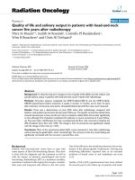
Fig. 1 TILs in the sections of metastatic tumours, using H&E staining, at 400× magnification. a: low level of lymphocytic infiltration; b: high level of
lymphocytic infiltration. Low level of TILs defined as ≤60% of tumour nests infiltrated by lymphocytes. High level of TILs defined as > 60% of tumour
with a pCR in the metastatic ALNs (p = 0.010) (Fig. 2: g,
h). There was, however, no significant association between the level of FOXP3+ and CTLA-4+ T cells and a
pCR in metastatic ALNs following NAC (Fig. 2: e, f ).
Table 2 documents the median number of cells per
HPF found intra-tumourally in metastatic deposits in
the tumour-draining ALNs. It shows the predominance
of the CD4+ and CD8+ T cell subsets, the much lower
but still prominent level of infiltration by FOXP3+ T
cells and the low level of infiltration by CTLA-4+ T cells
and CD56+ NK cells (Fig. 2: a, b; c, d; e, f; g, h).
Higher levels of tumour infiltration by FOXP3+ and CTLA-4+
T cells, and CD56+ NK cells in ALN metastases compared
with corresponding primary breast Tumours (n = 20): No
difference in infiltration by TILs, CD4+ and CD8+ T cells
The levels of intra-tumoural TILs, CD4+ and CD8+ T
cell subsets in ALN metastatic tumour deposits were
comparable with the levels in the corresponding primary
breast cancers (Additional file 3: Table S1 and Table 3).
There were, however, significantly higher levels of
tumour-infiltrating FOXP3+ and CTLA-4+ T cells in
ALN metastases compared with the levels in the corresponding primary breast cancers (p = 0.026, p = 0.036,
respectively). The level of tumour-infiltrating CD56+ NK
cells was also significantly increased (p = 0.006) (Table
3). The CD8+: FOXP3+ T cell ratio, on the other hand,
was not significantly different between the primary
breast tumours and the metastatic tumours in the ALNs.
Positive correlation between tumour-infiltrating lymphocyte
subsets (CD8+, CD56 +) in primary breast Tumours and
metastatic Tumours in ALNs in women with LLABCs
There was a positive correlation between CD8+ T and
CD56+ NK cells infiltrating primary breast cancers and
the tumour deposits in metastatic ALNs (rho = 0.514,
p = 0.020; rho = 0.721, p < 0.001, respectively). There was
no correlation, however, between CD4+, FOXP3+ and
CTLA-4+ T cells infiltrating the primary and metastatic
tumours (Additional file 3: Table S2).
Table 2 High Levels of T Effector (CD4+, CD8+) and CD56+ NK Cells in Pre-NAC (a) ALN(b) Metastatic Tumours: Association with a PCR
Following NAC
Lymphocyte Subsets (n = 20)
Groups
Tumour Infiltration
Median (range)(c)
P Value(d)
(PCR Versus Non PCR)
CD4+
Pathological Complete Response (PCR, n = 9)
65.0 (19.4-157.4)
0.004e
Non Pathological Complete Response (Non PCR, n = 11)
13.2 (0.6-100.8)
Pathological Complete Response (PCR, n = 9)
99.2 (33.2-160.8)
CD8+
Non Pathological Complete Response (Non PCR, n = 11)
11.6 (0.4-93.0)
FOXP3+
Pathological Complete Response (PCR, n = 9)
18.0 (5.0-73.6)
Non Pathological Complete Response (Non PCR, n = 11)
6.4 (1.0-20.4)
CTLA-4+
Pathological Complete Response (PCR, n = 9)
2.6 (0.4-11.6)
Non Pathological Complete Response (Non PCR, n = 11)
0.8 (0.0-2.2)
CD56+
Pathological Complete Response (PCR, n = 9)
2.2 (1.0-26.8)
Non Pathological Complete Response (Non PCR, n = 11)
1.0 (0.0-2.2)
(a)
NAC: Neoadjuvant chemotherapy;
U test; e Statistically significant
(b)
ALN: Axillary lymph node;
(c)
0.001e
0.152
0.112
0.010e
Average cell count per 400× high-power field (see Materials and Methods); (d) Mann-Whitney
Kaewkangsadan et al. BMC Cancer (2018) 18:123
Page 6 of 14
Table 3 Comparison of Tumour-infiltrating Lymphocyte Subsets
in Primary Breast Tumours and Pre-NAC(a)ALN(b) Metastatic Tumours
in Women with LLABCs(c)
Lymphocyte
Subsets
(n = 20)
Primary Tumours Metastatic Tumours P Value(e)
in Breast Median in ALNs(b) Median (Primary Versus
(Range)(d)
Metastases)
(Range)(d)
CD4+
12.8 (0.6-166.2)
26.1 (0.6-157.4)
0.313
CD8+
27.4 (0.4-112.6)
37.1 (0.4-160.8)
0.117
+
5.5 (0.4-96.8)
7.2 (1.0-73.6)
0.026f
+
0.4 (0.0-2.2)
0.8 (0.0-11.6)
0.036f
0.8 (0.0-3.2)
1.5 (0.0-26.8)
0.006f
3.91 (0.18-45.00)
3.29 (0.40-21.92)
0.167
FOXP3
CTLA-4
CD56+
+
+
CD8 :FOXP3
ratio
(a)
NAC: Neoadjuvant chemotherapy: (b)ALNs: Axillary lymph nodes
(corresponding ipsilateral); (c)LLABCs: Large and locally advanced breast
cancers; (d)Average cell count per 400× high-power field (see Materials and
Methods); (e)Wilcoxon signed rank test; fStatistically significant
metastatic ALNs (Table 4). Fig. 3 documents CD8+ (A, B)
and FOXP3+ T cells (C, D) and CD56+ NK cells in the
para-cortical compartment of ALNs.
High levels of CD8+ and low levels of FOXP3+ T cell
subsets in the Para-cortical compartment (tumour-free) of
metastatic ALNs are associated with a PCR following NAC
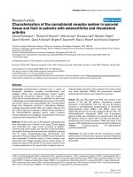
Fig. 2 CD4+ (a, b), CD8+ (C, D) T lymphocytes, FOXP3+ Tregs (e, f)
and CD56+ NK cells (G, H) in the sections of metastatic tumours,
using IHC staining, at 400× magnification. Briefly, heat-mediated
antigen retrieval was performed using citrate buffer, pH 6 (20 mins).
The sections were then incubated with MAbs to CD4 (Dako, M7310)
at a 1:80 dilution for 30 mins at RT, MAbs to CD8 (Dako, M7103) at a
1:100 dilution for 30 mins at RT, MAbs to FOXP3 (Abcam, ab20034)
at a concentration of 20 μg/ml for 30 mins at RT, MAbs to CD56
(Dako, M7304) at a 1:50 dilution for 30 mins at RT. Polymeric HRP-linker
antibody conjugate was used as secondary antibody. DAB chromogen
was used to visualize the staining. The sections were counterstained
with haematoxylin. a, c, e, g low level of CD4+, CD8+ T cell, FOXP3+
Treg, CD56+ NK cell infiltration respectively; b, d, f, h: high level of CD4
+
, CD8+ T cell, FOXP3+ Treg, CD56+ NK infiltration respectively. The
average number of brown membrane-stained cells (CD4+, CD8+ T cells,
CD56+ NK cells) and brown nuclear-stained cells (FOXP3+ Tregs)
regardless of intensity, in contact with tumour cells or within tumour
cell nests per HPF was counted. MTu: Metastatic tumour nest; LN:
Lymphoid tissue
No difference in the lymphocyte profiles (T Effector [CD4
+
, CD8+], T regulatory [FOXP3+, CTLA-4+, PD1+] and NK
[CD56+] cells) in metastatic and non-metastatic ALNs in
women with LLABCs
There were no significant differences in the levels (%) of T
effector (CD4+, CD8+), T regulatory (FOXP3+, CTLA-4+,
PD1+) and NK (CD56+) cells in the tumour-free para-cortical compartments of metastatic ALNs and non-
Comparison between metastatic ALNs with a pCR and
without a pCR following NAC demonstrates a significantly high level of CD8+ T cells (p = 0.048) and low
level of FOXP3+ T cells (p = 0.019) in the para-cortical
compartment (tumour-free) of the ALNs. There was no
difference in the levels of CD4+ and CTLA-4+ T cells,
nor CD56+ NK cells in these ALN response groups
(Table 5).
High CD8+: FOXP3+ T cell ratio in ALNs and association
with a PCR following NAC
A high CD8+: FOXP3+ T cell ratio in the para-cortex
(tumour-free) of metastatic ALNs was significantly associated with a pCR following NAC. A median of 7.24 was
found in ALNs with a pCR versus 3.19 in ALNs without
a pCR (p = 0.006). Comparison of the CD8+: FOXP3+ T
cell ratios in metastases in ALNs with and without a
pCR, however, just failed to reach statistical significance
(p = 0.080). Moreover, this ratio in corresponding
primary tumours was also higher in the pCR group compared with the non-pCR group (7.40 versus 1.48, p =
0.002) (Table 6) (data from Kaewkangsadan et al. [13]).
Expression of cytokines (TH1, TH2, TH17, TGF-β) and biological
molecules (IDO, PD-L1, VEGF) in ALNs
A wide range of cytokines and biological molecules were
studied in ALNs (metastatic and non-metastatic) (Fig. 4).
Significantly higher levels of expression of the Th1 cytokine, IL-2, was found in non-metastatic ALNs (88.9%)
compared with metastatic ALNs (14.3%) (p = 0.003). A
Kaewkangsadan et al. BMC Cancer (2018) 18:123
Page 7 of 14
Table 4 Analyses of Lymphocyte Subsets in ALNs(a) in Women
with LLABCs(b) Undergoing NAC(c): Comparison of Metastatic
and Non Metastatic ALNs
Lymphocyte
Subsets
(n = 33)
Groups
ALN Median
(Range)
P Value(g)
CD4+
Non metastatic ALNs
(n = 9)
63.0 (43.0-74.0)(e)
0.796
Metastatic ALNs
(n = 24)
68.0 (32.0-75.0)
Non metastatic ALNs
(n = 9)
26.0 (15.4-34.0)(e)
Metastatic ALNs
(n = 24)
20.5 (10.4-40.0)
Non metastatic ALNs
(n = 9)
4.4 (2.9-8.6)(e)
Metastatic ALNs
(n = 24)
4.6 (0.2-10.8)
Non metastatic ALNs
(n = 9)
16.8 (5.2-100.4)(f)
Metastatic ALNs
(n = 24)
11.0 (0.6-38.6)
Non metastatic ALNs
(n = 9)
6.4 (1.4-36.0)(f)
Metastatic ALNs
(n = 7)
12.6 (2.0-72.6)
Non metastatic ALNs
(n = 9)
17.8 (15.8-52.8)(f)
Metastatic ALNs
(n = 24)
18.3 (2.2-60.4)
CD8+
FOXP3+
CTLA-4+
PD-1+ (d)
CD56+
0.121
0.736
0.193
0.408
0.437
(a)
ALNs: Axillary lymph nodes (paracortical areas: tumour deposits are
excluded if present); (b) LLABCs: Large and locally advanced breast cancers;
NAC: Neoadjuvant chemotherapy; (d) PD-1+: Programmed death-1 (n = 16);
(e)
Average percentage of positively stained cells out of all the lymphoid cells in
the ALN sections examined; (f) Average cell count of positively stained cells per
400× high-power field in the ALN sections examined; (g) Mann-Whitney U test
(c)
similar profile was seen with the Th1 cytokine, IFN-ϒ
(72.8% versus 20%, p = 0.049) (Table 7). In contrast, the
Th2 cytokine, IL-10, was significantly higher in metastatic compared with non-metastatic ALNs (71.4% versus 22.2%, p = 0.049). There were no significant
differences in the levels of expression of transforming
growth factor-beta (TGF-β), IL-17, programmed death
ligand 1 (PD-L1), indole-amine 2, 3-dioxygenase (IDO)
and vascular endothelial growth factor (VEGF) between
metastatic and non-metastatic ALNs (Table 7). Thus
there was a polarisation from a Th1 to a Th2 profile in
tumour-draining metastatic ALNs.
Discussion
There is ample evidence that a pCR in the primary
breast cancer following NAC is significantly associated
with high levels of tumour infiltration by TILs, CD4+
and CD8+ T effector cells, CD56+ NK cells and high
CD8+: FOXP3+ T cell ratios [9, 21, 40, 22, 11, 10, 23, 41,
24–26, 13]. The contribution of the various TIL subsets,
however, is inadequately studied and data for several of
the subsets is poorly documented.
Droesser et al. [42] found that CD4+ T cells infiltrating
breast cancer were not a prognostic indicator. Heys et al.
[43] reported low levels of CD4+ T cells to be significantly associated with a better response to NAC. In contrast, we documented that high levels of CD4+ T cells,
intratumoural and stromal, in LLABCs were significantly
associated with a pCR following NAC [13]. Mahmoud
et al. [44] described that high levels of CD8+ T cells were
independently associated with longer breast cancerspecific survival. Matkowski et al. [45], however, showed
that a high level of CD8+ T cells in specific types of
breast cancers (high tumour grade, metastatic spread to
ALNs) was associated with a poor prognosis. A small
number of studies, including our own, have found that
high levels of CD8+ T cells in primary breast cancers
were associated with a pCR following NAC [22, 13]; a
high CD8+: FOXP3+ T cell ratio was also significantly
associated with a pCR [21, 13].
The role of TILs, T effector (CD8+, CD4+) and T regulatory (FOXP3+, CTLA-4+, PD-1+) cells and CD56+ NK
cells in ALNs is even less well studied and their contribution to the induction of immune-mediated tumour
cell death in ALN metastases poorly documented. A
small number of studies have been carried out in sentinel lymph nodes (SLNs) and non SLNs from the axilla
in women with breast cancer. SLNs are the first group of
lymph nodes draining the primary tumour in the breast
and are thus the first immune barrier to disseminating
cancer cells [31–33].
Korht et al. [31] showed that increased levels of CD4+
and CD8+ T effector cells in both SLNs and ALNs correlated with an improved 5 year DFS. The ALN but not
the SLN immune profile, on the other hand, was independent of the presence of metastatic disease in ALNs
[31]. Mansfield et al. [33] documented enhanced CD8+
T cell levels in SLNs, with or without metastases. We
did not perform SLN biopsies in the women undergoing
surgery post-NAC in our study group. Our study showed
no differences in the CD4+ and CD8+ T cell subsets between metastatic (tumour-free areas) and non-metastatic
ALNs and is in agreement with the findings described
above.
CD4+ T cells consist of different T helper cells (Th1,
Th2, Th17), secreting a wide range of pro- and antiinflammatory cytokines, as well as producing natural
and inducible CD4+CD25+FOXP3+ Tregs, and show a
degree of plasticity in terms of function [46]. CD8+ T
cells also consist of different subsets - naïve, memory
and activated CD8+ cytotoxic T lymphocytes (CTLs) and
weak suppressor cells [47]. It was not possible to attribute precisely the contribution of the different CD4+
/CD8+ subsets to the pCR with NAC.
Kaewkangsadan et al. BMC Cancer (2018) 18:123
Page 8 of 14
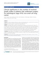
Fig. 3 CD8+ T cells (a, b), FOXP3+ Tregs (c,dD) and CD56+ NK cells (E, F) in the sections of axillary lymph nodes (ALNs), using IHC staining, at 400×
magnification. Briefly, heat-mediated antigen retrieval was performed using citrate buffer pH 6 (20 mins). The sections were then incubated with MAbs to
CD8 (Dako, M7103) at a 1:100 dilution for 30 mins at RT, MAbs to FOXP3 (Abcam, ab20034) at a concentration of 20 μg/ml for 30 mins at RT, MAbs to
CD56 (Dako, M7304) at a 1:50 dilution for 30 mins at RT. Polymeric HRP-linker antibody conjugate was used as secondary antibody. DAB chromogen was
used to visualize the staining. The sections were counterstained with haematoxylin. a, c, e: low percentage of CD8+ T cells, FOXP3+ Tregs and low number
of CD56+ NK cells respectively; B, d, d: high percentage of CD8+ T cells, FOXP3+ Tregs and high number of CD56+ NK cells respectively. The positively
brown membrane-stained cells (CD8+ T cells) and brown nuclear-stained cells (FOXP3+ Tregs) in non-metastatic paracortical areas of ALNs were quantified
as the average % of all cells (5 HPFs). CD56+ NK cells were quantified as average number of cell count per HPF in non-metastatic para-cortical areas of ALNs
with the greatest accumulation of the positively brown membrane-stained cells
We have documented recently the important contribution of CD56+ NK cells to a pCR with NAC in LLABCs.
High levels of CD56+ NK cell concentration in the primary tumour, intra-tumoural or stromal, were associated
with good pathological responses and pCRs, and shown
to be an independent predictor for a pCR [26]. In the
current study, high levels of CD56+ NK cells infiltrating
metastatic deposits in ALNs were found to be similarly
significantly associated with pCRs following NAC.
Interestingly, there was no difference in the CD56
Table 5 Analyses of Lymphocyte Subsets in Metastatic ALNs(a) in Women with LLABCs(b) Undergoing NAC(c): Comparison of
Metastatic ALNs with a PCR with those without a PCR
Lymphocyte Subsets (n = 24)
Groups
ALN Median (Range)
+
CD4
Pathological Complete Response (PCR, n = 10)
61.0 (32.0-75.0)
Non Pathological Complete Response (Non PCR, n = 14)
69.0 (36.0-74.0)
CD8+
Pathological Complete Response (PCR, n = 10)
27.0 (13.4-40.0)(d)
Non Pathological Complete Response (Non PCR, n = 14)
19.5 (10.4-30.0)
Pathological Complete Response (PCR, n = 10)
3.1 (0.2-6.9)(d)
FOXP3+
(d)
Non Pathological Complete Response (Non PCR, n = 14)
6.5 (1.7-10.8)
CTLA-4+
Pathological Complete Response (PCR, n = 10)
5.7 (0.6-29.6)(e)
Non Pathological Complete Response (Non PCR, n = 14)
11.2 (3.2-38.6)
CD56+
Pathological Complete Response (PCR, n = 10)
19.7 (2.2-60.4)(e)
Non Pathological Complete Response (Non PCR, n = 14)
15.9 (6.8-39.0)
(a)
P Value(f)
0.172
0.048g
0.019g
0.341
0.472
ALNs: Axillary lymph nodes (paracortical areas: tumour deposits excluded); (b)LLABCs: Large and locally advanced breast cancers; (c)NAC: Neoadjuvant chemotherapy;
(d)
Average percentage of positively stained cells out of all the lymphoid cells in the ALN sections (CD4+ and CD8+ and FOXP3+ T cells); (e)Average cell count of positively
stained cells per 400× high-power field in the ALN sections (CTLA-4+ T cells and CD56+ NK cells); (f)Mann-Whitney U test; g Statistically significant
Kaewkangsadan et al. BMC Cancer (2018) 18:123
Page 9 of 14
Table 6 The Association Between CD8+: FOXP3+ T Cell Ratio (Breast Tumour, ALN Metastases, ALNs(a) and Subsequent PCR
Following NAC(b)
Sites
Groups
Median (Range)(c)
P Value(d) (PCR Versus Non PCR)
Primary breast tumours, n = 33 (CD8+: FOXP3+ T cell ratio)
Tumours with pCR
7.40 (0.27-45.00)
0.002e
Tumours with non pCR
1.48 (0.18-6.04)
Metastatic tumours with pCR
5.87 (1.35-21.92)
Metastatic tumours with non pCR
1.93 (0.40-7.20)
ALNs with pCR
7.24 (3.33-75.00)
ALNs with non pCR
3.19 (1.78-8.00)
ALN metastatic tumours, n = 20 (CD8 : FOXP3 T cell ratio)
+
ALNs, n = 24 (%CD8+: %FOXP3+ T cell ratio)
(a)
+
ALNs: Axillary lymph nodes (metastatic but tumour-free paracortical area);
Methods); (d)Mann-Whitney U test; eStatistically significant
+
(b)
NAC: Neoadjuvant chemotherapy;
NK cell subset present in the para-cortical compartment
of metastatic (tumour-free areas) and non-metastatic
ALNs. To the best of our knowledge, these findings in
ALNs in human breast cancer have not previously been
described.
CD56+ NK cells have been shown to play an important
role in tumour immune surveillance, in the prevention
of progressive tumour growth and in the defence against
metastatic dissemination [26]. Most human solid tumours have low levels of infiltration by CD56+ NK cells.
A prominent infiltration, however, is usually associated
with an improved prognosis and reduction of tumour recurrence [48–51]. Our results in the current study are in
agreement with these published findings, as a pCR in
tumour and lymph nodes in breast cancer is a surrogate
marker of a good clinical outcome [27, 28].
T regulatory cells are generated during the immune
response and suppress the function of a wide range of
immune cells (T effector [CD4+, CD8+], NK and DCs)
[52, 53]. Blood and tumour-infiltrating Tregs (FOXP3+,
CTLA-4+, PD-1+) play a crucial role in controlling the
anti-cancer cellular immune responses in the circulation
and tumour microenvironment [54, 13]. High levels of
FOXP3+ T cells have been reported infiltrating invasive
breast cancers and to be significantly increased in both
HER2 positive and triple-negative breast cancers [55–59, 13].
Oda et al. [22] documented that high levels of FOXP3
+
T cells in the primary tumour prior to NAC were associated with high pCR rates. Moreover, Demir et al. [38]
stated that high levels of FOXP3+ T cell infiltration postNAC correlated with enhanced rates of pCR. In contrast,
we have shown that NAC reduced both blood and
tumour FOXP3+ T cells concurrently in patients with
LLABCs and that high levels of FOXP3+ T cells in blood
and tumour following NAC were associated with a poor
pathological response [13]. In breast cancer, NAC has
been well documented to significantly reduce tumourinfiltrating FOXP3+, CTLA-4+ and PD-1+ T cells (but
not CD8+ T cells) [60, 61, 38, 25, 13].
The profile and the function of FOXP3+ T cells in
tumour-draining ALNs is less well studied. FOXP3+ T
0.080
0.006e
(c)
Ratio of CD8+:FOXP3+ T cells (see Materials and
cells have been shown to be increased in numbers in
SLNs, in particular in metastatic nodes; even micrometastatic disease was associated with increased levels
of FOXP3+ T cells [62, 63, 32, 33]. In our study, high
levels of FOXP3+ and CTLA-4+ T cells were documented in metastatic tumours in the ALNs and were
higher than the levels in the corresponding primary tumours. A low % of FOXP3+ T cells (and high % of CD8+
T cells) in para-cortical (tumour-free) areas of metastatic
ALNs was significantly associated with ALN pCRs. Such
findings have not been documented in the literature.
CTLA-4 is a co-inhibitory receptor molecule found on
activated and exhausted T cells and Tregs and negatively
regulates T cell interaction with CD80/CD86 ligand
binding sites [64, 65]. In primary breast cancers there is
an increased expression of CTLA-4, compared with normal
breast tissue [66]. High levels of CTLA-4 mRNA in primary breast cancers were shown to be associated with
ALN metastases and advanced tumours [66, 67]. We have
previously demonstrated high levels of CTLA-4+ T cells in
the blood of women with LLABCs [54]. Although high
levels of tumour-infiltrating FOXP3+ T cells (and PD-1+
lymphocytes) were not associated with a pCR following
NAC, tumour stromal infiltration by high levels of CTLA-4
+
T cells were. The in situ CTLA-4+ expression was likely
to be due to activated T cells [13]. In our study in ALNs,
higher levels of CTLA-4+ T cells were demonstrated in
ALN metastases than in the corresponding primary tumours. In contrast to the findings in the primary breast
cancers, high levels of CTLA-4+ T cells in ALNs were not
significantly associated with a pCR following NAC. There is,
however, a dearth of publications regarding CTLA-4+ T cells
and breast cancer, either in the primary tumour or ALNs.
PD-1 is expressed on activated and exhausted T cells,
Tregs, NK cells and DCs [68, 16, 69]. On interacting
with PD-L1/L2 in a co-inhibitory pathway in tissues it
down-regulates activated T cells resulting in T cell tolerance and prevention of auto-immunity [70]. The PD-1/
PD-L1 pathway is a key immune check-point exploited
by malignant cells to escape anti-cancer immune
defences [71].
Kaewkangsadan et al. BMC Cancer (2018) 18:123
Page 10 of 14
Table 7 Expression of Cytokines (Th1, Th2 and Th17), IDO(a),
PD-L1(b)and VEGF(c) in ALNs(d) in Women with
LLABCs(e)Undergoing NAC(f)
(n = 16) Groups
Low/Negative High
Pearson
P
Expression (n) Expression (n) Chi-Square Value
Value
IL-1
Non
metastatic
ALNs (n = 9)
3
6
Metastatic
ALNs (n = 7)
2
5
Non
metastatic
ALNs (n = 9)
1
8
Metastatic
ALNs (n = 7)
6
1
Non
metastatic
ALNs (n = 9)
3
6
Metastatic
ALNs (n = 7)
3
4
Non
metastatic
ALNs (n = 9)
7
2
Metastatic
ALNs (n = 7)
2
5
Non
metastatic
ALNs (n = 9)
1
8
Metastatic
ALNs (n = 7)
3
4
Non
metastatic
ALNs (n = 9)
2
7
Metastatic
ALNs (n = 7)
4
3
Non
metastatic
ALNs (n = 9)
7
2
Metastatic
ALNs (n = 7)
5
2
Non
metastatic
ALNs (n = 9)
1
8
Metastatic
ALNs (n = 7)
4
3
TGF-β(g) Non
metastatic
ALNs (n = 9)
5
4
Metastatic
ALNs (n = 7)
5
2
Non
metastatic
ALNs (n = 9)
6
3
Metastatic
ALNs (n = 7)
6
1
IL-2
IL-4
IL-10
IL-17
IDO(g)
Fig. 4 IL-2 (a, b), IL-10 (c, d), IL-17 (e, f) and IFN-γ (g, h) expression in the
sections of axillary lymph nodes (ALNs), using IHC staining, at 400×
magnification. Briefly, heat-mediated antigen retrieval was performed
using citrate buffer pH 6 (20 mins). The sections were then incubated
with MAbs to IL-2 (Abcam, ab92381) at a 1:500 dilution for 30 mins at RT,
polyclonal Abs to IL-10 (Abcam, ab34843) at a 1:400 dilution for 30 mins
at RT, polyclonal Abs to IL-17 (Abcam, ab9565) at a 1:100 dilution for 30
mins at RT, polyclonal Abs to IFN-γ (Abcam, ab9657) at a concentration
of 4 μg/ml for 30 mins at RT. Polymeric HRP-linker antibody conjugate
was used as secondary antibody. DAB chromogen was used to visualize
the staining. The sections were counterstained with haematoxylin. a, c, e,
g: low level of expression; b, d, f, h: high level of expression. The H score
[% of positive cells (brown membrane/cytoplasmic-stained cells) x
intensity of staining (1 to 3)] was used to assess the level of expression;
low was ≤100 and high was > 100. Scoring performed on non-metastatic
areas of a whole ALN section (7-10 HPFs)
High levels of PD-1+ lymphocytes have been shown to
have a significant correlation with reduced patient survival [72]. In our primary breast cancer study the levels
of PD-1+ cells were low and there was no association
with a subsequent pCR following NAC [13]. Comparable
findings were documented in ALN metastases. PD-1+ T
cell subsets have not previously been described in ALNs
in breast cancer; we found no difference in the T
regulatory profiles between metastatic and non-metastatic ALNs.
PD-L1
IFN-γ
VEGF
(a)
0.042
0.838
8.905
0.003h
0.152
0.696
3.874
0.049h
2.116
0.146
2.049
0.152
0.085
0.771
3.883
0.049h
0.423
0.515
0.762
0.383
IDO: Indoleamine 2,3-dioxygenase; (b)PDL-1: Programmed death ligand
1; (c)VEGF: Vascular endothelial growth factor; (d)ALNs: Axillary lymph
nodes; (e)LLABCs: Large and locally advanced breast cancers; (f)NAC:
Neoadjuvant chemotherapy; (g)IDO and TGF-β were scored as negative
and positive; hStatistically significant
Kaewkangsadan et al. BMC Cancer (2018) 18:123
In various human cancers malignant cells and host infiltrating cells express and secrete a range of Th1, Th2 and
Th17 cytokines and TGF-β which enhance or suppress
the in situ anti-cancer immune responses [73–77]. In such
studies the semi-quantitative methods used did not discriminate between the tumour-infiltrating immune and
malignant cells, nor quantify the contribution of the different cells to the cytokine profiles in the tumour [13].
We have previously demonstrated a polarisation of Th2
production in vitro by blood lymphocytes from women
with LLABCs; this polarisation persisted post-NAC [54].
Our current study has also revealed the presence of a Th2
cytokine polarisation in ALNs with metastases. There was
a high level of expression of the Th2 suppressive cytokine
IL-10 and low level of expression of the Th1 inflammatory
cytokines INF-ϒ and IL-2, when compared with nonmetastatic ALNs. Interestingly, Matsuura et al. [32] noted
in breast cancer that micro-metastases in SLNs stimulated
Th1 responses (T-box family of transcription factors)
whilst macro-metastases enhanced Th2 responses (GATA
family of zinc finger proteins). This area of immune
reactivity in ALNs is poorly studied.
The role of IL-17 in human cancer is not well defined.
In a study in breast cancer, the level of Th17 cells was
shown to be increased and associated with a good clinical outcome [76]. In human cancers, TGF-β expression
is usually upregulated. It induces the production of
FOXP3+ Tregs, inhibiting the generation and activity of
innate and adaptive immunity [53, 78]. High levels of expression of IL-10 and IL-17 in breast cancer following
NAC have been shown to be significantly associated with
failure to achieve a pCR [13]. In our current study with
ALNs we did not demonstrate any significant changes in
expression of IL-17 and TGF-β, as well as PD-L1, VGF
and IDO, between tumour-free and metastatic ALNs.
Although most chemotherapeutic agents inhibit elements
of innate and adaptive immunity, they can enhance both
components, resulting in immune-mediated tumour cell
death [79, 17, 20]. Chemotherapy induces cancer cell stress
and damage which results in the release of “danger signals”
and immunogenic tumour-associated antigens (TAAs).
Danger signals activate innate immune cells whilst TAAs
are taken up by DCs resulting in the release of proinflammatory cytokines and the production of anti-cancer
CTLs. Anthracyclines, in particular, induce tumour cell
damage and release/expose calreticulin and other endoplasmic proteins [79, 80, 19]. The NAC combination used in
our trial consisted of anthracycline, cyclophosphamide, taxane, ± capecitabine. These chemotherapeutic agents are
known to have immunomodulatory effects. Doxorubicin
(anthracycline) can enhance the production of TAA-specific CD8+ CTLs and induce tumour infiltration by CD8+ T
cells [16, 81]. Cyclophosphamide inhibits the generation
and function of FOXP3+ Tregs [15, 82]. Docetaxel (taxane)
Page 11 of 14
has been shown to increase serum IFN-ϒ, IL-2 and IL-6
levels and NK cell activity in blood [83, 14]. Capecitabine is
enzymatically converted to 5-fluorouracil (5-FU) and this
enhances the expression of TAAs on tumour cells and
antibody-dependent cell-mediated cytotoxicity [84, 85].
Thus NAC induces a range of anti-cancer immune responses which contribute to the damage and eradication of
malignant cells.
Our current and previous findings suggest that the immune milieu in the breast and ALNs in patients with
LLABCs plays a key role in inducing tumour cell death,
both in the primary cancer and ALN metastases in patients undergoing NAC. High levels of TILs, CD4+ and
CD8+ T cells and NK cells in the primary and ALN metastases were associated with significant pCRs. There was no
alteration in levels of infiltration by TILs and a positive
correlation between CD8+ T and NK cells infiltrating primary and metastatic tumours [26]. To the best of our
knowledge, there are no publications regarding TILs,
Tregs (FOXP3+, CTLA-4+) and NK cells in metastatic tumours in ALNs, nor comparisons with corresponding primary breast cancers. In our NAC trial in LLABCs
concurrent pCRs at both sites was infrequent but were associated with the best clinical outcome; a less beneficial
outcome was seen with a pCR in the breast alone [28].
Conclusions
Our study of tumour-draining ALNs in women with
LLABCs undergoing NAC has demonstrated new and
important findings, complementing the results previously documented in the primary tumours. We have
characterised further the key contributions of tumourinfiltrating TILs, T effector (CD4+, CD8+), T regulatory
(FOXP3+, CTLA-4+) and CD56+ NK cells to pCRs in
ALN metastases. High levels of CD8+ T cells and low
levels of FOXP3+ T cells in para-cortical areas (tumourfree) were associated with pCRs following NAC. Th2 polarisation (high IL-10, low IL-2 and IFN-ϒ) was present
in ALNs with metastases. In LLABCs, the close interrelationship between a pCR in breast and ALNs and the
concomitant immune changes induced by NAC suggests
that immune-mediated cell death may be a crucial component of NAC-associated tumour cell destruction and
removal.
Additional files
Additional file 1: Table S3. Patient and Tumour Characteristics,
Responses to Neoadjuvant Chemotherapy (n = 33). (DOCX 56 kb)
Additional file 2: Table of Patient Characteristics. (DOCX 15 kb)
Additional file 3: Table S1. Comparison of Tumour-infiltrating Lymphocytes (TILs) in Primary Breast Tumours and Pre-NAC(1) ALN(2) Metastatic
Tumours in Women with LLABCs(3). There was no significant difference
between the levels of TILs in primary breast tumours and axillary
Kaewkangsadan et al. BMC Cancer (2018) 18:123
metastatic tumour deposits. Table S2. Correlations of Tumour-infiltrating
Lymphocyte Subsets in Primary Breast Tumours and ALN (1) Metastatic
Tumours in Women with LLABCs(2) [Spearman’s Correlation Coefficient
(rho)] (n = 20). There was a positive correlation between CD8+ T and
CD56+ NK cells infiltrating primary breast cancers and the tumour
deposits in metastatic ALNs (rho=0.514, p =0.020; rho=0.721, p < 0.001,
respectively). There was no correlation, however, between CD4+, FOXP3+
and CTLA-4+ T cells infiltrating the primary and metastatic tumours.
(DOCX 26 kb)
Abbreviations
5-FU: 5-fluorouracil; A: Adriamycin; ALN: Axillary lymph node;
C: Cyclophosphamide; CD: Cluster of differentiation; CTL: Cytotoxic T
lymphocyte; CTLA-4: Cytotoxic T lymphocyte antigen 4; DAB: Diamino-benzidine; DC: Dendritic cell; DFS: Disease-free survival; ER: Oestrogen
receptor; FOXP3: Forkhead box protein 3; H&E: Haematoxylin and eosin;
HER2: Human epidermal growth factor receptor 2; HPF: High-power field;
HRP: Horseradish peroxidase; IFN-γ: Interferon-gamma;
IHC: Immunohistochemistry; IL: Interleukin; LLABC: Large locally advanced
breast cancer; MAb: Monoclonal antibody; NAC: Neoadjuvant chemotherapy;
NK: Natural killer; OS: Overall survival; pCR: Pathological complete response;
PD-1: Programmed death 1; PD-L1: Programmed death ligand 1; RT: Room
temperature; SLN: Sentinel lymph node; T: Docetaxel; TAA: Tumour-associated
antigen; TGF-β: Transforming growth factor-beta; Th: T helper;
TIL: Tumour-infiltrating lymphocyte; Treg: T regulatory cell; X: Capecitabine
Acknowledgments
We wish to acknowledge Mr. Christopher Nolan (Academic Unit of Clinical
Oncology, City Hospital, University of Nottingham) for his advice and help
with the IHC assays. The clinical trial, from which patients’ tissue specimens
and blood samples were collected for the study, was supported by
educational grants from Sanofi-Aventis UK, Roche UK and Chughai UK.
Funding
The authors wish to acknowledge the financial support provided for this
study by a grant from the Nottinghamshire, Derbyshire and Lincolnshire
Research Alliance, and Candles Charity. The funding body had no role in the
design of the study and collection, analysis, and interpretation of data and in
writing the manuscript.
Availability of data and materials
Data of patient and tumour characteristics, responses to neoadjuvant
chemotherapy is available in Additional file 1: Table S3.
Authors’ contributions
Conception and Design: VK, CV, JE, GC, OE. Data Acquisition: VK, CV, JE, GC,
OE. Data Analysis and Interpretation: VK, CV, JE, GC, MI, OE. Laboratory Assays:
VK, CV, GC. Writing of Manuscript: VK, CV, JE, OE. Review of and Final
Approval of Manuscript: VK, CV, JE, GC, MI, OE. All authors read and approved
the final manuscript.
Ethics approval and consent to participate
The study was given approval by the Leicestershire, Northamptonshire &
Rutland Research Ethics Committee 1: Reference Number 07/H0406/260;
Favourable Opinion 24/01/2008. All patients enrolled in the study gave
informed consent to participate in and to publish the results of the study.
The study Registration is ISRCTN00407556.
Consent for publication
All patients enrolled in the study gave informed consent to participate in
and to publish the results of the study.
Competing interests
The authors declare that they have no competing interests.
Publisher’s Note
Springer Nature remains neutral with regard to jurisdictional claims in
published maps and institutional affiliations.
Page 12 of 14
Author details
1
Division of Gastrointestinal Surgery, Nottingham Digestive Diseases Centre,
Faculty of Medicine and Health Sciences, University of Nottingham, E Floor
West Block, Queen’s Medical Centre, Derby Rd, Nottingham NG7 2UH, UK.
2
Research & Development Department, Lincoln Breast Unit, Lincoln County
Hospital, Greetwell Road, Lincoln LN2 5QY, UK. 3Department of Pathology,
PathLinks, Lincoln County Hospital, Greetwell Road, Lincoln LN2 5QY, UK.
4
Academic Department of Pathology, Faculty of Medicine and Health
Sciences, University of Nottingham, A Floor West Block, Queens Medical
Centre, Derby Road, Nottingham NG7 2UH, UK. 5Department of Surgery,
Phramongkutklao Hospital and College of Medicine, 315 Rajavithi Road,
Bangkok 10400, Thailand.
Received: 26 September 2017 Accepted: 24 January 2018
References
1. Aloysius M, Walker L, Eremin O. Cancer and the immune response Ch 4. In:
Eremin O, Sewell H, editors. Essential immunology for surgeons. Oxford:
OUP; 2011. p. 237–302.
2. Fridman WH, Galon J, Pages F, Tartour E, Sautes-Fridman C, Kroemer G.
Prognostic and predictive impact of intra- and peritumoral immune
infiltrates. Cancer Res. 2011;71(17):5601–5. />CAN-11-1316.
3. Galon J, Angell HK, Bedognetti D, Marincola FM. The continuum of cancer
immunosurveillance: prognostic, predictive, and mechanistic signatures.
Immunity. 2013;39(1):11–26. />4. Teng MW, Ngiow SF, Ribas A, Smyth MJ. Classifying cancers based on T-cell
infiltration and PD-L1. Cancer Res. 2015;75(11):2139–45. />1158/0008-5472.CAN-15-0255.
5. Zhang L, Conejo-Garcia JR, Katsaros D, Gimotty PA, Massobrio M, Regnani G,
et al. Intratumoral T cells, recurrence, and survival in epithelial ovarian
cancer. N Engl J Med. 2003;348(3):203–13. />NEJMoa020177.
6. Pages F, Berger A, Camus M, Sanchez-Cabo F, Costes A, Molidor R, et al.
Effector memory T cells, early metastasis, and survival in colorectal cancer.
N Engl J Med. 2005;353(25):2654–66. />7. Fridman WH, Pages F, Sautes-Fridman C, Galon J. The immune contexture
in human tumours: impact on clinical outcome. Nat Rev Cancer. 2012;12(4):
298–306. />8. Angell H, Galon J. From the immune contexture to the Immunoscore: the
role of prognostic and predictive immune markers in cancer. Curr Opin
Immunol. 2013;25(2):261–7. />9. Denkert C, Loibl S, Noske A, Roller M, Muller BM, Komor M, et al. Tumorassociated lymphocytes as an independent predictor of response to
neoadjuvant chemotherapy in breast cancer. J Clin Oncol. 2010;28(1):105–13.
/>10. Yamaguchi R, Tanaka M, Yano A, Tse GM, Yamaguchi M, Koura K, et al.
Tumor-infiltrating lymphocytes are important pathologic predictors for
neoadjuvant chemotherapy in patients with breast cancer. Hum Pathol.
2012;43(10):1688–94. />11. Ono M, Tsuda H, Shimizu C, Yamamoto S, Shibata T, Yamamoto H, et al.
Tumor-infiltrating lymphocytes are correlated with response to neoadjuvant
chemotherapy in triple-negative breast cancer. Breast Cancer Res Treat.
2012;132(3):793–805. />12. Dieci MV, Criscitiello C, Goubar A, Viale G, Conte P, Guarneri V, et al.
Prognostic value of tumor-infiltrating lymphocytes on residual disease after
primary chemotherapy for triple-negative breast cancer: a retrospective
multicenter study. Ann Oncol. 2014; />mdt556.
13. Kaewkangsadan V, Verma C, Eremin JM, Cowley G, Ilyas M, Eremin O. Crucial
contributions by T lymphocytes (Effector, regulatory, and checkpoint
inhibitor) and cytokines (TH1, TH2, and TH17) to a pathological complete
response induced by Neoadjuvant chemotherapy in women with breast
cancer. J Immunol Res. 2016;2016:4757405. />4757405.
14. Tsavaris N, Kosmas C, Vadiaka M, Kanelopoulos P, Boulamatsis D. Immune
changes in patients with advanced breast cancer undergoing
chemotherapy with taxanes. Br J Cancer. 2002;87(1):21–7. />1038/sj.bjc.6600347.
Kaewkangsadan et al. BMC Cancer (2018) 18:123
15. Ghiringhelli F, Menard C, Puig PE, Ladoire S, Roux S, Martin F, et al.
Metronomic cyclophosphamide regimen selectively depletes CD4+CD25+
regulatory T cells and restores T and NK effector functions in end stage
cancer patients. Cancer Immunol Immunother. 2007;56(5):641–8. https://doi.
org/10.1007/s00262-006-0225-8.
16. Park JY, Jang MJ, Chung YH, Kim KY, Kim SS, Lee WB, et al. Doxorubicin
enhances CD4(+) T-cell immune responses by inducing expression of CD40
ligand and 4-1BB. Int Immunopharmacol. 2009;9(13-14):1530–9. https://doi.
org/10.1016/j.intimp.2009.09.008.
17. Barbon CM, Yang M, Wands GD, Ramesh R, Slusher BS, Hedley ML, et al.
Consecutive low doses of cyclophosphamide preferentially target Tregs and
potentiate T cell responses induced by DNA PLG microparticle immunization.
Cell Immunol. 2010;262(2):150–61. />18. Aloysius M, Verma C, Eremin O. Therapy and host defences Ch 7. In: Eremin
O, Sewell H, editors. Essential immunology for surgeons. Oxford: OUP; 2011.
p. 379–402.
19. Menger L, Vacchelli E, Adjemian S, Martins I, Ma Y, Shen S, et al. Cardiac
glycosides exert anticancer effects by inducing immunogenic cell death. Sci
Transl Med. 2012;4(143):143ra99. />3003807.
20. Kroemer G, Galluzzi L, Kepp O, Zitvogel L. Immunogenic cell death in
cancer therapy. Annu Rev Immunol. 2013;31:51–72. />annurev-immunol-032712-100008.
21. Ladoire S, Mignot G, Dabakuyo S, Arnould L, Apetoh L, Rebe C, et al. In situ
immune response after neoadjuvant chemotherapy for breast cancer predicts
survival. J Pathol. 2011;224(3):389–400. />22. Oda N, Shimazu K, Naoi Y, Morimoto K, Shimomura A, Shimoda M, et al.
Intratumoral regulatory T cells as an independent predictive factor for
pathological complete response to neoadjuvant paclitaxel followed by 5FU/epirubicin/cyclophosphamide in breast cancer patients. Breast Cancer
Res Treat. 2012;136(1):107–16. />23. Lee HJ, Seo JY, Ahn JH, Ahn SH, Gong G. Tumor-associated lymphocytes
predict response to neoadjuvant chemotherapy in breast cancer patients. J
Breast Cancer. 2013;16(1):32–9. />24. Seo AN, Lee HJ, Kim EJ, Kim HJ, Jang MH, Lee HE, et al. Tumour-infiltrating
CD8+ lymphocytes as an independent predictive factor for pathological
complete response to primary systemic therapy in breast cancer. Br J
Cancer. 2013;109(10):2705–13. />25. Garcia-Martinez E, Gil GL, Benito AC, Gonzalez-Billalabeitia E, Conesa MA,
Garcia Garcia T, et al. Tumor-infiltrating immune cell profiles and their
change after neoadjuvant chemotherapy predict response and prognosis of
breast cancer. Breast Cancer Res. 2014;16(6):488. />s13058-014-0488-5.
26. Verma C, Kaewkangsadan V, Eremin JM, Cowley GP, Ilyas M, El-Sheemy MA,
et al. Natural killer (NK) cell profiles in blood and tumour in women with
large and locally advanced breast cancer (LLABC) and their contribution to
a pathological complete response (PCR) in the tumour following
neoadjuvant chemotherapy (NAC): differential restoration of blood profiles
by NAC and surgery. J Transl Med. 2015;13:180. />s12967-015-0535-8.
27. von Minckwitz G, Untch M, Blohmer JU, Costa SD, Eidtmann H, Fasching PA, et al.
Definition and impact of pathologic complete response on prognosis after
neoadjuvant chemotherapy in various intrinsic breast cancer subtypes. J Clin
Oncol. 2012;30(15):1796–804. />28. Eremin J, Cowley G, Walker LG, Murray E, Stovickova M, Eremin O. Women
with large (>=3 cm) and locally advanced breast cancers (T3, 4, N1, 2, M0)
receiving neoadjuvant chemotherapy (NAC: cyclophosphamide,
doxorubicin, docetaxel): addition of capecitabine improves 4-year diseasefree survival. SpringerPlus. 2015;4:9.
29. Carter CL, Allen C, Henson DE. Relation of tumor size, lymph node status,
and survival in 24,740 breast cancer cases. Cancer. 1989;63(1):181–7.
30. Recht A, Houlihan MJ. Axillary lymph nodes and breast cancer: a review.
Cancer. 1995;76(9):1491–512.
31. Kohrt HE, Nouri N, Nowels K, Johnson D, Holmes S, Lee PP. Profile of immune
cells in axillary lymph nodes predicts disease-free survival in breast cancer.
PLoS Med. 2005;2(9):e284. />32. Matsuura K, Yamaguchi Y, Osaki A, Ohara M, Okita R, Emi A, et al. FOXP3
expression of micrometastasis-positive sentinel nodes in breast cancer
patients. Oncol Rep. 2009;22(5):1181–7.
33. Mansfield AS, Heikkila P, von Smitten K, Vakkila J, Leidenius M. Metastasis to
sentinel lymph nodes in breast cancer is associated with maturation arrest of
Page 13 of 14
34.
35.
36.
37.
38.
39.
40.
41.
42.
43.
44.
45.
46.
47.
48.
49.
50.
51.
52.
dendritic cells and poor co-localization of dendritic cells and CD8+ T cells.
Virchows Arch. 2011;459(4):391–8. />Cimino-Mathews A, Ye X, Meeker A, Argani P, Emens LA. Metastatic triplenegative breast cancers at first relapse have fewer tumor-infiltrating
lymphocytes than their matched primary breast tumors: a pilot study. Hum
Pathol. 2013;44(10):2055–63. />Ogston KN, Miller ID, Payne S, Hutcheon AW, Sarkar TK, Smith I, et al. A new
histological grading system to assess response of breast cancers to primary
chemotherapy: prognostic significance and survival. Breast. 2003;12(5):320–7.
Walker LG, Eremin JM, Aloysius MM, Vassanasiri W, Walker MB, El-Sheemy M,
et al. Effects on quality of life, anti-cancer responses, breast conserving
surgery and survival with neoadjuvant docetaxel: a randomised study of
sequential weekly versus three-weekly docetaxel following neoadjuvant
doxorubicin and cyclophosphamide in women with primary breast cancer.
BMC Cancer. 2011;11:179. />Salgado R, Denkert C, Demaria S, Sirtaine N, Klauschen F, Pruneri G, et al.
The evaluation of tumor-infiltrating lymphocytes (TILs) in breast cancer:
recommendations by an international TILs working group 2014. Ann Oncol.
2015;26(2):259–71. />Demir L, Yigit S, Ellidokuz H, Erten C, Somali I, Kucukzeybek Y, et al.
Predictive and prognostic factors in locally advanced breast cancer: effect of
intratumoral FOXP3+ Tregs. Clinical & experimental metastasis. 2013; https://
doi.org/10.1007/s10585-013-9602-9.
Mansfield AS, Heikkila P, von Smitten K, Vakkila J, Leidenius M. The presence
of sinusoidal CD163(+) macrophages in lymph nodes is associated with
favorable nodal status in patients with breast cancer. Virchows Arch. 2012;
461(6):639–46. />West NR, Milne K, Truong PT, Macpherson N, Nelson BH, Watson PH. Tumorinfiltrating lymphocytes predict response to anthracycline-based
chemotherapy in estrogen receptor-negative breast cancer. Breast Cancer
Res. 2011;13(6):R126. />Issa-Nummer Y, Darb-Esfahani S, Loibl S, Kunz G, Nekljudova V, Schrader I,
et al. Prospective validation of immunological infiltrate for prediction of
response to neoadjuvant chemotherapy in HER2-negative breast cancer–a
substudy of the neoadjuvant GeparQuinto trial. PLoS One. 2013;8(12):
e79775. />Droeser R, Zlobec I, Kilic E, Guth U, Heberer M, Spagnoli G, et al. Differential
pattern and prognostic significance of CD4+, FOXP3+ and IL-17+ tumor
infiltrating lymphocytes in ductal and lobular breast cancers. BMC Cancer.
2012;12:134. />Heys SD, Stewart KN, McKenzie EJ, Miller ID, Wong SY, Sellar G, et al.
Characterisation of tumour-infiltrating macrophages: impact on response and
survival in patients receiving primary chemotherapy for breast cancer. Breast
Cancer Res Treat. 2012;135(2):539–48. />Mahmoud SM, Paish EC, Powe DG, Macmillan RD, Grainge MJ, Lee AH, et al.
Tumor-infiltrating CD8+ lymphocytes predict clinical outcome in breast cancer.
J Clin Oncol. 2011;29(15):1949–55. />Matkowski R, Gisterek I, Halon A, Lacko A, Szewczyk K, Staszek U, et al. The
prognostic role of tumor-infiltrating CD4 and CD8 T lymphocytes in breast
cancer. Anticancer Res. 2009;29(7):2445–51.
Sewell H. Basic immunology Ch 1. In: Eremin O, Sewell H, editors. Essential
immunology for surgeons. Oxford: OUP; 2011. p. 1–160.
Mayer CT, Floess S, Baru AM, Lahl K, Huehn J, Sparwasser T. CD8+ Foxp3+ T
cells share developmental and phenotypic features with classical CD4+
Foxp3+ regulatory T cells but lack potent suppressive activity. Eur J
Immunol. 2011;41(3):716–25. />Coca S, Perez-Piqueras J, Martinez D, Colmenarejo A, Saez MA, Vallejo C,
et al. The prognostic significance of intratumoral natural killer cells in
patients with colorectal carcinoma. Cancer. 1997;79(12):2320–8.
Ishigami S, Natsugoe S, Tokuda K, Nakajo A, Che X, Iwashige H, et al.
Prognostic value of intratumoral natural killer cells in gastric carcinoma.
Cancer. 2000;88(3):577–83.
Villegas FR, Coca S, Villarrubia VG, Jimenez R, Chillon MJ, Jareno J, et al.
Prognostic significance of tumor infiltrating natural killer cells subset CD57
in patients with squamous cell lung cancer. Lung Cancer. 2002;35(1):23–8.
Cao FM, Zhang XH, Yan X, Wang JL, Wang XL, Zhang ZG, et al. Prognostic
significances of natural killer cells and dendritic cells infiltrations in
esophageal squamous cell carcinoma. Ai zheng = Aizheng = Chinese
journal of cancer. 2005;24(2):232–6.
Shevach EM. Mechanisms of foxp3+ T regulatory cell-mediated suppression.
Immunity. 2009;30(5):636–45. />
Kaewkangsadan et al. BMC Cancer (2018) 18:123
53. Nishikawa H, Sakaguchi S. Regulatory T cells in tumor immunity.
International journal of cancer Journal international du cancer. 2010;127(4):
759–67. />54. Verma C, Eremin JM, Robins A, Bennett AJ, Cowley GP, El-Sheemy MA, et al.
Abnormal T regulatory cells (Tregs: FOXP3+, CTLA-4+), myeloid-derived
suppressor cells (MDSCs: monocytic, granulocytic) and polarised T helper
cell profiles (Th1, Th2, Th17) in women with large and locally advanced
breast cancers undergoing neoadjuvant chemotherapy (NAC) and surgery:
failure of abolition of abnormal treg profile with treatment and correlation
of treg levels with pathological response to NAC. J Transl Med. 2013;11:16.
/>55. Wolf AM, Wolf D, Steurer M, Gastl G, Gunsilius E, Grubeck-Loebenstein B.
Increase of regulatory T cells in the peripheral blood of cancer patients. Clin
Cancer Res. 2003;9(2):606–12.
56. Bates GJ, Fox SB, Han C, Leek RD, Garcia JF, Harris AL, et al. Quantification of
regulatory T cells enables the identification of high-risk breast cancer
patients and those at risk of late relapse. J Clin Oncol. 2006;24(34):5373–80.
/>57. Perez SA, Karamouzis MV, Skarlos DV, Ardavanis A, Sotiriadou NN, Iliopoulou
EG, et al. CD4+CD25+ regulatory T-cell frequency in HER-2/neu (HER)positive and HER-negative advanced-stage breast cancer patients. Clin
Cancer Res. 2007;13(9):2714–21. />58. Ladoire S, Arnould L, Apetoh L, Coudert B, Martin F, Chauffert B, et al.
Pathologic complete response to neoadjuvant chemotherapy of breast
carcinoma is associated with the disappearance of tumor-infiltrating foxp3+
regulatory T cells. Clin Cancer Res. 2008;14(8):2413–20. />1158/1078-0432.CCR-07-4491.
59. Merlo A, Casalini P, Carcangiu ML, Malventano C, Triulzi T, Menard S, et al.
FOXP3 expression and overall survival in breast cancer. J Clin Oncol. 2009;
27(11):1746–52. />60. Aruga T, Suzuki E, Saji S, Horiguchi S, Horiguchi K, Sekine S, et al. A low
number of tumor-infiltrating FOXP3-positive cells during primary systemic
chemotherapy correlates with favorable anti-tumor response in patients
with breast cancer. Oncol Rep. 2009;22(2):273–8.
61. Liu F, Li Y, Ren M, Zhang X, Guo X, Lang R, et al. Peritumoral FOXP3(+) regulatory
T cell is sensitive to chemotherapy while intratumoral FOXP3(+) regulatory T cell
is prognostic predictor of breast cancer patients. Breast Cancer Res Treat. 2012;
135(2):459–67. />62. Matsuura K, Yamaguchi Y, Ueno H, Osaki A, Arihiro K, Toge T. Maturation of
dendritic cells and T-cell responses in sentinel lymph nodes from patients
with breast carcinoma. Cancer. 2006;106(6):1227–36. />cncr.21729.
63. Mansfield AS, Heikkila PS, Vaara AT, von Smitten KA, Vakkila JM, Leidenius
MH. Simultaneous Foxp3 and IDO expression is associated with sentinel
lymph node metastases in breast cancer. BMC Cancer. 2009;9:231. https://
doi.org/10.1186/1471-2407-9-231.
64. Teft WA, Kirchhof MG, Madrenas J. A molecular perspective of CTLA-4
function. Annu Rev Immunol. 2006;24:65–97. />annurev.immunol.24.021605.090535.
65. Teft WA, Madrenas J. Molecular determinants of inverse agonist activity of
biologicals targeting CTLA-4. J Immunol. 2007;179(6):3631–7.
66. Mao H, Zhang L, Yang Y, Zuo W, Bi Y, Gao W, et al. New insights of CTLA-4
into its biological function in breast cancer. Curr Cancer Drug Targets. 2010;
10(7):728–36.
67. de la Cruz-Merino L, Barco-Sanchez A, Henao Carrasco F, Nogales Fernandez
E, Vallejo Benitez A, Brugal Molina J, et al. New insights into the role of the
immune microenvironment in breast carcinoma. Clinical & developmental
immunology. 2013;2013:785317. />68. Shinohara T, Taniwaki M, Ishida Y, Kawaichi M, Honjo T. Structure and
chromosomal localization of the human PD-1 gene (PDCD1). Genomics.
1994;23(3):704–6. />69. Topalian SL, Drake CG, Pardoll DM. Targeting the PD-1/B7-H1(PD-L1)
pathway to activate anti-tumor immunity. Curr Opin Immunol. 2012;24(2):
207–12. />70. Ostrand-Rosenberg S, Horn LA, Haile ST. The programmed death-1
immune-suppressive pathway: barrier to antitumor immunity. J Immunol.
2014;193(8):3835–41. />71. Criscitiello C, Curigliano G. Immunotherapeutics for breast cancer. Curr Opin
Oncol. 2013;25(6):602–8. />
Page 14 of 14
72. Sun S, Fei X, Mao Y, Wang X, Garfield DH, Huang O, et al. PD-1(+) immune
cell infiltration inversely correlates with survival of operable breast cancer
patients. Cancer Immunol Immunother. 2014;63(4):395–406. />10.1007/s00262-014-1519-x.
73. Reichert TE, Watkins S, Stanson J, Johnson JT, Whiteside TL. Endogenous
IL-2 in cancer cells: a marker of cellular proliferation. J Histochem Cytochem.
1998;46(5):603–11.
74. Schweyer S, Soruri A, Peters J, Wagner A, Radzun HJ, Fayyazi A. Malignant
germ cell tumours of the testis express interferon-gamma, but are resistant
to endogenous interferon-gamma. Br J Cancer. 2003;89(5):915–21. https://
doi.org/10.1038/sj.bjc.6601209.
75. Llanes-Fernandez L, Alvarez-Goyanes RI, Arango-Prado Mdel C, AlcocerGonzalez JM, Mojarrieta JC, Perez XE, et al. Relationship between IL-10 and
tumor markers in breast cancer patients. Breast. 2006;15(4):482–9. https://
doi.org/10.1016/j.breast.2005.09.012.
76. Yang L, Qi Y, Hu J, Tang L, Zhao S, Shan B. Expression of Th17 cells in breast
cancer tissue and its association with clinical parameters. Cell Biochem
Biophys. 2012;62(1):153–9. />77. Liu Q, Russell MR, Shahriari K, Jernigan DL, Lioni MI, Garcia FU, et al.
Interleukin-1beta promotes skeletal colonization and progression of
metastatic prostate cancer cells with neuroendocrine features. Cancer Res.
2013;73(11):3297–305. />78. Flavell RA, Sanjabi S, Wrzesinski SH, Licona-Limon P. The polarization of
immune cells in the tumour environment by TGFbeta. Nat Rev Immunol.
2010;10(8):554–67. />79. Casares N, Pequignot MO, Tesniere A, Ghiringhelli F, Roux S, Chaput N, et al.
Caspase-dependent immunogenicity of doxorubicin-induced tumor cell
death. J Exp Med. 2005;202(12):1691–701. />20050915.
80. Garg AD, Krysko DV, Verfaillie T, Kaczmarek A, Ferreira GB, Marysael T, et al. A
novel pathway combining calreticulin exposure and ATP secretion in
immunogenic cancer cell death. EMBO J. 2012;31(5):1062–79. https://doi.
org/10.1038/emboj.2011.497.
81. Mattarollo SR, Loi S, Duret H, Ma Y, Zitvogel L, Smyth MJ. Pivotal role of
innate and adaptive immunity in anthracycline chemotherapy of
established tumors. Cancer Res. 2011;71(14):4809–20. />1158/0008-5472.CAN-11-0753.
82. Le DT, Jaffee EM. Regulatory T-cell modulation using cyclophosphamide in
vaccine approaches: a current perspective. Cancer Res. 2012;72(14):3439–44.
/>83. Tong AW, Seamour B, Lawson JM, Ordonez G, Vukelja S, Hyman W, et al.
Cellular immune profile of patients with advanced cancer before and after
taxane treatment. Am J Clin Oncol. 2000;23(5):463–72.
84. Vincent J, Mignot G, Chalmin F, Ladoire S, Bruchard M, Chevriaux A, et al.
5-fluorouracil selectively kills tumor-associated myeloid-derived suppressor
cells resulting in enhanced T cell-dependent antitumor immunity. Cancer
Res. 2010;70(8):3052–61. />85. Weir GM, Liwski RS, Mansour M. Immune modulation by chemotherapy or
immunotherapy to enhance cancer vaccines. Cancers. 2011;3(3):3114–42.
/>
Submit your next manuscript to BioMed Central
and we will help you at every step:
• We accept pre-submission inquiries
• Our selector tool helps you to find the most relevant journal
• We provide round the clock customer support
• Convenient online submission
• Thorough peer review
• Inclusion in PubMed and all major indexing services
• Maximum visibility for your research
Submit your manuscript at
www.biomedcentral.com/submit