Outcome of inflammatory breast cancer in Moroccan patients: Clinical, molecular and pathological characteristics of 219 cases from the National Oncology Institute (INO)
Bạn đang xem bản rút gọn của tài liệu. Xem và tải ngay bản đầy đủ của tài liệu tại đây (761.73 KB, 9 trang )
Slaoui et al. BMC Cancer (2018) 18:713
/>
RESEARCH ARTICLE
Open Access
Outcome of inflammatory breast cancer in
Moroccan patients: clinical, molecular and
pathological characteristics of 219 cases
from the National Oncology Institute (INO)
Meriem Slaoui1,2* , Abdou Azaque Zoure3,4,5, Fatima Zahra Mouh1,2, Youssef Bensouda6, Mohammed El Mzibri2,
Youssef Bakri7 and Mariam Amrani1
Abstract
Background: Usually misdiagnosed, Inflammatory Breast Cancer (IBC) is the most aggressive form of non-metastatic
breast cancer. This orphan disease is more frequent in North Africa. Despite intensive treatment, the survival rate
remains very low.
Methods: We have retrospectively studied all breast cancer cases diagnosed at the National Oncology Institute
(INO), Rabat between 2005 and 2010. We have collected 219 cases of women with metastatic and non-metastatic
IBC. Data have been obtained from patients’ personal medical files over a follow-up period of 5 years. We have
described IBC’s clinicopathological features and analyzed its clinical outcome using SPSS software. HR (hazard Ratio)
was calculated using Cox regression analysis.
Results: The frequency of IBC cases is 4.05%. The majority of our patients (65.3%) were under 50 years old. The
most prevalent molecular subtype was Luminal A (38.7%) followed by Luminal B HER2+ (27.9%) and Triple negative
(21.6%).
During the follow-up period, 72 patients (32.9%) had recurrence and 40 patients (18.3%) died. The 3-year OS
(Overall Survival) and EFS (Event Free Survival) of non-metastatic patients were 70.4 and 46.5% respectively, while in
the metastatic disease, the 3-year OS was only 41.9%. In non-metastatic women, we observed a higher rate of EFS
associated to Selective estrogen receptor modulation treatment (p = 0.01), and a lower rate EFS in triple negative
breast cancer patients (p = 0.02). In univariate analysis, we found that EFS rate is lower in patients presenting Triple
Negative tumors when compared to other molecular subtypes (HR: 3.54; 95%CI: 1.13–11.05; p = 0.02). We also found
that Selective estrogen receptor modulation treatment is associated with higher EFS rate (HR: 0.48; 95%CI: 0.07–0.59;
p = 0.01).
Conclusions: IBC in Morocco shows similar characteristics to those in North African countries; however, survival
rates are still the highest when compared with neighboring countries. Collaborative studies with prospective
aspects are warranted to establish the epidemiological profile and understand the high frequencies of IBC in North
Africa. More studies on molecular markers are also needed to improve IBC patients’ management and eventually
their survival rate.
Keywords: Inflammatory breast cancer, Molecular subtypes, Morocco, Overall survival, Event-free survival
* Correspondence:
1
Equipe de recherche ONCOGYMA, Faculty of Medicine and Pharmacy of
Rabat, University Mohamed V Rabat, Avenue Mohammed Belarbi El Alaoui –
Souissi – BP, 6203 Rabat, Morocco
2
Unité de Biologie et Recherche Médicale, Centre National de l’Energie, des
Sciences et des Techniques Nucléaires, Rabat, Morocco
Full list of author information is available at the end of the article
© The Author(s). 2018 Open Access This article is distributed under the terms of the Creative Commons Attribution 4.0
International License ( which permits unrestricted use, distribution, and
reproduction in any medium, provided you give appropriate credit to the original author(s) and the source, provide a link to
the Creative Commons license, and indicate if changes were made. The Creative Commons Public Domain Dedication waiver
( applies to the data made available in this article, unless otherwise stated.
Slaoui et al. BMC Cancer (2018) 18:713
Background
Breast cancer is the most prevalent malignancy in
women with more than a million and half new cases diagnosed annually [1].
Inflammatory breast cancer (IBC) is however uncommon, and considered as a rare type of breast cancer. Usually misdiagnosed, IBC is the most aggressive form of
non-metastatic breast cancer [2]. IBC is characterized by
rapid proliferation and several skin changes such as redness, orange skin, edema, ulceration and warmth [3, 4].
The diagnosis of this disease is based on clinical characteristics. Despite all intensive treatments, this study population still shows a very low survival rate [5]. IBC is usually
associated with negative hormone receptors especially
Estrogen receptor, positive Human Epidermal Growth
Factor Receptor-2 (HER2), advanced stages and more
metastasis [6].
IBC is more frequent in North Africa with 5% in
Morocco, 6% in Tunisia and 11% in Egypt, while in America, only 2.5% of breast cancers are classified as IBC [6–9].
These striking differences in IBC frequencies around the
world are still misunderstood. In spite of all the scientific
advances in medical research tackling this disease, the
identification of risk factors directly related to IBC is
inconclusive. Studies suggest that infectious agents, primarily Mouse Mammary Tumor Virus, represent the
most probable etiology [10, 11]. Various studies have reported the suspicion of risk factors such as exposure to
exogenous hormones, high fat intake, ethnicity, young
age, heredity and socio-economic level [12–15]. Still, none
of these etiological factors have been proven to be directly
linked to IBC.
IBC is still under-studied in Morocco, and to our
knowledge, only one published study on this special
breast cancer entity is counted [7]. For this reason,
we have conducted this relatively large retrospective study
of inflammatory breast cancer patients diagnosed at the
National Oncology Institute (INO) in Rabat. This study
aims at describing clinicopathological features, molecular
characteristics and risk factors in a set of Moroccan inflammatory breast cancer patients over a period of 5 years
and at analyzing prognostic factors and survival.
Methods
Study design and population
Our study population consists of Moroccan women diagnosed with breast cancer and/or followed up at the National Oncology Institute in Rabat, Morocco from January
2005 until December 2010. A total of 5400 breast cancer
patients has been recorded. Medical files have been
reviewed, and confirmed inflammatory breast cancer cases
have been selected for the purpose of this study. At the
end, we have collected 219 cases of women diagnosed with
metastatic and non-metastatic inflammatory breast cancer.
Page 2 of 9
Inclusion criteria: all Moroccan women diagnosed with
IBC during the study period at the National Oncology
Institute. We have excluded patients with incomplete
medical files and patients without histological confirmation of breast cancer.
Patients’ ages ranged between 26 to 75 years. The
mean age of women at diagnosis was 47 ± 10.3.
Data collection
Data has been obtained from patients’ personal medical
files. The medical records have then been retrospectively
reviewed and collected using SPSS-software 13.0. For
each case, we have collected all information on age, parity, body mass index, hormonal status, familial history of
breast cancer, clinical as well as pathological data, and
follow-up.
Histological type has been updated according to the
WHO classification of breast tumors of 2012 (World
Health Organization) [16]. Tumor pTNM (pathological
Tumor Node Metastasis) staging is consistent with the
seventh edition of AJCC classification (American Joint
Committee on Cancer) of 2009. Tumor grade has been
assessed according to Scarff-Bloom & Richardson (SBR)
grading system, amended by Ellis and Elston [17].
Estrogen and Progesterone receptors (ER and PR)
were considered positive when at least 10% of the tumor
cells showed nuclear expression.
Immuno-histo-chemical expression of Her 2 has been
defined based on cytoplasmic membrane staining of the
infiltrative component according to the American Society of Clinical Oncology (ASCO) [18]. Fluorescent in
situ hybridization (FISH) has been performed to assess
Her 2 amplification in 2+ borderline cases.
According to ER, PR and Her2 status, breast cancer cases
have been classified into five subgroups: Luminal A (ER
+/PR+/Her2-), Luminal B Her2- (ER+/PR- or lower than
20% /Her2-), Luminal B Her+ (ER+/PR+ or - /Her2+),
Her2 (ER-/PR-/Her2+) and triple negative (ER-/PR-/Her2-)
[19].
Treatment data such as: surgery type (total mastectomy/
Partial mastectomy), chemotherapy, radiotherapy, targeted
therapy and hormone therapy have been collected from
patients’ medical files. During the study period, selective
estrogen receptor modulators (SERM) were being used as
hormone therapy.
Follow-up
Patients were followed up until December 2012. Event
free survival (EFS) was calculated from the date of neoadjuvant chemotherapy to the date of loco-regional recurrence or distant metastasis. Overall survival (OS) was
calculated from the date of histological diagnosis to the
date of death. The follow-up was carried out by checking
the status of patients in their personal medical files.
Slaoui et al. BMC Cancer (2018) 18:713
Page 3 of 9
Statistical analysis
Table 1 Clinical data in all inflammatory breast patients
Statistical analysis has been assessed by SPSS 13.0 software
(IBM), while descriptive variables have been expressed as
means ± SD. Calculation of survival rates has been performed by the Kaplan-Meier method and compared using
the Log-rank test. Patients lost to follow-up were considered as a censored event.
Hazard ratios have been calculated using Cox regression analysis and assumptions of Cox proportional hazards regression were checked graphically using “log-log”
plots.
Variables
Results
Clinical and pathological data
The mean age in our series was 47 ± 10.3 years with extreme ages of 26 years and 75 years. Clinical and pathological results are listed in Tables 1 and 2. The majority of
our patients (65.3%) were aged under 50 years. Only
31.4% were nulliparous and almost half of the patients had
more than three full-term pregnancies. Pre-menopausal
women were as many as post-menopausal women; only
35.3% of patients had normal body mass index, while
63.5% were overweight or obese.
At the time of diagnosis, sixty-six women had metastatic disease (30.1%). The most prevalent molecular
subtype was Luminal A (38.7%) followed by Luminal B
HER2+ (27.9%) and Triple negative (21.6%).
Mean tumor size was 6.27 cm, and the majority of patients (52.3%) had tumors sized more than 5 cm. Vascular invasion was found in 119 patients (54.3%). High
SBR (SBR II and SBR III) grades were observed in 92.9%
of the tumors, and most of patients had invaded axillary
lymph nodes (69.9%).
Treatment
Neoadjuvant chemotherapy was administered to 95.4%
of patients: 70.3% received Anthracyclines-based chemotherapy, 23.9% received Anthracyclines and taxanes regimen and only 5.7% took taxanes only. 125 women
(57.1%) underwent radical surgery. Adjuvant chemotherapy and Herceptine were administered respectively in
22.8 and 17.4% of the cases. After surgery, 47.5% of the
patients received radiotherapy while only 28.3% received
SERM (Table 3).
Survival and outcome
Median follow-up was 13 months with a range of 1–
63 months. During the follow-up period, 72 patients
(32.9%) had recurrence and 40 patients (18.3%) died,
while 19 patients (8.67%) were lost to follow-up. The
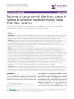
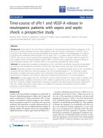
results of Kaplan-Meier analysis are reported in Fig. 1.
The 3-year OS and EFS of non-metastatic patients were
70.4 and 46.5% respectively, while in metastatic disease,
the 3-year OS was only 41.9% (Fig. 1). In non-metastatic
Number of patients
percentage (%)
˂30y
13
5.9
31–40
46
21.0
41–50
84
38.4
˃50y
76
34.7
Yes
66
31.4
No
144
68.6
Unknown
9
–
Age
Nulliparity
Number of full-term pregnancies
0
66
31.5
1–2
43
20.6
3–4
48
23.0
≥5
52
24.9
Unknown
10
–
Pre-menopausal
113
51.6
Post-menopausal
106
48.4
Yes
28
26.2
No
79
73.8
Unknown
112
–
Underweight
2
1.2
Normal
60
35.3
Overweight
50
29.4
Obese
58
34.1
Unknown
49
–
Yes
155
70.8
No
64
29.2
Yes
45
20.5
No
174
79.5
Yes
100
54.3
No
119
45.7
Yes
55
25.1
No
164
74.9
Right breast
102
46.6
Left breast
115
52.5
Bilateral
2
0.9
Menopausal staus
Familial history of BC
BMI
Peau d’orange
Oedema
Redness
Palpable mass
Side
Slaoui et al. BMC Cancer (2018) 18:713
Page 4 of 9
Table 1 Clinical data in all inflammatory breast patients
(Continued)
Table 2 pathological data in inflammatory breast cancer tumors
Variables
ER
Number of patients
percentage (%)
Yes
66
30.1
No
153
69.9
Metastatic disease
Variables
Number of patients
Percentage (%)
Positive
99
55.6
Negative
79
44.4
Unknown
41
–
Positive
124
69.7
Negative
54
30.3
Unknown
41
–
Positive
41
35.3
Negative
75
64.7
Unknown
103
–
Luminal A
43
38.7
Luminal B Her2 –
5
4.5
Luminal B Her2 +
31
27.9
Her2
8
7.2
Triple negative
24
21.6
Unknown
108
–
PgR
women, we observed a higher EFS rate associated to
SERM treatment with a significant difference (p = 0.01),
and a lower EFS rate in TNBC patients (p = 0.02), while
the other parameters did not show significant results in
Kaplan-Meier analysis.
Univariate and multivariate analysis of EFS and OS are
represented in Table 4. In univariate analysis, we found
that EFS rate is lower in patients presenting left breast tumors or bilateral tumors (HR: 1.92; 95%CI: 1.07–3.44; p =
0.02 - HR: 10.32; 95%CI: 1.32–80.47; p = 0.02), and TNBC
tumors when compared to other molecular subtypes (HR:
3.54; 95%CI: 1.13–11.05; p = 0.02). We also found that
SERM treatment is associated with a higher EFS rate (HR:
0.48; 95%CI: 0.07–0.59; p = 0.01). The multivariate model
shows that the EFS rate in non-metastatic patients is
higher in women aged more than 50 years (HR: 0.06;
95%CI: 0.00–0.61; p = 0.01) and in patients treated with
SERM (HR: 0.09; 95%CI: 0.01–0.72; p = 0.02). Univariate
analysis for OS did not demonstrate significant associations and no parameter showed close statistical significance (Table 4).
Discussion
In this study, we have intended to investigate IBC’s clinical, molecular and pathological features, and analyze
survival in Moroccan patients diagnosed with IBC between 2005 and 2010.
IBC is more frequent in North African countries, especially in Tunisia and Egypt where frequencies are 5 and
6% respectively. In our series, the frequency of IBC cases
was 4.05%, which agrees with a previous study conducted at the same institute where authors have found
an occurrence of 5% of all breast cancer cases [7].
A number of important epidemiological studies have found
that IBC occurs at a younger age than non-inflammatory
breast cancer [10]. Indeed, 65.3% of our IBC patients
were younger than 50 years, while in Algeria the percentage was 59.8%. On the other hand, the National
Cancer Institute’s Surveillance, Epidemiology, and End Results (SEER) program has shown that only 34.7% of IBC
patients were aged less than 50 years [20]. We have also
noted some differences in median age between Algerian,
Tunisian, Moroccan and American IBC series. Tunisian
patients represent the youngest age with a median age of
43.5 years [21], followed by Moroccan and Algerian patients with a median age of 47 years and 48.5 years,
Her2
Molecular subtype
Tumor size
≤ 20 mm
27
15.7
21–50 mm
55
32.0
> 50 mm
90
52.3
Unknown
47
–
N0
66
30.1
N1
84
38.4
N2
45
20.5
N3
24
11.0
Invasive carcinoma of NST
212
96.8
Invasive lobular carcinoma
4
1.8
Others
3
1.4
Yes
119
54.3
No
100
45.7
SBR I
15
7.1
SBR II
110
52.1
SBR III
86
40.8
Unknown
8
–
Lymph nodes
Histological type
Vascular invasion
SBR grade
Slaoui et al. BMC Cancer (2018) 18:713
Page 5 of 9
Table 3 Treatment data for IBC cases
Treatment
Number of patients
Percentage (%)
Neoadjuvant Chemotherapy
Yes
209
95.4
No
10
4.6
Yes
125
57.1
No
94
42.9
Mastectomy
Adjuvant Chemotherapy
Yes
50
22.8
No
169
77.2
Yes
38
17.4
No
181
82.6
Yes
104
47.5
No
115
52.5
Yes
62
28.3
No
157
71.7
Herceptine
Radiotherapy
SERM treatment
respectively [7, 22]. Whereas American patients from the
SEER program have shown the higher median age, 56 years
[20]. These comparisons show that IBC might occur at
younger age in North African populations compared to
the American one. We may explain these differences by
the possible viral etiology especially Mouse Mammary
Tumor Virus Like (MMTV-Like) as described in previous
studies led in this area [23, 24].
IBC diagnosis is entirely clinical and well established
by AJJC; it is based on the presence of inflammatory
signs especially diffuse erythema and oedema of the
breast with or without an underlying mass. In the
present study, palpable mass was detected in only 25.1%
as compared to the Algerian series where it was detected
in 31.9% of patients, while in Tunisian patients, the
majority of women (76%) had palpable mass at the time
of diagnosis [21, 22]. Once again, the Tunisian population
shows a different aspect from the Algerian and Moroccan
populations.
High BMI is considered as a risk factor for IBC and
has been analyzed in several studies but the results are
not conclusive [12, 21, 25, 26]. In the Tunisian series,
42% of IBC patients were obese while in our study we
have registered a percentage of 34.1%. Data from the
Breast Cancer Surveillance Consortium (BCSC) shows
that 32.2% of IBC patients had a high BMI [26]. In a
French study, we note that IBC patients are less obese,
and only 21% of patients presented high BMI [12].
Furthermore, results from a comparative study between
North-African series show no significant difference in
BMI between IBC and non-IBC patients, but the authors
still insist on the need for further studies because of the
increasing incidence of obesity among women in North
Africa [27].
IBC is known to show pejorative pathological characteristics. Therefore, we have found that 84.3% of the
tumors measured more than 2 cm in greatest diameter,
which joins the Algerian study findings with 88% of large
sized tumors [22]. High SBR grades (SBR II and SBR III)
were found in 92.9% of our IBC patients, 80.2% of SEER
population [20], 76% of Tunisian patients [21], and 100%
of Algerian and Egyptian patients [22, 27]. The comparative study between North African countries (Egypt,
Tunisia and Morocco) demonstrate no statistical difference regarding SBR grades [27]. At the molecular level,
many studies have documented that IBC is usually correlated to negative hormone receptors and positive
HER2 status, which confers to this disease its aggressiveness [2]. The Tunisian study has shown that 52% of IBC
tumors were ER-/PR- [28], while in Egypt only 38.9% of
the tumors were negative for hormone receptors [27].
The lack of expression of hormone receptors in the Algerian study was 26.7% for ER and 71.8% for PR [22],
while in our study IBC tumors were ER- in 44.4% and
PR- in 30.3%. According to the comparative study, these
disparities between North African countries did not
show a significant difference [27].
Studies suggest that about 20~ 40% of IBC cases are
triple negative breast cancers [2, 22, 29], which has a
worse prognosis and lower survival rates than other
breast cancer subtypes. Our study has shown the same
range with 21.6% of TNBC tumors, and EFS was also at
a lower rate in the TNBC subgroup compared to the
other molecular subgroups with a significant difference
(p = 0.02). The investigation of the seven triple negative
subtypes, as described in Lehmann study (basal-like 1
(BL1), basal-like 2 (BL2), immunomodulatory (IM), mesenchymal (M), mesenchymal stem-like (MSL), luminal
androgen receptor (LAR), and unstable (UNS)), could
contribute to resolving the differing clinical behavior
when IBC and TNBC coexist [30, 31].
Interestingly and as in the Algerian study [22], the
most prevalent subtype in our series was Luminal A
followed by luminal B HER2+, unlike the Tunisian study
where the most prevalent subtype was TNBC followed
by HER2 subtype [32]. Molecular differences between
these neighboring countries might be due to environmental and genetic factors that vary from an area to another. Further collaborative studies between these
countries are needed.
The role of adjuvant endocrine therapy in the survivorship of IBC patients was clearly investigated in several
clinical trials and concluded that SERM treatment is as efficient as chemotherapy in premenopausal breast cancer
Slaoui et al. BMC Cancer (2018) 18:713
Page 6 of 9
Fig. 1 Outcomes (OS and/or EFS) in metastatic and non-metastatic IBC patients (a, b and c), EFS in TNBC patients (e), and impact of Hormone
therapy and Radiotherapy (d and f). (OS: Overall survival; EFS: Event-Free Survival)
patients [21, 33]. Our study as well as the Tunisian one
shows a significant better EFS in IBC patients who received adjuvant SERM treatment [21].
Contrastingly, the survival rates are higher in our
series compared to the Tunisian study. In fact, the
3-year OS and EFS in our series were 70.4 and 46.5% respectively, while in Tunisia rates were 44 and 28%, respectively. This difference is mostly due to the lack of
supportive care services and the absence of access to
new drugs such as taxanes during the 1990’s, which corresponds to the period of study in Tunisian series [21].
Our study has several strengths. First, the number of patients with IBC is relatively large. Second, the large period
that was taken to select participants extended over 6 years.
Furthermore, our study represents the first large study including clinical, epidemiological, pathological and molecular characteristics of IBC in Moroccan patients.
This study has also limitations due to its retrospective
aspect. Lack of data in some parameters is the major limitation. In addition, the study has been conducted in a single
institution. Although it is the reference center of oncology
in Morocco, our patients are not representative of the
population. We also believe that short median follow-up
and loss to follow-up rates could have influenced our
survival rates. Finally, socioeconomic conditions have not
been investigated, which might have limited access to some
drugs like taxanes and Trastuzumab.
Conclusions
IBC in Morocco shows similar characteristics to those in
North African countries; however, survival rates are still
the highest when compared with neighboring countries.
Collaborative studies with prospective aspects are warranted to establish the epidemiological profile and
Slaoui et al. BMC Cancer (2018) 18:713
Page 7 of 9
Table 4 Univariate and Multivariate Cox analysis for Overall survival and Event-Free Survival in non-metastatic patients
Parameters
Event Free Survival
Overall Survival
Univariate analysis
HR
Multivariate analysis
95% CI
p
HR
Univariate analysis
95% CI
p
HR
95% CI
p
Side
Right breast
1
Left breast
1.92
1.07–3.44
0.02
2.87
1
0.98–8.42
0.05
1.36
1
0.61–3.01
0.44
Bilateral
10.32
1.32–80.47
0.02
–
–
–
5.14
0.65–40.65
0.12
0.72–2.54
0.33
1.45
0.55–3.85
0.44
1.80
0.76–4.27
0.17
Obesitya
No
1
Yes
1.36
1
1
SBR Grade
I
1
1
II
0.63
0.18–2.16
0.46
III
1.33
0.40–4.38
0.63
–
–
–
0.67
0.14–3.09
0.61
1.00
0.22–4.48
0.99
0.61–3.91
0.35
0.32–20.76
0.36
N status
N-
1
N+
1.55
1
0.79–3.04
0.19
–
0.29–2.67
0.84
0.24
–
–
1.55
0.03–1.86
0.17
2.60
Agea
˂30y
1
31–40
0.89
1
1
41–50
0.66
0.22–1.97
0.46
0.20
0.02–1.60
0.12
1.29
0.16–10.42
0.81
˃50y
0.62
0.20–1.95
0.46
0.06
0.00–0.61
0.01
2.18
0.25–18.36
0.47
Negative
1
Positive
0.69
0.85–4.98
0.11
0.42–2.53
0.94
0.04–3.81
0.44
0.78–2.60
0.98
ER
1
0.38–1.27
0.23
–
–
–
2.05
0.46–1.66
0.69
–
–
–
1.03
PgR
Negative
1
Positive
0.88
1
Her2
Negative
1
Positive
0.77
1
0.35–1.70
0.53
0.16
–
–
–
0.15–4.82
0.86
0.42
Molecular subtypea
Luminal A
1
Luminal B Her2 –
2.57
0.68–9.64
1
Luminal B Her2 +
1.78
0.59–5.34
0.29
0.31
0.07–1.34
0.11
0.43
0.04–4.23
0.47
Her2
0.57
0.06–4.91
0.61
0.11
0.01–1.18
0.06
0.57
0.05–3.81
0.98
Triple negative
3.54
1.13–11.05
0.02
1.72
0.47–6.31
0.41
1.99
1.10–10.00
0.96
0.39–1.39
0.34
1.13
0.37–3.49
0.82
0.62
0.23–1.70
0.36
0.81–1.87
0.32
0.86
1
2.57
Surgerya
No
1
Yes
0.73
1
1
a
Radiotherapy
No
1
Yes
1.20
1
0.9–1.6
0.21
0.88
1
0.28–2.73
0.82
1.23
SERM treatment
No
1
1
1
Slaoui et al. BMC Cancer (2018) 18:713
Page 8 of 9
Table 4 Univariate and Multivariate Cox analysis for Overall survival and Event-Free Survival in non-metastatic patients (Continued)
Parameters
Event Free Survival
Overall Survival
Univariate analysis
Yes
Multivariate analysis
Univariate analysis
HR
95% CI
p
HR
95% CI
p
HR
95% CI
p
0.48
0.07–0.59
0.01
0.09
0.01–0.72
0.02
1.01
0.69–1.49
0.92
: variables being adjusted for the multivariate model; significant p values are in boldface
a
understand the high frequencies of IBC in North Africa.
More studies on molecular markers are also needed to
improve IBC patients’ management and eventually their
survival rate.
Health Sciences Research, (IRSS)/ Department of Biomedical and Public
Health, Ouagadougou, Burkina Faso. 6Faculty of Medicine and Pharmacy of
Rabat, University Mohamed V Rabat, Avenue Mohammed Belarbi El Alaoui –
Souissi – BP, 6203 Rabat, Morocco. 7Biochemistry-Immunology Laboratory,
Faculty of Sciences Rabat, University Mohammed V – Agdal, Rabat, Morocco.
Abbreviations
AJCC: American Joint Committee on Cancer; BC: Breast cancer;
CI: Confidence interval; EFS: Event free survival; ER: Estrogen receptor;
FISH: Fluorescent in situ hybridization; Her2: Human epidermal growth factor
receptor 2; HR: Hazard ratio; IBC: Inflammatory Breast Cancer;
IHC: Immunohistochemistry; LN: Lymph nodes; NST: No Special Type;
PgR: Progesterone receptor; pTNM: Pathological Tumor Node Metastasis;
SBR: Scarff-Bloom Richardson classification; SERM: Selective estrogen receptor
modulation; TNBC: Triple negative breast cancer; WHO: World Health
Organization
Received: 24 October 2017 Accepted: 25 June 2018
Acknowledgements
We thank Dr. Erraki Mohamed from the epidemiology unit at the National
Institute of Oncology and his team for providing us necessary medical
records needed for the study.
Availability of data and materials
The datasets used and/or analysed during the current study are available
from the corresponding author on reasonable request.
Authors’ contributions
SM exploited data, analyzed data, conducted statistical analysis, wrote and
drafted the manuscript; AAZ and FZM co-exploited data and drafted the
manuscript; YB co-exploited data and interpreted data; MEM and YB contributed revising and critical drafting of the manuscript; MA conceived and coordinated the study, and drafted the manuscript. All authors have approved
the final manuscript for publication.
Ethics approval and consent to participate
The Ethical Committee of Biological Research, Faculty of Medicine and
Pharmacy – Rabat, approved the study under the reference number 325/13,
and no consent was needed because of the retrospective aspect of the
study. The present publication does not compromise anonymity or
confidentiality or breach local data protection laws.
Consent for publication
Not applicable.
Competing interests
The authors declare that they have no competing interests.
Publisher’s Note
Springer Nature remains neutral with regard to jurisdictional claims in
published maps and institutional affiliations.
Author details
1
Equipe de recherche ONCOGYMA, Faculty of Medicine and Pharmacy of
Rabat, University Mohamed V Rabat, Avenue Mohammed Belarbi El Alaoui –
Souissi – BP, 6203 Rabat, Morocco. 2Unité de Biologie et Recherche Médicale,
Centre National de l’Energie, des Sciences et des Techniques Nucléaires,
Rabat, Morocco. 3Pietro Annigoni Biomolecular Research Center (CERBA)/
LABIOGENE, University of Ouaga 1 Joseph KI ZERBO, UFR/SVT, Ouagadougou,
Burkina Faso. 4Laboratory of Biochemistry and Immunology, Faculty of
Sciences, University of Mohammed V-Rabat, Rabat, Morocco. 5Institute of
References
1. GLOBOCAN: Estimated incidence, mortality and prevalence worldwide in
2012. 2012.
2. Dawood S, Merajver S, Viens P, Vermeulen P, Swain S, Buchholz T, Dirix L,
Levine P, Lucci A, Krishnamurthy S. International expert panel on inflammatory
breast cancer: consensus statement for standardized diagnosis and treatment.
Ann Oncol. 2011;22(3):515–23. />3. Jaiyesimi IA, Buzdar AU, Hortobagyi G. Inflammatory breast cancer: a review.
J Clin Oncol. 1992;10(6):1014–24.
4. Tabbane F, Bahi J, Rahal K, May AE, Riahi M, Cammoun M, Hechiche M, Jaziri
M, Mourali N. Inflammatory symptoms in breast cancer. Correlations with
growth rate, clinicopathologic variables, and evolution. Cancer. 1989;64(10):
2081–9.
5. Gonzalez-Angulo AM, Hennessy BT, Broglio K, Meric-Bernstam F, Cristofanilli M,
Giordano SH, Buchholz TA, Sahin A, Singletary SE, Buzdar AU. Trends for
inflammatory breast cancer: is survival improving? Oncologist. 2007;12(8):904–12.
6. Hance KW, Anderson WF, Devesa SS, Young HA, Levine PH. Trends in
inflammatory breast carcinoma incidence and survival: the surveillance,
epidemiology, and end results program at the National Cancer Institute.
J Natl Cancer Inst. 2005;97(13):966–75.
7. Ismaili N, Elyaakoubi H, Bensouda Y, Errihani H. Demographic, clinical,
pathological, molecular, treatment characteristics and outcomes of
nonmetastatic inflammatory breast cancer in Morocco: 2007 and 2008.
Exp Hematol Oncol. 2014;3(1):1.
8. Boussen H, Bouzaiene H, Ben Hassouna J, Dhiab T, Khomsi F, Benna F,
Gamoudi A, Mourali N, Hechiche M, Rahal K. Inflammatory breast cancer in
Tunisia. Cancer. 2010;116(S11):2730–5.
9. Soliman AS, Banerjee M, Lo A-C, Ismail K, Hablas A, Seifeldin IA, Ramadan M,
Omar HG, Fokuda A, Harford JB. High proportion of inflammatory breast
cancer in the population-based Cancer registry of Gharbiah, Egypt. Breast J.
2009;15(4):432.
10. Levine PH, Veneroso C. The epidemiology of inflammatory breast cancer. In:
Seminars in oncology: 2008, vol. 35. Elsevier; 2008. p. 11–6.
11. Pogo BGT, Holland JF, Levine PH. Human mammary tumor virus in
inflammatory breast cancer. Cancer. 2010;116(S11):2741–4.
12. Lê MG, Arriagada R, Bahi J, Pfeiffer F, Cammoun M, Tabbane F, Rubino C.
Are risk factors for breast cancer similar in women with inflammatory breast
cancer and in those with non-inflammatory breast cancer? Breast. 2006;
15(3):355–62.
13. Chang S, Parker SL, Pham T, Buzdar AU, Hursting SD. Inflammatory breast
carcinoma incidence and survival. Cancer. 1998;82(12):2366–72.
14. Jimenez AM, Growney A, Behrens G, Corbridge C, Chapman DD, Usha L.
Hereditary inflammatory breast cancer associated with BRCA2 mutation: a
rare disease presentation in mother and daughter. Clin Adv Hematol Oncol.
2012;10(6):402–4.
15. Duke TJ, Jahed NC, Veneroso CC, Da Roza R, Johnson O, Hoffman D, Barsky
SH, Levine PH. A cluster of inflammatory breast cancer (IBC) in an office
setting: additional evidence of the importance of environmental factors in
IBC etiology. Oncol Rep. 2010;24(5):1277–84.
16. Sinn HP, Kreipe H. A brief overview of the WHO classification of breast
tumors, 4th edition, focusing on issues and updates from the 3rd edition.
Breast Care (Basel). 2013;8(2):149–54.
Slaoui et al. BMC Cancer (2018) 18:713
17. Singletary SE, Allred C, Ashley P, Bassett LW, Berry D, Bland KI, Borgen PI,
Clark G, Edge SB, Hayes DF. Revision of the American joint committee on
Cancer staging system for breast cancer. J Clin Oncol. 2002;20(17):3628–36.
18. Wolff AC, Hammond MEH, Hicks DG, Dowsett M, McShane LM, Allison KH,
Allred DC, Bartlett JM, Bilous M, Fitzgibbons P. Recommendations for
human epidermal growth factor receptor 2 testing in breast cancer:
American Society of Clinical Oncology/College of American Pathologists
clinical practice guideline update. Arch Pathol Lab Med. 2013;138(2):241–56.
19. Goldhirsch A, Winer EP, Coates AS, Gelber RD, Piccart-Gebhart M,
Thürlimann B, Senn H-J, Members Panel. Personalizing the treatment of
women with early breast cancer: highlights of the St Gallen international
expert consensus on the primary therapy of early breast Cancer 2013. Ann
Oncol. 2013;24(9):2206–23.
20. Dawood S, Lei X, Dent R, Gupta S, Sirohi B, Cortes J, Cristofanilli M, Buchholz
T, Gonzalez-Angulo A. Survival of women with inflammatory breast cancer:
a large population-based study. Ann Oncol. 2014;25(6):1143–51.
21. Labidi SI, Mrad K, Mezlini A, Ouarda MA, Combes JD, Abdallah MB,
Romdhane KB, Viens P, Ayed FB. Inflammatory breast cancer in Tunisia in
the era of multimodality therapy. Ann Oncol. 2008;19(3):473–80.
22. Chaher N, Arias-Pulido H, Terki N, Qualls C, Bouzid K, Verschraegen C,
Wallace AM, Royce M. Molecular and epidemiological characteristics of
inflammatory breast cancer in Algerian patients. Breast Cancer Res Treat.
2012;131(2):437–44.
23. Slaoui M, El Mzibri M, Razine R, Qmichou Z, Attaleb M, Amrani M. Detection
of MMTV-like sequences in Moroccan breast cancer cases. Infectious Agents
and Cancer. 2014;9(1):37.
24. Levine PH, Pogo BGT, Klouj A, Coronel S, Woodson K, Melana SM, Mourali N,
Holland JF. Increasing evidence for a human breast carcinoma virus with
geographic differences. Cancer. 2004;101(4):721–6.
25. Moslehi R, Freedman E, Zeinomar N, Veneroso C, Levine PH. Importance
of hereditary and selected environmental risk factors in the etiology of
inflammatory breast cancer: a case-comparison study. BMC Cancer.
2016;16(1):1.
26. Schairer C, Li Y, Frawley P, Graubard BI, Wellman RD, Buist DS, Kerlikowske K,
Onega TL, Anderson WF, Miglioretti DL. Risk factors for inflammatory breast
cancer and other invasive breast cancers. J Natl Cancer Inst. 2013;105(18):
1373–84.
27. Soliman AS, Kleer CG, Mrad K, Karkouri M, Omar S, Khaled HM, Benider A-L,
Ayed FB, Eissa SS, Eissa MS, et al. Inflammatory breast cancer in North Africa:
comparison of clinical and molecular epidemiologic characteristics of
patients from Egypt, Tunisia, and Morocco. Breast Dis. 2011;33(4):159–69.
28. Mejri N, Boussen H, Labidi S, Bouzaiene H, Afrit M, Benna F, Rahal K.
Inflammatory breast cancer in Tunisia from 2005 to 2010: epidemiologic
and anatomoclinical transitions from published data. Asian Pac J Cancer
Prev. 2014;16(3):1277–80.
29. Li J, Gonzalez-Angulo AM, Allen PK, Yu TK, Woodward WA, Ueno NT, Lucci
A, Krishnamurthy S, Gong Y, Bondy ML, et al. Triple-negative subtype
predicts poor overall survival and high Locoregional relapse in inflammatory
breast Cancer. Oncologist. 2011;16(12):1675–83.
30. Lehmann BD, Bauer JA, Chen X, Sanders ME, Chakravarthy AB, Shyr Y,
Pietenpol JA. Identification of human triple-negative breast cancer subtypes
and preclinical models for selection of targeted therapies. J Clin Invest.
2011;121(7):2750–67.
31. Masuda H, Baggerly KA, Wang Y, Iwamoto T, Brewer T, Pusztai L, Kai K,
Kogawa T, Finetti P, Birnbaum D. Comparison of molecular subtype
distribution in triple-negative inflammatory and non-inflammatory breast
cancers. Breast Cancer Res. 2013;15(6):R112.
32. Ben Hamida A, Labidi IS, Mrad K, Charafe-Jauffret E, Ben Arab S, Esterni B,
Xerri L, Viens P, Bertucci F, Birnbaum D, et al. Markers of subtypes in
inflammatory breast cancer studied by immunohistochemistry: prominent
expression of P-cadherin. BMC Cancer. 2008;8(1):1–8.
33. Low JA, Berman AW, Steinberg SM, Danforth DN, Lippman ME, Swain SM.
Long-term follow-up for locally advanced and inflammatory breast cancer
patients treated with multimodality therapy. J Clin Oncol. 2004;22(20):4067–74.
Page 9 of 9