Intra-arterial ethanol embolization augments response to TACE for treatment of HCC with portal venous tumor thrombus
Bạn đang xem bản rút gọn của tài liệu. Xem và tải ngay bản đầy đủ của tài liệu tại đây (3.25 MB, 10 trang )
Yang et al. BMC Cancer (2018) 18:101
DOI 10.1186/s12885-018-3989-2
RESEARCH ARTICLE
Open Access
Intra-arterial ethanol embolization
augments response to TACE for treatment
of HCC with portal venous tumor thrombus
Biao Yang1†, Chun-Lin Li1†, Wen-hao Guo1, Tian-qiang Qin2, He Jiao3, Ze-jun Fei3, Xuan Zhou3, Lin-jia Duan3
and Zheng-yin Liao1*
Abstract
Background: The prognosis of hepatocellular carcinoma with portal vein tumor thrombus remains extremely poor.
This pilot study aimed to evaluate the technical feasibility, effectiveness and safety of transcatheter chemoembolization
for tumors in the liver parenchyma plus intra-arterial ethanol embolization for portal vein tumor thrombus.
Methods: A pilot study was carried out on 31 patients in the treatment group (transcatheter chemoembolization plus
intra-arterial ethanol embolization) and 57 patients in the control group (transcatheter chemoembolization alone).
Enhanced computed tomography/magnetic resonance images were repeated 4 weeks after the procedure to assess
the response. Overall survival and complications were assessed until the patient died or was lost to follow-up.
Results: Median survival was 10.5 months in the treatment group (2.4 ± 1.7 courses) and 3.9 months in the control
group (1.9 ± 1 courses) (P = 0.001). Patients in the treatment group had better overall survival (at 3, 6 and 12 months,
respectively), compared to patients in the control group (90.3% vs. 59.6%, 64.5% vs. 29.8%, and 41.9% vs. 10.6%;
p = 0.001). Furthermore, the rate of portal vein tumor thrombus regression was higher in the treatment group (93.1%)
than in the control group (32.1%) (P < 0.001).
Conclusions: Based on the results of this study, transcatheter chemoembolization combined with intra-arterial
ethanol embolization may be more effective than transcatheter chemoembolization alone for treating hepatocellular
carcinoma with portal vein tumor thrombus. Intra-arterial ethanol embolization for treating portal vein tumor thrombus
is safe, feasible and prolongs overall survival.
Keywords: Portal vein tumor thrombus, Transcatheter arterial chemoembolization, Hepatocellular carcinoma,
Cone-beam computed tomography
Background
Hepatocellular carcinoma (HCC) is the fifth most frequently diagnosed cancer worldwide and the third most
frequent cause of cancer death [1, 2]. Unfortunately, HCC
has a propensity to invade the portal vein and cause portal
vein tumor thrombus (PVTT) [3], and this can be detected in 30-62% of patients with HCC [4]. PVTT is considered as an adverse prognosis factor [3]. Although liver
* Correspondence:
†
Equal contributors
1
Department of Abdominal Oncology, Cancer Center and State Key
Laboratory of Biotherapy, West China Hospital, West China Medical School,
Sichuan University, Guoxue Lane No. 37, Chengdu, Sichuan Province 610041,
People’s Republic of China
Full list of author information is available at the end of the article
resection and liver transplantation are accepted as the only
potential curative treatment for HCC patients, HCC with
PVTT has been considered a contraindication to surgery
due to poor prognosis and high surgical risk [5]. Both percutaneous ethanol injection (PEI) and radiofrequency ablation have not been shown to improve survival in cases of
HCC with neoplastic involvement of major branches of the
portal vein or main portal trunk (Vp3/Vp4), and median
survival ranged from 2.4 to 4.8 months [6]. For patients
with PVTT, sorafenib is suggested as the standard therapy
of care in the Barcelona Clinic Liver Cancer (BCLC) staging system [7, 8]. However, the median overall survival
(OS) gain with sorafenib is 5.6 months, and better treatment modalities are clearly required [9]. Yamada et al. [10]
© The Author(s). 2018 Open Access This article is distributed under the terms of the Creative Commons Attribution 4.0
International License ( which permits unrestricted use, distribution, and
reproduction in any medium, provided you give appropriate credit to the original author(s) and the source, provide a link to
the Creative Commons license, and indicate if changes were made. The Creative Commons Public Domain Dedication waiver
( applies to the data made available in this article, unless otherwise stated.
Yang et al. BMC Cancer (2018) 18:101
performed TACE in nine patients with PVTT (Vp4), and
1-month mortality was 55.5%. Among those patients, 33%
of patients died of hepatic insufficiency. Based on this
study, they concluded that TACE was contraindicated in
HCC patients with PVTT (Vp4). Recently, two studies
have indicated that transarterial chemoembolization
(TACE) could be safely performed in such patients with
no increase in morbidity or mortality [11, 12]. Most
importantly, all methods described in these studies are targeting intrahepatic lesions, and none of these focused on
treating PVTT itself. In addition to treating intrahepatic
lesions, it is our hypothesis that a therapeutic approach
including the treatment of the portal vein thrombus itself
could provide benefits in terms of OS.
Ethanol can produce an embolization effect by causing
endothelial damage and thrombus of the arteriolar lumen
of tumor feeder vessels and tumor vasculature, thereby
leading to tumor infarction [13]. Intra-arterial lipiodolethanol mixture embolization has been shown to be effective for treating HCC [14, 15]. Si et al. [16] revealed that the
feeding vessels of PVTT are complex. However, in their
study, 92.3% of PVTT had the same blood supply characteristics as intrahepatic lesions, indicating that most nutrient vessels of PVTT correspond to liver arteries. C-arm
cone beam computed tomography (CACT) angiography
could be helpful to identify the PVTT-feeding artery and
embolize the PVTT by lipiodol-ethanol mixture. In
addition, CACT provides a good method for evaluating
iodized oil deposition during the procedure. Based on these
data, we considered that intraarterial ethanol embolization
for PVTT could be feasible and effective. In the present
study, we present a new lipiodol-ethanol mixture technique,
wherein, intraarterial ethanol embolization and TACE are
combined to treat HCC patients with PVTT (Vp3/Vp4).
Page 2 of 10
computer tomography (CT)/magnetic resonance imaging
(MRI), while some patients underwent abdominal angiography. The extent of the tumor thrombus to the portal vein
was accurately assessed through these imaging techniques.
Inclusion criteria
(1) patients with unresectable HCC with PVTT (Vp3/Vp4);
(2) patients with no history of any disease-specific treatment including surgery in the past 6 months; (3) patients
who had both an international normalized ratio of < 1.5
and Child-Pugh class A/B cirrhosis; (4) patients with
an Eastern Cooperative Oncology Group (ECOG)
score ≤ 2; (5) patients who provided an informed consent; (6) patients who had no serious concurrent
medical illness; (7) patients with histologically or cytologically proven HCC, except for lesions > 2 cm, with
typical features of one dynamic imaging technique
and α-fetoprotein level > 40 ng/mL; (8) patients who had
a tumor size of up to 18 cm in the largest dimension; (9)
patients with treatment failure with sorafenib or those
who refused to receive sorafenib as treatment for advanced HCC; (10) patients who were allergic to ethanol
and refused to be included in the treatment group, and
were thereby included in the control group.
Exclusion criteria
(1) patients who were ≥ 75 years old or < 18 years old; (2)
females who were pregnant; (3) patients who had a history
of variceal bleeding within the past 3 months; (4) patients
with active hepatitis B (HBV-DNA > 1000 copies/ml); (5)
patients who have a history of acute tumor rupture with
hemoperitoneum; (6) patients with concurrent ischemic
heart disease or heart failure; (7) patients with a history of
hepatic encephalopathy; (8) patients with thrombosis of
the target hepatic artery.
Methods
Study design
Procedure
This cohort study was approved by the Local Ethics
Committee of West China Hospital, Sichuan University. A
written informed consent was obtained from each patient
after being informed of the purpose and investigational nature of the present study. This study was conducted according to the Declaration of Helsinki, and strictly adhered to
the CONSORT guidelines. Participants were recruited from
June 2014 to November 2016. Patients were stratified into
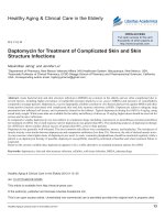
two groups according to their willingness (Fig. 1). These patients were followed up until the date of analysis in January
2017. Among these patients, 31 patients received TACE
plus intraarterial ethanol embolization (treatment group),
while 57 patients received TACE only (control group).
Intra-arterial ethanol embolization for PVTT
Eligibility criteria
All these patients were preoperatively evaluated by abdominal ultrasonography and thoracoabdominal dynamic
All TACE and intra-arterial ethanol embolization procedures were performed by two operators using the
same angiographic system (Allura Xper FD20, Philips
Healthcare). Prophylactic antibiotic treatment was not
given. The treatment procedure was performed under local
anesthesia with 3–5 mL of 1% lidocaine (Lidocaine
Hydrochloride Injection, Taiji, Chongqing, China) administered at the groin, and started with hepatic arteriography to
identify the tumor. Intra-arterial ethanol embolization
procedures were performed using a 2-F tip microcatheter
(Progreat α; Terumo Clinical Supply, Gifu, Japan) through
a 4F catheter, in order to identify the potential PVTTfeeding artery under digital subtraction angiography (DSA).
Furthermore, an angiographic unit with a 38 × 30 cm2 flat
panel detector (FPD) was used to obtain CACT images and
confirm the PVTT-feeding artery. For each CACT scan,
Yang et al. BMC Cancer (2018) 18:101
Page 3 of 10
Fig. 1 Study flow chart
312 projection images with X-ray parameters of 120 kV
and 200-300 mAs were acquired with the motorized Carm, covering a 200° clockwise arc at a rotation speed
of 20° per second. Then, 6-20 mL of contrast material
(370 mg I/mL; Omnipaque, Bracco-Sine, Shanghai,
China) were injected under 100-300 MPa over 6-10 s with
a 2-s delay. After confirmation of the PVTT-feeding artery
and before delivery of the ethiodized oil–ethanol solution,
1 mL of 1% lidocaine was instilled intra-arterially through
the microcatheter at each site of the solution administration for pain control. Lipiodol-ethanol mixture
(1:1 ratio by volume up to 15 mL) was injected at a rate of
0.5-1 ml/min until the PVTT-feeding artery was nearly
occluded, followed by embolization with 0.2-0.5-mm
gelatin-sponge particles (Gelfoam; Fukangseng, Guilin,
China) using a three-way stopcock valve and two 2.5-mL
syringes. The agents were delivered under fluoroscopic
control until the vasculature of all tumors was entirely
filed, as shown by the fluoroscopic evidence of intraarterial flow stasis or until the maximum dose was
reached. In case of acute severe abdominal pain, the procedure was temporarily suspended or stopped.
Intra-arterial ethanol embolization in managing different
types of PVTT
It was observed that repeating the treatment at 3-4 weeks
after the first treatment was often necessary to achieve
good results, since achieving complete intra-arterial
ethanol embolization in a single session was difficult in
most patients. Further treatment sessions were administered when there was CT evidence of residual tumors or
occurrence of new hepatic tumors. There was no limit
on the total number of treatment sessions.
Simple type (PVTT with only one or two feeding arteries)
Intra-arterial embolization was performed in most patients to treat the PVTT-feeding artery. The lipiodolethanol mixture was slowly injected in the feeding-artery,
followed by a gelatin sponge mixed with contrast material.
If the patient experienced acute intense pain, further injection was stopped or delayed. Two advantages of using a
gelatin sponge were observed. First, it avoids lipiodolethanol mixture regurgitation, and decreases the rate of
cholecystitis and bile leakage. Second, it stays within the
target tissue for a long time and at a higher concentration,
Yang et al. BMC Cancer (2018) 18:101
accounting for better lipiodol deposition on postprocedure CT scan. Then, an epirubicin-lipiodol mixture
was injected into the intrahepatic lesions through TACE
with a mixture of lipiodol (10 ml) and epirubicin (50 mg;
Pfizer, Wuxi, China), followed by a gelatin sponge mixed
with contrast material, until the vasculature of all tumors
was entirely filed, as shown by fluoroscopic evidence of
intra-arterial flow stasis.
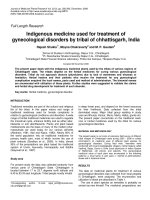
Brush type
Some patients with PVTT had several small tortuous
feeding-arteries (Fig. 2, A1-A3). Hence, it was difficult to
directly insert the microcatheter into the feeding-artery
due to anatomical variations in its location. In our technique, for this type of PVTT, TACE was first performed
in intrahepatic lesions until stasis distal to small tortuous
feeding arteries. Then, the lipiodol-ethanol mixture was
injected in the nearby PVTT-feeding artery, followed by
a gelatin sponge. Throught this method, high concentrations of lipiodol-ethanol mixture flowing through the
PVTT-feeding artery could be achieved.
Large arteriovenous fistula type
In patients with large arteriovenous fistulas, after measuring the diameter of the vessel, the distal outflow vessel
Page 4 of 10
was embolized using a larger diameter gelatin sponge before injecting the lipiodol-ethanol mixture (Fig. 2, B1-B3),
avoiding the mixture from flowing out too fast. This
helped to maintain the high lipiodol-ethanol mixture
concentration in the feeding-artery for a longer time,
providing enough time for diffusion to the PVTT.
TACE for tumors in the liver parenchyma
TACE with a mixture of lipiodol and epirubicin (50 mg of
epirubicin; Pfizer, Wuxi, China) was performed for intrahepatic lesions, followed by gelatin-sponge embolization
under DSA without CACT scan. Before the catheter was
removed from the artery, diluted heparin was injected
(50 IU/ml, 10 ml).
Follow-up and assessment indices
Primary outcomes were the overall response of PVTT to
therapy and OS. Adverse events were considered as
secondary outcomes. The latest version of the Response
Evaluation Criteria In Solid Tumors (RECIST) guidelines
(version 1.1) were used to assess the tumor response of
intrahepatic lesions to therapy [17]. Considering that the
tissue organization of postoperative residual thrombi
without viability can be persistent for months or years,
and that PVTT is always accompanied by benign
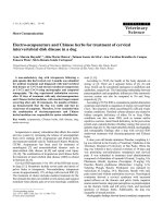
Fig. 2 Intra-arterial ethanol embolization procedure for different types of PVTT. (A1) A microcatheter was inserted into place: (1) epirubicin
injection followed by a gelatin sponge. (A2-A3) The microcatheter was withdrawn from its location: (2) lipiodol-ethanol mixture injection (1 ml/s),
followed by a gelatin sponge. (B1) Same method as described in A1. (B2) A microcatheter was placed to permit the gelatin sponge to block the
draining vessel. (B3) Lipiodol-ethanol mixture injection followed by gelatin sponge is shown
Yang et al. BMC Cancer (2018) 18:101
thrombus [18], the investigators decided not to adopt
the RECIST guidelines in assessing the efficiency on
PVTT. Therefore, the following four grades were proposed to classify PVTT response to therapy: grade 3, recanalization of the portal vein trunk or, right or left
portal vein; grade 2, decreased PVTT diameter without
recanalization of any branch of the portal vein; grade 1,
neither shrinkage to qualify for grade 2 nor increase to
qualify for grade 0; grade 0, PVTT diameter increased
by 20%. Regression of PVTT to grade3/grade2 and
complete/partial response of intrahepatic lesions based
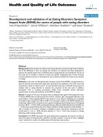
on the modified RECIST criteria were considered as significant responses to interventional therapy. At 2-7 days
after the procedure, CT scan revealed lipiodol deposits
within PVTT (Fig. 3, C1-D1). Enhanced CT/MRI
Page 5 of 10
images, which were evaluated by two experienced radiologists, were repeated at 4 weeks after the procedure, in
order to assess the response. OS was defined as the period
from the date of first treatment to the date of death, or
censorship at the date of last follow-up if the patient is still
alive. Repeated TACE was performed if lesion diameter increased or new lesions were found. Repeated intra-arterial
ethanol embolization was suspended if PVTT diameters
did not increase, or complete embolization of the visible
PVTT-feeding artery was achieved.
Statistical analyses
Continuous baseline characteristic variables were compared by Student t-test, ranked data was compared by
rank-sum test, and categorical variables were compared
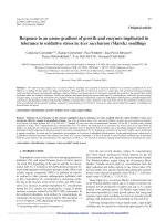
Fig. 3 Intraarterial ethanol embolization with TACE in a 64-year-old male with HCC and PVTT (Vp3). (A1, A2) CT scan in the portal venous phase
highlighting PVTT in the right portal vein (arrow) is shown; (A3) PVTT-feeding artery identified on CT. (B1) PVTT-feeding artery identified on DSA
by superselective catheterization of the feeding artery using a microcatheter; (B2, B3) enhanced C-arm CT was performed to further confirm the
PVTT-feeding artery; (C1-C3) axial CT showing lipiodol-ethanol mixture deposition within PVTT. (D1-D3) Follow-up images showing stable lipiodol-ethanol
mixture deposition within PVTT at 3, 6, and 12 months after the operation
Yang et al. BMC Cancer (2018) 18:101
by χ2-test. Survival curves were estimated using the
Kaplan-Meier method. Data were analyzed with SPSS
version 20.0 (SPSS Inc., Chicago, IL, USA). All statistical
tests used were two-sided, and P < 0.05 was considered
statistically significant.
Page 6 of 10
in 7%, 31%, 41.3% and 20.7% of patients in the treatment
group, compared to 11.3%, 9.4%, 66.1%, and 13.2% of patients in the control group, respectively (P = 0.61, Table 2).
PVTT radiographic response rate to therapy was significantly higher in the treatment group (37.9%), compared
with the control group (13.2%) (P < 0.001, Table 2).
Results
Patient demographics
In the treatment group, the mean age of patients (n = 31)
was 54.3 ± 11.9 years old. These patients received TACE
with 50 mg of epirubicin dissolved in 10 ml of lipiodol for
intrahepatic lesions combined with intra-arterial ethanol
embolization (alcohol/lipiodol, 7.8 ± 3.9 ml) for PVTT
(Fig. 3). In the control group, the mean age patients
(n = 57) was 54.2 ± 13.4 years old. These patients received
TACE with 50 mg epirubicin alone (Table 1). The mean
course of procedures in the treatment and control groups
was 2.4 ± 1.7 and 1.9 ± 1.0, respectively (P = 0.18). Results
related to the end-points of the present study are summarized in Table 2.
Survival
Thirty-eight patients died during follow-up: 14 patients in
the treatment group and 24 patients in the control group.
Median survival was 10.5 months and mean survival was
11.5 ± 8.5 months (95% CI: 8.6-15.3) in the treatment
group, while median survival was 3.8 months and mean
survival was 5.0 ± 4.0 months (95% CI: 4-6) in the control
group (Fig. 4). Probabilities of survival at 3, 6 and
12 months were significantly higher in the treatment
group than in the control group (90.3% vs. 59.6%, 64.5%
vs. 29.8%, and 41.9% vs. 10.6%) (P = 0.001, Table 2). The
mean survival of patients classified as Vp3 and Vp4 in the
treatment and control group was 16.4 ± 10.8 vs. 9.2 ± 6.1
(P = 0.004) and 5.9 ± 4.7 vs. 4.3 ± 3.7 (P < 0.001). The mean
survival of patients classified as Child-Pugh A and
Child-Pugh B in the treatment and control groups
was 13.2 ± 12.6 vs. 4.0 ± 3.0 (P < 0.001) and 11.0 ± 9.4
vs. 5.2 ± 4.4 (P = 0.003), respectively.
Safety
Ninety complications occurred in the treatment group,
while 125 complications occurred in the control group.
The duration of the procedure was significantly longer in
the treatment group than in the control group (1.9 ± 0.6 h
vs. 0.6 ± 0.2 h, respectively; P < 0.001). No treatmentrelated death, pulmonary embolism, renal damage, renal
failure, respiratory failure, cholecystitis, or cholangitis
were observed during follow-up in both groups. The other
main adverse events are presented in Table 3.
Significant clinical response
Complete response/partial response/stable/progressive
disease of intrahepatic lesions at 1 month was observed
Discussion
PVTT is an independent prognostic factor for patients
with HCC. The reported median survival for untreated
HCC patients with PVTT (Vp3/Vp4) was 2.7 months,
whereas survival in patients without PVTT was
24.4 months [3]. Intra-arterial ethanol embolization has
been used in HCC cases in a similar approach to TACE,
which has exhibited a higher 1-year OS rate (93.3% vs.
73.3%) and greater lipiodol retention (89.5% vs. 47.5%);
but its specific impact on PVTT has not been previously
studied [19]. The present study demonstrated that our
therapeutic approach may be more effective than TACE
in HCC patients with PVTT (Vp3/Vp4). The percentage
of patients with more than three nodes or diffused
tumors, or huge tumors in the treatment group was
higher than in the control group. This selection bias
may be due to the patient’s decision, and may have lessened the possibility to evaluate the advantages in the
treatment group. Despite this limitation, there was a significant trend toward OS improvement vs. the control
group. We found that occluding the arterial supply to
PVTT with the help of intraarterial ethanol embolization
not only resulted in the recanalization of the portal vein,
but also significantly improved survival in this patient
group. In the treatment group, patients had more complications compared with those in the control group,
which was possibly correlated to the destruction of the
PVTT-feeding artery induced by ethanol. The potential
risk of thrombus at the infusion port was higher due to
longer procedure time in the treatment group. This is
why some blood was extracted from both the infusion
port and connected catheter before removing the needle
from the infusion port, followed by flushing with diluted
heparin. The duration of post-procedure abdominal pain
was longer in the treatment group than in the control
group, which was possibly due to the ethanol itself.
Yamada et al. [10] first reported that HCC patients with
Vp3/Vp4 PVTT treated with TACE had a 28.6% 1-year
survival rate. Recently, Chung et al. [20] reported a
30% 1-year survival rate and 28.2% PVTT response rate.
Georgiades et al. [12] and Takayasu et al. [21] reported a
25% and 35% 1-year survival rate, respectively. Peng et al.
[11] reported that PVTT had a 36.1% 1-year survival rate.
In contrast, in the present study, the 1-year survival rate
was 41.9% for patients in the treatment group vs. 10.6%
for patients in the control group, which show that patients
in the treatment group had a higher OS rate than those
Yang et al. BMC Cancer (2018) 18:101
Page 7 of 10
Table 1 Baseline patient characteristics
Table 1 Baseline patient characteristics (Continued)
Variable
Treatment group
(n = 31)
Control group
(n = 57)
Agea (year)
54.29 ± 11.87
54.16 ± 13.42
Genderb
0.96
Variable
Female
Treatment group
(n = 31)
P-value
Total bilirubin levela (μmol/L)
22.15 ± 12.63
20.91 ± 9.19
0.60
39.22 ± 8.12
41.68 ± 5.91
0.11
< 200
10 (23.8)
22 (38.6)
25 (80.6)
49 (86)
Albumin levela (g/L)
6 (19.4)
8 (14)
α-Fetoproteinb (ng/mL)
b
Control group
(n = 57)
Laboratory Tests
0.52
Male
Classification of PVTT
P-value
0.37
0.34
Vp3
10 (32.3)
24 (42.1)
200–1000
4 (29)
11 (19.3)
Vp4
21 (67.7)
33 (57.9)
> 1000
17 (42.9)
24 (42.1)
Prothrombin timea (seconds)
12.86 ± 1.66
13.01 ± 1.17
0.42
19.41 ± 2.35
19.71 ± 1.23
0.63
ECOG performanceb
0.43
0
17 (54.8)
34 (59.6)
1
9 (29.1)
20 (35.1)
5(16.1)
3(5.3)
2
Cause of liver disease
b
a
Thrombin time (seconds)
Mean ± standard deviation (SD); bn (%)
a
1.00
HBV
31 (100)
57 (100)
Other
0 (0)
0 (0)
Absent
8 (25.8)
13 (22.8)
Present
23 (74.2)
44 (77.2)
Lung
2 (6.5)
2 (3.5)
Others
0 (0)
0 (0)
Liver Cirrhosisb
0.75
Distant metastasisb
0.53
Ascitesb
0.71
Absent
26 (83.9)
46 (80.7)
Mild
1(3.2)
3(5.3)
Moderate
3(9.7)
7(12.3)
Massive
1(3.2)
1(1.7)
A
25 (80.6)
33 (57.9)
B
6 (19.4)
24 (42.1)
Child-Pugh scoreb
0.03
Tumor descriptionb
0.83
Number of tumors
1
6 (19.4)
11(19.3)
2
4 (12.9)
9(15.8)
≥3
21 (67.7)
37 (64.9)
7.00
8.5
Size of largest tumor,
median (cm)
0.34
Angiography of PVTT feeding-artery
Timea (seconds)
8.65 ± 1.74
–
Pressurea (MPa)
243.55 ± 92.86
–
Speeda (ml/s)
1.63 ± 0.43
–
1.92 ± 0.62
0.62 ± 0.18
Alcohola (ml)
7.77 ± 3.95
–
Epirubicina (mg)
50.00
50.00
Number of sessions
2.42 ± 1.71
1.89 ± 0.98
Table 2 Comparison of primary and secondary outcomes
between the treatment and control groups
Outcome
Treatment group Control group P-value
Tumor responseb
n = 29
n = 53
Complete response
2 (7)
6 (11.3)
Partial response
9 (31)
5 (9.4)
Stable disease
12 (41.3)
35 (66.1)
Progressive disease
6 (20.7)
7 (13.2)
Portal vein tumor thrombus n = 29
responseb
< 0.001
0.18
n = 53
Grade 3
6 (20.7)
1 (1.9)
Grade 2
15 (51.7)
6 (11.3)
Grade 1
6(20.7)
10 (18.9)
Grade 0
Procedure
Total procedure
Timea (hour)
reported in literature [11, 12]. In the present study, PVTT
response rate was higher than those were reported by
Yamada et al. [10]. Percutaneous ethanol injection has been
reported to be efficient for treating PVTT on the basis that
ethanol is diffused within cells [22]. Nevertheless, ethanol
in PEI is limited both in terms of diffusion and of adapting
the ethanol dose. In contrast, intra-arterial ethanol
embolization, a method based on the infusion of ethanol
into the artery, can achieve a more efficient diffusion without damaging the normal liver parenchyma, and allows the
ethanol dose to be easier controlled according to the pain
degree of the patient or the distribution of ethanol.
Diagnosing PVTT remains difficult. Percutaneous
puncture biopsy is invasive, and is associated with a high
risk of tumor seeding along the needle track. Color
2 (6.9)
36 (67.9)
n = 31
n = 57
At 3-months
90.3
59.6
At 6-months
64.5
29.8
At 12-months
41.9
10.6
Overall survival (%)
0.61
< 0.001
0.001
Values are depicted as n (%)
b
Before the statistics were performed, two patients died in treatment group
and control group, respectively
Yang et al. BMC Cancer (2018) 18:101
Page 8 of 10
Fig. 4 A graphical representation of the overall survival of patients in the two groups by the Kaplan-Meier method. a The total overall survival
curve in the two groups. b The overall survival of patients diagnosed with Vp3 in the two groups. c The overall survival of patients with Vp4 in
the two groups
Doppler sonography (CDS) has been widely used with
the method of pulsatile flow. PVTT has been diagnosed
in approximately 62% of patients using the presence of a
pulsatile flow as its diagnostic criterion [23]. DSA
together with CACT combines the advantages of DSA
and contrast-enhanced CT, which can provide slice imaging and dynamic flow information. Wallace et al. [24]
reported that 60% of CACT images contain information
that were not found in DSA, and influenced the treatment procedure in 19% of cases. CACT detects HCC
with greater accuracy and sensitivity than both DSA and
CT [25]. In addition, CACT data sets can be viewed in
three-dimension and slice-images. These information
provides a more effective therapy by delivering an increased amount of lipiodol-ethanol mixture to the target,
while sparing uninvolved parenchyma exposure from
toxic agents such as the gallbladder.
In an animal experiment carried out by Kan et al. [13]
and the use of absolute ethanol, the endothelial cell was
denuded from the vascular wall, its protoplasm precipitated and a fracture in the vascular wall to the level of
the internal elastic lamina was formed, followed by the
Table 3 Comparison of adverse events related to the procedure
Adverse event
Treatment group
(n = 31)
Control group
(n = 57)
Fatigue
16
18
Gastrointestinal hemorrhage
2
1
Fever
14
19
Abdominal pain
31
56
Vomiting
8
15
Chest pain
2
0
Per procedure vomiting
3
1
Back pain
12
7
Loss of appetite
2
8
Total
90
125
shrinking of lesions. Hence, ethanol has been widely
used for vascular malformations [16]. Ethanol is a better
embolic agent than lipiodol, and can lead to vascular
endothelial destruction. However, ethanol is not radioopaque, and its flow and speed are difficult to visualize.
In contrast, lipiodol-ethanol mixture (in a 1:1 ratio) is
visible during injection. At the same time, it maintains
the potency of absolute ethanol in the target vasculature,
and is not diluted by aqueous solutions; which are necessary to avoid regurgitation and ectopic embolization
[15]. From the fluoroscopic observation on an animal
model, dual embolization could be induced by the slow infusion of an insoluble substance such as the lipiodolethanol mixture, which appears as small droplets passing
through the hepatic sinusoids and to the portal vein [14].
This achieves complete embolization in both arteries that
supply the tumor and its adjacent parenchymal portal
veins [14]. The long-lasting embolization of both the
arterioles and portal venules is highly effective in causing
infarction of the whole tumor including the tumor border,
which is commonly supplied by portal venules [26]. The
treatment group, unlike a gelatin sponge, not only induces
tumor ischemia and hypoxia, but also diffuses within
tumor cells [15, 27, 28]. Ischemia and hypoxia may be potent stimulators of angiogenesis and carcinogenesis, which
promote collateral circulation and the restoration of
tumor blood supply; and these may eventually lead to
tumor proliferation and recurrence [29, 30].
Limitations
The main limitations of the present study are small
sample size, non-randomized controls, relatively short
follow-up, and a single center experience. Therefore, performing prospective randomized studies are warranted to
confirm these results. Any new treatment should ideally
be compared with the reference standard for the disease
at that stage. The evidence based standard of care for
locally advanced HCC is sorafenib. However, few Chinese
Yang et al. BMC Cancer (2018) 18:101
people able to bear the high cost, especially in developing
countries [31]. Moreover, there is no standard treatment
for patients with treatment failure with sorafenib. A recent
study [32] demonstrated that patients with PVTT (Vp3),
who received TACE or sorafenib, had a poor 1-year OS
(35.7 vs. 26.5 months). Hence, for this group of patients,
we propose TACE treatment. Although, there was no statistical significance between the compared groups in terms
of treatment courses, more number of courses of repeated
TACE in HCC provided better results in the treatment
group. Clearly, a higher proportion of Child-Pugh A
patients in the treatment group may contribute to explain
the longer OS.
Conclusion
Although intra-arterial ethanol embolization combined
with TACE does not represent a cure for HCC with
PVTT, the principal goals of significant safety, effectiveness and OS could be achieved. In the present pilot
study, considering the higher survival rate for TACE plus
intra-arterial ethanol embolization compared with TACE
alone, this therapeutic approach may be the treatment of
choice for HCC patients with PVTT (Vp3/Vp4). However further prospective studies are needed to confirm
the present data.
Abbreviations
CACT: C-arm cone beam computed tomography; CDS: Color doppler
sonography; CT: Computer tomography; DSA: Digital subtraction
angiography; ECOG: Eastern Cooperative Oncology Group; FPD: Flat panel
detector; HCC: Hepatocellular carcinoma; MRI: Magnetic resonance imaging;
OS: Overall survival; PEI: Percutaneous ethanol injection; PVTT: Portal vein
tumor thrombus; RECIST: Response evaluation criteria in solid tumors;
TACE: Transarterial chemoembolization
Acknowledgements
Not applicable
Funding
This work was supported by the National Nature Science Foundation of
China (Grant no. 81470141).
Availability of data and materials
The datasets used and/or analyzed during the present study are available
from the corresponding author on reasonable request.
Authors’ contributions
BY and CLL participated in the study design, the collection, analysis and
extraction of data, and in writing the manuscript. WHG provided great help
in terms of the study design and ethical application. LJD resolved all
discrepancies as an intercessor. TQQ, a statistician, contributed to the
interpretation of data, statistical analysis and the SPSS software. HJ, ZJF and
XZ, who are radiologists, provided help in evaluating medical images as well
as in performing CT/MRI/DSA scans and the acquisition of data. The authors
would also like to thank Dr. LZY for providing support in terms of the study
design and coordination, founding (National Nature Science Foundation of
China to LZY), and the draft revision throughout the entire duration of the
study. All authors read and approved the final manuscript.
Ethics approval and consent to participate
This cohort study was approved by the Local Ethics Committee of West
China Hospital, Sichuan University.
Page 9 of 10
Consent for publication
Written informed consent was obtained from each patient after being
informed of the purpose and investigational nature of this study.
Competing interests
The authors declare that they have no competing interests.
Publisher’s Note
Springer Nature remains neutral with regard to jurisdictional claims in
published maps and institutional affiliations.
Author details
1
Department of Abdominal Oncology, Cancer Center and State Key
Laboratory of Biotherapy, West China Hospital, West China Medical School,
Sichuan University, Guoxue Lane No. 37, Chengdu, Sichuan Province 610041,
People’s Republic of China. 2Chinese Evidence-Based Medicine Centre, West
China Hospital, West China Medical School, Sichuan University, Chengdu,
People’s Republic of China. 3Department of Radiology, West China Hospital,
West China Medical School, Sichuan University, Chengdu, People’s Republic
of China.
Received: 19 April 2017 Accepted: 15 January 2018
References
1. El-Serag HB, Rudolph KL. Hepatocellular carcinoma: epidemiology and
molecular carcinogenesis. Gastroenterology. 2007;132(7):2557–76.
2. Zhu K, Chen J, Lai L, Meng X, Zhou B, Huang W, Cai M, Shan H.
Hepatocellular carcinoma with portal vein tumor thrombus: treatment with
transarterial chemoembolization combined with sorafenib–a retrospective
controlled study. Radiology. 2014;272(1):284–93. />radiol.14131946. Epub 14132014 Apr 14131946
3. Llovet JM, Bustamante J, Castells A, Vilana R, Ayuso Mdel C, Sala M, Bru C,
Rodes J, Bruix J. Natural history of untreated nonsurgical hepatocellular
carcinoma: rationale for the design and evaluation of therapeutic trials.
Hepatology. 1999;29(1):62–7.
4. Esnaola NF, Mirza N, Lauwers GY, Ikai I, Regimbeau JM, Belghiti J, Yamaoka Y,
Curley SA, Ellis LM, Nagorney DM, et al. Comparison of clinicopathologic
characteristics and outcomes after resection in patients with hepatocellular
carcinoma treated in the United States, France, and Japan. Ann Surg.
2003;238(5):711-719.
5. Chen JS, Wang Q, Chen XL, Huang XH, Liang LJ, Lei J, Huang JQ, Li DM,
Cheng ZX. Clinicopathologic characteristics and surgical outcomes of
hepatocellular carcinoma with portal vein tumor thrombosis. J Surg Res.
2012;175(2):243–50.
6. Ohkubo T, Yamamoto J, Sugawara Y, Shimada K, Yamasaki S, Makuuchi M,
Kosuge T. Surgical results for hepatocellular carcinoma with macroscopic
portal vein tumor thrombosis. J Am Coll Surg. 2000;191(6):657–60.
7. Song Do S, Song MJ, Bae SH, Chung WJ, Jang JY, Kim YS, Lee SH, Park JY,
Yim HJ, Cho SB, et al. A comparative study between sorafenib and hepatic
arterial infusion chemotherapy for advanced hepatocellular carcinoma with
portal vein tumor thrombosis. J Gastroenterol. 2015;50(4):445–54.
8. Bruix J, Sherman M, Llovet JM, Beaugrand M, Lencioni R, Burroughs AK,
Christensen E, Pagliaro L, Colombo M, Rodes J. Clinical management of
hepatocellular carcinoma. Conclusions of the Barcelona-2000 EASL
conference. European Association for the Study of the Liver. J Hepatol.
2001;35(3):421-430.
9. Yu SJ, Kim YJ. Effective treatment strategies other than sorafenib for the
patients with advanced hepatocellular carcinoma invading portal vein.
World J Hepatol. 2015;7(11):1553.
10. Yamada R, Sato M, Kawabata M, Nakatsuka H, Nakamura K, Takashima S.
Hepatic artery embolization in 120 patients with unresectable hepatoma.
Radiology. 1983;148(2):397–401.
11. Peng ZW, Guo RP, Zhang YJ, Lin XJ, Chen MS, Lau WY. Hepatic resection
versus transcatheter arterial chemoembolization for the treatment of
hepatocellular carcinoma with portal vein tumor thrombus. Cancer.
2012;118(19):4725–36.
12. Georgiades CS, Hong K, D’Angelo M, Geschwind JF. Safety and efficacy
of transarterial chemoembolization in patients with unresectable
hepatocellular carcinoma and portal vein thrombosis. J Vasc Interv Radiol.
2005;16(12):1653–9.
Yang et al. BMC Cancer (2018) 18:101
13. Kan Z, Wallace S. Transcatheter liver lobar ablation: an experimental trial in
an animal model. Eur Radiol. 1997;7(7):1071–5.
14. Park JH, Han JK, Chung JW, Choi BI, Han MC, Kim YI. Superselective
transcatheter arterial embolization with ethanol and iodized oil for
hepatocellular carcinoma. J Vasc Interv Radiol. 1993;4(3):333–9.
15. Gu Y-K. Transarterial embolization ablation of hepatocellular carcinoma with
a lipiodol-ethanol mixture. World J Gastroenterol. 2010;16(45):5766.
16. Si Q, Huang SX, Tong W, Qian XL, Lv XP, Huang YL. Clinical study of blood
perfusion characteristics in liver cancer and portal vein tumor thrombosis by
CEUS and CDUS. Military Medical Journal of Southeast China. 2011;13(1):20–2.
17. Eisenhauer E, Therasse P, Bogaerts J, Schwartz L, Sargent D, Ford R, Dancey J,
Arbuck S, Gwyther S, Mooney M. New response evaluation criteria in solid
tumours: revised RECIST guideline (version 1.1). Eur J Cancer. 2009;45(2):228–47.
18. Yoon JH, Kim HC, Chung JW, Yoon JH, Jae HJ, Park JH. CT findings of
completely regressed hepatocellular carcinoma with main portal vein tumor
thrombosis after transcatheter arterial chemoembolization. Korean J Radiol.
2010;11(1):69–74.
19. Yu SC, Hui JW, Hui EP, Mo F, Lee PS, Wong J, Lee KF, Lai PB, Yeo W.
Embolization efficacy and treatment effectiveness of transarterial therapy for
unresectable hepatocellular carcinoma: a case-controlled comparison of
transarterial ethanol ablation with lipiodol-ethanol mixture versus transcatheter
arterial chemoembolization. J Vasc Interv Radiol. 2009;20(3):352–9.
20. Chung J, Park JH, Han JK, Choi B, Han M. Hepatocellular carcinoma and
portal vein invasion: results of treatment with transcatheter oily
chemoembolization. AJR Am J Roentgenol. 1995;165(2):315–21.
21. Takayasu K, Arii S, Ikai I, Omata M, Okita K, Ichida T, Matsuyama Y,
Nakanuma Y, Kojiro M, Makuuchi M, et al. Prospective cohort study of
transarterial chemoembolization for unresectable hepatocellular carcinoma
in 8510 patients. Gastroenterology. 2006;131(2):461–9.
22. Gao F, Gu YK, Fan WJ, Zhang L, Huang JH. Evaluation of transarterial
chemoembolization combined with percutaneous ethanol ablation for large
hepatocellular carcinoma. World J Gastroenterol. 2011;17(26):3145–50.
23. Richard L, Eichner L, Santiguida LA. Portal vein thrombosis in patients with
cirrhosis: does Sonographic detection of lntrathrombus flow allow differentiation
of benign and malignant thrombus? Am J Surg. 1988;155(1):70–5.
24. Wallace MJ, Murthy R, Kamat PP, Moore T, Rao SH, Ensor J, Gupta S, Ahrar K,
Madoff DC, SE MR, et al. Impact of C-arm CT on hepatic arterial interventions
for hepatic malignancies. J Vasc Interv Radiol. 2007;18(12):1500–7.
25. Higashihara H, Osuga K, Onishi H, Nakamoto A, Tsuboyama T, Maeda N,
Hori M, Kim T, Tomiyama N. Diagnostic accuracy of C-arm CT during
selective transcatheter angiography for hepatocellular carcinoma:
comparison with intravenous contrast-enhanced, biphasic, dynamic MDCT.
Eur Radiol. 2012;22(4):872–9. />Epub 02011 Nov 00326
26. Matsui O, Kadoya M, Yoshikawa J, Gabata T, Arai K, Demachi H, Miyayama S,
Takashima T, Unoura M, Kogayashi K. Small hepatocellular carcinoma:
treatment with subsegmental transcatheter arterial embolization. Radiology.
1993;188(1):79–83.
27. Tanaka K, Okazaki H, Nakamura S, Endo O, Inoue S, Takamura Y, Sugiyama M,
Ohaki Y. Hepatocellular carcinoma: treatment with a combination therapy of
transcatheter arterial embolization and percutaneous ethanol injection.
Radiology. 1991;179(3):713–7.
28. Tanaka K, Nakamura S, Numata K, Okazaki H, Endo O, Inoue S, Takamura Y,
Sugiyama M, Ohaki Y. Hepatocellular carcinoma: treatment with percutaneous
ethanol injection and transcatheter arterial embolization. Radiology.
1992;185(2):457–60.
29. Mathupala SP, Rempel A, Pedersen PL. Glucose catabolism in cancer cells:
identification and characterization of a marked activation response
of the type II hexokinase gene to hypoxic conditions. J Biol Chem.
2001;276(46):43407–12. Epub 42001 Sep 43413
30. Liu C, Lu P, Lu Y, Xu H, Wang S, Chen J. Clinical implications of metastatic
lymph node ratio in gastric cancer. BMC Cancer. 2007;7:200.
31. Liu L, Zhang C, Zhao Y, Qi X, Chen H, Bai W, He C, Guo W, Yin Z, Fan D, et al.
Transarterial chemoembolization for the treatment of advanced hepatocellular
carcinoma with portal vein tumor thrombosis: prognostic factors in a singlecenter study of 188 patients. Biomed Res Int. 2014;2014:194278.
32. Lee JM, Jang BK, Lee YJ, Choi WY, Choi SM, Chung WJ, Hwang JS, Kang KJ,
Kim YH, Chauhan AK, et al. Survival outcomes of hepatic resection compared
with transarterial chemoembolization or sorafenib for hepatocellular carcinoma
with portal vein tumor thrombosis. Clinical and molecular hepatology.
2016;22(1):160–7.
Page 10 of 10
Submit your next manuscript to BioMed Central
and we will help you at every step:
• We accept pre-submission inquiries
• Our selector tool helps you to find the most relevant journal
• We provide round the clock customer support
• Convenient online submission
• Thorough peer review
• Inclusion in PubMed and all major indexing services
• Maximum visibility for your research
Submit your manuscript at
www.biomedcentral.com/submit