3,6-Dihydroxyflavone regulates microRNA34a through DNA methylation
Bạn đang xem bản rút gọn của tài liệu. Xem và tải ngay bản đầy đủ của tài liệu tại đây (1.14 MB, 9 trang )
Peng et al. BMC Cancer (2017) 17:619
DOI 10.1186/s12885-017-3638-1
RESEARCH ARTICLE
Open Access
3,6-Dihydroxyflavone regulates microRNA34a through DNA methylation
Xiaoli Peng2, Hui Chang1, Junli Chen1, Qianyong Zhang1, Xiaoping Yu1,2* and Mantian Mi1*
Abstract
Background: Breast cancer is the common cancer in China. In previous study, we determined that 3,6dihydroxyflavone (3,6-DHF) increases miR-34a significantly in breast carcinogenesis, but the mechanism remains
unclear.
Methods: We used qRT-PCR to analyze miR-34a and ten-eleven translocation (TET)1, TET2, TET3 levels in breast
cancer cells. With a cellular breast carcinogenesis model and an experimental model of carcinogenesis in rats, TET1
levels were evaluated by western blot analysis and immunofluorescence. TET1 and 5hmC (5-hydroxymethylcytosine)
levels were evaluated by immunofluorescence in nude mouse xenografts of MDA-MB-231 cells. Chromatin
immunoprecipitation(ChIP) assayed for TET1 on the TET1 promoter, and dot blot analysis of DNA 5hmC was
performed in MDA-MB-231 cells. We evaluated the mechanism of 3,6-DHF on the expression of tumor suppressor
miR-34a by transfecting them with DNA methyltransferase (DNMT)1 plasmid and TET1 siRNA in breast cancer cells.
Methylation-specific PCR detected methylation of the miR-34a promoter.
Results: First, we found that 3,6-DHF promotes the expression of TET1 during carcinogen-induced breast
carcinogenesis in MCF10A cells and in rats. 3,6-DHF also increased TET1 and 5hmC levels in MDA-MB-231 cells.
Further study indicated that TET1 siRNA and pcDNA3/Myc-DNMT1 inhibited the 3,6-DHF reactivation effect on
expression of miR-34a in breast cancer cells. Methylation-specific PCR assays indicated that TET1 siRNA and
pcDNA3/Myc-DNMT1 inhibit the effect of 3,6-DHF on the demethylation of the miR-34a promoter.
Conclusions: Our study showed that 3,6-DHF effectively increases TET1 expression by inhibiting DNMT1 and DNA
hypermethylation, and consequently up-regulates miR-34a in breast carcinogenesis.
Keywords: Breast cancer, Carcinogenesis, 3,6-Dihydroxyflavone, TET1, DNMT1, miR-34a, Methylation
Background
Breast cancer is a common cancer and the leading cause
of cancer deaths in China [1]. Current chemotherapy
treatments for breast cancer cause serious side effects;
plant-based bioactive compounds are desired as chemotherapeutic drugs in cancer treatment due to their
minimal side effects. Dietary flavonoids have been identified for cancer therapy and prevention because of their
ability to suppress cancer cell proliferation [2], induce
cell-cycle arrest and promote apoptosis [3]. In our previous experiment, we have identified that 3,6-DHF has the
effect to inhibit breast carcinogenesis [4]. In the present
* Correspondence: ;
1
Research Center for Nutrition Correspondence and Food Safety, Third
Military Medical University, Chongqing Key Laboratory of Nutrition and Food
Safety, 30 Gaotanyan Street, Shapingba District, Chongqing 400038, China
Full list of author information is available at the end of the article
study, we investigate the mechanism of 3,6-DHF’s
anti-carcinogenesis property in the context of breast
carcinogenesis.
Phytochemicals extracted from plants may modulate
and reverse gene transcription, aberrant epigenetic
changes, including DNA methylation, histone modification and non-coding RNA (miRNA) alteration [5]. DNA
methylation change patterns can occur throughout the
life of an individual; some changes can be a physiological
response to environmental changes, whereas others
might be associated with a pathological process such as
oncogenic transformation [6]. DNA methylation dysregulation contribute to silencing tumor suppressor genes
or activating oncogenes in tumor progression [7, 8].
DNA methyltransferases (DNMTs) play key roles in
epigenetic methylation of DNA. DNMTs overexpression
© The Author(s). 2017 Open Access This article is distributed under the terms of the Creative Commons Attribution 4.0
International License ( which permits unrestricted use, distribution, and
reproduction in any medium, provided you give appropriate credit to the original author(s) and the source, provide a link to
the Creative Commons license, and indicate if changes were made. The Creative Commons Public Domain Dedication waiver
( applies to the data made available in this article, unless otherwise stated.
Peng et al. BMC Cancer (2017) 17:619
results in hypermethylation and DNMT1 deletion leads
to DNA demethylation [9]. The ten-eleven translocation
(TET) family (TET1/2/3) are Fe(II)- and 2-oxoglutarate
(2OG)-dependent dioxygenases that convert 5-methylcyt
osine to 5-hydroxymethylcytosine(5hmC), and play potential roles in epigenetic through DNA demethylation
[10]. Dysfunction of TET and DNMT activity is considered an epigenetic hallmark of human cancers [11, 12];
Disruption in DNA methylation and demethylation
dynamics is intimately implicated in carcinogenesis [13].
Our previous research found that 3,6-DHF inhibits
DNMT1 effectively. We propose that 3,6-DHF would have
an effect on the balance of methylation and demethylation
in breast carcinogenesis and breast cancer cells.
DNA hypermethylation is a major epigenetic event which
is associated with tumor suppressor gene silencing. MiR-34a
is a miRNA regulated by the p53 network at the transcriptional level and has been shown to be remarkably down
regulated in a variety of cancers. Research shows that the
miR-34a promoter hypermethylation leads to its epigenetic
inactivation [14–17]. MiR-34a may counteract the p53 response to DNA damage [18] and miR-34a hypermethylation
occurs in pre-cancerous lesions in tumor formation [19].
Upregulating miR-34a changes its target genes expression
involving in multiple signal transduction pathways, represses
tumor growth significantly [20, 21], and may be an efficient
strategy for cancer treatment. In our previous research, we
observed that 3,6-DHF up-regulates the miR-34a and overexpressed miR-34a promoted cytotoxicity and apoptosis in
breast cancer cells induced by 3,6-DHF [22]. In this paper,
we explored how DNA methylation and demethylation
influence the effect of 3,6-DHF on miR-34a.
In this paper, we demonstrate that 3,6-DHF demethylases the miR-34a promoter by inhibiting DNMT1 activity and increasing TET1 expression. We also show that
3,6-DHF increases TET1 expression partially by inhibiting the activity of DNMT1. These results suggest that
3,6-DHF can modulate the expression of anticancer
genes by regulating the imbalance of DNA methylation
and demethylation. Furthermore, our findings provide a
novel epigenetic mechanism contributing to breast cancer chemoprevention by flavonoids.
Methods
Chemicals and reagents
3,6-DHF was purchased from Alfa Aesar (Massachusetts,
US); FBS and DMEM/F12 medium were from HyClone
(Beijing, China); Trizol reagent, Lipofectamine 2000, gentamicin, insulin, Opti-Mem and horse serum were from Invitrogen (Carlsbad, CA, USA); all antibodies were from Cell
Signaling Technology (Danvers, MA, USA). 4-(methylnitrosamino)-1-(3-pyridyl)-1-butanone (NNK), benzo[a]pyrene
(B[a]P), 1-methyl-1-nitrosourea (MNU) and other chemicals were from Sigma-Aldrich (St. Louis, MO, USA). The
Page 2 of 9
pcDNA3/Myc-DNMT1 (Plasmid 36,939) plasmid was provided by Addgene (MA, USA). TET1 siRNA(sc-154,204)
was from Santa Cruz Biotechnology. The cell lines were obtained from the Institute of Biochemistry and Cell Biology,
Chinese Academy of Sciences (Shanghai, China).
Animals and treatment
Mammary gland and tumor samples used in this study
were obtained in previously published carcinogenesis and
cancer cell grafting experiments. Animal experiments performed as previously described [22]. BALB/c nude mice
(aged 42–48 days, 15–20 g) and Female Sprague–Dawley
(SD) rats (aged 42–48 days, 145–165 g) were bred and
maintained in accordance with our institutional guidelines. All of the animal procedures were approved by the
Animal Ethics Committee of the Third Military Medical
University. Experimental model of carcinogenesis in
rats: Rat carcinogenesis model was established as previously described [22]. The rats were fed 3,6-DHF (20 mg/
kg/day; n = 12) in the 3,6-DHF administration group,fed
the vehicle alone in the control group. All rats were
injected MNU (50 mg/kg). The rats were sacrificed at the
end of the experiment. Xenograft in nude mice: Female
BALB/c nude mice were implanted with MDA-MB-231
cells at a density of 2 × 10⁶ cells/ml s.c. into the right axilla, and randomly divided into the control(normal saline;
n = 6) and 3,6-DHF administration groups(20 mg/kg/day;
n = 6). Mice were sacrificed at the end of the experiment.
Western blot analysis
Protein was extracted using RIPA buffer with protease
and phosphatase inhibitors. Equal amounts of proteins
were electrophoresed and transferred to a nitrocellulose
membrane. After immunoblotted with antibodies, the
antigen-antibody complexes on the filters were detected
by chemiluminescence.
Immunohistochemistry
Breast tissues and the tumors of MNU-treated rats, xenografted breast tumors of MDA-MB-231 cells in athymic
mice were all obtained in a previous study [22]. As previously described [22], immunohistochemical staining was
performed with antibodies against TET1 and 5hmC (dilution 1:200) as the primary antibodies. After applied secondary biotinylated antibody, the signal was developed using a
modified avidin-biotin complex immunoperoxidase staining procedure according to the manufacturer’s instruction.
Stained cells were quantified per high-power field (hpf),
and 10 hpf were averaged for each tissue section. At least
three sections were analyzed for each sample.
Transfection of MDA-MB-231 cells
For DNMT1 overexpression, the pcDNA3/Myc-DNMT1
(Plasmid 36,939) plasmid was used. MDA-MB-231 cells
Peng et al. BMC Cancer (2017) 17:619
were transfected with TET1 siRNA(sc-154,204) for silencing experiments. MDA-MB-231 cells were transfected
with Lipofectamine2000 reagent according to the
protocol. The cells were collected for the subsequent
experiments after 48 h transfection.
qRT-PCR analysis
Total cellular RNA was isolated using Biozol adopting the
manufacturer’s manual. BioRT cDNA First Strand Synthesis Kit, BioEasy SYBR Green I Real Time PCR Kit with
specific primers, which were synthesized by Invitrogen
were used to quantify the TET1, TET2 and TET3 miRNA
transcripts in our study. Each sample was run in triplicate.
qRT-PCR analysis for miR-34a
Total RNA was extracted. The miRNA first-strand
cDNA synthesis kit and miRNA Real-Time PCR Assay
kit (Aidlab, Beijing) were applied to quantify the miRNA
transcripts. U6 small nucleolar RNA was used as reference. Each reaction sample was run in triplicate. The
relative expression level of miRNA was calculated using
the comparative CT method (2−ΔΔCt).
Bisulfite modification and methylation-specific PCR (MSP)
The sodium bisulfite modified DNA was used for
MSP. The PCR primers used to detect the CpGmethylation of the miR-34a promoter were previously
established [16, 17, 22]. Methylated-MSP: forward, 5′GGTTTTGGGTAGGCGCGTTTC-3′, reverse, 5′-TCCTC
ATCCCCTTCACCGCCG-3′; unmethylated-MSP: forward,
5′-IIGGTTTTGGGTAGGTGTGTTTT-3′, reverse, 5′-AA
TCCTCATCCCCTTCACCACCA-3′. The PCR primers
Page 3 of 9
used to detect the CpG-methylation of the TET1 promoter
were designed with MethPrimer. Methylated-MSP: forward,
5′-TGATAAAATTTTGATATTTTTTTACGT-3′, reverse:
5′-ATAAAACTAAAACTCTACCTTCGCT-3′; unmethyla
ted-MSP: forward, 5′-TGATAAAATTTTGATATTTTTTTATGT3–3′, reverse, 5’AATAAAACTAAAACTCTACCT
TCACT-3′. The reactions were carried out as previously
[16, 17, 22]. The gel was directly visualized under UV
illumination after electrophoresis. Bisulfite template DNA
of miR-34a and TET1 were also detected by quantitative
PCR (qPCR).
Chromatin immunoprecipitation(ChIP) assay for TET1 on
TET1 promoter
ChIP was performed following the instructions of the EZChIP™ Chromatin immunoprecipitation kit (Millipore).
Briefly, MDA-MB-231 cells were treated with 3,6-DHF
(20 μM) for 24 h, then washed and crosslinked with 1% formaldehyde for 10 min. The unreacted formaldehyde was
quenched with glycine. After sonicated, all samples were
chosen with the mean size of DNA fragments maintained
at 500 bp. Immunoprecipitation with the indicated antibodies, pre-immune mouse IgG (as a negative control) or
anti-RNA Polymerase (as a positive control) was carried
out for 24 h with Protein G Agarose. The input (20 μl) and
immunoprecipitates were washed and eluted, and the
crosslinking was later reversed. After ChIP, qRT-PCR was
used to detect the DNA precipitated by the target antibody.
Relative data quantification was performed using the 2−ΔΔCt
method, and the result was calculated in the form of %
Input: %Input = 2(Ctinput−CtChIP) × input dilution factor × 100.
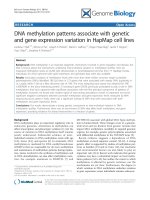
Fig. 1 3,6-DHF decreases global DNA methylation levels and promotes the expression of TET1 in breast cancer cells. a MDA-MB-231 breast cancer
cells were treated with 3,6-DHF (10, 20 μM). The results are expressed as a percentage of vehicle (DMSO)-treated control. b Effects of 3,6-DHF
treatment (0, 5, 10, and 20 μM) for 24 h on TET1, TET2 and TET3 in MDA-MB-231 cells as detected by qRT-PCR. The data are presented as the
mean ± SD (n = 3). *P < 0.05 compared with the MDA-MB-231 cells treated with 0 μM 3,6-DHF for 24 h. c Western blots showing levels of TET1
in MDA-MB-231 breast cancer cells. d Anti-5hmC dot blot for DNA extracted from MDA-MB-231 cells treated with 3,6-DHF
Peng et al. BMC Cancer (2017) 17:619
The purified DNAs were amplified with the following
primer pairs [23]:
TET1 Site-1 Forward(5′-3′):TTGGGAACCGACTCC
TCACCT.
TET1 Site-1 Reverse(5′-3′): TCGGGCAAACTTTCC
AACTCGC.
TET1 Site-2 Forward(5′-3′): ACGCTGGGCATTTCT
GATCCACTA.
TET1 Site-2 Reverse(5′-3′): TATTGTGCAGCTCGTT
TAGTGCCC.
TET1 Site-3 Forward(5′-3′): ACTTTGACCTCCCAA
AGTGCTGGA.
TET1 Site-3 Reverse(5′-3′):ACCTGAGTGATGCTGA
GACTTCCT.
Dots blot analysis of DNA 5hmC
Genomic DNA samples were extracted from cultured
cells. DNA samples were diluted to equal concentrations.
Page 4 of 9
After added 0.1 M NaOH, DNA samples were denatured
at 95 °C for 5 min, and neutralized with 6.6 M ammonium
acetate. The samples were spotted onto a nitrocellulose
membrane, then fixed by baking for 30 min at 80 °C. After
blocking with 5% skim milk, the membrane was incubated
with antibody specific to 5hmC (1:500) followed by incubation with secondary antibody (1:500). The dot signal
was visualized with the ECL Plus chemiluminescence
assay kit (Fusion FX).
Statistical analysis
The experimental data are presented as the means ± the
standard deviation (SD). The results are from at least
three independent experiments. The data were analyzed
by one-way ANOVA. Tukey’s test was used for multiple
comparisons. Differences were considered statistically
significant for P < 0.05.
Fig. 2 3,6-DHF promotes the expression of TET1 during carcinogens-induced breast carcinogenesis. a Western blots showing levels of TET1 in the
cellular breast carcinogenesis model. b The level of TET1 in breast tissues (0, 2 w) and tumors (18 w) in MNU-treated rats with 3,6-DHF administration
(20 mg/kg, i.g.), as detected by immunohistochemistry and western blot c Immunohistochemistry of TET1 in xenografted breast tumors in breast
tumors in athymic mice. d Western blos showing levels of TET1 in breast tissues (0, 2 w) and tumors (18 w) in MNU-treated rats, and in xenografted
breast tumors in athymic mice. Immunostaining density was quantified using image J analysis. The data are presented as the mean ± SD
(n = 3).*P < 0.05, **P < 0.01 compared with control. #P < 0.05 compared with 0 W
Peng et al. BMC Cancer (2017) 17:619
Results
3,6-DHF increases TET1 in breast cancer cells
We examined the effect of 3,6-DHF on global DNA
methylation in breast cancer MDA-MB-231 cells. As
shown in Fig. 1a, after treatment with 10 or 20 μM 3,6DHF for 24 h, the global DNA methylation showed no
significant change. Since the TET family plays potential
roles in epigenetic regulation, we detected Tet1, Tet2
and Tet3 mRNA levels in MDA-MB-231 cells. The results (Fig. 1b) indicated that Tet1 mRNA expression was
significantly increased after 3,6-DHF treatment for 24 h,
while Tet2 and Tet3 showed no notable changes. Western
blot detection (Fig. 1c) confirmed that 3,6-DHF increased
the level of TET1 and TET1 siRNA blocked the
effect(Fig. 3a) in MDA-MB-231 cells. Dot blot analysis
demonstrated that 3,6-DHF treatment increased the
level of 5hmC(Fig. 1d). There was no detectable effect
of knocking down TET1 on global increase of 5hmC
level after 3,6-DHF treatment(Fig. 1d).
3,6-DHF promotes the expression of TET1 in breast
carcinogenesis
TET1 and 5hmC down-regulation has been observed more
frequently in tumorigenesis [24]. We assessed the TET1 expression in breast carcinogenesis in vitro by chronic exposure to NNK and B[a]P. Our data showed that the levels of
TET1 significantly decreased in breast cell carcinogenesis,
and 3,6-DHF co-treatment counteracted the decrease of
TET1 (Fig. 2a). Then, we detected the expression of TET1
in MNU-treated rats with immunohistochemistry and
western blotting. The results (Fig. 2b, d) showed that TET1
levels significantly decreased in breast carcinogenesis in
Page 5 of 9
vivo, while 3,6-DHF administration (20 mg/kg, i.g.) could
effectively up-regulate the expression of TET1. Furthermore, we found that 3,6-DHF administration promotes the
levels of TET1 in xenografted breast tumors derived from
MDA-MB-231 cells (Fig. 2c, d).
3,6-DHF reactivates the tumor suppressor miR-34a via
promoting TET1
Our previous study revealed that 3,6-DHF increases the
level of miR-34a in breast cell carcinogenesis and breast
cancer cells. However, the mechanism is unclear. We
blocked TET1 expression by siRNA to evaluate the role
of TET1 in 3,6-DHF-induced up-regulation of miR-34a
in MDA-MB-231 cells (Fig. 3a, b).The results showed
that inhibition of TET1 significantly suppresses the
effects of 3,6-DHF on miR-34a (Fig. 3c). MSP assays
showed that 3,6-DHF decreases the methylation of the
miR-34a promoter, and that TET1 inhibition could
counteract the effect of 3,6-DHF on the miR-34a promoter (Fig. 4a, b). These data suggests that 3,6-DHF upregulates miR-34a by increasing TET1 expression and
thus demethylation of miR-34a promoter.
3,6-DHF improves the level of TET1 by repressing DNMT1
Our previous study observed that 3,6-DHF is an effective
DNMT1 inhibitor and decreases DNMT activity in
MDA-MB-231 cells [22]. In this study, we evaluated the
effect of DNMT1 on 3,6-DHF-induced promotion of
TET1 by transfecting DNMT1 plasmids in MDA-MB231 cells. As expected, over-expression of DNMT1
significantly down-regulated TET1 and reduced the promotional effect of 3,6-DHF on TET1 (Fig. 5a, b). MSP
Fig. 3 3,6-DHF reactivates the expression of tumor suppressor miR-34a through increasing TET1 level in breast cancer cells. a Western blots
showing levels of TET1 in MDA-MB-231 cells after transfecting TET1 siRNA. b The effect of 3,6-DHF (20 μM) on the levels of TET1 in MDA-MB-231
cells after transfecting TET1 siRNA, detected by Western blotting. c The effect of 3,6-DHF (0, 20 μM) on the levels of miR-34a in MDA-MB-231 cells
after transfecting TET1 siRNA or pcDNA3/Myc-DNMT1(DNMT1) as detected by qRT-PCR. The data are presented as the mean ± SD (n = 3).
*
P < 0.05, **P < 0.01 compared with the control
Peng et al. BMC Cancer (2017) 17:619
Page 6 of 9
Fig. 4 The methylation status of miR-34a and TET1 promoters. a The methylation status of miR-34a promoter in MDA-MB-231 cells with 3,6-DHF
(20 μM) treatment for 24 h, or transfecting TET1 siRNA before 3,6-DHF (20 μM) treatment for 24 h. or transfecting pcDNA3/Myc-DNMT1 before
3,6-DHF (20 μM) treatment for 24 h. b The level of the DNA methylation of miR-34a promoters in MDA-MB-231 cells as determined by qPCR according
to Fig. 4a. c The methylation status of the TET1 promoter in MDA-MB-231 cells after 3,6-DHF (20 μM) treatment for 24 h, or transfecting of pcDNA3/
Myc-DNMT1 before 3,6-DHF (20 μM) treatment. d The level of the DNA methylation of TET1 promoters in MDA-MB-231 cells as determined by qPCR
according to Fig. 4c. Methylation status was detected by MSP; methylation levels are also detected with qPCR. M: methylated; U: unmethylated. The
data are presented as the mean ± SD (n = 3). *P < 0.05 compared with the control or compared with 0 μM
detection indicated that DNMT1 over-expression inhibits
the effect of 3,6-DHF on methylation of the TET1 promoter (Fig. 4c, d). The results also showed that DNMT1
over-expression significantly reduces 3,6-DHF activation
of miR-34a (Fig. 3c) and inhibits the demethylation effect
of 3,6-DHF on the miR-34a promoter (Fig. 4a, b). Because
TET1 may bind to its own promoter region directly, we
analyzed whether 3,6-DHF influenced the autoregulation
of TET1. ChIP assays showed that 3,6-DHF did not increase the binding of TET1 on its own promoter (Fig. 5c).
These findings indicate that 3,6-DHF increases TET1 expression by demehylation of the TET1 promoter partially
through the inhibition of DNMT1.
Discussion
Investigate the factors that relate to carcinogenesis may
contribute to strategies for cancer treatment and
prevention [25]. As epigenetic aberrations occur and initiate events in tumorigenic processes, epigenetic treatment is a promising strategy for cancer prevention [26].
Some phytochemicals are shown to modulate epigenetic
modifications. Several phytochemicals such as resveratrol [27], curcumin [28], tea phenols [29], genistein [30]
and sulforaphane [31] inhibit DNA methyltransferases
and alter DNA methylation of some genes. Phytochemicals, such as EGCG [32], organosulfur compounds [33]
and resveratrol [34], have critical roles in histone acetylation or deacetylation. Elagitannins, EGCG, genistein,
indole-3-carbinol and resveratrol also have effects on
miRNAs oncogenic relating with carcinogenesis [35]. In
our research, we observed that 3,6-DHF could reverse
the global down-regulation of miR-34a in breast carcinogenesis by regulating the miR-34a promoter methylation. Regulation of the cytosine methylation status of
Peng et al. BMC Cancer (2017) 17:619
Page 7 of 9
Fig. 5 3,6-DHF improves the expression of TET1 by repressing DNMT1 activity. a Western blots showing the levels of DNMT1 and TET1 in MDAMB-231 cells after transfecting pcDNA3/Myc-DNMT1. b The effect of 3,6-DHF (20 μM) on the levels of TET1 after transfecting pcDNA3/Myc-DNMT1,
detected by western blot analysis. c The level of TET1 binding to its own promoter in MDA-MB-231 cells as determined by a ChIP assay with
anti-TET1 antibody followed by qPCR; Site-3 is a negative control. The data are presented as the mean ± SD (n = 3). d Flow chart illustrating
mechanism of 3,6-DHF in regulating DNA methylation of the miR-34a promoter
promoters could contribute to the epigenetic control of
3,6-DHF in carcinogenesis. This finding prompted us to
further study the mechanism of 3,6-DHF in regulating
DNA methylation of the miR-34a promoter.
Considerable attention has been focused recently on the
crucial role of DNA methylation in tumorigenesis, and
demonstrates its potential as a disease biomarker and
therapeutic cancer target. DNMT1 is the most abundant
DNMT which maintains the DNA methylation pattern.
The expression levels of DNMT1 are reportedly elevated
in various cancers [36]; reduction of DNMT1 also blocks
tumorigenesis [37]. In our previous research, we found
3,6-DHF inhibits the activity of DNMT1, and now we
further confirmed the effect of 3,6-DHF on DNMT1 by
expression of DNMT1 plasmids. DNMT1 over-expression
blocked the effect of 3,6-DHF on increasing miR-34a
mRNA and miR-34a promoter demethylation, suggesting
that 3,6-DHF could reactivate tumor suppressor genes
silenced by promoter methylation during tumorigenesis
by repressing DNMT1 activity.
TET protein expression and its dominant enzymatic
product (5hmC) are markedly reduced in breast tumors
[38]. Researchers found that decreased 5hmC or TET
levels have a close correlation with robust tumor growth
and metastasis. Increasing TET activity could be a useful
strategy in cancer treatment [39]. For example, vitamin
C has the role of increasing DNA demethylation through
enhancing TET activity in cancer cells [40]. In our research, we found that 3,6-DHF treatment increased
TET1 level in MDA-MB-231 cells, and had no effect on
TET2 and TET3. By immunohistochemistry, we found
that the level of TET1 significantly decreased during
carcinogen-induced breast carcinogenesis in MCF10A
cells and rats, and that 3,6-DHF administration could effectively up-regulate the expression of TET1. 3,6-DHF
administration also promoted the levels of TET1 and
5hmC in xenografted breast tumors derived from MDAMB-231 cells, confirming the effect of 3,6-DHF on
TET1. TET1 inhibition with siRNA in MDA-MB-231
cells blocked the effect of 3,6-DHF on increasing
miR-34a mRNA and miR-34a promoter demethylation, suggesting that the increase of TET1 could be
one of the mechanisms of breast cancer prevention
by 3,6-DHF. Furthermore, DNMT1 over-expression in
part blocked the effect of TET1 on miR-34a by TET1
promoter demethylation. Thus we can conclude that
3,6-DHF inhibits DNMT1 activity, modulates the imbalance of DNA methylation and demethylation status, increases TET1 expression, re-expresses miR-34a,
and as a consequence, prevents breast carcinogenesis.
MiR-34a levels are not only determined by transcriptional regulation, but also by processes relating to
miRNA biogenesis. We will continue this interesting
research in further studies.
Peng et al. BMC Cancer (2017) 17:619
Conclusions
Our study showed that 3,6-DHF increases TET1 expression during carcinogenesis and up-regulates miR-34a
level by regulating the methylation status of DNA.
Abbreviations
3,6-DHF: 3,6-dihydroxyflavone; 5hmC: 5-hydroxymethylcytosine;
B[a]P: benzo[a]pyrene; BC: Breast cancer; ChIP assay: Chromatin
immunoprecipitation assay; DNMTs: DNA methyltransferases; MSP: Bisulfite
Modification and Methylation-Specific PCR; NNK: 4-(methylnitrosamino)-1-(3pyridyl)-1-butanone; SAM: Methyl donor S-adenosyl-methionine; TET: Teneleven translocation
Acknowledgements
The authors thank Elsevier WebShop for the English language editing of the
article.
Funding
The design of the study and collection of data were supported by the
Chongqing Fundamental and Advanced Research Project
(cstc2013jcyjA10083). The analysis and interpretation of data, manuscript
writing and publishing were supported by research grants from the National
Natural Science Foundation of China (81,372,974, 81,402,675).
Availability of data and materials
The datasets generated and analysed during the current study are available
from the corresponding author on reasonable request.
Authors’ contributions
XLP and JLC carried out experiments, acquisition of data. HC made
substantial contributions to carry out experiments, analysis and interpretation
of data; QYZ carried out experiments and made substantial contributions to
conception and design; MTM drafted the manuscript; XPY revised the
drafted manuscript critically for important intellectual content. All authors
have participated in this research, agreed to be accountable for all aspects of
the work in ensuring that questions related to the accuracy or integrity of
any part of the work are appropriately investigated and resolved. All authors
approved the final manuscript. XPY and MTM contributed equally to this
work and should be considered co-corresponding authors.
Ethics approval
Since there was no human subject in this experiment, written human
subject consent was not necessary.
The animal experiments were approved by the Institutional Animal Care and
Use Committee of the Third Military Medical University (Permit No. SCXK(army)-2007–015). The experiments were proceed according to the
guidelines for the care and use of experimental animals.
Consent for publication
This manuscript does not contain any patient details.
Competing interests
The authors declare that they have no competing interests.
Publisher’s Note
Springer Nature remains neutral with regard to jurisdictional claims in
published maps and institutional affiliations.
Author details
1
Research Center for Nutrition Correspondence and Food Safety, Third
Military Medical University, Chongqing Key Laboratory of Nutrition and Food
Safety, 30 Gaotanyan Street, Shapingba District, Chongqing 400038, China.
2
Department of Public Health, School of Preclinical Medicine, Chengdu
Medical College, Chengdu, China.
Page 8 of 9
Received: 21 January 2016 Accepted: 29 August 2017
References
1. Chen W, Zheng R, Zhang S, Zhao P, Zeng H, Zou X. Report of cancer
incidence and mortality in China, 2010. Ann Transl Med. 2014;2:61.
2. Shike M, Doane AS, Russo L, Cabal R, Reis-Filo J, Gerald W, et al. The effects
of soy supplementation on gene expression in breast cancer: a randomized
placebo-controlled study. J Natl Cancer Inst. 2014;106
3. Bishayee K, Ghosh S, Mukherjee A, Sadhukhan R, Mondal J, Khuda-Bukhsh
AR. Quercetin induces cytochrome-c release and ROS accumulation to
promote apoptosis and arrest the cell cycle in G2/M, in cervical carcinoma:
signal cascade and drug-DNA interaction. Cell Prolif. 2013;46:153–63.
4. Hui C, Yujie F, Lijia Y, Long Y, Hongxia X, Yong Z, et al. MicroRNA-34a and
microRNA-21 play roles in the chemopreventive effects of 3,6dihydroxyflavone on 1-methyl-1-nitrosourea-induced breast carcinogenesis.
Breast Cancer Res. 2012;14:R80.
5. Vanden Berghe W. Epigenetic impact of dietary polyphenols in cancer
chemoprevention: lifelong remodeling of our epigenomes. Pharmacol Res.
2012;65:565–76.
6. Pan MH, Chiou YS, Chen LH, Ho CT. Breast cancer chemoprevention by
dietary natural phenolic compounds: specific epigenetic-related molecular
targets. Mol Nutr Food Res. 2015;59(1):21–35.
7. Gao F, Xia Y, Wang J, Lin Z, Ou Y, Liu X, et al. Integrated analyses of DNA
methylation and hydroxymethylation reveal tumor suppressive roles of
ECM1, ATF5, and EOMES in human hepatocellular carcinoma. Genome Biol.
2014;15:533.
8. Faam B, Ghaffari MA, Ghadiri A, Azizi F. Epigenetic modifications in human
thyroid cancer. Biomed Rep. 2015;3:3–8.
9. Pandey M, Shukla S, Gupta S. Promoter demethylation and chromatin
remodeling by green tea polyphenols leads to re-expression of GSTP1 in
human prostate cancer cells. Int J Cancer. 2010;126:2520–33.
10. Ko M, An J, Pastor WA, Koralov SB, Rajewsky K, Rao A. TET proteins and
5-methylcytosine oxidation in hematological cancers. Immunol Rev.
2015;263:6–21.
11. Lian CG, Xu Y, Ceol C, Wu F, Larson A, Dresser K, et al. Loss of 5hydroxymethylcytosine is an epigenetic hallmark of melanoma. Cell. 2012;
150:1135–46.
12. Fernandez AF, Huidobro C, Fraga MF. De novo DNA methyltransferases:
oncogenes, tumor suppressors, or both? Trends Genet. 2012;28:474–9.
13. Rawłuszko-Wieczorek AA, Siera A, Jagodziński PP. TET proteins in cancer:
Current 'state of the art'. Crit Rev Oncol Hematol. 2015;96(3):425–36.
14. Vogt M, Munding J, Grüner M, Liffers ST, Verdoodt B, Hauk J, et al. Frequent
concomitant inactivation of miR-34a and miR-34b/c by CpG methylation in
colorectal, pancreatic, mammary, ovarian, urothelial, and renal cell
carcinomas and soft tissue sarcomas. Virchows Arch. 2011;458:313–22.
15. Cui X, Zhao Z, Liu D, Guo T, Li S, Hu J, et al. Inactivation of miR-34a by
aberrant CpG methylation in Kazakh patients with esophageal carcinoma. J
Exp Clin Cancer Res. 2014;33:20.
16. Lodygin D, Tarasov V, Epanchintsev A, Berking C, Knyazeva T, Körner H, et al.
Inactivation of miR-34a by aberrant CpG methylation in multiple types of
cancer. Cell Cycle. 2008;7:2591–600.
17. Siemens H, Neumann J, Jackstadt R, Mansmann U, Horst D, Kirchner T, et al.
Detection of miR-34a promoter methylation in combination with elevated
expression of c-met and β-catenin predicts distant metastasis of colon
cancer. Clin Cancer Res. 2013;19:710–20.
18. Stankevicins L, Almeida da Silva AP, Ventura Dos Passos F, Dos Santos
Ferreira E, Menks Ribeiro MC, G David M, et al. MiR-34a is up-regulated in
response to low dose, low energy X-ray induced DNA damage in breast
cells. Radiat Oncol. 2013;8:231.
19. Wong KY, Yu L, Chim CS. DNA methylation of tumor suppressor miRNA
genes: a lesson from the miR-34 family. Epigenomics. 2011;3:83–92.
20. Rokavec M, Öner MG, Li H, Jackstadt R, Jiang L, Lodygin D, et al. IL-6R/
STAT3/miR-34a feedback loop promotes EMT-mediated colorectal cancer
invasion and metastasis. J Clin Invest. 2014;124:1853–67.
21. Di Martino MT, Campani V, Misso G, Gallo Cantafio ME, Gullà A, Foresta U, et
al. In vivo activity of miR-34a mimics delivered by stable nucleic acid lipid
particles (SNALPs) against multiple myeloma. PLoS One. 2014;9:e90005.
22. Peng X, Chang H, Gu Y, Chen J, Yi L, Xie Q, Zhu J, Zhang Q, Mi M. 3,6Dihydroxyflavone suppresses breast carcinogenesis by epigenetically
regulating miR-34a and miR-21. Cancer Prev Res (Phila). 2015;8(6):509–17.
Peng et al. BMC Cancer (2017) 17:619
Page 9 of 9
23. Sun M, Song CX, Huang H, Frankenberger CA, Sankarasharma D, Gomes S,
et al. HMGA2/TET1/HOXA9 signaling pathway regulates breast cancer
growth and metastasis. Proc Natl Acad Sci U S A. 2013;110:9920–5.
24. Chen HF, Wu KJ. Epigenetics, TET proteins, and hypoxia in epithelialmesenchymal transition and tumorigenesis. Biomedicine (Taipei). 2016;6(1):
1.
25. Verma M. Cancer control and prevention: nutrition and epigenetics. Curr
Opin Clin Nutr Metab Care. 2013;16(4):376–84.
26. Stahl M, Kohrman N, Gore SD, Kim TK, Zeidan AM, Prebet T. Epigenetics in
cancer: a hematological perspective. PLoS Genet. 2016;12(10):e1006193.
27. Singh B, Shoulson R, Chatterjee A, Ronghe A, Bhat NK, Dim DC, et al.
Resveratrol inhibits estrogen-induced breast carcinogenesis through
induction of NRF2-mediated protective pathways. Carcinogenesis.
2014;35:1872–80.
28. Zheng J, Wu C, Lin Z, Guo Y, Shi L, Dong P, et al. Curcumin up-regulates
phosphatase and tensin homologue deleted on chromosome 10 through
microRNA-mediated control of DNA methylation–a novel mechanism
suppressing liver fibrosis. FEBS J. 2014;281:88–103.
29. Henning SM, Wang P, Carpenter CL, Heber D. Epigenetic effects of green
tea polyphenols in cancer. Epigenomics. 2013;5:729–41.
30. Xie Q, Bai Q, Zou LY, Zhang QY, Zhou Y, Chang H, et al. Genistein inhibits
DNA methylation and increases expression of tumor suppressor genes in
human breast cancer cells. Genes Chromosomes Cancer. 2014;53:422–31.
31. Wong CP, Hsu A, Buchanan A, Palomera-Sanchez Z, Beaver LM, Houseman
EA, et al. Effects of sulforaphane and 3,3′-diindolylmethane on genomewide promoter methylation in normal prostate epithelial cells and prostate
cancer cells. PLoS One. 2014;9:e86787.
32. Hu Q, Chang X, Yan R, Rong C, Yang C, Cheng S, et al. (−)-Epigallocatechin3-gallate induces cancer cell apoptosis via acetylation of amyloid precursor
protein. Med Oncol. 2015;32:390.
33. Altonsy MO, Habib TN, Andrews SC. Diallyl disulfide-induced apoptosis in a
breast-cancer cell line (MCF-7) may be caused by inhibition of histone
deacetylation. Nutr Cancer. 2012;64:1251–60.
34. Venturelli S, Berger A, Böcker A, Busch C, Weiland T, Noor S, et al.
Resveratrol as a pan-HDAC inhibitor alters the acetylation status of histone
[corrected] proteins in human-derived hepatoblastoma cells. PLoS One.
2013;8:e73097.
35. Gerhauser C. Cancer chemoprevention and nutriepigenetics: state of the art
and future challenges. Top Curr Chem. 2013;329:73–132.
36. Mirza S, Sharma G, Parshad R, Gupta SD, Pandya P, Ralhan R. Expression of
DNA methyltransferases in breast cancer patients and to analyze the effect
of natural compounds on DNA methyltransferases and associated proteins.
J Breast Cancer. 2013;16:23–31.
37. Jin H, Chen JX, Wang H, Lu G, Liu A, Li G, et al. NNK-induced DNA
Methyltransferase 1 in lung tumorigenesis in a/J mice and inhibitory effects
of (−)-Epigallocatechin-3-Gallate. Nutr Cancer. 2014;1:1–10.
38. Tian YP, Zhu YM, Sun XH, Lai MD. Multiple functions of ten-eleven
translocation 1 during tumorigenesis. Chin Med J. 2016;129(14):1744–51.
39. Yang H, Liu Y, Bai F, Zhang JY, Ma SH, Liu J, et al. Tumor development is
associated with decrease of TET gene expression and 5-methylcytosine
hydroxylation. Oncogene. 2013;32:663–9.
40. Huang Y, Rao A. Connections between TET proteins and aberrant DNA
modification in cancer. Trends Genet. 2014;30:464–74.
Submit your next manuscript to BioMed Central
and we will help you at every step:
• We accept pre-submission inquiries
• Our selector tool helps you to find the most relevant journal
• We provide round the clock customer support
• Convenient online submission
• Thorough peer review
• Inclusion in PubMed and all major indexing services
• Maximum visibility for your research
Submit your manuscript at
www.biomedcentral.com/submit

![Tài liệu Báo cáo khoa học: The stereochemistry of benzo[a]pyrene-2¢-deoxyguanosine adducts affects DNA methylation by SssI and HhaI DNA methyltransferases pptx](https://media.store123doc.com/images/document/14/br/gc/medium_Y97X8XlBli.jpg)