MiR-9, miR-21, and miR-155 as potential biomarkers for HPV positive and negative cervical cancer
Bạn đang xem bản rút gọn của tài liệu. Xem và tải ngay bản đầy đủ của tài liệu tại đây (724.02 KB, 8 trang )
Park et al. BMC Cancer (2017) 17:658
DOI 10.1186/s12885-017-3642-5
RESEARCH ARTICLE
Open Access
MiR-9, miR-21, and miR-155 as potential
biomarkers for HPV positive and negative
cervical cancer
Sunyoung Park1†, Kiyoon Eom1†, Jungho Kim1, Hyeeun Bang2, Hye-young Wang2, Sungwoo Ahn1, Geehyuk Kim1,
Hyoungsoon Jang1, Sunghyun Kim3, Dongsup Lee4, Kwang Hwa Park5* and Hyeyoung Lee1*
Abstract
Background: Cervical cancer is the second leading cause of death among female patients with cancer in the world.
High risk human papillomavirus has causal roles in cervical cancer initiation and progression by deregulating several
cellular processes. However, HPV infection is not sufficient for cervical carcinoma development. Therefore, other genetic
and epigenetic factors may be involved in this complex disease, and the identification of which may lead to better
diagnosis and treatment. Our aim was to analyze the expression of microRNAs in cervical cancer cases positive or
negative for HPV E6/E7 mRNA, and to assess their diagnostic usefulness and relevance.
Methods: The expression of three different microRNAs (miR-9, miR-21, and miR-155) in 52 formalin-fixed
paraffin-embedded (FFPE) primary cervical cancer tissue samples and 50 FFPE normal cervical tissue samples
were evaluated.
Results: MiR-9, miR-21, and miR-155 were significantly overexpressed in cervical cancer tissues compared to
normal tissues (P < 0.001). MiR-21 and miR-155 expression combined with the HPV E6/E7 mRNA assay in HPV
E6/E7 negative cervical cancer showed increased AUC of 0.7267 and 0.7000, respectively (P = 0.01, P = 0.04),
demonstrating their potential as diagnostic tools. Moreover, miR-21 and miR-155 were predictors showing a 7
fold and 10.3 fold higher risk for HPV E6/E7 negative patients with cervical cancer (P = 0.024 and P = 0.017,
respectively) while miR-155 was a predictor showing a 27.9 fold higher risk for HPV E6/E7 positive patients
with cervical cancer (P < 0.0001).
Conclusions: There is a strong demand for additional, alternative molecular biomarkers for diagnosis and
management of precancer patients. MiR-21 and miR-155 may be helpful in the prediction of both HPV
positive and HPV negative cases of cervical cancer.
Keywords: Cervical cancer, microRNA, HPV E6/E7, RT-qPCR, Molecular diagnosis
Background
Cervical cancer is the third most common malignancy in
women worldwide [1]. High risk human papillomavirus
(HR-HPV) infection is recognized as the most important
risk factor in cervical cancer. Persistent over-expression of
* Correspondence: ;
†
Equal contributors
5
Department of Pathology, Wonju College of Medicine, Yonsei University
Wonju College of Medicine, 20 Ilsan-ro, Wonju-si, Gangwon-do 26426,
Republic of Korea
1
Department of Biomedical Laboratory Science, College of Health Sciences,
Yonsei University, Wonju-si, Gangwon-do 26493, Republic of Korea
Full list of author information is available at the end of the article
the E6 and E7 oncogenes encoded in the HPV genome
have a critical role in the development of cervical cancer
by causing genetic and epigenetic instability [2]. HPV E6
leads to the degradation of p53, which is a critical tumor
suppressor that regulates abrogation of cell growth arrest.
Furthermore, HPV E7 binds and deactivates another important tumor suppressor, the retinoblastoma protein
(pRb), thereby interfering with cell cycle regulation [3–6].
Recently, several studies reported the development of
cervical cancers that are HPV negative despite increased
sensitivity of HR-HPV detection methods. Through
meta-analyses of HPV detection methods, both Tjalma,
© The Author(s). 2017 Open Access This article is distributed under the terms of the Creative Commons Attribution 4.0
International License ( which permits unrestricted use, distribution, and
reproduction in any medium, provided you give appropriate credit to the original author(s) and the source, provide a link to
the Creative Commons license, and indicate if changes were made. The Creative Commons Public Domain Dedication waiver
( applies to the data made available in this article, unless otherwise stated.
Park et al. BMC Cancer (2017) 17:658
et al. and Giorgi, et al. found that 4.2 to 8.2% of cases
were HPV negative in 574 invasive cervical cancers and
3162 invasive cervical cancers [7, 8]. A large international retrospective cross-sectional study including
10,575 cases with invasive cervical cancer found that
15% (1598 cases) were negative for HPV DNA [9]. Similarly, our previous study also found that 15% of patients
with cervical cancer (100 cases) were HPV negative [10].
Epigenetic instability is affected by microRNAs (miRNA
or miR-). MiRNAs are 19 to 25 nucleotides (nt) in length,
and have a role in transcriptional and epigenetic regulation through binding the 3′-UTR of the target-mRNA
[11, 12]. It is now widely known that miRNA dysregulation is associated with a wide variety of human malignancies, such as breast cancer, lung cancer, colon cancer, and
gastric cancer [13–16].
Many miRNAs studies have tried to confirm the
utility of each miRNAs in cervical cancers with different methods. Lui et al. used miRNA direct sequencing analysis with six human cervical carcinoma cell
lines and frozen cervical tumor tissues [17]. Lee et al.
had miRNA expression profiling with 157 panel analyses with frozen cervical tumor tissues [18]. Gocze et al.
utilized quantitative real time polymerase chain reaction
(RT-qPCR) of eight miRNAs (miR-21, miR-27a, miR-34a,
miR-146a, miR-155, miR-196a, miR-203, miR-221)
individually [19].
Among these several miRNAs, three miRNAs (miR-9,
miR-21, and miR-155) having their revealed targets that
might be related to cancer were selected. Ma et al.
showed miR-9 increased cell motility and invasiveness
by targeting Cadherin 1(CDH1) and lead to cancer metastasis [20]. Asangani et al. and Bumrungthai et al.
showed miR-21 promoted invasion and cell proliferation
targeting programmed cell death 4(PDCD4) [21, 22].
MiR-155 expression promotes the proliferation targeting
liver kinase B1 (LKB1) [23, 24].
Although the roles of these three miRNAs (miR-9,
miR-21, and miR-155) have been studied in cervical cancer, their potential diagnostic or prognostic value in a
clinical setting has not been examined. In addition, it is
not known whether there is an association between
these three miRNAs and HR-HPV infection status in
clinical tissue specimens. Therefore, the purpose of this
study was to investigate miR-9, miR-21, and miR-155 expression levels in cervical cancer and normal tissue samples, and determine their possible relation to HR-HPV
E6/E7 oncogene expression.
Methods
Clinical samples
A total of 52 FFPE cervical cancer tissue samples and 50
FFPE normal cervical tissue samples were used from the
Department of Pathology, Yonsei University Wonju
Page 2 of 8
Severance Christian Hospital, Wonju, Republic of Korea,
between January 2010 and December 2014 (Table 1). Institutional Ethics Committee at Yonsei University Wonju
College of Medicine approved the study protocol (approval no. YWMR-12-4-010) and all subjects provided
written informed consent. Cases with tissue biopsies
available were reviewed by two pathologists. The 52
cervical cancer samples consisted of tissue samples from
50 squamous cell carcinomas and 2 adenocarcinomas.
Deparaffinization of FFPE tissues and total RNA extraction
Three to four 10-μm thick sections of FFPE cervical tissue were used for total RNA extraction. To remove paraffin from FFPE tissue, 160 μL of Deparaffinization
solution (Qiagen, Hilden, Germany) was added and vortexed, followed by incubation for 3 min at 56 °C. RNA
extraction was performed using the Qiagen RNeasy
FFPE kit (Qiagen, Hilden, Germany) according to the
manufacturer’s protocol. Total RNA purity and concentration were determined by measuring the ratio of the
absorbance at 260 and 280 nm using an Infinite 200
spectrophotometer (Tecan, Salzburg, Austria). All preparation and handling procedures were conducted under
RNase-free conditions. Isolated total RNA was stored at
−70 °C until used.
cDNA synthesis
Complementary DNA (cDNA) was synthesized using a
TaqMan microRNA Reverse Transcriptase kit (Applied
Biosystems by Life Technologies, Foster City, CA, USA)
according to manufacturer’s instructions. Briefly, 5 to
10 ng of total RNA was used for cDNA synthesis. The
reverse transcriptase (RT) reaction mixture contained
0.15 μL of 100 mM dNTP mix (100 mM each dATP,
dGTP, dCTP, and dTTP at a neutral pH), 1 μL of 50 U/μL
reverse transcriptase, 1.5 μL of 10× reverse transcriptase
buffer, 0.19 μL of 20 U/μL RNase inhibitor, and adjusted
the total reaction volume to 15 μL with nuclease free
Table 1 Sample information in cervical cancer and normal
Variables
Cancer, n (%)
Normal, n (%)
< 50 years
18 (34.6)
31 (62.0)
≥ 50 years
34 (65.4)
19 (38.0)
Age
Histology
SCC
50 (96.2)
ADC
2 (3.8)
HPV E6/E7 mRNA expression
Positive
37 (71.2)
0 (0)
Negative
15 (28.8)
50 (100)
52 (100)
50 (100)
Total
SCC Squamous cell carcinoma, ADC Adenocarcinoma
Park et al. BMC Cancer (2017) 17:658
water. The cDNA synthesis reaction was performed
as follows: 16 °C for 30 min followed by 42 °C for
30 min, and 85 °C for 5 min.
MiRNA analysis using RT-qPCR
MiRNA expression was quantified by determining the cycle
threshold (CT) which is the number of PCR cycles required
for the fluorescence to exceed a value significantly higher
than the background fluorescence, using the TaqMan small
RNA assay (Applied Biosystems by Life Technologies) with
miRNA specific primers according to manufacturer’s instructions. Briefly, 1.4 μL of cDNA was added to 10 μL of
probe qPCR mix and 7.6 μL of nuclease free water. The following TaqMan small RNA assay (Applied Biosystems)
primers were used: hsa-miR-9-5p, hsa-miR-21-5p, hsa-miR155-5p, and RNU6B. All analyzed miRNAs are of human
(Homo sapiens) origin and therefore, the prefix “hsa” is
omitted throughout the text. RT-qPCR reactions were performed using a CFX96 Real-Time PCR System Detector
(Bio-Rad, Hercules, CA, USA). Samples were run in duplicate for each experiment. Data were analyzed using the
comparative Ct (2-ΔΔCT) method using the small nuclear
RNA, RNU6B, as an endogenous control. To monitor
reagent contamination, negative controls were included for
each primer pair. PCR cycling conditions were as follows: 95 °C for 3 min 40 cycles of 95 °C for 15 s and
60 °C for 60 s.
HPV E6/E7 mRNA analysis using RT-qPCR
To detect HPV E6/E7 mRNA in FFPE cervical tissues,
multiplex RT-qPCR was performed using the TaqMan
assay with the OPTIMYGENE HPV E6/E7 mRNA RTqDx assay kit (Optipharm, Osong, Republic of Korea).
PCR primers and the corresponding TaqMan probes
were designed for three different sets of HPV regions,
with each set of probes targeting their conserved sequence (FAM: HPV genotypes 16, 31, 33, 35, 52, and 58;
CY5: HPV genotypes 18, 39, 45, 51, 59, and 68; and
HEX: HPV genotypes 53, 56, 66, and 69).
RT-qPCR reactions consisted of 10 μL of 2 × Thunderbird
probe qPCR mix (Toyobo, Osaka, Japan), 5 μL of primers
and TaqMan probe mixture, 2 μL of template cDNA, and
distilled water for a final reaction volume of 20 μL. The
multiplex RT-qPCR assay detected the HPV E6 and E7
genes simultaneously in a single tube by incorporating two
targets (E6 and E7) using specific TaqMan probes, which
were labeled with different fluorophores (FAM, HEX,
and Cy5). Positive and negative controls were included
throughout the procedure. PCR cycling conditions were
as follows: 95 °C for 3 min 45 cycles of 95 °C for 20 s and
60 °C for 40 s. To avoid false negatives because of mRNA
degradation, glyceraldehyde-3-phosphate dehydrogenase
(GAPDH) was used as an endogenous control.
Page 3 of 8
Statistical analysis
Statistical analysis was performed using GraphPad
Prism software version 5.02 (GraphPad, La Jolla, CA,
USA) and MedCalc 9.0 software (MedCalc Software
Inc., Mariakerke, Belgium). Student’s t-test and Mann
Whitney U test were used to determine statistical significance between cervical cancer and normal cervical
tissue samples as well as investigate miRNA expression in patients according to HPV infection status.
Receiver operating characteristic (ROC) curves were
generated to assess diagnostic accuracy of each
miRNA, and the area under the ROC curve (AUC)
was calculated to measure discriminatory capacity.
The best sensitivity/specificity pair was selected based
on the maximum likelihood ratio. Univariate and
multivariate logistic regression by odds ratio (OR) and
95% confidential interval (95% CI) were performed to
assess predictors for cervical cancer diagnosis using
the XLSTAT software (Addinsoft, New York, USA).
All statistical tests were two-sided, and a P value
≤0.05 was considered statistically significant.
Results
HPV E6/E7 mRNA expression in cervical cancer tissues
Prior to investigating miRNA expression levels, we first
examined HPV E6/E7 mRNA expression in 52 FFPE cervical cancer tissue samples and 50 FFPE normal control
samples. Fifteen (28.8%) of the 52 FFPE cervical cancer
tissue samples were negative for HR-HPV E6/E7 expression (termed HR-HPV E6/E7-negative), while 37 (71.2%)
samples were positive (termed HR-HPV E6/E7-positive).
We found that all 50 FFPE normal cervical control samples were negative for HPV E6/E7 mRNA expression
(Table 1).
MiRNA expression levels in cervical cancer and normal
tissues
Expression levels of miR-9, miR-21, and miR-155 were
investigated in our 52 FFPE cervical cancer tissue samples and 50 FFPE normal cervical tissue controls. All
three miRNAs were significantly up regulated in FFPE
cervical cancer tissues compared to FFPE normal cervical tissues (P < 0.0001) (Fig. 1a-c). The AUC was
0.7565 [95% confidence interval (CI) = 0.6624–0.8507]
in miR-9, 0.8325 (95% CI = 0.7530–0.9120) in miR-21,
and 0.8492 (95% CI = 0.7736–0.9249) in miR-155, all of
which indicate these miRNAs may be used as potential
biomarkers for cervical cancer (Fig. 1d-f ).
Diagnostic value of miR-9, miR-21, and miR-155 miRNAs
To assess the potential diagnostic value of these three
miRNAs, the performance characteristics sensitivity,
specificity, positive predictive value, and negative
Park et al. BMC Cancer (2017) 17:658
Page 4 of 8
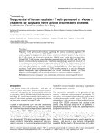
Fig. 1 MiR-9, miR-21, and miR-155 expression levels in formalin-fixed paraffin-embedded (FFPE) cervical cancer and normal tissue samples. a MiR-9,
b miR-21, and c miR-155 expression levels in 52 FFPE cervical cancer tissue samples were significantly different compared to that found in 50 FFPE
normal cervical tissue samples (P < 0.0001 for all three comparisons). Receiver operating characteristic (ROC) curve analysis showed that d miR-9 had
an area under the ROC curve (AUC) value of 0.7565 [95% confidence interval (CI) = 0.6624–0.8507], while e miR-21, and f miR-155 had AUC values of
0.8325 (95% CI = 0.7530–0.9120) and 0.8492 (95% CI = 0.7736–0.9249), respectively
predictive value were determined and evaluated. The
cut-off values of miR-9, miR-21, and miR-155 as determined using the likelihood ratio, were 4.035, 1.975, and
3.880 respectively, for optimal sensitivity and specificity.
The sensitivity of miR-9, miR-21, and miR-155 was
67.3% (95% CI = 52.9–79.7), 82.7% (95% CI = 69.7–
91.8), and 65.4% (95% CI = 50.9–78.0) respectively, while
the specificity was 80.0% (95% CI = 66.3–90.0) for miR9, 72.0% (95% CI = 57.5–83.8) for miR-21, and 96.0%
(95% CI = 86.3–99.5) for miR-155. Positive predictive
values (PPVs) of miR-9, miR-21, and miR-155 were
77.8%, 75.4%, and 94.3% respectively, while the respective negative predictive values (NPVs) were 70.2%, 80.0%,
and 71.6% (Table 2).
MiR-9, miR-21, and miR-155 in HPV E6/E7-positive and
-negative cervical cancer
To investigate the expression of miR-9, miR-21, and
miR-155 with cervical cancer cases that were HPV E6/
E7 mRNA-positive or -negative for HPV E6/E7 mRNA,
expression levels of the three miRNAs were analyzed in
three groups: HPV E6/E7-positive cancer samples, HPV
E6/E7-negative cancer samples, and normal samples. We
found that all three miRNAs were significantly up regulated in HR-HPV E6/E7-positive cancer tissue samples
compared to normal tissue samples (P < 0.0001), while
miR-21 and miR-155 were up regulated in HPV E6/E7negative cancer tissue samples compared to normal
controls (P = 0.0079 and P = 0.0384, respectively). We
Table 2 Clinical cut-off values, sensitivity, and specificity of miRNAs
Cut-off value
Sensitivity, % (95% CI)
Specificity, % (95% CI)
PPV, % (95% CI)
NPV, % (95% CI)
Likelihood ratio
miR-9
>4.035
67.3 (52.9–79.7)
80.0 (66.3–90.0)
77.8 (62.9–88.8)
70.2 (56.6–81.6)
3.4
miR-21
>1.975
82.7 (69.7–91.8)
72.0 (57.5–83.8)
75.4 (62.2–85.9)
80.0 (65.4–90.4)
3.0
miR-155
>3.880
65.4 (50.9–78.0)
96.0 (86.3–99.5)
94.3 (80.8–90.6)
71.6 (59.3–82.0)
15.9
95% CI 95% confidence interval, PPV positive predictive value, NPV negative predictive value
Park et al. BMC Cancer (2017) 17:658
found no significant difference in miR-9 expression
levels between HR-HPV E6/E7-negative cancer samples
and normal cervical samples (Fig. 2).
Page 5 of 8
Table 3 Diagnostic values of miRNAs in conjunction with HPV
E6/E7 for cervical cancer
AUCa
95% CI
P-value
HPV E6/E7
0.8558
0.7773–0.9343
<0.0001
HPV E6/E7 + miR-9
0.8135
0.7257–0.9013
<0.0001
HPV E6/E7 + miR-21
0.8215
0.7349–0.9082
<0.0001
HPV E6/E7 + miR-155
0.8935
0.8243–0.9626
<0.0001
HPV E6/E7
0.5000
0.3321–0.6679
1.00
HPV E6/E7 + miR-9
0.6000
0.4284–0.7716
0.24
HPV E6/E7 + miR-21
0.7267
0.5776–0.8757
0.01
HPV E6/E7 + miR-155
0.7000
0.5152–0.8648
0.04
All cases
Diagnostic value of miR-9, miR-21, and miR-155 miRNAs
in conjunction with the HPV E6/E7 mRNA assay
The effectiveness of each three miRNA combined with
HPV E6/E7 mRNA assay was investigated by analysis of
AUC. In all cases of cervical cancer, the value of AUC was
0.8558 (95% CI 0.7773–0.9343), 0.8135 (95% CI 0.7257–
0.9013), 0.8215 (95% CI 0.7349–0.9082), 0.8935 (95% CI
0.8243–0.9626) in HPV E6/E7, HPV E6/E7 + miR-9, HPV
E6/E7 + miR-21, and HPV E6/E7 + miR-155, respectively.
Especially, in HPV negative cervical cancer, miR-21 and
miR-155 in conjunction with the HPV E6/E7 mRNA was
shown increased AUC value of 0.7267 (95% CI 0.5776–
0.8757) and 0.7000 (95% CI 0.5152–0.8646) respectively,
compared to HPV E6/E7 assay (Table 3).
MiRNA predictors for diagnosing cervical cancer in HPV
E6/E7-negative cases
We analyzed the following risk predictors of cervical
cancer: age, HPV E6/E7 mRNA-expressing status, and
miR-9, miR-21, and miR-155 expression. We found that
the highest risk factor was HPV E6/E7 mRNA expression (OR = 244.4, 95% CI = 13.6–4376.4) followed by expression of miR-155 (OR = 41.7, 95% CI = 9.1–191.2),
miR-21 (OR = 12.3, 95% CI = 4.8–31.7), and miR-9
(OR = 8.2, 95% CI = 3.3–20.3). Ages below 60 years
were not a significant risk factor but being over the age
of 60 years did confer a higher risk to cervical cancer
(Table 4).
HPV negative cases
AUC an area under the ROC curve
a
To investigate the predictive power for diagnosing cervical cancer in HPV E6/E7 mRNA negative cases, the
odds ratio of the miRNAs was determined based on
HPV E6/E7 mRNA expression using multivariate analysis. We found that miR-21 and miR-155 in HPV E6/
E7-negative cervical cancer patients conferred a 7.0
(95% CI = 1.3–37.6) and 10.3 (95% CI = 1.5–70.7) fold
risk to cervical cancer, respectively (Table 5).
Discussion
HPV DNA detection for cervical cancer screening is
widely used as an early diagnostic guideline to prevent
the progression of cervical cancer. Nevertheless, there is
an ongoing need for studies investigating the molecular
mechanisms related to cervical cancer carcinogenesis
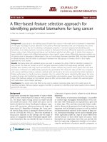
Fig. 2 Box and whisker plots of comparisons between three miRNA expression levels in normal cervical tissues and cancer tissues with (+) or without
(−) HPV E6/E7 mRNA expression. a MiR-9, b miR-21, and c miR-155 expression levels in high risk human papillomavirus (HR-HPV) E6/E7-positive cervical
cancer tissues were significantly up regulated compared to that found in normal cervical tissue samples (P < 0.0001 for all three comparisons). MiR-21
and miR-155 expression levels in HR-HPV E6/E7-negative cervical cancer tissues were significantly up regulated compared to that found in normal
cervical tissue samples (P = 0.0079 and P = 0.00384, respectively). NS, not significant
Park et al. BMC Cancer (2017) 17:658
Page 6 of 8
Table 4 The diagnostic utility of predictors for cervical cancer
(univariate analysis)
OR
95% CI
P value
Ages
< 30 years
1
31–40 years
1.4
0.4–5.1
0.580
41–50 years
1.7
0.4–6.5
0.442
51–60 years
3.5
0.8–16.4
0.109
> 60 years
35.2
3.6–344.2
0.002
13.6–4376.4
<0.0001
3.3–20.3
<0.0001
4.8–31.7
<0.0001
9.1–191.2
<0.0001
HPV E6/E7
negative
1
positive
244.4
miR-9
negative
1
positive
8.2
miR-21
negative
1.0
positive
12.3
miR-155
negative
1.0
positive
41.7
OR Odds ratio, 95% CI 95% confidential interval
because most high-risk HPV infections that present
without any symptoms go away within one to two years,
and there have been reports of several HPV negative
cases of cervical cancer [7–9, 25]. In our previous study,
we found that 15 out of 100 FFPE cervical cancer tissue
samples were HPV negative using an E6/E7 mRNA assay
as well as testing for the HPV L1 genotype (data not
shown).
Several oncogenic miRNAs are associated with cervical
cancer tumorigenesis [17–23]. However, the results from
those studies were not comprehensively evaluated using
clinical specimens, and those studies have not tested for
an association between miRNA expression levels and
HR-HPV E6/E7 mRNA expression in clinical specimens.
Table 5 MiRNA predictors of cervical cancer according to HPV
E6/E7 mRNA expression status in patients (multivariate analysis)
OR
95% CI
P value
3.3
0.6–18.7
0.173
HPV positive
miR-9
miR-21
1.8
0.3–11.1
0.515
miR-155
27.9
5.0–155.7
<0.0001
0.4
0.1–2.6
0.341
HPV negative
miR-9
miR-21
7.0
1.3–37.6
0.024
miR-155
10.3
1.5–70.7
0.017
OR Odds ratio, 95% CI 95% confidential interval
The aim of this study was to explore the potential clinical relevance of miR-9, miR-21, and miR-155 by investigating their expression levels in 52 FFPE cervical cancer
tissue samples and in 50 FFPE normal cervical tissue
samples. Evidence of association between these three
miRNAs and HR-HPV E6/E7 mRNA expression was
also investigated.
All three miRNAs (miR-9, miR-21, and miR-155)
showed significantly higher expression in cervical cancer
tissues compared to that found in normal cervical tissues
(P < 0.0001) (Fig. 1). This finding supports the possibility
that these three miRNAs may be implicated in cervical
cancer development in clinical samples. Although four
previous studies using cervical cancer cell lines and clinical samples found that miR-21, miR-155, and miR-9
were up regulated in cervical cancer, they did not validate these three miRNAs in terms of diagnostic value
[13, 18, 26, 27]. This study is the first to assess these
miRNAs as putative biomarkers and their possible
discriminatory capacity in FFPE tissues.
Differences in expression levels of the three miRNAs between HR-HPV E6/E7-positive cervical cancer tissue samples and HR-HPV E6/E7–negative cervical cancer tissue
samples showed the strongest association was between
miR-9 expression and HR-HPV E6/E7-positive cancer
cases compared to that found with the other miRNAs
(Fig. 2). Similarly, Weijun Liu et al. found miR-9 and HPV
E6 caused increased cell motility by down regulating follistatin like 1 (FSTL1) and activated leukocyte cell adhesion
molecule (ALCAM) mRNAs, both of which are involved
in cell migration [28].
Both miR-21 and miR-155 had reported other mechanism related to immune response as well as HR-HPV
E6/E7 expression. Bumrungthai et al. found that miR-21
is correlated with increased expression of α-smooth
muscle actin (α-SMA) and interleukin 6 (IL-6) and decreased expression of PDCD4 in cell proliferation and
initiates inflammation-associated carcinogenesis via nuclear factor kappa-light chain-enhancer of activated B
cells (NF-kB) and interleukin-6 (IL-6) signaling pathways
in colon and cervical cancer cells and Asangani et al.
found miR-21 down-regulates PDCD4 in colon cancer
and functions as stimulating invasion, intravasation, and
metastasis [21, 22, 29]. For miR-155, Guoying Lao et al.
found that miR-155 regulates LKB1 expression, which
functions as embryonic polarity, metabolism, and cell
growth and up-regulation of miR155 promotes proliferation of cervical cancer cells [23].
In terms of diagnostic value, miR-21, and miR-155 expression in combination with the HPV E6/E7 mRNA
assay may be useful in the diagnosis of cervical cancer
independent of HPV infection because these miR-21 and
miR-155 may have the discriminatory power to detect
HPV negative cervical cancer cases (Table 3). In
Park et al. BMC Cancer (2017) 17:658
particular, miR-21 was independent of HPV status being
consistently up regulated in cervical cancer (Fig. 2). We
analyzed the odds ratios to assess the effects of the predictors age, HPV E6/E7 mRNA expression, and expression of miR-9, miR-21, and miR-155 on cervical cancer
compared to normal controls. While we found that all of
the predictors were significantly associated with cervical
cancer, miR-21 and miR-155 expression were identified
as predictors for high risk in HPV negative cervical
cancer tissues compared to normal cervical tissues
(Tables 4 and 5).
Some studies have investigated miRNAs for association with HPV infection as potential diagnostic and
prognostic indicators. Xiaohong Wang et al. revealed
that miR-92a and miR-378 expression was associated
with cancer progression in HPV positive tissue samples
[30]. Similarly, we found that miR-155 overexpression is
associated with increased risk of cervical cancer in HPV
E6/E7 mRNA positive tissues. Moreover, miR-21 and
miR-155 overexpression in HPV E6/E7 mRNA negative
tissue samples could complement approaches for cervical cancer diagnosis and prediction of progression.
Conclusions
Our findings showed that miRNA RT-qPCR assays for
specific miRs may be useful tools in the diagnosis of
cervical cancer and especially, HPV negative cases of
cervical cancer. In addition, these findings are important
towards determining the possible role of miRNA expression in cervical cancer development, and the relationship
between miRNA and HPV infection. Further study is
needed in pre-cancer lesions to understand the role of
miRNAs in tumor carcinogenesis, and more tests using
normal and cervical cancer samples will be necessary to
clearly demonstrate the potential utility of these assays
for cervical cancer screening and diagnosis.
Abbreviations
95% CI: 95% confidential interval; ALCAM: Activated leukocyte cell adhesion
molecule; AUC: Area under the ROC curve; FFPE: Formalin-fixed paraffinembedded; FSTL1: Follistatin like 1; GAPDH: Glyceraldehyde-3-phosphate
dehydrogenase; HR-HPV: High risk human papillomavirus; IL-6: Interleukin-6;
LKB1: Liver kinase B1; miRNA: microRNA; NPV: Negative predictive value;
OR: Odds ratio; PPV: Positive predictive value; pRb: Retinoblastoma protein;
ROC: Receiver operating characteristic; α-SMA: α-smooth muscle actin
Funding
This research was supported by Basic Science Research Program through the
National Research Foundation of Korea (NRF) funded by the Ministry of
Science, ICT and future Planning (2015R1A2A2A04004455).
Availability of data and materials
All relevant materials are described in the manuscript. Additional data sets
supporting the conclusions of this article are available at request from the
corresponding author.
Authors’ contributions
SP, KE, and JK participated in the design of the study and performed the
statistical analysis and manuscript. KE and HJ and HW carried out the data
extraction. HY and KP and HB conceived of the study, and participated in its
Page 7 of 8
design and coordination and helped to draft the manscripts. GK, DL, and SK
helped to assemble the clinical data and performed the initial data
interpretation together with HB and organize the experiment and sample
collection. All authors read and approved the final manuscript.
Ethics approval and consent to participate
Institutional Ethics Committee at Yonsei University Wonju College of
Medicine approved the study protocol (approval no. YWMR-12-4-010) and all
subjects provided written informed consent.
Consent for publication
Not applicable.
Competing interests
The authors declare that they have no competing interests.
Publisher’s Note
Springer Nature remains neutral with regard to jurisdictional claims in
published maps and institutional affiliations.
Author details
1
Department of Biomedical Laboratory Science, College of Health Sciences,
Yonsei University, Wonju-si, Gangwon-do 26493, Republic of Korea.
2
Optipharm M&D, Inc., Wonju Eco Environmental Technology Center,
Wonju-si, Gangwon-do 26493, Republic of Korea. 3Department of Clinical
Laboratory Science, College of Health Sciences, Catholic University of Pusan,
Pusan, Republic of Korea. 4Department of Clinical Laboratory Science,
Hyejeon College, Hongseoung, Republic of Korea. 5Department of Pathology,
Wonju College of Medicine, Yonsei University Wonju College of Medicine, 20
Ilsan-ro, Wonju-si, Gangwon-do 26426, Republic of Korea.
Received: 27 August 2016 Accepted: 5 September 2017
References
1. McGuire S. World Cancer Report 2014. Geneva, Switzerland: World Health
Organization, International Agency for Research on Cancer, WHO Press.
2015. Adv Nutr. 2016;7:418–9.
2. Crosbie EJ, Kitchener HC. Human papillomavirus in cervical screening and
vaccination. Clin Sci (Lond). 2006;110:543–52.
3. Munger K, Werness BA, Dyson N, Phelps WC, Harlow E, Howley PM.
Complex formation of human papillomavirus E7 proteins with the
retinoblastoma tumor suppressor gene product. EMBO J. 1989;8:4099–105.
4. Koivusalo R, Mialon A, Pitkanen H, Westermarck J, Hietanen S. Activation of
p53 in cervical cancer cells by human papillomavirus E6 RNA interference is
transient, but can be sustained by inhibiting endogenous nuclear exportdependent p53 antagonists. Cancer Res. 2006;66:11817–24.
5. Liu X, Clements A, Zhao K, Marmorstein R. Structure of the human
Papillomavirus E7 oncoprotein and its mechanism for inactivation of the
retinoblastoma tumor suppressor. J Biol Chem. 2006;281:578–86.
6. Moody CA, Laimins LA. Human papillomavirus oncoproteins: pathways to
transformation. Nat Rev Cancer. 2010;10:550–60.
7. Giorgi Rossi P, Sideri M, Carozzi FM, Vocaturo A, Buonaguro FM, Tornesello
ML, Burroni E, Mariani L, Boveri S, Zaffina LM, et al. HPV type distribution in
invasive cervical cancers in Italy: pooled analysis of three large studies.
Infect Agent Cancer. 2012;7:26.
8. Tjalma WA, Fiander A, Reich O, Powell N, Nowakowski AM, Kirschner B,
Koiss R, O’Leary J, Joura EA, Rosenlund M, et al. Differences in human
papillomavirus type distribution in high-grade cervical intraepithelial
neoplasia and invasive cervical cancer in Europe. Int J Cancer.
2013;132:854–67.
9. de Sanjose S, Quint WG, Alemany L, Geraets DT, Klaustermeier JE, Lloveras B,
Tous S, Felix A, Bravo LE, Shin HR, et al. Human papillomavirus genotype
attribution in invasive cervical cancer: a retrospective cross-sectional
worldwide study. Lancet Oncol. 2010;11:1048–56.
10. Wang HY, Kim G, Cho H, Kim S, Lee D, Park S, Park KH, Lee H. Diagnostic
performance of HPV E6/E7, hTERT, and Ki67 mRNA RT-qPCR assays on
formalin-fixed paraffin-embedded cervical tissue specimens from women
with cervical cancer. Exp Mol Pathol. 2015;98:510–6.
11. Bartel DP. MicroRNAs: genomics, biogenesis, mechanism, and function. Cell.
2004;116:281–97.
Park et al. BMC Cancer (2017) 17:658
12. Esquela-Kerscher A, Slack FJ. Oncomirs - microRNAs with a role in cancer.
Nat Rev Cancer. 2006;6:259–69.
13. Guo J, Miao Y, Xiao B, Huan R, Jiang Z, Meng D, Wang Y. Differential
expression of microRNA species in human gastric cancer versus nontumorous tissues. J Gastroenterol Hepatol. 2009;24:652–7.
14. Iorio MV, Ferracin M, Liu CG, Veronese A, Spizzo R, Sabbioni S, Magri E,
Pedriali M, Fabbri M, Campiglio M, et al. MicroRNA gene expression
deregulation in human breast cancer. Cancer Res. 2005;65:7065–70.
15. Takamizawa J, Konishi H, Yanagisawa K, Tomida S, Osada H, Endoh H,
Harano T, Yatabe Y, Nagino M, Nimura Y, et al. Reduced expression of the
let-7 microRNAs in human lung cancers in association with shortened
postoperative survival. Cancer Res. 2004;64:3753–6.
16. Michael MZ, O’Connor SM, van Holst Pellekaan NG, Young GP, James RJ.
Reduced accumulation of specific microRNAs in colorectal neoplasia. Mol
Cancer Res. 2003;1:882–91.
17. Lui WO, Pourmand N, Patterson BK, Fire A. Patterns of known and novel
small RNAs in human cervical cancer. Cancer Res. 2007;67:6031–43.
18. Lee JW, Choi CH, Choi JJ, Park YA, Kim SJ, Hwang SY, Kim WY, Kim TJ,
Lee JH, Kim BG, Bae DS. Altered MicroRNA expression in cervical
carcinomas. Clin Cancer Res. 2008;14:2535–42.
19. Gocze K, Gombos K, Juhasz K, Kovacs K, Kajtar B, Benczik M, Gocze P,
Patczai B, Arany I, Ember I. Unique microRNA expression profiles in cervical
cancer. Anticancer Res. 2013;33:2561–7.
20. Ma L, Young J, Prabhala H, Pan E, Mestdagh P, Muth D, Teruya-Feldstein J,
Reinhardt F, Onder TT, Valastyan S, et al. miR-9, a MYC/MYCN-activated
microRNA, regulates E-cadherin and cancer metastasis. Nat Cell Biol. 2010;
12:247–56.
21. Bumrungthai S, Ekalaksananan T, Evans MF, Chopjitt P, Tangsiriwatthana T,
Patarapadungkit N, Kleebkaow P, Luanratanakorn S, Kongyingyoes B,
Worawichawong S, Pientong C. Up-regulation of miR-21 is associated with
cervicitis and human papillomavirus infection in cervical tissues. PLoS One.
2015;10:5.
22. Asangani I, Rasheed S, Nikolova D, Leupold J, Colburn N, Post S, Allgayer H.
MicroRNA-21 (miR-21) post-transcriptionally downregulates tumor
suppressor Pdcd4 and stimulates invasion, intravasation and metastasis in
colorectal cancer. Oncogene. 2008;27:2128–36.
23. Lao G, Liu P, Wu Q, Zhang W, Liu Y, Yang L, Ma C. Mir-155 promotes
cervical cancer cell proliferation through suppression of its target gene
LKB1. Tumour Biol. 2014;35:11933–8.
24. Turner M, Vigorito E. Regulation of B- and T-cell differentiation by a single
microRNA. Biochem Soc Trans. 2008;36:531–3.
25. Schiffman M, Castle PE, Jeronimo J, Rodriguez AC, Wacholder S. Human
papillomavirus and cervical cancer. Lancet. 2007;370:890–907.
26. Wang X, Tang S, Le SY, Lu R, Rader JS, Meyers C, Zheng ZM. Aberrant
expression of oncogenic and tumor-suppressive microRNAs in cervical
cancer is required for cancer cell growth. PLoS One. 2008;3:e2557.
27. Pereira PM, Marques JP, Soares AR, Carreto L, Santos MA. MicroRNA
expression variability in human cervical tissues. PLoS One. 2010;5:e11780.
28. Liu W, Gao G, Hu X, Wang Y, Schwarz JK, Chen JJ, Grigsby PW, Wang X.
Activation of miR-9 by human papillomavirus in cervical cancer. Oncotarget.
2014;5:11620–30.
29. Deftereos G, Corrie SR, Feng Q, Morihara J, Stern J, Hawes SE, Kiviat NB.
Expression of mir-21 and mir-143 in cervical specimens ranging from
histologically normal through to invasive cervical cancer. PLoS One.
2011;6:e28423.
30. Wang X, Wang HK, Li Y, Hafner M, Banerjee NS, Tang S, Briskin D, Meyers C,
Chow LT, Xie X, et al. microRNAs are biomarkers of oncogenic human
papillomavirus infections. Proc Natl Acad Sci U S A. 2014;111:4262–7.
Page 8 of 8
Submit your next manuscript to BioMed Central
and we will help you at every step:
• We accept pre-submission inquiries
• Our selector tool helps you to find the most relevant journal
• We provide round the clock customer support
• Convenient online submission
• Thorough peer review
• Inclusion in PubMed and all major indexing services
• Maximum visibility for your research
Submit your manuscript at
www.biomedcentral.com/submit